báo cáo khoa học: "Deficiency of maize starch-branching enzyme i results in altered starch fine structure, decreased digestibility and reduced coleoptile growth during germination" pot
Bạn đang xem bản rút gọn của tài liệu. Xem và tải ngay bản đầy đủ của tài liệu tại đây (6.22 MB, 13 trang )
RESEARCH ARTICLE Open Access
Deficiency of maize starch-branching enzyme i
results in altered starch fine structure, decreased
digestibility and reduced coleoptile growth
during germination
Huan Xia
1,3
, Marna Yandeau-Nelson
2,4
, Donald B Thompson
3
and Mark J Guiltinan
4*
Abstract
Background: Two distinct starch branching enzyme (SBE) isoforms predate the divergence of monocots and
dicots and have been conserved in plants since then. This strongly suggests that both SBEI and SBEII provide
unique selective advantages to plants. However, no phenotype for the SBEI mutation, sbe1a, had been previously
observed. To explore this incongruity the objective of the present work was to characterize functional and
molecular phenotypes of both sbe1a and wild-type (Wt) in the W64A maize inbred line.
Results: Endosperm sta rch granules from the sbe1a mutant were more resistant to digestion by pancreatic a-
amylase, and the sbe1a mutant starch had an altered branching pattern for amylopectin and amylose. When
kernels were germinated, the sbe1a mutant was associated with shorter coleoptile length and higher residual
starch content, suggesting that less efficient starch utilization may have impaired growth during germination.
Conclusions: The present report documents for the first time a molecular phenotype due to the absence of SBEI,
and suggests strongly that it is associated with altered physiological function of the starch in vivo. We believe that
these results provide a plausible rationale for the conservation of SBEI in plant s in both monocots and dicots, as
greater seedling vigor would provide an important surviv al advantage when resources are limited.
Background
The starch granule is a highly-ordered structure with
alternating crystalline and amorphous g rowth rings
[1,2]. Starch molecules are biopolymers of anhydroglu-
cose units linked by a-1,4 and a-1,6 glycosidic bonds.
They are composed of two glucan polymers, the gener-
ally linear fraction, amylose, and the branched fraction,
amylopectin. The constituent amylopectin chains can be
mainly categorized into A chains (not bearing any
branches) and B chains (bearing one or more branches)
[3]. The main physiological functions of starch include
high-density storage of energy and the controlled release
of this energy during starch degradation.
Starch-branching enzyme (SBE) plays an important
role in starch biosynthesis by introducing branch point s,
the a-1,6 linkages in starch. Boyer and Preiss [4] identi-
fied three major SBE isoforms in developing maize ker-
nels: SBEI, SBEIIa, and SBEIIb. The SBE isoforms have
been shown to be encoded by different genes [5-8]. Phy-
logenetic analyses of SBE sequences from a number of
plantspecieshaveshownthattheSBEIandSBEIIiso-
forms are conserved among most plants, and that
SBEIIa and SBEIIb isoforms are conserved among most
monocots [9-13]. Furthermore, genes belonging to both
the SBEI and SBEII families can be identified in various
lineages of green alga, which supports the theory that
these two families of genes evolved approximately a bil-
lion years ago [14]. These examples of extreme evolu-
tionary conservation are strong evidence for a specific
and vital role for each enzyme isoform in starch
biosynthesis.
In vitro biochemical analyses have documented that
the SBEI and SBEII isoform activities are not identical
[15,16], but these studies do not necessarily indicate
* Correspondence:
4
Department of Horticulture, The Pennsylvania State University, University
Park, Pennsylvania 16802-5807, USA
Full list of author information is available at the end of the article
Xia et al. BMC Plant Biology 2011, 11:95
/>© 2011 Xia et al; licensee BioMed Central Ltd. This is an Open Access article distributed under the terms of the Creative Commons
Attribution License ( which permits unrestricted use, distribution, and reproduction in
any medium, provided the original work is properly cited.
their actio n in vivo, as starch biosynthesis occurs i n the
presence of starch synthases and debranching enzymes.
Studies have suggested that multi-pro tein starch synthe-
sizing complexes exist, and that interactions within
these complexes could modulate the intricate structure
of a developing starch granule [17-33]. Whether there
are functional differences among SBE isoforms in vivo
remains to be addressed.
Insight into a possible in vivo function of an SBE may
be gained from the study of sbe mutants deficient in
one or more SBE isoform activities. The maize amylose
extender (ae) mu tant, which is deficient in SBEIIb, has a
profound effect on starch structure, leading to an
increased amylose proportion and a reduced branching
density of endosperm amylopectin [5,33-35]. More
recently, studies of a maize sbe2a mutant showed that
deficiency of the SBEIIa isoform decreased plant fitness
and resulted in lower kernel yield, but there was m ini-
mal effect on kernel starch properties [11,36]. Previous
work showed no effe ct of SBEI deficiency (in the sbe1a
mutation) on starch molecular size and on chain length
distribution after debranching [26,37]. Subsequently,
preliminary analysis of susceptibility of sbe1a endosperm
starch to pancreatic a-amylase digestion, using the
AOAC procedure (2002.02) to determine enzyme-resis-
tant starc h (RS), indicated that sbe1a mutant endosperm
starch had a greater resistance to digestion [38]. We rea-
soned that it was likely that the deficiency in SBEI led to
reduced susceptib ility to enzymatic digestion by altering
the starch structure in some way. Thus, in this work we
sought to conf irm this initial observation and to explore
more subtle aspe cts of starch struc ture in the sbe1a
mutant. The objective of the present work was to char-
acterize functional and molecular phenotypes of both
sbe1a and wild-type (Wt) in the W64A maize inbred
line.
Results
Starch Molecular Structure
To study the functional role of SBEI on molecular struc-
ture of amylose and amylopectin, Wt Sbe1a starch and
mutant sbe1a starch were fractionated from mature ker-
nels. The maize sbe1a mutant contains a Mu transposon
in the 14th exon of the Sbe1a gene, and was previously
shown to be null for the expression of SBEI transcript
and protein [37]. The proportions, iodine binding prop-
erties, and size-exclusio n chromatograms for the amylo-
pectin and amylose fractions were similar for Wt and
sbe1a starch (data not shown). To study the molecular
fine structure, b-amylolysis and subsequent isoamylase
and pullulanase debranching were applied to both the
amylopectin and amylose fractions from Wt an d sbe1a.
Despite a similar chain length (CL) profile observed for
both fractions from the two genotypes (see Additional
File 1 online), the CL distribution after various extents
of b-amylolysis showed differences for Wt and sb e1a
(Figure 1A; see Additional File 2 online).
For the amylop ectin fr action from both genotypes,
hydrolysis with b-amylase caused a dramatic change in
CL distribution wit hin the first 10 min (Figure 1A): A
major increase was observed below degree of polymeri-
zation (DP) ~10. In this region for Wt, the change in
the CL distribution from 10 min to 24 h of b-amylolysis
was primarily a reduction of the DP 4 stubs to DP 2
stubs; however, for the sbe1a sample no further reduc-
tion in DP 4 was observed after 10 min (Figure 1A).
After 24 h of b-amylolysis, conditions necessary to pro-
duce b-limit dextrin (b-LD) [39,40], the sbe1a sample
had a much smaller proportion of the DP 2 chains and
a much larger proportion of DP 4 chains than the Wt
sample (Figure 1A; see Additional File 2 &3 online).
For the amylose fraction from both genotypes, b-LD
was produced. Analysis of the CL distribution of isoa-
mylase-debranched b-LDs showed a higher proportion
of chains of DP ≥ 100 and lower proportions of other
chains (DP < 100), before and after pullulanase addition
(Figure 1B; see Additional File 4 online). The subse-
quent pullulanase debranching led to an increase in
both the DP 3 and DP 2 areas for both genotypes, and
this increase was greater in sbe1a (Figure 1B; see Addi-
tional File 4 online). The subsequent pullulanase deb-
ranching also led to a decrease in chain s of
approximately DP 8-9 for both genotypes (Figure 1B).
Starch Digestibility In vitro by Pancreatic a-Amylase
Starch hydrolysis is an important feature of starch func-
tion both in the plant and when the plant is used for
human f ood. Hydrolysis of starch ingested as food can
vary both with respect to the rate and the extent of
digestion by pancreatic a-amylase. In the human diges-
tive tract, the undigested starch that reaches the colon is
termed RS; the level of RS is a measure of the extent of
digestion by this enzyme. An official in vitro method
(AOAC 2002.02) is used for determination of the RS
level. This method was modified to allow study of both
the digestio n rate and the extent of digestion [41,42]. F-
tests performed for a fully nested analysis of variance
(ANOVA) showed an effect of genotype (p = 0.000), but
no effect of biological replication (p = 0.334). The RS
value was higher in the sbe1a mutant starch (13.2%)
than in the Wt starch (1.6%) from measures of 3 biolo-
gical replications (p < 0.05).
The digestion pattern was similar among the three
biological replications for each genotype (data not
shown). For graphic illustration of the digestion time-
course, curves for one biological replication for each
genotype are shown in Figure 2. The kinetics of d iges-
tion were analyzed using a five-parameter, double-
Xia et al. BMC Plant Biology 2011, 11:95
/>Page 2 of 13
exponential decay model (see “Materials and Methods”),
and the calculated parameters are presented in Table 1.
Ahighery
0
(the limit of digestion as determined using
the model) was found in sbe1a than for Wt (Table 1),
consistent with the higher limit of digestion given by
the RS value for this genotype.
Granular Morphology of Native Starch and Residual
Starch after Digestion
Scanning electron micro scopy was used to image starch
granules from Wt and sbe1a mutant plants in native
form and after the 16 h in vitro digestion with pancrea-
tic a-amylase for determination of the RS value. Prior to
digestion, native starch granules from Wt and sbe1a had
similar morphology (Figure 3A) with an average dia-
meter of 10.2 μmforWtand9.8μmforsbe1a.How-
ever, after digestion, differences were observed between
the two genotypes (Figure 3B; see Additional File 5 &6
online). Samples of the sbe1a RS contained many resi-
dual granules with distinct holes in the surface and hol-
low interiors, whereas for Wt only small fragments of
residual granules were seen (Figure 3B). The Wt frag-
ments also showed evident alternating layers on the
edge of the pieces, which was less evidently prese nt in
sbe1a samples (Figure 3B; see Additional File 6 online).
Light micrographs of io dine-stained native granules
are shown in Figure 3C. For both genotypes, all native
starch granules were stained blue and produced a
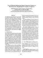
Figure 1 Amylopectin and amylose structure of Wt and sbe1a mutant starch samples by HPSEC analysis. A. Proportions of chains
1
from
debranched
2
b-dextrins during time course of b-amylolysis of amylopectin from Wt (——) and sbe1a mutant (- - -) starch using b-amylase (250
U/mL). B. Chromatograms
1
of isoamylase-debranched and isoamylase-plus-pullulanase-debranched b-limit dextrins
3
from amylose fraction from
Wt and sbe1a mutant starch.
1
Chromatographic regions were divided as in [40]. Proportions of DP ≥ 18, DP 8-17, DP 5-7, DP 4, DP 3 and DP 2 were
calculated as the areas for DP ≥ 17.5, 7.5 ≤ DP ≤ 17.5, 4.5 ≤ DP ≤ 7.5, 3.5 ≤ DP ≤ 4.5, 2.5 ≤ DP ≤ 3.5, and DP ≤ 2.5, respectively, as in [40].
Proportions of chains in each region for B are presented in Additional File 4. Calculation was based on representative chromatograms for starch from
one biological replication. Values are percentage by weight.
2
Debranching was performed successively with isoamylase for 24 h and pullulanase for 24
h.
3
b-Limit dextrin was obtained after 3 times of 24-h b-amylolysis on amylose.
Xia et al. BMC Plant Biology 2011, 11:95
/>Page 3 of 13
characteristic Maltese Cross when viewed in the polar-
ized light microscope; however, sbe1a native starch
showed more heterogeneity in staining as compared to
Wt, as there were more relatively dark -stained granules
in sbe1a than in Wt native starch (24.3% and 8.7%).
Starch Utilization during Kernel Germination
As endosperm starch from the sbe1a mutant has a lower
susceptibility to pancreatic a-amylase, we suspected that
the sbe1a endosperm starch might be l ess readily uti-
lized during kernel germination. To study the effect of
sbe1a on kernel germination, starch utilization and
coleoptile growth during germinat ion of Wt and sbe1a
mutant kernels were examined.
All the kernels from three different ears of both Wt and
sbe1a genotypes were germinated, demonstrating no
differences in germination rate. The coleoptile length of
each genotype was measured daily over 11 days (Figure 4).
The avera ge length of sbe1a coleoptiles was shorter than
Wt from Day 7 onward (Figure 4). For both genotypes the
endosperm starch content decreased over time (Figure 4).
On Days 6, 8, and 11, the starch content was higher in
sbe1a germinating endosperm as compared to Wt, sug-
gestin g less utilization of starch. This trend is consistent
with the reduced growth of sbe1a coleoptiles after Day 6.
Discussion
Starch Molecular Structure
In the present study, rapid degradation of chains DP
≥18 and DP 8-17 were observed for both Wt and sb e1a
samples in the first 10 min of b-amylolysis (Figure 1A).
As b-amylase cannot bypass branch points to hydrolyze
starch chains, a plausible interpretation for the less
extensive degradation of DP 8-17 in sbe1a would be
that the B chains (those chains w ith other chains
attached) [43] would have slightly longer internal seg-
ments and shorter external chains. For the second stage
of b-amylolysis [44], a slow reduction in the amount of
DP 4 chains was observed in Wt samples over the per-
iodof10minto24hbutnotinsbe1a samples (Figure
1A), suggesting differences in the proportion of branch
points that would differentially limit access of the
enzyme to glycosidic linkages [40].
Amylopectin branching pattern models for both sbe1a
and Wt are presented to account for this difference in
b-amylase action on DP 4 stubs (Figure 5A). In the
model for sbe1a, DP 4 stubs would be difficult for b-
amylase to hydrolyze to DP 2 when closely associated
branch points present a steric barrier to binding of b-
amylase. Although most of the DP 4 is from residual A
chains [43], some DP 4 chains f rom residual B chains
would result from short B chains with short internal
segments. The incomplete hydrolysis of DP 4 in sbe1a
suggests that A chains a re preferentially localized near
another b ranch point, leading to 1) hindered hydrolysis
of residual A chains of DP 4 to DP 2 due to steric con-
straint, and 2) more residual B chains with DP 4 due to
incidence of short internal segments (Figure 5A). In the
model for Wt, the DP 4 stubs would be slowly hydro-
lyzed to DP 2, as there is less steric hindrance from
proximal branch points. According to the two models,
sbe1a amylopectin contains a higher proportion of clo-
sely associated branch points than Wt. Furthermore,
based on CL profiles (see Additional File 1 online), the
calculated overall average branching density is similar in
the two amylopectins. Thus, we suggest that the effect
of the sbe1a mutation is to incr ease the local concentra-
tion of branch points but not to influence the overall
amount of branch points in amylopectin.
Figure 2 Time-course of digestio n of the resistant starch assay
for Wt and sbe1a mutant starch. Results shown were from one
biological replication. Curves shown are best fits of analysis of
combined data from two independent digestions.
Table 1 Kinetics of digestion
1
of the resistant starch
assay for Wt and sbe1a mutant starch
2
Starch y
0
(%) S
1
(%) k
1
(min
-1
) S
2
(%) k
2
(min
-1
)
Wt -5.4 ±
2.3
a
85.9 ±
3.5
b
1.4 ± 0.1
a
(×10
-2
)
17.9 ±
5.4
a
0.9 ± 0.2
a
(×10
-3
)
sbe1a 13.7 ±
2.8
b
59.8 ±
3.0
a
1.8 ± 0.1
b
(×10
-2
)
24.3 ±
2.4
a
3.0 ± 1.1
b
(×10
-3
)
1
Kinetic parameters are obtained from model fit using the double
exponential decay equation:
y
=
y
0
+ S
1
e
−k
1
x
+ S
2
e
−k
2
x
where y is % NDS, x is the time, y
0
is the y-value that the model
asymptotically approaches, S
1
and S
2
are the concentrations of the two
different substrate components, and k
1
and k
2
are the reaction rate constants
for the decay of the two different components.
2
Values are expressed as mean ± SD for three biological replications. Values
for each biological replication were obtained from fit of combined data from
two independent digestions. Significant differences (p < 0.05) in the same
column, as determined by one-way ANOVA analysis, are indicated by different
superscripts.
Xia et al. BMC Plant Biology 2011, 11:95
/>Page 4 of 13
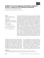
Figure 3 Micrographs of Wt and sbe1a mutant starch samples. A. Scanning electron micrographs of native starch from Wt (left) and sbe1a
mutant (right). Scale bars represent 10 μm at the top of the graphs. B. Scanning electron micrographs of residual starch after 16-h a-amylase
digestion from Wt and sbe1a mutant. Scale bars represent 10 μm, 5 μm, or 1 μm at the top of the graphs. C. Bright field (left) and polarized
light (right) micrographs of native Wt and sbe1a mutant starch. The specimen were stained with 0.04% iodine and viewed within 5 min. Arrows
point to dark stained granules.
Xia et al. BMC Plant Biology 2011, 11:95
/>Page 5 of 13
In the debranched b-LDs from the amylose fraction
(but not in intact amylose), a higher proportion of long
chains of DP ≥ 100 was observed i n sbe1a (Figure 1B
and Additional File 1 online). The higher proportion of
longer chains in b-LDs of amylose from sbe1a can be
expl ained by branch points that tend to be closer to the
non-reducing ends, so that longer internal chains result.
When debranching of b-LDs from amylose w as per-
formed with isoamylase without subsequent pullulanase
digest ion, there were fewer DP 2 than DP 3 chains (Fig-
ure 1B; see Additional F ile 4 online). For b-LD from
amylopectin, all of the DP 3 and some of the DP 2
chains are known to be debranched by isoa mylase [40].
However, our results of b-LD from amylose for both
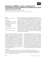
Figure 4 Germination analysis of Wt and sbe1a mutant kernels.
The lengths of the emerged coleoptiles were measured on
successive days during the incubation period
1
. Starch content in the
germinating endosperm was quantified at Day 1, 6, 8, 11, and
percentage of starch content at each day against the dry weight of
Day 1 kernels was plotted
1
.
1
Each data point is mean ± standard
error of measurements of kernels from three biological replications.
As 2 kernels were removed at Day 1, 6, 8, 11 for quantifying starch
content, 15, 13, 11, and 9 kernels from three biological replications
were used for coleoptile measurement Day 1, 2-6, 7-8, 9-11,
respectively. Comparison between two genotypes for each day was
made by one-way ANOVA analysis and a significant difference was
marked by an asterisk (p < 0.05).
Figure 5 Branching pattern models. A. Branching pattern models
for amylopectin from sbe1a and Wt starches. Shown are b-dextrins
approaching the limit of digestion by b-amylase, with differences in
the amount of DP 4 stubs. All circles indicate glucose units. Dotted
line indicates more glucose units. Dotted circles indicate glucose
hydrolyzed by b-amylase. Solid black circles indicate branch points.
Circles with a slash indicate reducing ends. Circles in an ellipse
indicate glucose units that would result in a DP 4 chain. Arrows
indicate the action sites of b-amylase. Arrows with a cross indicates
that action of b-amylase is prevented by closely associated branch
points nearby. Fast and slow indicate the first and second stage of
b-amylolysis, respectively. B. Branching pattern models for a region
of the amylose from sbe1a and Wt starches. Shown are b-limit
dextrins that are consistent with difference in action of isoamylase.
All circles indicate glucose units. Dotted lines indicate more glucose
units. Solid black circles indicate branch points. Circles with a slash
indicate reducing ends. Arrows indicate the action sites of
isoamylase. Arrows with a cross indicates that action of isoamylase
is prevented by closely associated branch points nearby. The model
does not consider the presence of B chains. C. Proposed overall
amylose branching pattern models for sbe1a and Wt starches,
consistent with the differences in actions of b-amylase and
isoamylase. All lines indicate glucose chains. Solid black circles
indicate branch points. Circles with a slash indicate reducing ends.
Xia et al. BMC Plant Biology 2011, 11:95
/>Page 6 of 13
genotypes s uggest that even some DP 3 chains are not
debranched by isoamylase. Comparing sbe1a to Wt,
moreofDP2andDP3chainsarenotdebranchedby
isoamylase in b-LD from sbe1a amylose (Figure 1B). As
the structures escaping isoamylase debranching may
have closely associated branch points and those struc-
tures can be debranched by pullulanase [40], a greater
increase in both DP 2 and DP 3 by subsequent pullula-
nase treatment suggests that a higher proportion o f
these structures are resistant to isoamylase in amylose
from sbe1a. Amylose branching pattern models are pre-
sented in Figure 5B to account for the difference in iso-
amylase action. In the model for sbe1a, A chains are
preferentially attached by branch points close to each
other whereas in Wt, A chains are not, leading to less
hindered isoamylase debranching.
Our data sugge st that amylose of sbe1a mutant starch
has1)longerinternalchainsand2)moreAchains
attached by branch points close to each other. This evi-
dence can be used to create an overall model for amy-
lose branching patterns of sbe1a and Wt (Figure 5C).
The models are drawn taking into account s imilar CL
profiles (see Addition al File 1 online) and as suming that
~50% of amylose molecules are branche d, with ~5-6
branches per molecule [45]. According to the proposed
model, for sbe1a, A chains are closer to each other, and
the location of the chains tends to be more towards
non-reducing end. For Wt, A chains are farther away
from each other, and the location of the chains is more
random and thus more distributed.
Starch Digestion
Kine tic analysis shows that the y
0
value for Wt starch is
effectively zero (Table 1), in agreement with the RS
value for Wt starch (1.6%), and the y
0
and RS values for
sbe1a starch are also in good agreement.
The kinetic model is based on the presence of two
general types of starch substrate: a rapidly-digested sub-
strate ( S
1
), and a slowly-digested substrate (S
2
) [41,42].
The t wo genotypes differ both in the proportio ns of S
1
and S
2
and the reaction rate constants f or these two
components. The S
1
components of Wt and sbe1a
starch were 85.9% and 59.8% respectively. This suggests
that the sbe1a mu tation altered the starch structure and
this resulted in less rapidly-digested component. Consis-
tent with our results, Ao et al . [46] found that increased
branch density l ed to a decreased proportion of RDS
(analogous to our S
1
) and an increa sed proportion of
SDS (analogous to our S
2
).
Starch Granular Structure
Two microscopic techniques, scanning electron micro-
scopy (SEM) and light microscopy (LM), were employed
to obser ve granular structure before and after RS
digestion b y pancreatic a-amylase. Native starch gran-
ules from Wt and sb e1a appear similar in size, shape,
degree of birefringence, and morphology, as described in
a previous report for wx and sbe1a wx granules [47].
Polarized l ight microscopy (see Additional File 5 online)
showsthatalmostallofthedigestedWtgranuleshad
lost their birefringence, while for sbe1a, many digested
granules had maintained some birefringence in the per-
ipheral area of the granules, which indicates that the
center of the digested sbe 1a granules is either gone or
no longer crysta lline enough to show birefringence. The
presence of a hollow interior in the digested sbe1a gran-
ules was confirmed by SEM (Figure 3B), indicating a
relatively greater resistance to digestion for the exterior
portion of the sbe1a granule.
Most of the recovered RS from Wt were represented
by small granule fragments. However, the sbe1a RS
showed variations in morphology, from small fragments
to hollow granules. The difference in digestion of indivi-
dual granules may be due to differences in heterogeneity
in granule structure, as a higher proportion of relatively
dark-stained granules were observed in sbe1a than in
Wt native starch (Figure 3C). SEM revealed the p re-
sence of alternating layers in the Wt residual fragments
(Figure 3 B), which probably reflect the residual growth
rings after digestion.
By observing the sbe1a RS by SEM (Figure 3B) , one
may roughly estimate that, for the recovered granules,
approximately 40% of granule content has escaped
digestion. However the RS value for sbe1a starch is
approximately 13%. Therefore, some of the sbe1a gran-
ules were likely to have been digested completely. The
heterogeneity found among sbe1a granules (Figure 3C)
may account for different degree of digestion of indivi-
dual granules. Thus, it can be reasoned that the micro-
graphs of the sbe1a RS may disproportionately represent
the more resistant granules.
A distinct feature of the recovered sbe1a RS is the
presence of holes on the s urfac e of th e peripheral por-
tion of the granules. These holes are possibly from t he
enlargement of the surface pores in native granules by
a-amylase hydrolysis [48]. The presence of these holes
on the shell is consistent with previous studies demon-
strating that digestion of normal granules starts wit h
surface pores and proceeds through deeper hydrolysis in
channels [49-52], followed by fragmentat ion [48]. In the
current study, the presence of remaining shells with
holes in the sbe1a RS indicates continuing difficulty in
digestion by a-amylase. Neither holes nor shells were
observed in the Wt RS, indicating a more complete
digestion.
As observed under microscopy, the RS from Wt con-
sists m ostly of portions of residual growth rings, while
the RS o f sbe1a is mostly residual peripheral regions.
Xia et al. BMC Plant Biology 2011, 11:95
/>Page 7 of 13
The kinetic anal ysis shows t hat the digestion of sbe1a
starch reached a plateau by 16 h, suggesting the RS
from sbe1a is not further digested. When the RS i s
observed by SEM, one can conclude that some of the
peripheral regions in sbe1a starch granules cannot be
further digested. Enrichme nt of amylose has been found
by some to exist toward the granule peripheral region
[53,54]. SEM s howed that the pe ripheral regions were
more resistant to a-amylase digestion in sbe1a granules.
It is possible that these differences may be preferentia lly
localized in the peripheral region of the granules, where
starch synthesis may be more influenced by deficiency
of SBEI [10]. The CL distribution of residual starch col-
lected after a-amylase digestion showed some small dif-
ferences between Wt and sbe1a (see Additional File 7
online). However, no direct evidence was obtained in
the current study about whether the molecular structure
in the peripheral regions was different in sbe1a.
Starch Utilization during Kernel Germination
An endogenous a-amylase is considered to be responsi-
ble for attac king the starch granule and initiating starch
hyd rolysis in germinating cereal endosperm [55]. Starch
hydrolysis continues by the action of limit dextrinase, a-
amylase, b-amylase, and a-glucosidase to produce mal-
tose and gl ucose for plant utilization [55]. The observed
reductioninstarchhydrolysisduringthelaterstagesof
germination raises the possibility that continued hydro-
lysis of a-amylase-hydrolyzed glucans is hindered in t he
sbe1a mutant. The altered carbon metabolism could
then cause a deficiency in general plant growth charac-
teristicssuchascoleoptilelength [23]. The structural
analysis of sbe1a starch suggests that the decreased
starch utilization of sbe1a seeds is due to an altered
starch branching pattern.
Consideration of SBEI Function in the Context of
Pleiotropic Effects
Differences in SBE activity in sbe mutants could be sim-
ply due to the amount of a remaining SBE isoform or to
biochemical or physical interactions that modulate the
activities of an isoform; for the latter possibility SBEI
may be regulated through complex interactions with
other starch synthetic enzymes. Colleoni et al [21]
showed that two migratory forms of SBEI are missing in
maize endosperm of the maize ae mutant, indicating a
possible interaction of SBEI and SBEIIb. Seo et al. [24]
found that when SBEs were heterologously expressed in
a yeast system, SBEIIa and/or SBEIIb appear to act
before SBEI on synthesizing glucan structure. The s tu-
dies of Yao et al. [25,26] suggest that in the absence of
SBEIIb, a reciprocal inhibition exists between SBEI and
SBEIIa, and that the presence of eith er SBEI or SBEIIa
increases amylopectin branching as opposed to the pre-
sence of SBEI and SBEIIa together.
Direct evidence for protein-protein interactions
between SBEs and different members of all the proteins
involved in starch biosynthesis has also been reported
by several groups, based on co-immunoprecipitation and
affinity purification methods. Tetlow et al. [27] reported
that SBEI from wheat amyloplasts was present in a high
molecular weight complex with starch phosphorylase
and SBEIIb. A separate study [56] using maize amylo-
plasts showed that eliminat ing SBEIIb caused significant
increases in the abundance of SBEI, BEIIa, SSIII, and
starch phosphorylase in the granule, without affecting
SSI or SSIIa. Hennen-Bierwagen [30] reported that SBEI
and SSI were shown to interact in one of three indepen-
dent methods tested; SBEI did not interact with any of
the other proteins in their study (SSIIa, SSIII, SBEIIa,
SBEIIb), and unlike the other five proteins in their
study, SBEI was the only protein to exist as a monomer
in gel permeation chromatography.
In pr esent study, the sbe1a mutant line is nearly iso-
genic w ith the Wt control. Most if not all mutant phe-
notypes a re likely the resul t of many effects, direct and
indirect, on the overall growth, development and phy-
siology of the plant, so it is impossible to truly isolate a
primary effect of the mutation when looking at a whole
plant level phenotype, even the starch structure pheno-
type. Modifying SBE activity may induce modifications
in the distribution of phosphate groups within amy lo-
pectin such as in potato [57,58]. This may alter accessi-
bility of amylase (a or b) to its substrate and may
induce differences in digestibili ty. Nonetheless, there is
value in observing and characterizing the phenotype of
these plants, both at the macro and molecular levels as
we have presented. We have a sister paper [36] which
does inve stigate the effec t of various SBE mutations on
leaf starch which further sheds light on the SBEI func-
tion in the context of pleiotropic consequences.
Evolution and Function of Maize SBEI Isoform in Starch
Biosynthesis
This work for the first time reports a specific and
unique function for SBEI during the life cycle of maize.
Molecular structure analysis suggests an important func-
tion of SBEI in modulating the branching pattern in
normal starch by decreasing local clustering of amylo-
pectin branch points. Thompson [59] emphasized the
non-ran dom nature of the distribution of branch points
in starch. A specific type of non-random branching pat-
ternmayberequiredtooptimizebothstorageand
hydroly sis. It is reasonable to hypothesize that alteration
in the specific non-random branching pattern could lead
to an altered granule organization, rendering it more or
Xia et al. BMC Plant Biology 2011, 11:95
/>Page 8 of 13
less favorable to the plant for storage and/or for enzyme
hydrolysis during utilization. Our data from in vitro
starch digestibility and from plant germination analysis
support this hypothesis.
Gene duplication and neo-functionalization are well
known mechanisms by which specific genes can evolve
to express d ifferent isoforms of enzymes with slightly
specialized expression patterns or different enzymatic
activities [60-62]. With the evidence from current and
previous work, we can infer that an ancestral Sbe gene
has duplicated at least twice during the evolution of
maize, and these evolved t o express three d ifferent SBE
isoforms with highly specific functions in starch bio-
synthesis. A detailed phylogenetic analysis of the
branching enzymes was published by Deschamps et al.
[14]. This work demonstrated that genes belonging to
both the SBEI and SBEII families can be identified in
the green alga, which supports the theory that these two
families of genes evolved approximately a billion y ears
ago based on phylogenetic estimates of the divergence
between the Chlorophyta and Magnolippyta lineages
(estimates range from 729-1210 million years ago)
[63,64]. This example of extreme evolutionary conserva-
tion is strong evidence for a specific and vital role for
each enzyme isoform in starch biosynthesis. While most
plant species studied retain genes representing each sub-
family of SBE, Arabidopsis does not, suggesting that
somewhereinthelineageleadingtoArabidopsis,the
gene was lost with minimal consequences to the species
[65].
The evidence presented in this work strongly supports
the hypothesis that SBEI is required to synthesize endo-
sperm starch granules that allow normal hydrolysis and
utilization during g ermination. Considering plant survi-
val in the wild, optimal seedl ing vigor would be a strong
evo lutionary force to select for genotypes of plants with
starch granules optimized for molecular structure that
would lead to efficient storage and utilization. The
reduced seedling vigor of sbe1a mutant seeds observed
in this work provides powerful evidence for a specialized
and important role of SBEI in plant development, con-
sistent with the evol utionary conservation of SBEI in all
higher plants.
Conclusions
This work for the first time reports that a lack of SBEI
activity resulted in an observable effect, which was seen
on both starch molecular structure and starch function.
Structural and functional analysis of endosperm starch
deficient i n SBEI activity strongly supports the hypoth-
esis that SBEI is required to synthesize starch granules
for normal kernel development, allowing efficient hydro-
lysis and utilization.
Evidence from this work reveals a unique and essential
function of SBEI in normal plant development, consis-
tent with the evolutionary conservation of SBEI in all
higher plants.
The new knowledge generated in this work will con-
tribute to our understanding o f the function and evolu-
tion of the maize SBEs, and o f their roles in the
biosynthesis, hydrolysis and utilization of starch gran-
ules. Moreover, the novel sbe1a starch might have appli-
cation as a food ingredient with nutritional benefit.
Methods
Starch Material
Maize plants of Wt and sbe1a mutant were grown dur-
ing summer, 2007 at Penn State Horticultural Research
Farm (Rock Springs, PA). In order to compare starch
material within a highly similar genetic background,
homozygous Sbe1a/Sbe1a (i.e. Wt) and sbe1a/sbe1a
mutant siblings were identified from a single segregating
population derived from seeds of selfed Sbe1a/sbe1a
plants to obtain ears for endosperm analysis. Genotyp-
ing of Wt and sbe1a mutant plants followed Blauth et
al. [37]. The detected homozygous Wt and sbe1a
mutant plants were self-pollinated to produce ears for
endosperm analysis, and are segregated from a BC
4
F
3
population back crossed by Blauth et al. [11,37]. Starch
extraction from three different ears, considered as three
biological replications, for each genotype, was according
to Yao et al. [66]. Starch fractionation followed Klucinec
and Thompson [67].
b-Amylolysis of Amylopectin and Debranching of b-
Dextrins
b-Dextrins were prepared by the method of Xia and
Thompson [40] with slight modifications in sample
size. Amylopectin samples (48 mg) were dispersed in
480 μL of 90% dimethyl sulfoxide (DMSO) by heating
in a boiling water bath for 10 min. To the dispersion,
warm sodium acetate buffer (3.52 mL, 50°C 0.02M,
pH 6.0) was added. The mixture was heated in a bo il-
ing water bath for 10 min and cooled to 50°C. A 200-
μL aliquot of a b-amylase (from barley, Cat.No. E-
BARBL; Megazyme International Ireland, Ltd.) solu-
tion (250 U/mL, 0.02M sodium acetate, pH 6.0) was
added, and the samples were incubated at 50°C with
constant agitation (200 strokes/min). At approximately
10 min, 30 min, 1 h, 2 h, 6 h, and 24 h, a 0.5-mL ali-
quot of sample was removed and heated in a boiling
water bath for 10 min to stop the reaction. The proce-
dures for precipitating b-dextrins and debranching b-
dextrins by successive action of isoamylase (from
Pseudomonas sp., Cat.No. E-ISAMY; Megazyme) and
pullulanase (from Klebsiella planticola, Cat.No. E-
Xia et al. BMC Plant Biology 2011, 11:95
/>Page 9 of 13
PULKP; Megazyme) were the same as used previously
for b-LDs) [39,40].
Preparation of Isoamylase-Debranched and Isoamylase
plus Pullulanase-Debranched b-Limit Dextrins from
Amylose Fractions
The preparation and debranching of b-L Ds followed the
procedures in Klucinec and Thompson [39] with slight
modifications in sample size. After the b-LDs were deb -
ranched with isoamylase for 24 h, a 30-μL aliquot of the
digested solution was added to 270 μLofDMSOand
reserved for analysis by high-performance size-ex clusion
chromatography (HPSEC). Then the b-LDs were further
debranched with pullulanase for 24 h, afterwards
another 30-μL aliquot of the digested solution was
added to 270 μ L of DMSO for HPSEC analysis [40].
Resistant Starch Determination
The official method for in vitro RS determination
(AOAC 2002.02, AACC 32-40) was employed, which
was scaled-down and modified for direct analysis of the
digestion supernatant for total carbohydrate [41]. The
modification allowed analysis of digestion time-course
for small starch samples (~20 mg). For RS determina-
tion, after the 16 h digestion step at 37°C with porcine
pancreatic a-amylase and amyloglucosidase (enzymes
from RS Assay Kit, Cat.No. K-RSTAR, Megazyme), the
sample tube was removed from the water bath and to
an aliquot of each sample was added 1 volume of 95%
(v/v) ethanol with 0.5% (w/v) EDTA. After centr ifuga-
tion (1,500 × g, 10 min), the supernatant was analyzed
in duplicate for total carbohydrate using t he phenol sul-
furic acid method [68]. The percent non-dige sted starch
(% NDS) was calculated from this data and was the
basis for the calculation of the RS value. Starch isolated
from Wt and sbe1a mutant endosperm es from three
separate plants (triplicate biological replications) were
subjected to triplica te pancreatic a-amylase digestion,
for determining the RS values.
Digestion Time-Course Analysis
For determination of digestion time-course, the starch
samples were digested as described above. An aliquot
was removed at approximately 30 sec, 3 min, 6 min, 15
min, 30 min, 1 h, 2 h, 3 h, 4 h, 5 h, 7 h, 10 h, 13 h, and
twice at 16 h, and added to 1 volume of ethanol/EDTA
solution to ensure immediate deactivation of the
enzymes. After centrifugation the supernatants were
analyzed for total carbohydrate as described above.
Digestion time-course was analyzed following the
method developed by Rees [42] to obtain kinetic data. A
“Double, 5 parameter” regression model in SigmaPlot
(Systat Software, Inc.) was selected to fit the data using
the double exponential decay equation:
y
=
y
0
+ S
1
e
−k
1
x
+ S
2
e
−k
2
x
where y is % NDS, x is the time, y
0
is the y-value that
the model asymptotically approaches, S
1
and S
2
are the
concentrations of the two different substrate compo-
nents, and k
1
and k
2
are the reaction rate constants for
the decay of the two different components. The units
for y
0
, S
1
,andS
2
were % of initial starch, and the un its
for the rate const ants were min
-1
.Afterrunningthe
regression program, the software gives three possible
completion status messages depending on how well the
model fits the data:
(1) Converged, tolerance satisfied.
(2) Converged, tolerance satisfied. Parameter may
not be valid. Arra y numerically singular on fi nal
iteration.
(3) Didn’t c onverge, exceeded maxi mum number of
iterations.
Thedatawerekeptforfurtherregressionanalysisif
message 1 or 2 resulted, an d were discarded if message
3 resulted.
Digestion time-course analysis was performed for
three biological replications per genotype. For each bio-
logical replication, two technical replications were per-
formed. If both sets of data “converged” usin g the
model (message 1 or 2), no further analyses were per-
formed. If message 3 appeared, a new technical replica-
tion was done until the data “converged.” The data from
the two “converged” technical replications for each bio-
logical replication were combined, and the software pro-
gram was run on the combined data. For all samples,
the regression model fit for the combined data com-
pleted with convergence (Message 1), and generated
valid parameters for analysis. Using the combined data,
values for five parameters in the equation were deter-
mined for each biological replication. A mean and s tan-
dard deviation of the five parameters for each genotype
was then calculated, and comparisons among genotypes
were made by one-way ANOVA analysis.
Light Microscopy
Bright field and polarized light microscopy were per-
formed using a light microscope (BX51; Olympus) with
an attached digital camera (Spot II RT; Diagnostic
Instruments). 5 mg of native starch sample was mixed
with 0.5 mL of deionized water in a micro-centrifuge
tube. For the resistant starch samples, the supernatant
was removed after centrifugation of digestion solution
and 20 μL of deionized water was added to the pellets
to disperse the sample. To examine the sample under
the microscope, 20 μL of the dispersed sample was
added to a glass slide, and a cover slip was fi xed over
Xia et al. BMC Plant Biology 2011, 11:95
/>Page 10 of 13
the sample with fingernail polish. Examination of iodine-
stained starch followed the method in Evans and
Thompson [54]. 20 μL of iodine solution (0.08% I
2
,
0.12% KI) was placed onto 20 μL of the dispersed sam-
pletogiveafinalI
2
concentration of 0.04%. In order to
compare birefringence between granules, the camera’ s
automatic expos ure function was turned off, and the
exposure was set the same for all samples. The same
sample field was examined under bright field and p olar-
ized light.
Heterogeneity of iodine staining was evaluated quanti-
tatively by a volunteer panel. Differentially iodine-
stained starch granules were classified into two cate-
gories, dark or light stainedgranules,andweresorted
visually by five individual evaluators who were not
otherwise involved in the research. The evaluators were
trained to understand the difference between dark and
light stained granules, by observing granules in a portion
of a micrograph for sbe1a granules. Four micrographs
for each genotype (Wt or sbe1a) were used for sorting.
The evaluators were then given those eight micrographs,
unlabeled and in randomized order, and asked to sort
the granules into two categories. The proportion of dark
granules for each micrograph was calculated based on
the sorting results from all five evaluators, and a mean
proportion was obtained for each micrograph. For each
genotype, a mean was calculated from the means of the
four micrographs. Comparison between two genotypes
was made by one-way ANOVA.
Scanning Electron Microscopy
A thin layer of starch sample was applied to double-
sided sticky carbon tape on a specimen stub, and sput-
ter-coated wit h 10 nm A u/P d (BAL-TEC SCD 050; US-
TechnoTrade). Samples were then examined using a
scanning electron microscope (JSM-5400; JEOL Ltd.) at
an accelerating voltage of 20 keV and at different mag-
nification levels (1,500 ×, 3,500 ×, an d 10,000 ×). For
image collection, lower magnification was first employed
to examine the whole view of samples, and higher mag-
nification was then used to focus on sample areas that
were representative overall.
Kernel Germination Assay
A kernel germinat ion assay was performed according to
the method in Dinges et al. [23] with slight modifica-
tions. Mature, dried maize kernels were surface-ster i-
lized by immersion in 15 mL of 1% sodium hypochlorite
for 5 min and then washed three times with deionized
water. 15 kernels from each of three ears for each geno-
type w ere placed in Petri dishes containing three layers
of moist Whatman paper and incubated at 30°C in the
dark. The length of each coleoptile was measured by a
ruler on successive days throughout the 11-day
incubation period. To measure the amount of endo-
sperm starch remaining, the roots, coleoptiles, embryo,
and pericarps were removed fro m 2 kernels at days 1, 6,
8, and 11. The remaining endosperm was ground with a
mortar and pestle on ice. The powered tissue was
washed into a tube with deionized water and homoge-
nized with a Tissumizer (Model SDT 1810; Tekmar) at
20,000 rpm for 1 min. The ground tissue was washed
with deionized water, centrifuged at 1500 × g for 10
min, and suspended in 3 mL of deionized water. For cal-
culating the dry weight of samples, 1 mL of this suspen-
sion was dried a t 70°C overnight and weighed. The
remaining 2 mL of the suspension was boiled for 30
min, and the total glucan polysaccharide in the solubi-
lized solution was quantified in triplicates, using a com-
mercial assay kit that measures glucose released after
digest ion with a-amylase and amyloglucosidase (Cat.No.
K-TSTA; Megazyme). The quantified starch content was
normalized against the dry w eight for comparison
between genotypes.
Additional material
Additional file 1: Chromatograms of isoamylase-debranched
amylopectin and amylose fractions from Wt (——) and sbe1a
mutant (- - -) starch.
Additional file 2: Difference plots between sbe1a mutant and Wt
starch for the proportions of chains from debranched b-dextrins
during time course of b-amylolysis of amylopectin. Individual plots
for sbe1a mutant and Wt are presented in Figure 1A.
Additional file 3: Chain length distribution of isoamylase-
debranched and isoamylase-plus-pullulanase-debranched b-limit
dextrins from the amylopectin fraction from Wt and sbe1a mutant
starch.
Additional file 4: Chain length distribution of isoamylase-
debranched and isoamylase-plus-pullulanase-debranched b-limit
dextrins from the amylose fraction from Wt and sbe1a mutant
starch.
Additional file 5: Bright field (left) and polarized light (right)
micrographs of residual starch after 16 h a-amylase digestion from
Wt and sbe1a mutant. Arrows point to residual granules with dark
center.
Additional file 6: Transmission electron micrographs of residual
starch after 16 h a-amylase digestion from Wt (left) and sbe1a
mutant (right). Scale bars represent 5 μm at the top of the graphs.
Additional file 7: Chromatograms of isoamylase-debranched
resistant starch from Wt (——) and sbe1a mutant (- - -) starch
Acknowledgements
We thank Yuan Yao and Jihong Li for their contributions to the breeding of
maize sbe genotype into W64A background; we thank Missy Hazen at
Electron Microscopy Facility at PSU for assistance with microscopy
preparations; we thank fellow graduate students in Department of Food
Science at PSU for their help in evaluating differentially-stained starch
granules.
Author details
1
MARS Petcare US, 315 Cool Springs Boulevard, Franklin, Tennessee 37067,
USA.
2
Department of Biochemistry, Biophysics & Molecular Biology, Iowa
Xia et al. BMC Plant Biology 2011, 11:95
/>Page 11 of 13
State University, Ames, Iowa 50011-3260, USA.
3
Department of Food Science,
The Pennsylvania State University, University Park, Pennsylvania 16802-2504,
USA.
4
Department of Horticulture, The Pennsylvania State University,
University Park, Pennsylvania 16802-5807, USA.
Authors’ contributions
HX conceived of the study, performed the experiments, and drafted the
manuscript. MYN participated in maize genotype breeding and discussion of
experimental design and major results. DBT & MJG advised the conception
of the study, experimental design, result discussion, and revised the
manuscript. All authors read and approved the final manuscript.
Received: 10 December 2010 Accepted: 21 May 2011
Published: 21 May 2011
References
1. Buléon A, Ball S, Planchot V, Colonna P: Starch granules: structure and
biosynthesis. Int J Biol Macromol 1998, 23:85-112.
2. Gidley MJ: Starch structure/function relationships: achievements and
challenges. In Starch: advances in structure and function. Edited by: Tina, et
al. Royal Society of Chemistry (Great Britain). Food Chemistry Group;
2001:1-7.
3. Manners DJ: Recent developments in our understanding of amylopectin
structure. Carbohydr Polym 1989, 11:87-112.
4. Boyer CD, Preiss J: Multiple forms of (1,4)-α-D-glucan-6-glucosyl transferase
from developing Zea Mays L. kernels. Carbohydr Res 1978, 61:321-34.
5. Boyer CD, Preiss J: Evidence for independent genetic control of the
multiple forms of maize endosperm branching enzymes and starch
synthases. Plant Physiol 1981, 67:1141-5.
6. Gao M, Fisher DK, Kim KN, Shannon JC, Guiltinan MJ: Independent genetic
control of maize starch-branching enzymes IIa and IIb-Isolation and
characterization of a Sbe2a cDNA. Plant Physiol 1997, 114:69-78.
7. Kim KN, Fisher DK, Gao M, Guiltinan MJ: Genomic organization and
promoter activity of the maize Starch branching enzyme I gene. Gene
1998, 216:233-43.
8. Kim KN, Gao M, Fisher DK, Guiltinan MJ: Molecular cloning and
characterization of the Amylose-Extender gene encoding starch
branching enzyme IIB in maize. Plant Mol Biol 1999, 38:945-56.
9. Burton RA, Bewley JD, Smith AM, Bhattacharyya MK, Tatge H, Ring S, Bull V,
Hamilton WDO, Martin C: Starch branching enzymes belonging to
distinct enzyme families are differentially expressed during pea embryo
development. Plant J 1995, 7:3-15.
10. Gao M, Fisher DK, Kim KN, Shannon JC, Guiltinan MJ: Evolutionary
conservation and expression patterns of maize starch branching enzyme
I and IIb genes suggests isoform specialization. Plant Mol Biol 1996,
30:1223-32.
11. Blauth SL, Yao Y, Klucinec JD, Shannon JC, Thompson DB, Guiltinan MJ:
Identification of Mutator insertional mutants of starch-branching
enzyme 2a in corn. Plant Physiol 2001, 125:1396-405.
12. Rahman S, Regina A, Li Z, Mukai Y, Yamamoto M, Kosar-Hashemi B,
Abrahams S, Morell MK: Comparison of starch-branching enzyme genes
reveals evolutionary relationships among isoforms. Characterization of a
gene for starch-branching enzyme IIa from wheat D genome donor
Aegilops tauschii. Plant Physiol 2001, 125:1314-24.
13. Xu JH, Messing J: Organization of the prolamin gene family provides
insight into the evolution of the maize genome and gene duplication in
grass species. Proc
Natl Acad Sci 2008, 105:14330-35.
14. Deschamps P, Moreau H, Worden AZ, Dauvillée D, Ball SG: Early Gene
Duplication Within Chloroplastida and Its Correspondence With
Relocation of Starch Metabolism to Chloroplasts. Genetics 2008,
178:2373-87.
15. Guan HP, Preiss J: Differentiation of the properties of the branching
isozymes from maize (Zea mays). Plant Physiol 1993, 102:1269-73.
16. Takeda Y, Guan HP, Preiss J: Branching of amylose by the branching
isoenzymes of maize endosperm. Carbohydr Res 1993, 240:253-63.
17. Boyer CD, Preiss J: Properties of citrate-stimulated starch synthesis
catalyzed by starch synthase I of developing maize kernels. Plant Physiol
1979, 64:1039-42.
18. Gao M, Wanat J, Stinard PS, James MG, Myers AM: Characterization of dull
1, a maize gene coding for a novel starch synthase. Plant Cell 1998,
10:339-412.
19. Beatty MK, Rahman A, Cao H, Woodman W, Lee M, Myers AM, James MG:
Purification and molecular genetic characterization of ZPU1, a
pullulanase-type starch-debranching enzyme from maize. Plant Physiol
1999, 119:255-66.
20. Nishi A, Nakamura Y, Tanaka N, Satoh H: Biochemical and genetic effects
of amylose-extender mutation in rice endosperm. Plant Physiol 2001,
127:459-72.
21. Colleoni C, Myers AM, James MG: One- and two-dimensional native PAGE
activity gel analyses of maize endosperm proteins reveal functional
interactions between specific starch metabolizing enzymes. J Appl
Glycosci 2003, 50:207-12.
22. Dinges JR, Colleoni C, Myers AM, James MG: Molecular structure of three
mutations at the maize sugary1 locus and their allele-specific
phenotypic effects. Plant Physiol 2001, 125:1406-18.
23. Dinges JR, Colleoni C, James MG, Myers AM: Mutational analysis of the
pullulanase-type debranching enzyme of maize indicates multiple
functions in starch metabolism. Plant Cell 2003, 15:666-80.
24. Seo BS, Kim S, Scott MP, Singletary GW, Wong KS, James MG, Myers AM:
Functional interactions between heterologously expressed starch-
branching enzymes of maize and the glycogen synthases of Brewer’s
yeast. Plant Physiol 2002, 128:1189-99.
25. Yao Y, Thompson DB, Guiltinan MJ: Starch biosynthesis in maize
endosperm: in the absence of SBEIIb, the deficiency of SBEIIa leads to
increased amylopectin branching. Presentation at 2003 AACC conference,
Portland, OR .
26. Yao Y, Thompson DB, Guiltinan MJ: Maize starch branching enzyme (SBE)
isoforms and amylopectin structure: in the absence of SBEIIb, the future
absence of SBEIa leads to increased branching. Plant Physiol
2004,
106:293-316.
27.
Tetlow IJ, Wait R, Lu Z, Akkasaeng R, Bowsher CG, Esposito S, Kosar-
Hashemi B, Morell MK, Emes MJ: Protein phosphorylation in amyloplasts
regulates starch branching enzyme activity and protein-protein
interactions. Plant Cell 2004, 16:694-708.
28. Tetlow IJ, Beisel KG, Cameron S, Makhmoudova A, Liu F, Bresolin NS, Wait R,
Morell MK, Emes MJ: Analysis of protein complexes in wheat amyloplasts
reveals functional interactions among starch biosynthetic enzymes. Plant
Physiol 2008, 146:1878-91.
29. Deschamps P, Colleoni C, Nakamura Y, Suzuki E, Putaux JL, Buleon A,
Haebel S, Ritte G, Steup M, Falcon L, Moreira D, Loffelhardt W, Nirmal RJ,
Plancke C, d’Hulst C, Dauvillee D, Ball S: Metabolic symbiosis and the birth
of the plant kingdom. Mol Biol Evol 2008, 25:536-48.
30. Hennen-Bierwagen TA, Liu F, Marsh R, Kim S, Gan Q, Tetlow IJ, Emes MJ,
James MG, Myers AM: Starch biosynthetic enzymes from developing Zea
mays endosperm associate in multisubunit complexes. Plant Physiol 2008,
146:1892-908.
31. Hennen-Bierwagen TA, Lin Q, Grimaud F, Planchot V, Keeling PL, James MG,
Myers AM: Proteins from multiple metabolic pathways associate with
starch biosynthetic enzymes in high molecular weight complexes: a
model for regulation of carbon allocation in maize amyloplasts. Plant
Physiol 2009, 149:1541-59.
32. Kötting O, Kossmann J, Zeeman SC, Lloyd JR: Regulation of starch
metabolism: the age of enlightenment? Current Opinion in Plant Biology
2010, 13:321-9.
33. Liu F, Makhmoudova A, Lee EA, Wait R, Emes MJ, Tetlow IJ: The amylose
extender mutant of maize conditions novel protein-protein interactions
between starch biosynthetic enzymes in amyloplasts. J Exp Bot 2009,
60:4423-40.
34. Garwood DL, Shannon JC, Creech RG: Starches of endosperms possessing
different alleles at the amylose-extender locus in Zea mays L. Cereal Chem
1976, 53:355-64.
35. Boyer CD, Daniels RR, Shannon JC: Starch granule (amyloplast)
development in endosperm of several Zea mays L. genotypes affecting
kernel polysaccharides. Amer J Bot 1977, 64:50-56.
36. Yandeau-Nelson MD, Laurens L, Shi Z, Xia H, Smith AM, Guiltinan MJ:
Starch Branching Enzyme IIa is required for proper diurnal cycling of
starch in leaves of Zea mays. Plant Physiology Online Open Access 2011
[ />pdf+html].
37. Blauth SL, Kim K, Klucinec JD, Shannon JC, Thompson DB, Guiltinan MJ:
Identification of Mutator insertional mutants of starch branching
enzyme 1 (sbe1) in Zea mays L. Plan Mol Bio 2002, 48:287-97.
Xia et al. BMC Plant Biology 2011, 11:95
/>Page 12 of 13
38. Xia H: Structure and function of endosperm starch from maize mutants
deficient in one or more starch-branching enzyme isoform activities. Ph.
D. thesis The Pennsylvania State University, University Park, PA; 2009.
39. Klucinec JD, Thompson DB: Note: Structure of amylopectins from ae-
containing maize starches. Cereal Chem 2002, 79:19-23.
40. Xia H, Thompson DB: Debranching of β-limit dextrins with isoamylase or
pullulanse to explore the Branching Pattern of Amylopectins from Three
Maize Genotypes. Cereal Chem 2006, 83:668-76.
41. Evans A, Thompson DB: Enzyme susceptibility of high-amylose starch
precipitated from sodium hydroxide dispersions. Cereal Chem 2008,
85:480-7.
42. Rees E: Effect of a heat-moisture treatment on alpha-amylase
susceptibility of high amylose maize starches. MS thesis The Pennsylvania
State University, University Park, PA; 2008.
43. Hizukuri S: Polymodal distribution of the chain lengths of amylopectins,
and its significance. Carbohydr Res 1986, 147:342-7.
44. Lee EYC: The action of sweet potato β-amylase on glycogen and
amylopectin: formation of a novel limit dextrin. Arch Biochem Biophys
1971, 146:488-92.
45. Takeda Y, Hizukuri S, Takeda C, Suzuki A: Structures of branched
molecules of amyloses of various origins, and molar fractions of
branched and unbranched molecules. Carbohydr Res 1987, 165:139-45.
46. Ao Z, Simsek S, Zhang G, Venkatachalam M, Reuhs BL, Hamaker BR: Starch
with slow digestion property produced by altering its chain-length,
branch density and crystalline structure. J Agri Food Chem 2007,
55:4540-7.
47. Li JH, Thompson DB, Guiltinan M: Mutation of the maize sbe1a and ae
genes alters morphology and physical behavior of wx-type endosperm
starch granules. Carbohydr Res 2007, 342:2619-27.
48. Zhang G, Ao Z, Hamaker BR: Slow digestion property of native cereal
starches. Biomacromolecules 2006, 7:3252-8.
49. Leach HW, Schoch TJ: Structure of the starch granule. II. Action of various
amylase on granular starches. Cereal Chem 1961, 38:34-46.
50. Valetudie JC, Colonna P, Bouchet B, Gallant DJ: Hydrolysis of tropical tuber
starches by bacterial and pancreatic α-amylases. Starch/Stärke
1993,
45:270-6.
51. Helbert W, Schulein M, Henrissat B: Electron microscopic investigation of
the diffusion of bacillus licheniformis α-amylase into corn starch
granules. Int J Biol Macromol 1996, 19:165-9.
52. Gallant DJ, Bouchet B, Buleon A, Perez S: Physical characteristics of starch
granules and susceptibility to enzymatic degradation. Eur J Clin Nutr
1992, 46:S3-S16.
53. Planchot V, Colonna P, Gallant DJ, Bouchet B: Extensive degradation of
native starch granules by alpha-amylase from Apergillus fumigatus. J
Cereal Sci 1995, 21:163-71.
54. Evans A, Thompson DB: Resistance to α-Amylase digestion in four native
high-amylose maize starches. Cereal Chem 2004, 81:31-7.
55. Smith AM, Zeeman SC, Smith SM: Starch Degradation. Annu Rev Plant Biol
2005, 56:73-98.
56. Grimaud F, Rogniaux H, James MG, Myers AM, Planchot V: Proteome and
phosphoproteome analysis of starch granule-associated proteins from
normal maize and mutants affected in starch biosynthesis. J Exp Bot
2008, 59:3395-406.
57. Blennow A, Hansen M, Schulz A, Jørgensen K, Donald AM, Sanderson J: The
molecular deposition of transgenically modified starch in the starch
granule as imaged by functional microscopy. J Struct Biol 2003,
143:229-41.
58. Blennow A, Wischmann B, Houborg K, Ahmt T, Jørgensen K, Engelsen SB,
Bandsholm O, Poulsen P: Structure function relationships of transgenic
starches with engineered phosphate substitution and starch branching.
Int J Biol Macromol 2005, 36:159-68.
59. Thompson DB: On the non-random nature of amylopectin branching.
Carbohydr Polym 2000, 40:223-39.
60. Gingerich DJ, Hanada K, Shiu SH, Vierstra RD: Large-scale, lineage-specific
expansion of a bric-a-brac/tramtrack/broad complex ubiquitin-ligase
gene family in rice. Plant Cell 2007, 19:2329-48.
61. Prokhnevsky AI, Peremyslov VV, Dolja VV: Overlapping functions of the
four class XI myosins in Arabidopsis growth, root hair elongation, and
organelle motility. Proc Natl Acad Sci 2008, 105:19744-9.
62. Saleh A, Alvarez-Venegasm R, Yilmaz M, Le O, Hou G, Sadder M, Al-
Abdallat A, Xia Y, Lu G, Ladunga I, Avramova Z: The Highly Similar
Arabidopsis homologs of trithorax ATX1 and ATX2 encode proteins with
divergent biochemical functions. Plant Cell 2008, 20:568-79.
63. Hedges SB, Dudley J, Kumar S: TimeTree: A public knowledge-base of
divergence times among organisms.
Bioinformatics 2006, 22:2971-2.
64. Chaw SM, Chang CC, Chen HL, Li WH: Dating the monocot-dicot
divergence and the origin of core eudicots using whole chloroplast
genomes. J Mol Evol 2004, 58:424-41.
65. Dumeza S, Wattebleda F, Dauvilleea D, Delvallea D, Planchotb V, Ball SG,
D’Hulsta C: Mutants of Arabidopsis Lacking Starch Branching Enzyme II
Substitute Plastidial Starch Synthesis by Cytoplasmic Maltose
Accumulation. Plant Cell 2006, 18:2694-709.
66. Yao Y, Guiltinan MJ, Shannon JC, Thompson DB: Single kernel sampling
method for maize starch analysis while maintaining kernel vitality. Cereal
Chem 2002, 79:757-762.
67. Klucinec JD, Thompson DB: Fractionation of high amylose maize starches
by differential alcohol precipitation and chromatograph of the fractions.
Cereal Chem 1998, 75:887-96.
68. Dubois M, Gilles KA, Hamilton JK, Rebers PA, Smith F: Colorimetric method
for determination of sugars and related substances. Anal Chem 1956,
28:350-6.
doi:10.1186/1471-2229-11-95
Cite this article as: Xia et al.: Deficiency of maize starch-branching
enzyme i results in altered starch fine structure, decreased digestibility
and reduced coleoptile growth during germination. BMC Plant Biology
2011 11:95.
Submit your next manuscript to BioMed Central
and take full advantage of:
• Convenient online submission
• Thorough peer review
• No space constraints or color figure charges
• Immediate publication on acceptance
• Inclusion in PubMed, CAS, Scopus and Google Scholar
• Research which is freely available for redistribution
Submit your manuscript at
www.biomedcentral.com/submit
Xia et al. BMC Plant Biology 2011, 11:95
/>Page 13 of 13