báo cáo khoa học: "Brachypodium distachyon: a new pathosystem to study Fusarium head blight and other Fusarium diseases of wheat" pdf
Bạn đang xem bản rút gọn của tài liệu. Xem và tải ngay bản đầy đủ của tài liệu tại đây (3.74 MB, 14 trang )
RESEARC H ARTIC L E Open Access
Brachypodium distachyon: a new pathosystem to
study Fusarium head blight and other Fusarium
diseases of wheat
Antoine Peraldi
1*
, Giovanni Beccari
2
, Andrew Steed
1
and Paul Nicholson
1
Abstract
Background: Fusarium species cause Fusarium head blight (FHB) and other important diseases of cereals. The
causal agents produce trichothecene mycotoxins such as deoxynivalenol (DON). The dicotyledonous model species
Arabidopsis thaliana has been used to study Fusarium-host interactions but it is not ideal for mode l-to-crop
translation. Brachypodium distachyon (Bd) has been proposed as a new monocotyledonous model species for
functional genomic studies in grass species. This study aims to assess the interacti on between the most prevalent
FHB-causing Fusarium species and Bd in order to develop and exploit Bd as a genetic model for FHB and other
Fusarium diseases of wheat.
Results: The ability of Fusarium graminearum and Fusarium culmorum to infect a range of Bd tissues was examined
in various bioassays which showed that both species can infect all Bd tissues examined, including intact foliar
tissues. DON accumulated in infected spike tissues at levels similar to those of infected wheat spikes. Histological
studies revealed details of infection, colonisation and host response and indicate that hair cells are important sites
of infection. Susceptibility to Fusarium and DON was assessed in two Bd ecotypes and revealed variation in
resistance between ecotypes.
Conclusions: Bd exhibits characteristics of susceptibility highly similar to those of wheat, including susceptibility to
spread of disease in the spikelets. Bd is the first reported plant species to allow successful infection on intact foliar
tissues by FHB-causing Fusarium species. DON appears to function as a virulence factor in Bd as it does in wheat.
Bd is proposed as a valuable model for undertaking studies of Fusarium head blight and other Fusarium diseases
of wheat.
Keywords: Fusarium, Brachypodium distachyon, wheat, deoxynivalenol, model-to-crop translation, disease resistance,
host-pathogen interaction
Background
Several Fusarium species are globally important patho-
gens of wheat (Triticum aestivum). These fungi infect
floral tissues as well as seedlings, stem bases and roots
causing Fusarium head blight (FHB), seedling blight,
crown rot and root rot, respectively [1,2]. Of these, FHB
is the one of greatest significance worldwide being one
of the most destructive diseases of wheat, with economic
and health impacts [3,4]. The predominant Fusarium
speci es associated with FHB are Fusarium graminearum
(Fg) (teleomorph: Gibberella zeae)andFusarium
culmorum (Fc) which are also the most economically
relevant [5,3].
FHB is of primary concern because Fg and Fc produce
a number of secondary metabolites within infected grain
that are toxic to human and animal consumers. The
most prevalent Fusarium mycotoxins in wheat are tri-
chothecenes such as deoxynivalenol (DON) and niva le-
nol (NIV) [6]. Experiments using mutants of Fg unable
to produce DON showed that this mycotoxin functions
as a virulence factor in wheat, enhancing spread of the
disease within heads but in contrast plays no discernable
* Correspondence:
1
Department of Disease and Stress Biology, John Innes Centre, Colney Lane,
Norwich, NR4 7UH, UK
Full list of author information is available at the end of the article
Peraldi et al. BMC Plant Biology 2011, 11:100
/>© 2011 Peraldi et al; licensee BioMed Central Ltd. This is an Open Access art icle distr ibuted unde r the terms of the Creativ e Commons
Attribution License ( which permits unre stricted use, distribution, and reproduction in
any medium, provided the original work is properly cited.
role in barley [7]. Studies on trichothecene toxicity indi-
cate that DON inhibits protein synthesis by binding to
the 60S ribosomal subunit, activating a cel lular signal-
ling pathway resulting in a form of programmed cell
death [8,9]. The phytotoxic effects of DON in wheat
are chlorosis, necrosis and wilting, often leading to
thebleachingofthewholeheadabovetheinoculation
point [10].
The use of resistant wheat cultivars is considered to
be the most effective strategy to prevent FHB epidemics
and contamination of grain with trichothecenes [11].
FHB resistance in wheat has been broadly classifie d into
two different types: resistance to initial penetration (type
I) and resistance to pathogen spread within the head
(type II) [12]. However, other types of resistance have
also been propose d; resistance to kernel infection (type
III), tolerance against FHB and trichothecenes (type IV)
[13] and tolerance to trichothecene accumulation (type
V) by two m eans: chemical modification of trichothe-
cenes (type V-1) and inhibition of trichothecene synth-
esis (type V-2) [14]. Over a hundred quantitative trait
loci (QTL) f or FHB resistance in wheat have been reli-
ably identified [11], but to date, only four loci have been
shown to exhibit Mendelian inheritance [15-18]. Fhb1,
derived from the resistant Chinese cultivar ‘Sumai-3’ is
the only locus for which a molecular mechanism has
been proposed. Wheat lines containing this QTL are
able to convert DON into less phytotoxic DON-3-O-gly-
coside (type V-1) indicating that Fhb1 is either encoding
a DON-glycosyltransferase or a modulator of the expres-
sion or activity of such an enzyme [10].
Wheat is not readily amenable for undertaking genetic
studies of complex traits because of its large allohexa-
ploid genome (three ancestral genomes totalling about
17,000 Mbp) which greatly hinders th e complete genetic
characterization of FHB-resistance QTLs. Because of the
inherent difficulties associated with wheat, a number of
alternative hosts have been proposed as models with
which to investigate host-pat hogen interactions in FHB.
Although its genome is not yet fully sequenced, barley
(H ordeum vulgare ) presents the advantage of h aving a
diploid genome, whilst also being a m onocotyledonous
plant naturally infected by Fu sarium spp. However, bar-
ley has an inherent FHB-type II resistance [3] which can
be a hindrance for studying the mechanisms underlying
FHB-resistance in wheat. Rice (Oryza sativa)wasthe
first monocotyledonous plant to have its genome
sequenced and is a natural host for Fusarium spp. How-
ever, certain characteristics of rice and its interaction
with Fusarium fungi reduce its potential for modelling
FHB of wheat: rice is a tropical plant adapted to an
aquatic environment at an early stage of development
and is predominantly infected by Fusarium spp. other
than those that cause FHB of wheat [19].
Several researchers have used the best studied plant
model available, Arabidopsis thaliana, because i t is ide-
ally suited to laboratory studies and there are extensive
gene tic and genomic resources available [20]. Floral and
foliar bioassays have been reported for studies of the
interaction between Fg and Fc with Arabidopsis [21,22].
Such assays have demonstrated that NPR1 and EDS11
contribute to resistance of Arabidopsis against Fc [23]
and that over-expression of the GLK transcriptional
activator confers resistanc e to Fg [24]. However, to date,
translation of findings on the genetic mechanisms
involved in host resistance to Fusarium infection from
Arabidopsis to cereal crops is scarce. One example is
Chen et al. [25]whodemonstratedthatFgexploitsthe
ethylene (ET) signalling pathway to colonise Arabidopsis
and showed that ET signalling also contributes to sus-
ceptibility of wheat to FHB. Despite the numerous
advantages of using Arabidopsis as a model for FHB, it
is not a natural host of Fusarium, and it displays differ-
ent floral symptoms to those that occur on wheat [21].
Consequently, the identification of a model, genetically
tractable, monocot system that is more closely related to
wheat is highly desirable.
Brachypodium distachyon (Bd) is a temperate mono-
cotyledonous plant of the grass family which has been
proposed as a new model species for functional geno-
mics in grasses [26]. The inbred line Bd21 has been
recently sequenced to an 8 fold coverage [27]. Several
aspects of Bd make it a very attractive model for tempe-
rate small grain cereals, including wheat. Bd has one of
the smallest genomes found in grasses [28] comprising 5
chromosomes spanning over 272 Mbp in which about
25,000 protein-coding sequences are predicted [27]. Bd
diverged just prior to the clade of the ‘core pooid’ gen-
era that contain the majority of the temperate cereals,
including wheat, making it potentially useful for func-
tional genomics [26]. There is extensive chromosomal
synteny between Bd and other cereals with the strongest
syntenic relationship being with wheat for which about
77% of Bd genes have strong Triticeae EST matches
[28]. In addition, it is possible to obtain genetic/physical
locations in the wheat genome directly using Bd mar-
kers as demonstrated in the fine mapping of the com-
plex Ph1 locus region in wheat [29]. A further
advantageofBdisthatitisaself-fertile,inbreeding
annual with a rapid life cycle of around 8-10 weeks [26]
depending on the environmental growth conditions. In
addition, this species is small in size (approximately 30
cm at maturity) and has undemanding growth require-
ments. Furthermore, resources are being developed to
permit functional genetic studies to be undertaken in
Bd. Several mutant collections exist including EMS and
T-DNA insertional mutants [.
usda.gov, BrachyTAG.org, 30], as well as a segregating
Peraldi et al. BMC Plant Biology 2011, 11:100
/>Page 2 of 14
population using Bd21 and Bd3-1 as parental lines
[].
The current study aims to examine the potential of Bd
asamodeltostudyinteractionswithFusarium species
and a base from which to undertake model to crop
translational investigations.
Results
Floral infection
FHB is the disease of greatest significance in whea t and,
if Bd is to be useful as a model, it is imperative that it
expresses symptoms simila r to those of wheat. Spikes of
Bd were spray inoculated to assess the susceptibility of
Bd to Fg and Fc and to compare symptoms to those of
FHB on wheat (Figure 1a,b). Optimum infection was
achieved by placing plants into 8 h darkness immedi-
ately following inoculation (plants were inoculated at
the start of the dark period). Similar to the situation for
FHB of wheat, Bd spikes appeared to be most suscepti-
ble to infection by Fusarium spp at the period around
mid-anthesis [4,31]. Symptom development was mark-
edly restricted when Bd spikes were inoculated either
prior to or after mid-anthesis.
Mycelial growth was detectable on the host surface
from between 12 and 36 hpi and light brown, water-
soaked lesions appeared proximally on the outer surface
of the lemma, between 24 and 48 hpi (results not
shown). From 48 h to 96 h, florets lost their green
appearance and became bleached in a manner highly
reminiscent of the bleaching symptoms exhibited by
wheat heads with FHB (compare Figure 1a,b with Figure
1c,d). Following s pray inoculation, whole spikelets
became bleached and, between 96 and 144 hpi, necrotic
symptoms spread down the rachis and into neighbour-
ing spikelets above and below (Figure 1d). Disease con-
tinued to develop and between 7 and 14 days post
inoculation (dpi), whole spikes became bleached and
necrosis spread down into the peduncule (Figure 1e). If
humidity was not maintained following inoculation,
infection was reduced or even unsuccessful, leading to
the total arrest of symptom development after 24 to 48
hours post inoculation (hpi), (results not shown) . In
contrast, maintaining high humidity for longer than 48
hpi resulted in the extensive growth of a erial mycelium
which often covered the whole spike (Figure 1h).
Although floral symptoms on Bd21 and Bd3-1 were
similar following spray inoculation with either Fg or Fc
(data not shown) disease generally developed more
rapidly on Bd3-1 than on Bd21, particularly following
inoculation with the Fg isolates.
Point inoculation was carried out to det ermine
whether, like wheat, Bd exhibits susceptibility to spread
within the spikelet (type II susceptibility sensu Schroeder
and Christensen [12]). Fo llowing point inoculation,
bleaching of the floral tissues tended to spread from the
inoculation site towards the upper end of the spikelet
with less pronounced disease progression below the
point of inoculation (Figure 1f (2 dpi), 1 g (4 dpi)). Con-
tamination of wheat grain with DON is the most impor-
tant aspect of FHB with respect to food safety. The
ability of Fg to produce DON within Bd tissues was
investigated following spray inoculation of Bd21 spikes
with Fg. Very large amounts of DON were detected in
infected spikes with concentrations up to 1815 mg/kg of
fresh tissue when conditions were highly conducive to
infection and fungal growth (Figure 1h).
Detached Bd21 florets inoculated with Fg were studied
3dpi under a light microscope to investigate the early
phase of infection in regards to pathogen penetration
and early host response. Adaxial (lemma) and abaxial
(palea) foliar tissues were dissected and observed indivi-
dually. Extensive hyphal growth a nd branching was
observed on the external surface of the lemma, anchor-
ing and branching on voluminous macro-hairs (Figure
1i, arrows). Closer observation suggested that hyphae
coiled around the base of macro-hairs (Figure 1j, arrow)
and formed globose structures (Figure 1k, arrow) the
presence of which was correlated with an amber-brown
discolouration of the host tissue. At early stages of inter-
action, hyphae formed aggregated structures around the
base of macro-hairs (BMH) with little or no discoloura-
tion of the host tissues (Figure 1m). However, at late
stages of interaction, extensive hyphal growth around
the BMH was correlated with intense discolouration and
collapse of the host tissues (Figure 1n). Similar observa-
tions were made on the external surface of the palea
where globose hyphal structures were associated with
BMH and nearby cells of corrugated circular shape (Fig-
ure 1o,p) and strong amber-brown discolouration.
Macro-hairs are absent from the internal surface of the
palea. However, amber-brown discolouration and cell
death was observed among these corrugated circular
cells which we interpret to be developmentally arrested
hair primordia (Figure 1l).
Foliar infection
Spray inoculation of whole Bd21 plants was first per-
formed to identify tissues compatible with Fusarium
infection. Brown, water soaked necrotic lesions devel-
oped between 48 and 72hpi on leaves (Figure 2a) fol-
lowed at later stages by a surrounding chlorotic area
(Figure 2b). Deta ched leaf assays were also performed to
study symptom development on both intact and
wounded foliar tissues inoculated with Fg or Fc. Follow-
ing wound i noculation, dark-brown , water-soaked
necrotic lesions appeared initially at the wound site
between 24 and 48 hpi and extended primarily along
the vascular bundles towards both the leaf tip and base
Peraldi et al. BMC Plant Biology 2011, 11:100
/>Page 3 of 14
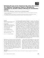
Figure 1 Fusarium head blight symptoms and penetration sites on Bd spikes. a) Typical early FHB symptoms on point inoculate d wheat
spike. b) Typical late FHB symptoms on point inoculated wheat spike displaying bleaching. c - e) FgUK1 spray inoculation symptoms: 3, 7 and
14 dpi, respectively. f & g) FgUK1 point inoculation, same spike 2 and 4 dpi, respectively. h) FgUK1 symptoms following spray inoculation with
maintained high humidity. Scale bars a-h = 1 cm. i-p) Light microscope images of detached Bd21 florets, 3dpi with Fg, cleared and stained with
aniline blue. i) External surface of lemma showing hyphal contact on macro-hairs (arrows). j & k) are close ups of picture i) taken at different
focal planes. j) shows hyphal strands enveloping the macro-hair and k) shows a globose fungal structure formed at the base of the macro-hair
(bmh). l) Internal surface of the palea showing hyphal colonization, necrosis and accumulation of phenolic compounds in corrugated circular
cells (arrow). m & n) Macro-hair base of lemma at early stage of fungal colonization showing aggregated hyphal structure, n) Macro-hair base of
lemma at late stage of fungal colonization showing extensive hyphal strands enveloping the base of the macro-hair, intense phenolic
compound accumulation and collapse of the macro-hair. o-p) External surface of the palea showing the base of a macro-hair and neighbouring
corrugated circular cell (arrow head) accumulating phenolic compounds (o) in response to hyphal contact (p), Upper arrow points at globose
structure located above the corrugated circular cell and lower arrow pointing at hyphal strands in contact with the base of the macro-hair. Scale
bars i-p = 20 μm.
Peraldi et al. BMC Plant Biology 2011, 11:100
/>Page 4 of 14
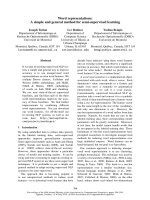
Figure 2 Fusarium symptoms and penetration sites on Bd21 f oliar tissue. a & b) FgUK1symptoms on Bd21 leaves after whole plant spray.
Scale bars: k = 0.5 cm, m = 1 cm: early and late symptoms, respectively. c & d) Fg symptoms on intact Bd21 detached leaf: c & e) 96hpi, and d)
144hpi. Scale bars: c & d = 0.25 cm, e = 250 μm. f) SEM image of Bd21 intact leaf surface showing Bd epidermis cell types (bc: bulliform cell,
mh: macro-hair, bmh: base of macro-hair, g: girder, p: prickle cell, hp: hooked prickle, s: stomata). Scale bar = 50 μm. g and h) Light microscope
images of chlorophyll cleared Bd21 leaves infected with Fg UK1, 120 hpi stained with trypan blue. Scale bars g & h = 50 μm. i) Fluorescent
microscope image of Bd21 foliar macro-hair base 96hpi with GFP1-Fc. Arrow head shows macro hair endogenous fluorescence. Arrows show
GFP1-Fc fluorescent hyphae forming globose structures at the bmh. Scale bar = 50 μm. j) Confocal laser scanning microscope (CLSM) image of
GFP1-Fc infection on intact Bd21 detached leaf, 72 hpi, showing chlorophyll-less cells above the vascular bundles and GFP1-Fc hyphae in the
cell directly beneath the bmh (bmh not in focal plane). Scale bar = 20 μm. k & l) SEM images of intact Bd21 leaf infection with FgS1, 48hpi. k)
Fg hyphae enveloping a prickle cell. Scale bar = 20 μm. l) Fg hyphae aggregating near the bmh, penetrating (arrow) and growing underneath
the cuticule. Scale bar = 10 μm.
Peraldi et al. BMC Plant Biology 2011, 11:100
/>Page 5 of 14
(Additional file 1). Following inoculation of intact Bd
foliar tissues, very small necrotic spots appeared on the
leaf beneath the inoculum droplet (Figure 2c,e) followed
by the appearance of more widespread necrosis. Chloro-
tic areas subsequently developed around these lesions
(Figure 2d). Symptoms developed in a similar manner to
those on the wound-inoculated leaves although progres-
sion was generally retarded by approximately 48 hours.
When studying infection processes it is important to
consider the structure of the tissues. The foliar epider-
mis of Bd is characterised by distinct c ell types orga-
nized in a succession of parallel ribs and furrows (Figure
2f). Ribs are voluminous structures which overlay the
vascular bundles. They comprise different cell types
organised along the longitudinal axis centred on succes-
sive wave-edged girder cells intercalated by prickle cells
and voluminous macro-hairs (Figure 2f). On each side
of this axis are between two and four rows of elongated
cells between which lie stomata (towards the line of gir-
der cells) and prickle cells (towards the furrow). Furrows
are formed by bulliform cells.
Following inoculation onto intact leaf surfaces, Fg
conidia generally aggregated in furrows. Conidia germi-
nated between 12 and 36 hpi and hyphae grew in all
directions across t he leaf surface from the inoculation
site. Hyphae were observed to grow towards and over
stomatal apertures (results not shown) but evidence for
direct penetration was not obtained.
Hyphae were frequently observed to coil around
prickle cells (Figure 2k) and macro-hairs. Association
with the base of macro-hairs was frequently observed
(Figure 2g) and this correlated w ith the earliest visible
host response: an amber-brown discolouration of the
base of the macro-hair being particularly prominent in
the cells lying immediately alongside the macro-hair
(Figure 2g,h). In many instances hyphal growth was
extensive about macro-hairs and globose fungal struc-
tures developed at the base of hairs (Figure 2i) and
hyphae were observed with CLSM within the cell
directly beneath the base of a macro-hair (Figure 2j).
SEM revealed that hyphae growing on the macro-hairs
could penetrate the cuticle and continue to grow
beneath the cuticle towards the base of the macro-hair
(Figure 2l) at which point it appears that infection pro-
ceeds, possibly via the globose structures that formed at
the base of hairs (Figure 2i).
Infection on other Bd tissues
Additional assays were used to investigate the ability of
Fg and Fc to infect other tissues and assess the potential
of Bd as a model for other cereal diseases caused by
Fusarium species. Brown, water-soaked necrotic lesions
developed between 48 and 72 hpi on virtually all above-
ground plant parts including stems, stem nodes, leaf
sheaths and leaves. Infected stems and stem nodes dis-
played only dark necrotic lesions even at late stages of
the interaction (between 5 and 7 dpi) whereas necrotic
areas on leaf sheaths became surrounded by chlorosis
(Figure 3a).
Symptoms developed r apidly on roots of Bd21 with
amber-brown discolouration present at the site of contact
withtheinoculumby24hpi(Figure 3b). Discolouration
of roots continued and, from 48 hpi onwards, lesions
became dark brown. Root symptoms spread in both
directions along the root from the infection site until the
whole root was necrotic between 96 and 120 hpi.
The outermost cell layer in the primary root of Bd is
the rhizodermis, a single cell layer under which is
located the cortex, made of multiple cell layers. Internal
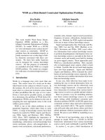
Figure 3 Analysis of Fusarium infection on Bd coleoptile and
root. a) FgUK1 symptoms on leaf sheath. Scale bar = 1 cm. b) FgUK1
symptoms on Bd21 roots (left) and mock inoculation control (right),
48 hpi. Scale bar = 0.5 cm. c-g) Light microscope images of Fg UK1
infection on Bd21 coleoptiles, 6 dpi, stained with trypan blue. c) Fg
hyphae penetration attempt via infection pegs (arrows) at the
junction between adjacent cells showing associated deposition of
phenolic compounds. d) Unsuccessful penetration attempt via
infection pegs (arrows) at the junction between adjacent cells which,
at lower focal plane (e), display intense deposition of phenolic
compounds beneath the attempted infection point. f) Successful
penetration attempt via infection pegs (arrows) at the junction
between adjacent cells which, at lower focal plane (g) appear to be
prised apart. Scale bars: c = 10 μm, d = 10 μm; e = 20 μm, f & g = 10
μm. h) Light microscope image of Fg UK1 at disease front of Bd21
root infection, 48 hpi stained with trypan blue. Scale bar = 20 μm. i)
CLSM image of GFP-expressing Fc at infection site of Bd21 root, 48
hpi. Arrow shows hyphal translocation between two adjacent cortical
cells. Scale bar = 10 μm.
Peraldi et al. BMC Plant Biology 2011, 11:100
/>Page 6 of 14
to the cortex and separated from it by the single cell
layer endodermis is the stele within which lie the central
metaxylem vessel and xylem vessels. Amber-brown dis-
colouration of the roots was observed at the site of
infection by 24 hpi, at which time intercellular and
intracellular presence of the fungus could only be
observed in the rhizodermis and the most external corti-
cal cell layer (Figure 3h). By 48 hpi, hyphae were colo-
nising, by both inter- and intracellular growth (Figure
3i), cortical cell layers and this was associated with the
amber-brown colouration of cortical cells.
Confocal microscopy confirmed that the fungus
invaded most internal layers of cortica l cells by 48 hpi
(Figure3i)buthyphaewereexcludedfromthestele
even after 96 hpi (results not shown). No symptoms
developed on roots following spray inoculation with Fg
conidia. However, mycelium grew externally to reach
the coleoptile where attempted penetration was fre-
quently observed at the junction between adjacent cells
and appeared to proceed via infection pegs (Figure 3c,
d). Attempted penetration was associated with localised
production of an amber-brown deposit within contacted
host cells at the site of contact/attempted penetration
(Figure 3d,e). In most instances fungal ingress was effec-
tively prevented while in some cases the cells appeared
to be prised apart allowing growth of the hypha between
them (Figure 3f,g).
Differential responses of Bd21 and Bd3-1 to Fg and DON
Two Bd ecotypes, parents to a mapping population
(modelcrop.org), were examined as a first step to deter-
mine the potential for natural variation for resistance to
Fusarium within Bd. Leaves of lines Bd21 and Bd3-1
were compared for their response to wound-inoculatio n
with Fg. Symptom development w as significantly m ore
rapid on Bd3-1 than on Bd21 (P = 0.016) (Figure 4).
Most strikingly, lesions on Bd3-1 were surrounded by
large areas of chlorosis w hereas those on Bd21 retained
their green colouration (Additional file 1). Conidial pro-
duction on Bd3-1 leaves was observed to be significantly
(P = 0.001) higher when compared to Bd21 leaves, 7dpi
(Additional file 2).
Bd21 and Bd3-1 were also compared to assess whether
they differed in type II resistance following single floret
point inoculation with Fg. Diseas e progress as deter-
mined by AUDPC was significantly (P < 0.05) greater in
Bd3-1 (31.92) than in Bd21 ( 20.16) (Additional file 3),
although there was no significant difference in conidial
production at 13 dpi, when the experiment was termi-
nated (data not shown).
In complementary experiments, single florets of Bd21
and Bd3-1 were detac hed, placed on moist filter paper in
Petri dishes and inoculated with conidial suspension
onto eith er the palea or lemma surface in order to study
infection of these tissues and to identify potential differ-
ences in susceptibility between t he Bd lines and between
the t issues. Conidial production on infected florets was sig-
nificantly greater (P < 0.001) when conidia were inoculated
onto the palea than o nto the lemma, in both Bd21 and
Bd3-1 ecotypes. In addition, conidial production on both
palea and lemma was higher in Bd3-1 (49,556 and 35,400
conidia/floret, respectively) than in Bd21 (37,533 and
23,200 conidia/floret, respectively) (Figure 5).
Lines B d21 and Bd3-1 were also assessed for suscept-
ibility to DON. Detached leaves were wound-inoculated
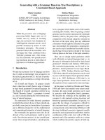
Figure 4 Comparison of Fusarium symptoms development on
Bd21 and Bd3-1 leaves inoculated with Fg. Development of
necrotic lesion area induced by Fg UK1 on wound-inoculated leaves
of Bd21 and Bd3-1 at 48, 72, 96 and 120 hpi. Means ± s.e. were
each calculated from measurements of twelve experimental
replicates. The data shown is representative of six independent
experiments.
Figure 5 Comparison of Fg conidial production on lemma and
palea of Bd21 and Bd3-1 detached spikelets. Conidial
production following inoculation of Fg UK1 onto palea or lemma
surface of Bd21 and Bd3-1 detached florets, 144 hpi. Means ± s.e.
were each calculated from measurements of twenty experimental
replicates. The data shown is representative of three independent
experiments.
Peraldi et al. BMC Plant Biology 2011, 11:100
/>Page 7 of 14
with a range of DON concentrations (15, 75 and 150
μM). At the highest DON concentration, an amber-
brown discolouration appeared around the wound site
of both Bd 21 and Bd3-1 from 72 hpi. Lesions spread
along the vascular bundles, becoming necrotic around
96 hpi. Lower DON concentrations did not result in the
spread of necrotic lesions (data not shown). The size of
the necrotic areas on Bd21 and Bd3-1 were not statisti-
cally different. However, chlorosis d eveloped on Bd3-1
at all DON concentrations, whilst none was observed on
Bd21 (Figure 6).
DON has been demonstrated to be a virulence fac-
tor for FHB and crown rot infection of w heat by Fg.
The influence of DON on Fusarium infection of Bra-
chypodium was examined on wound-inoculated
detached leaves to determine whether it enhanced
virulence for Fg and Fc. Amendment of conidial
inoculum with DON (75 μM) significantly increased
(P < 0.001) average lesion area for both Fg and Fc
(Figure 7a) and conidial production (Figure 7b) when
compared with infections using the conidia alone.
These results were strikingly similar to the effect of
DON amendment on lesion development on wheat
leaves (Additional file 4).
As shown above, symptom development on floral tis-
sues was greater in Bd3-1 than in Bd21 and additional
experiments were carried out to determine whether this
was also reflected in differences in accumula tion of
DON. Spikes of Bd21 and Bd3-1 were spray inoculated
with conidia of Fg and the DON content was assessed
21 dpi. No significant difference (P = 0.971) in DON
content was observed between Bd21 and Bd3-1 (620
mg/kg and 625 mg/kg of fresh tissue, respectively).
Discussion
The present study aimed to determine the potent ial for
Bd to act as a host to Fg and Fc and ascertain whether
this interaction might serve as a model of that between
Fusarium species and wheat. The results clearly demon-
strated the compatibility of interaction between Bd and
the two Fusarium species of greatest relevance to FHB,
the major Fusarium-associated disease of wheat. M ore-
over, t he development of disease symptoms closely
resembled those reported in wheat.
With respect to FHB, after a short asymptomatic per-
iod, Bd spikes spray inoculated with Fg co nidia dis-
played small brown spots, which first appeared at the
middle or base of the lemma, highly reminiscent of the
initial symptoms in wheat [4]. Lesions spread to infect
adjacent florets, often provoking the bleaching of the
upper part of the spikelet in a manner similar to that
observed in wheat [32,10] and infection extended down
the rachis to adjacent spikelets and even colonised ped-
uncles as seen during infecti on of wheat. Overall, Fusar-
ium infection of Bd spikes results in the development of
symptoms that strikingly resemble those described in
wheat heads infected with Fg and Fc [4].
Figure 6 Comparison of DON-induced lesions of Bd21 and
Bd3-1 detached leaves. Symptoms on leaves of Bd21 and Bd3-1,
120 hpi following wound-inoculation with water or DON (150 μM).
Means ± s.e. were each calculated from measurements of eight
experimental replicates. The data shown is representative of three
independent experiments.
Figure 7 Effect of DON treatment on Bd21 detached leaves
infected with Fg or Fc. a) Area under disease progress curve
(AUDPC, 6dpi) for lesions following wound-inoculation of leaves of
Bd21 with Fg UK1 or Fc GFP1 with or without amendment with
DON (75 μM). b) Conidial production (6dpi) on leaves of Bd21
following wound-inoculation with Fg UK1 or Fc GFP1 with or
without amendment with DON (75 μM). Means ± s.e. were each
calculated from measurements of three experimental replicates. The
data shown is representative of two independent experiments.
Peraldi et al. BMC Plant Biology 2011, 11:100
/>Page 8 of 14
Microscope analysis of floral tissues highlighted the
potential role played by specific epidermal cell types
during the early stages of infection. Fusarium hyphae
were repeatedly observed entwined about voluminous
macro-hairs that displayed a characteristic amber-brown
discolouration. Globose f ungal structures w ere repeat-
edly observed at the base of these hairs, suggesting that
these cell types are favoured targets for penetration.
Two components of resistance to FHB are widely recog-
nised; resistance to initial infection (type I) and resis-
tance to spread within the head (type II) [12]. The palea
and lemma tissues of barley have been shown to express
different levels of type I resistance with the former
being more susceptible than the latter [33]. Similar dif-
ferential type I susceptibility of the palea and lemma tis-
sues of Bd was observed in the present study along with
differences in type I susceptibility of the two tested
inbred lines. Type II resistance is assessed by point
inoculation of individual florets in wheat heads [4]. Fol-
lowing point inoculation of Bd florets, both Fg and Fc
successfully colonised Bd spikelet tissues and spread
through the rachis into neighbouring spikelets and
down the peduncle, closely resembling the pattern of
colonization in heads of susceptible wheat cultivars [4].
The bleaching of spikelets above the inoculation site in
wheat heads is another characteristic symptom of FHB
[10]. Bleaching has been correlated with the production
of DON by the fungus within infected wheat heads and
is also induced following injection of DON into wheat
heads [10]. Our observation of bleaching of infected
spikes of Bd thus resembles the situation in FHB of
wheat more closely than does barley, which has an
inherent type II resistance restricting Fusarium symp-
toms to the area of initial infection [3].
DON has been shown to function as a virulence factor
in wheat, inhibiting the development of cell wall fortifi-
cation within the rachis during FHB development [34]
and aiding stem colonisation during development of
crown rot [35]. In contrast DON appears to play no dis-
cernable role in disease development in heads of barley
[34,7] or floral tissues of Arabidopsis [36]. Amendment
of the conidial inoculum with DON significantly
enhanced both disease symptoms and conidial produc-
tion by Fg and Fc on wounded detached leaves of Bd.
DON amendment similarly influenced symptom devel-
opment and conidial production in detached wheat
leaves following inoculation with Fg and Fc (Additional
file 4). This strongly suggests that DON functions in Bd
as it does in wheat, where it is understood to act as a
virulence factor [34,35].
The detection of high concentrations of DON in Bd21
and Bd3-1 flowers following inoculation with Fg indi-
cates that these tissues support DON production in
Fusarium species. T he levels of DON in Bd spray-
inoculated spikes were similar to those reported pre-
viously following inoculation of wheat under controlled
conditions [37,38]. The high levels of DON observed in
floral tissues of Bd differs markedly from the situation
with Arabidopsis where the reported levels are generall y
extremely low [23,21]. Trichothecene production has
been shown not to be uniformly induced during infec-
tion of wheat but, rather, is tissue specific with induc-
tion in developing kernels and the rachis node [39]. It is
probable that the necessary components to induce tri-
chothecene production are present in Bd and wheat
whereas they are absent in Arabidopsis, making Bd an
attractive model for wheat. The current experiment
could not provide information on kernel resistance as
whole floral tissues were sampled because the h igh
infection pressure resulted in extremely shrivelled seeds.
However, reducing infection pressure and dissection of
floral parts could provide insight onto resistance to ker-
nel infection in future experiments.
Following spray inoculation of whole Bd plants, symp-
toms developed on virtually all above-ground plant parts
(stems, leaf sheaths and leaves). Unexpectedly, intact
leaves from spray inoculated plants also developed
necrotic and chlorotic symptoms as did inoculated
unwounded detached leaf sections. T he presence of
Fusarium within Bd tissues was confirmed by CLSM
observation of GFP-expressing fungus. This is, to our
knowledge, the first report to date of a successful infec-
tion on intact foliar tissue b y a Fusarium species.
Detache d leaf assays have been used previously to iden-
tify components of resistance related to FHB but these
experiments, although using unwounded inoculation,
were carried out using Microdochium majus, a non-
toxin producing FHB species [40]. We have determined
that Fg and Fc can infect floral and foliar tissues of Bd
allowing the mycotoxin-producing species to be used in
comparative assays on these tissues. The susceptibility
of intact Bd leaves therefore provides the first opportu-
nity to establish the relationship between foliar and
floral components of resistance to Fu sarium species and
identify those foliar components of relevance to FHB
resistance. The unique susceptibility of Bd to foliar
penetration by Fusarium spp. also provides the potential
to undertake high throughput genetic screening of Bd
mutant collections to identify lines altered in susceptibil-
ity to penetration. Having observed disease symptoms
on all tested Bd tissues, histological examination was
undertaken to determine how Fg and Fc gain entry into
this host. Direct stomatal penetration of wheat head tis-
sues by Fg and Fc has been previ ously reported [41-43].
Despite observing multiple instances of direct contacts
between Fg and Fc germination hyphae and stomatal
apertures, we did not obtain evidence for entry into Bd
via stomata. Overall, our results suggest tha t, although
Peraldi et al. BMC Plant Biology 2011, 11:100
/>Page 9 of 14
penetration may occur through stomatal apertures, it is
not likely t o be t he main mode of entry. In numerous
instance s, hyphal contact with stomata resulted in guard
cells becoming very dark brown, indicating the possible
deposition of phenolic compounds. Interestingly, pheno-
lic compounds have been previously shown to play a
role in FHB disease resist ance in wheat [44] and a simi-
lar situation may occur in the guard cells of Bd. Light
microscopy images of the first visible symptoms devel-
oping on leaves revealed a characteristic amber-brown
discolouration (distinct from the colour of contacted
guard cells), of the macro-hair base and directly adjacent
cells that was correlated with the presence of the fungus
and attempted penetration of the host. Although this
amber-brown colour is also indicative of phenolic com-
pounds, the results from coleoptile infection studies
showed that its accumulation at the site of attempted
fungal penetration is not effective in preventing infec-
tion. Similar appositions have been observed during
infection of wheat by Fg and were more pronounced in
resist ant than in susc eptible cultivars [45]. During infec-
tion of Bd coleoptiles Fg appeared to produce infection
pegs and gain entry via growth between cells. Again,
this is similar to infection observed on wheat [43]. SEM
analysis of intact Bd leaf surface indicated that penetra-
tion of hair cells may be the preferred route of entry for
the pathogen. We observed penetration of the cuticle,
growth and branching at the base of the macro-hair.
Macro- hairs are located above the vascular bundles, and
targeting their base for initial penetration provides the
pathogen almost direct ac cess to the vascular bundles
enabling rapid spread to adjacent tissues [46]. This is an
interesting finding in relation to previous studies made
on detached wheat glumes where Fg was observed to
penetrate and invade host tissue through short hair cells
(termed prickle hairs [47]). Associa tion between Fg
hyphae and prickle hairs (also referred to as papilla
cells) on wheat was also noted by Pritch and colleagues
[42], although they did not undertake detailed investiga-
tion of the interaction. The comparison of microscope
images of infected floral and foliar Bd tissues revealed
striking similarities. Fusarium hyphae were observed to
specifically target hairs i n both tissues, where globose
hyp hal structures developed about BMH. Accumulation
of phenolic compounds of unknown composition
occurred in both floral and foliar tissues as a host
response to penetration attempts. These similarities sup-
port the i dea that investigating the mechanisms of
Fusarium infection on foliar tissues may have direct
relevance to the mechanisms of resistance of the floral
tissues to FHB.
Root tissues were also successfully infected following
inoculation by contact with mycelial plugs. The infection
pressure generated by conidia, however, failed to induce
infection and it remains to be determined whether
infection can proceed directly from conidia or whether
infection requires hyphae. Infection was indicated by
discolouration and confirmed by observation of inter-
and intracellular fungal hyphae in the cortex at an early
stage of infection. Even at late stages of infection fungal
hyphae were excluded from the stele, a situation similar
to that recently reported in wheat [41]. Together with
observation of symptoms developing on the stem base,
these results suggest that Bd can also be used for mod-
elling crown rot and root rot.
Differential responses among Bd accessions to biotic
and abiotic stresses have been observed by others indi-
cating that naturally occurring allelic variation in Bd
accessions may provide insights into mechanisms under-
lying responses to agronomically important traits
[48,49]. Inoculation of Fg conidia on detached Bd florets
revealed quantitative differences in fungal development
betweenBd21andBd3-1lines. Interestingly, the two
lines also differed in susceptibility in the detached leaf
assay with the most notable difference between them
being the extensive chlorosis that developed in Bd3-1.
Interestingly, DON application to wounded Bd21 and
Bd3-1 leaves also resulted in a difference in response
with respect to the development of chlorosis indicating
that the differential response of the two lines to Fg is, at
least in part, a result of diffe rential susceptibility to
DON. The availability of the population derived from a
cross between Bd21 and Bd3-1 (http: //w ww.modelcro p.
org), will permit genetic mapping of the differential sus-
ceptibility of these lines to DON and foliar infection.
Additionally, investigating the wide range of di-, tetra-
and hexaploid Bd accessions would be expected to
reveal different levels and mechanisms of resistance to
Fusarium.
Bd was previously reported as a model for rice in
order to study resistance to Magnaporthe grisea [48].
The current study provides the first detailed report of
Bd as a potential model for a wheat disease caused by a
necrotrophic fungus.
Conclusions
We demonstrate herein a compatible reaction between
Fusarium species and Bd and establish a new pathosys-
tem with which to investigate mechanisms underlying
FHB resistance in a tractable monocotyledonous model
species. Disease symptoms on Bd spikes and the accu-
mulation of DON within floral tissues were highly simi-
lar to those on wheat heads. Futhermore, we identified
naturally-occurring variation for resistance to Fusarium
species among Bd accessions and report, for the first
time, successful Fusarium infection of intact foliar tis-
sues. Infection of both floral and foliar tissues were
highly similar, strongly suggesting direct relevance of
Peraldi et al. BMC Plant Biology 2011, 11:100
/>Page 10 of 14
findingsfromonetissuetotheother.Syntenybetween
Bd and wheat is very high making possible the direct
translation of information on the role of particular
genes in resistance in Bd to their counterparts in wheat.
This, taken together with the availability of a complete
genome sequence and an increasing number of
resources for functional genomics, gives Bd the potential
to become a significant model species with which to
investigate resistance to Fusarium species and provide
information of direct relevance to wheat and other cer-
eal crops.
Methods
Maintenance and preparation of Fusarium inoculum
DON-producing isolates o f Fg (UK1 and S1) from the
culture collection of the John Innes Centre were used
throughout. A DON-producing constitutive GFP expres-
sing isolate (FcGFP1) of Fc (kindly provided by Dr F.
Doohan, University College Dublin, Ireland) was used
for confocal microscopy. Conidial inoculum was pro-
duc ed in mung bean (MB) liquid medium and prepared
as described previously with shaking for 7 days at 25°C
[50]. To harvest conidia, the culture solution was filtered
through sterilized muslin and centrifuged at 3000 g for
5 min. The pellet was washed once an d re-suspended in
sterile distilled water (SDW) at a concentration of 1 ×
10
6
conidia ml
-1
and stored at -20°C until use.
Brachypodium lines and growth conditions
Brachypodium distachyon inbred lines Bd21 an d Bd3-1
[51] were used throughout. Bd seeds were germinated
and incubated for 5 days in Petri dishes on damp filter
paper in the dark at 5°C. Seed were then incubated in
darkness at 15°C for 24 h before exposing to a 16 h/8 h
light-dark cycle for 24 h at 20°C. Seeds were then
planted in 8 × 8 × 10 cm pots filled with 50% peat and
sand mixed with 50% John Innes number 2 loam com-
post, and placed in a climatically controlled chamber
with a relative humid ity (RH) of 70 % at 22°C. Foliar tis-
sue was obtained from plants grown under a 16 h/8 h
light-dark cycle while plants were grown under a 20 h/4
h light-dark cycle to obtain floral tissues.
Brachypodium spray, point, coleoptile and root
inoculations, incubation and symptom assessment
Whole Bd21 plants were sprayed with FgUK1 conidial
suspension (1 × 10
5
conidia ml
-1
), amended with 0.05%
Tween 20, using a handheld mister until run off.
Sprayed plants were placed under a plastic cover and
misted periodically with SDW to increase relative RH to
about 90%, until 3 days post inoculation (dpi) when cov-
ers were removed and misting ceased. Disease symp-
toms were photogr aphed using a Sams ung NV7 digital
camera. Floral point inoculations were performed by
inserting a piece (2 × 8 mm) of filter paper (Sartorius;
grade 292) between two adjacent florets. Conidial sus-
pension (5 μlof1×10
6
conidia ml
-1
)ofFgUK1was
carefully applied to the filter paper. Following inocula-
tion, plants were treated as for spray inoculation above.
Floral symptom development was quantified by visual
assessment and the number of infected florets was
counted at 2, 4 and 8 dpi.
Studies on infection of roots and coleoptiles were car-
ried out on Bd seedlings germinated as described above
and incubated for 5 days at 20°C under a 16 h/8 h light-
dark cycle. Seed lings used for root infection were inocu-
lated using a mycelium plug (5 mm dia) from the grow-
ing edge of a 14 day old colony grown on potato
dextrose agar (PDA) at 20°C and with a PDA plug for
the controls. Coleoptiles were inoculated by spraying 1
ml of conidial suspension (1 × 10
6
conidia ml
-1
)per
plate and with sterile water for the controls.
Bd21 and Bd3-1 flowers were harvested 21 days after
spray inoculation with Fg S1 or Fc GFP1 conidial sus-
pension (1 × 10
5
conidia ml
-1
), frozen in liquid N
2
and
ground to a fine powder. DON detection and quantifica-
tion was performed using an ELISA competitive immu-
noassay (AgraQuant
®
, Romer Labs Singapore Pte Ltd)
according to the manufacturer’s recommendation.
Inoculation, incubation and symptom assessment of
detached leaves
Leaves were removed from 21 days old plants, cut to 5
cm length and wounded in two positions 2 cm apart
and on opposite sides of the mid-rib by gentle compres-
sion with a glass Pasteur pipette on the adaxial surface.
Leaf sections were placed in 10 × 10 cm square plastic
boxes containing 0.8% water agar and treated as
reported previously for wheat and barley [25]. Each box
contained eight leaf sections from different plants. A
droplet (10 μl) of conidial suspension (1 × 10
6
conidia
ml
-1
), amended with 0.05% Tween 20, was deposited
onto each wound site. In other experiments the conidial
suspension was amended with DON (75 μM). Mock
inoculation was performed similarly using SDW
amended with 0.05% Tween 20 (10 μl).
In separate experiments, unwounded leaves were simi-
larly inoculated with Fg or treated by addition of DON
(15, 75 and 150 μM amended with 0.01% Tween 20).
The inner surface of the plate lid was misted with SDW
to maintain 100% RH and plates were incubated at 22°C
under a 16 h/8 h light-dark cycle. Disease symptoms
were recorded every 24 h and lesion sizes were mea-
sured using IMAGE-J software [52].
Light microscopy
Bd leaf sections and flowers were cleared in 70% ethanol
at 70°C for one hour to remove chlorophyll. Samples
Peraldi et al. BMC Plant Biology 2011, 11:100
/>Page 11 of 14
were stained for 1 min in trypan blue or aniline blue
(0.1%) in lactoglycerol (1:1:1, lactic acid: glycerol: H
2
O)
and rinsed in a 15 M solution of chloral hydrate. Sam-
ples were mounted in 40% glycerol, viewed with a
Nikon Eclipse 800 microscope and photographed with a
Pixera Pro ES 600 di gital camera. Inoculated palea a nd
lemma tissues were dissected, cleared of chlorophyll,
stained with aniline blue and observed under a light
microscope.
Confocal microscopy
Horizontal cross sections (50 μmthickness)ofroots
(inoculated and non-inoc ulated) were dissected 24, 48,
72 and 96 h post inoculation ( hpi) using a sectioning
sys tem (Vibratome 1000 plus) and placed between glass
slides in SDW. Root sections were analysed under a
confocal microscope (Leica DMR SP1) excited with a
488 nm Argon ion laser and detected at 505-555 nm.
Autofluorescence of cell walls and chloroplasts was
detected at 580-680 nm.
Scanning electron microscopy
Intact Bd leaf sections were mounted on an aluminum
stub by using O.C.T. compound (BDH), plunged into
liquid nitrogen slush, and then transferred onto the
cryostage of an ALTO 2500 cryo-transfer system
(Gatan) attached to a Zeiss Supra 55 VP F EG scanning
electron microscope. Samples were then sputter-coated
with platinum (90 s at 10 mA, -110°C), and imaged at 3
kV on the cryo-stage in the main chamber of the micro-
scope at -130°C. An alternative fixation method was
used to remove wax crystals from the surface of Bd
leaves and allow observation of sub-cuticular structures.
Leaf sections were fixed for approxi mately 4 hrs at 20°C
in FAA (3.7% formaldehyde, 5% acetic acid, 50% etha-
nol) and subsequently dehydrated t hrough an ethanol
series. After critical point drying, tissues w ere coated
with gold and examined in a Philips XL30 FEG micro-
scope using an acceleration voltage of 3 kV.
Statistical analysis
The disease severity, conidial production, DON accumu-
lation and lesion area data were analysed by generalised
linear modelling (GLM) using the software package
GENSTAT version 9.1 (Lawes Agricultural Trust,
Rothamsted Experimental Station, UK). Individual treat-
ments were compared with controls using the unpaired
t-tests within the GLMs.
Additional material
Additional file 1: Comparison of symptoms following Fg infection
on Bd21 and Bd3-1 leaves. Symptoms on leaves of Bd21 and Bd3-1,
120 h following wound inoculation with Fg UK1.
Additional file 2: Comparison of Fg conidial production on Bd21
and Bd3-1 detached leaves. Conidial production following inoculation
of Fg UK1 onto Bd21 and Bd3-1 detached leaves, 7 dpi.
Additional file 3: Comparison of necrotic symptoms development
following Fg point inoculation on Bd21 and Bd3-1 spikelets. Area
under disease progress curve (AUDPC) for lesions of Bd21 and Bd3-1
spikelets point inoculated with Fg UK1.
Additional file 4: Effect of DON treatment on detached wheat
leaves infected with Fg or Fc. a) Area under disease progress curve
(AUDPC) for lesions following wound-inoculation of wheat (cv. Paragon)
leaves with Fg UK1 and Fc GFP1 with or without amendment with DON
(75 μM). b) Conidial production (6dpi) on leaves of wheat (cv. Paragon)
following wound-inoculation with Fg UK1 and Fc GFP1 with or without
amendment with DON (75 μM).
Acknowledgements and Funding
We thank Dr Philippe Vain and Dr Vera Thole for providing Brachypodium
seeds. We are also thankful to Dr Thomas Girin and Mrs Kim Findlay for their
expertise in Scanning Electron Microscopy, as well as the UK Biotechnology
and Biological Science Research Council (BBSRC) for funding the research.
Author details
1
Department of Disease and Stress Biology, John Innes Centre, Colney Lane,
Norwich, NR4 7UH, UK.
2
Dipartimento di Scienze Agrarie e Ambientali,
Facoltà di Agraria, Università degli Studi di Perugia, Borgo XX Giugno 74,
Perugia, 06121, Italy.
Authors’ contributions
AP carried out all Bd spray and point inoculations, disease assessments and
detached leaf assays and normal light and SEM microscopy analysis. GB
carried out Bd root inoculations and CLSM analysis of root tissues and
participated to Bd detached leaf assays. AS and PN took part in designing
and supervising the study and participated in drafting the manuscript. All
authors have read and approved the final manuscript.
Received: 4 February 2011 Accepted: 3 June 2011
Published: 3 June 2011
References
1. Bottalico A: Fusarium diseases of cereals: Species complex and related
mycotoxin profiles, in Europe. Journal of Plant Pathology 1998, 80:85-103.
2. Desmond OJ, Manners JM, Stephens AE, Maclean DJ, Schenk PM,
Gardiner DM, Munn AL, Kazan K: The Fusarium mycotoxin deoxynivalenol
elicits hydrogen peroxide production, programmed cell death and
defence responses in wheat. Molecular Plant Pathology 2008, 9:435-445.
3. Foroud NA, Eudes F: Trichothecenes in cereal grains. International Journal
of Molecular Sciences 2009, 10:147-173.
4. Parry DW, Jenkinson P, McLeod L: Fusarium ear blight (scab) in small
grain cereals - a review. Plant Pathology 1995, 44:207-238.
5. Tóth B, Mesterházy A, Horváth A, Bartók T, Varga M, Varga J: Genetic
variability of central European isolates of the Fusarium graminearum
species complex. European Journal of Plant Pathology 2005, 113:35-45.
6. Placinta CM, D’Mello JPF, Macdonald AMC: A review of worldwide
contamination of cereal grains and animal feed with Fusarium
mycotoxins. Animal Feed Science and Technology 1999, 78:21-37.
7. Maier FJ, Miedaner T, Hadeler B, Felk A, Salomon S, Lemmens M, Kassner H,
Schäfer : Involvement of trichothecenes in fusarioses of wheat, barley
and maize evaluated by gene disruption of the trichodiene synthase
(Tri5) gene in three field isolates of different chemotype and virulence.
Molecular Plant Pathology 2006, 7:449-461.
8. Pestka JJ, Zhou HR, Moon Y, Chung YJ: Cellular and molecular
mechanisms for immune modulation by deoxynivalenol and other
trichothecenes: unravelling a paradox. Toxicology Letters 2004, 153:61-73.
9. Rocha O, Ansari K, Doohan FM: Effects of trichothecene mycotoxins on
eukaryotic cells: a review. Food Additives and Contaminants 2005, 22:369-378.
10. Lemmens M, Scholz U, Berthiller F, Dall’Asta C, Koutnik A, Schuhmacher R,
Adam G, Buerstmayr H, Mesterházy A, Krska R, Ruckenbauer P: The ability
to detoxify the mycotoxin deoxynivalenol colocalizes with a major
Peraldi et al. BMC Plant Biology 2011, 11:100
/>Page 12 of 14
quantitative trait locus for Fusarium head blight resistance in wheat.
Molecular Plant-Microbe Interaction 2005, 18:1318-1324.
11. Buerstmayr H, Ban T, Anderson JA: QTL mapping and marker-assisted
selection for Fusarium head blight resistance in wheat: a review. Plant
breeding 2009, 128:1-26.
12. Schroeder HW, Christensen JJ: Factors affecting resistance of wheat to
scab caused by Gibberella zeae. Phytopathology 1963, 53:831-838.
13. Mesterházy A: Types and components of resistance to Fusarium head
blight of wheat. Plant Breeding 1995, 114:377-386.
14. Boutigny AL, Richard-Forget F, Barreau C: Natural mechanisms for cereal
resistance to the accumulation of Fusarium trichothecenes. European
Journal Plant Pathology 2008, 121:411-423.
15. Cuthbert PA, Somers DJ, Thomas J, Cloutier S, Brulé-Babel A: Fine mapping
Fhb1, a major gene controlling fusarium head blight resistance in bread
wheat. Theoretical and Applied Genetics 2006, 112:1465-1472.
16. Cuthbert PA, Somers DJ, Brulé-Babel A: Mapping of Fhb2 on chromosome
6BS: a gene controlling Fusarium head blight field resistance in bread
wheat (Triticum aestivum L.). Theoretical and Applied Genetics 2007,
114:429-437.
17. Qi LL, Pumphrey MO, Friebe B, Gill BS: Molecular cytogenetic
characterization of alien introgressions with gene Fhb3 for resistance to
Fusarium head blight disease of wheat. Theoretical and Applied Genetics
2008, 117:1155-1166.
18. Xue S, Li G, Jia H, Xu F, Lin F, Tang M, Wang Y, An Y, Xu H, Zhang L,
Kong Z, Ma Z: Fine mapping Fhb4, a major QTL conditioning resistance
to Fusarium infection in bread wheat (Triticum aestivum L.). Theoretical
and Applied Genetics 2010, 121:147-156.
19. Amatulli MT, Spadaro D, Gullino ML, Garibaldi A: Molecular identification
of Fusarium spp. associated with bakanae disease of rice in italy and
assessment of their pathogenicity. Plant Pathology 2010, 59:839-844.
20. Huala E, Dickerman AW, Garcia-Hernandez M, Weems D, Reiser L, LaFond F,
Hanley D, Kiphart D, Zhuang M, Huang W, Mueller LA, Bhattacharyya D,
Bhaya D, Sobral BW, Beavis W, Meinke DW, Town CD, Somerville C,
Rhee SY: The Arabidopsis Information Resource (TAIR): a comprehensive
database and web-based information retrieval, analysis, and
visualization system for a model plant. Nucleic
Acids Research 2001,
29:102-105.
21. Urban M, Daniels S, Mott E, Hammond-Kosack K: Arabidopsis is susceptible
to the cereal ear blight fungal pathogens Fusarium graminearum and
Fusarium culmorum. The Plant Journal 2002, 32:961-973.
22. Chen X, Steed A, Harden C, Nicholson P: Characterization of Arabidopsis
thaliana-Fusarium graminearum interactions and identification of
variation in resistance among ecotypes. Molecular Plant Pathology 2006,
7:391-403.
23. Cuzick A, Lee S, Gezan S, Hammond-Kosack KE: NPR1 and EDS11
contribute to host resistance against Fusarium culmorum in Arabidopsis
buds and flowers. Molecular Plant Pathology 2008, 9:697-704.
24. Savitch LV, Subramaniam R, Allard GC, Singh J: The GLK1 ‘regulon’
encodes disease defense related proteins and confers resistance to
Fusarium graminearum in Arabidopsis. Biochemical and biophysical research
communications 2007, 359:234-238.
25. Chen X, Steed A, Travella S, Keller B, Nicholson P: Fusarium graminearum
exploits ethylene signalling to colonize dicotyledonous and
monocotyledonous plants. New Phytologist 2009, 182:975-983.
26. Draper J, Mur LAJ, Jenkins G, Ghosh-Biswas GC, Bablak P, Hasterok R,
Routledge APM: Brachypodium distachyon. A new model system for
functional genomics in grasses. Plant Physiology 2001, 127:1539-1555.
27. The International Brachypodium Initiative: Genome sequencing and
analysis of the model grass Brachypodium distachyon. Nature 2010,
463:763-768.
28. Huo N, Vogel JP, Lazo GR, You FM, Ma Y, McMahon S, Dvorak J,
Anderson OD, Luo MC, GuYQ : Structural characterization of
Brachypodium genome and its syntenic relationship with rice and
wheat. Plant Molecular Biology 2009, 70:47-61.
29. Griffiths S, Sharp R, Foote TN, Bertin I, Wanous M, Reader S, Colas I,
Moore G: Molecular characterization of Ph1 as a major chromosome
pairing locus in polyploid wheat. Nature 2006, 439:749-752.
30. Vain P, Worland B, Thole V, McKenzie N, Alves SC, Opanowicz M, Fish LJ,
Bevan MW, Snape JW: Agrobacterium
-mediated transformation of the
temperate
grass Brachypodium distachyon (genotype Bd21) for T-DNA
insertional mutagenesis. Plant Biotechnology Journal 2008, 6:941-941.
31. Xu XM, Nicholson P, Thomsett MA, Simpson D, Cooke BM, Doohan FM,
Brennan J, Monaghan S, Moretti A, Mule G, Hornok L, Beki J, Tatnell J,
Ritieni A, Edwards SG: Relationship between the fungal complex causing
Fusarium head blight of wheat and environmental conditions.
Phytopathology 2008, 98:69-78.
32. Weise MV: Compendium of Wheat Diseases. The American
Phytopathological Society Press, USA St., Paul, Minn 1987, 112.
33. Lewandowski SM, Bushnell WR, Evans CK: Distribution of mycelial colonies
and lesions in field-grown barley inoculated with Fusarium graminearum.
Phytopathology 2006, 96:567-581.
34. Jansen C, von Wettstein D, Schafer W, Kogel KH, Felck A, Maier FJ: Infection
patterns in barley and wheat spikes inoculated with wild-type and
trichodiene synthase gene disrupted Fusarium graminearum. Proceedings
of the National Academy of Sciences, USA 2005, 102:16892-16897.
35. Mudge AM, Dill-Macky R, Dong Y, Gardiner DM, White RG, Manners JM: A
role for the mycotoxin deoxynivalenol during crown rot disease of
wheat caused by Fusarium graminearum and Fusarium
pseudograminearum. Physiological and Molecular Plant Pathology 2006,
69:73-85.
36. Cuzick A, Urban M, Hammond-Kosack K: Fusarium graminearum gene
deletion mutants map1 and tri5 reveal similarities and differences in the
pathogenicity requirements to cause disease on Arabidopsis and wheat
floral tissue. New Phytologist 2008, 177:990-1000.
37. Savard ME, Sinha RC, Seaman WL, Fedak G: Sequential distribution of the
mycotoxin deoxynivalenol in wheat spikes after inoculation with
Fusarium graminearum. Canadian Journal of Plant Pathology 2000,
22:280-285.
38. Gosman N, Chandler E, thomsett M, Draeger R, Nicholson P: Analysis of the
relationship between parameters of resistance to Fusarium head blight
and in vitro tolerance to deoxynivalenol of the winter wheat cultivar
WEK0609®. European Journal of Plant Pathology 2005, 111:57-66.
39. Ilgen P, Hadeler B, Maier FJ, Schäfer W: Developing kernel and rachis node
induce the trichothecene pathway of Fusarium graminearum during
wheat head infection. Molecular Plant Microbe Interaction 2009, 8:899-908.
40. Browne RA, Mascher F, Golebiowska G, Hofgaard IS: Components of partial
disease resistance in wheat detected in a detached leaf assay inoculated
with Microdochium majus using first, second and third expanding
seedling leaves. Journal of Phytopathology 2006,
154:204-208.
41.
Beccari G, Covarelli L, Nicholson P: Infection processes and soft wheat
response to root rot and crown rot caused by Fusarium culmorum. Plant
Pathology .
42. Pritsch C, Muehlbauer GJ, Bushnell WR, Somers DA, Vance CP: Fungal
development and induction of defense response genes during early
infection of wheat spikes by Fusarium graminearum. Molecular Plant-
Microbe Interactions 2000, 13:159-169.
43. Kang K, Buchenhauer H: Cytology and ultrastructure of the infection of
wheat spikes by Fusarium culmorum. Mycological Research 2000,
104:1083-1093.
44. Boutigny AL, Barreau C, Atanasova-Penichon V, Verdal-Bonnin MN, Pinson-
Gadais L, Richard-Forget F: Ferulic acid, an efficient inhibitor of type B
trichothecene biosynthesis and Tri gene expression in Fusarium liquid
culture. Fungal Biology 2009, 113:746-753.
45. Kang Z, Buchenhauer H, Huang L, Han Q, Zhang H: Cytological and
immunocytochemical studies on responses of wheat spikes of the resistant
Chinese cv. Sumai 3 and the susceptible cv. Xiaoyan 22 to infection by
Fusarium graminearum. European Journal of Plant Pathology 2008, 120:383-396.
46. Kang K, Buchenauer H: Immunocytochemical localization of Fusarium
toxins in infected wheat spikes by Fusarium culmorum. Physiological and
Molecular Plant Pathology 1999, 55:275-288.
47. Rittenour WR, Harris SD: An In Vitro method for the analysis of infection-
related morphogenesis in Fusarium graminearum. Molecular Plant
Pathology 2010, 11:361-369.
48. Routledge APM, Shelley G, Smith JV, Talbot NJ, Draper J, Mur LAJ:
Magnaporthe grisea interactions with the model grass Brachypodium
distachyon closely resemble those with rice (Oryza sativa). Molecular Plant
Pathology 2004, 5:253-265.
49. Luo N, Liu J, Yu X, Jiang Y: Natural variation of drought response in
Brachypodium distachyon. Physiologia Plantarum 2010, 141:19-29.
50. Makandar R, Essig JS, Schapaugh MA, Trick HN, Shah J: Genetically
engineered resistance to Fusarium head blight in wheat by expression
of Arabidopsis NPR1. Molecular Plant-Microbe Interactions 2006, 19:123-129.
Peraldi et al. BMC Plant Biology 2011, 11:100
/>Page 13 of 14
51. Vogel JP, Gu YQ, Twigg P, Lazo GR, Laudencia-Chingcuanco D, Hayden DM,
Donze TJ, Vivian LA, Stamova B, Coleman-Derr D: EST sequencing and
phylogenetic analysis of the model grass Brachypodium distachyon.
Theoretical and Applied Genetics 2006, 113:186-195.
52. Abramoff MD, Magelhaes PJ, Ram SJ: Image processing with ImageJ.
Biophotonics International 2004, 11:36-42.
doi:10.1186/1471-2229-11-100
Cite this article as: Peraldi et al.: Brachypodium distachyon: a new
pathosystem to study Fusarium head blight and other Fusarium
diseases of wheat. BMC Plant Biology 2011 11:100.
Submit your next manuscript to BioMed Central
and take full advantage of:
• Convenient online submission
• Thorough peer review
• No space constraints or color figure charges
• Immediate publication on acceptance
• Inclusion in PubMed, CAS, Scopus and Google Scholar
• Research which is freely available for redistribution
Submit your manuscript at
www.biomedcentral.com/submit
Peraldi et al. BMC Plant Biology 2011, 11:100
/>Page 14 of 14