báo cáo khoa học: " The Arabidopsis translocator protein (AtTSPO) is regulated at multiple levels in response to salt stress and perturbations in tetrapyrrole metabolism" potx
Bạn đang xem bản rút gọn của tài liệu. Xem và tải ngay bản đầy đủ của tài liệu tại đây (6.26 MB, 17 trang )
RESEARCH ARTICLE Open Access
The Arabidopsis translocator protein (AtTSPO) is
regulated at multiple levels in response to salt
stress and perturbations in tetrapyrrole
metabolism
Emilia Balsemão-Pires
1,2
, Yvon Jaillais
2,3
, Bradley JSC Olson
2
, Leonardo R Andrade
4
, James G Umen
2
,
Joanne Chory
2,3*
and Gilberto Sachetto-Martins
1*
Abstract
Background: The translocator protein 18 kDa (TSPO), previously known as the peripheral-type benzodiazepine
receptor (PBR), is important for many cellular functions in mammals and bacteria, such as steroid biosynthesis,
cellular respiration, cell proliferation, apoptosis, immunomodulation, transport of porphyrins and anions. Arabidopsis
thaliana contains a single TSPO/PBR-related gene with a 40 amino acid N-terminal extension compared to its
homologs in bacteria or mammals suggesting it might be chloroplast or mitochondrial localized.
Results: To test if the TSPO N-terminal extension targets it to organelles, we fused three potential translational
start sites in the TSPO cDNA to the N-terminus of GFP (AtTSPO:eGFP). The location of the AtTSPO:eGFP fusion
protein was found to depend on the translational start position and the conditions under which plants were
grown. Full-length AtTSPO:eGFP fusion protein was found in the endoplasmic reticulum and in vesicles of
unknown identity when plants were gro wn in standard conditions. However, full length AtTSPO:eGFP localized to
chloroplasts when grown in the presence of 150 mM NaCl, conditions of salt stress. In contrast, when AtTSPO:eGFP
was truncated to the second or third start codon at amino acid position 21 or 42, the fusion protein co-localized
with a mitochondrial marker in standard conditions. Using promoter GUS fusions, qRT-PCR, fluorescent protein
tagging, and chloroplast fractionation approaches, we demonstrate that AtTSPO levels are regulated at the
transcriptional, post-transcriptional and post-translational levels in response to abiotic stress conditions. Salt-
responsive genes are increased in a tspo-1 knock-down mutant compared to wild type under conditions of salt
stress, while they are decreased when AtTSPO is overexpressed. Mutations in tetrapyrrole biosynthesis genes and
the application of chlorophyll or carotenoid biosynthesis inhibitors also affect AtTSPO expre ssion.
Conclusion: Our data suggest that AtTSPO plays a role in the response of Arabidopsis to high salt stress. Salt stress
leads to re-localization of the AtTSPO from the ER to chloroplasts through its N-terminal extension. In addition, our
results show that AtTSPO is regulated at the transcriptional level in tetrapyrrole biosynthetic mutants. Thus, we
propose that AtTSPO may play a role in transporting tetrapyrrole intermediates during salt stress and other
conditions in which tetrapyrrole metabolism is compromised.
Keywords: plant TSPO, subcellular localization, abiotic stress, regulation, chloroplast
* Correspondence: ;
1
Laboratório de Genômica Funcional e Transdução de Sinal, Departamento
de Genética, Universidade Federal do Rio de Janeiro, Rio de Janeiro, Brazil
2
Plant Biology Laboratory, The Salk Institute, 10010 North Torrey Pines Road,
La Jolla, CA 92037, USA
Full list of author information is available at the end of the article
Balsemão-Pires et al. BMC Plant Biology 2011, 11:108
/>© 2011 Balsemão-P ires et al; licensee BioMed Central Ltd. This is an Open Access article distr ibuted under the terms of the Creative
Commons Attribution License (http://creativecomm ons.org/licenses/by/2.0), which permits unrestricted use, distribution, and
reproduction in any medium, provided the original work is properly cited.
Background
Higher plants synthesize four major tetrapyrroles (chlor-
ophyll, haem, sirohaem and phytochromobilin) via a
common branched pathway [1-3] (Additional file 1). In
metazoans, heme and siroheme are synthesize d in mito-
chondria, but in plants tetrapyrrole biosynthesis is plas-
tid-localized, suggesting that tetrapyrroles are
transportedfromthechloroplast to the mitochondria.
This suggests that late stagesofthehemebiosynthetic
pathway are present in both chloroplasts and mitochon-
dria (Additional file 1). The concentration of tetrapyr-
role intermediates is tightly controlled because these
compounds are photoreactive and can generate reactive
oxygen species (ROS). Additionally, many of the
enzymes in this pathway are regulated by environmental
stimuli and development signals [4,5].
In mammals, an 18-kDa peripheral-type benzodiaze-
pine receptor (TSPO/PBR) is localized in the outer
mitochondrial membrane [6] where it binds other pro-
teins, such as the 34-kDa voltage-dependent anion chan-
nel and the inner membrane adenine nucleotide carrier
[7]. TSPO was originally named the “ peripheral benzo-
diazepine receptor” (PBR), however, it has more recently
been renamed “TSPO” reflecting its structural and func-
tional similarity to the bacterial tryptophan-rich sensory
protein [8].
TSPO primarily functions to transport heme, porphyr-
ins, steroids and anions [8-11]. However, TSPO proteins
are also import ant for cellular respiration [12], cell pro-
liferation [13] and apoptosis [14]. For example, in ery-
throids, in response to stress, TSPO is important for
trans porting porphyrins, which induce the expres sion of
heme biosynthesis genes. Likewise, in mouse erythroleu-
kemia cells TSPO has been shown to transport proto-
porphyrin IX playing a key role in tetrapyrrole and
heme biosynthesis [15].
In the a-proteobacterium Rhodobacter sphaeroides
TSPO is localized in the outer membrane and its
expression is induced by oxygen [16]. Under co nditions
of high oxygen, TSPO negatively regulates the expres-
sion of photosynthetic genes by exporting excess inter-
mediates of the tetrapyrrole pathway, such as Mg-
Protoporphyrin IX (Mg-ProtoIX) and MgProtoIX
Monomethyl ester [17]. The rat TSPO homol ogue com-
plements the Rhodobacter tspo mutant, suggesting that
the function of TSPO is conserved in R. sphaeroides and
metazoans [18].
Evidence for a functional TSPO protein in Arabidopsis
thaliana and other pla nts has been previously reported
[19]. Transport studies with the recombinant A rabidop-
sis TSPO in Escherichia coli revealed a benzodiazepine-
stimulated high-affinity uptake of protoporphyrin and
cholesterol, leading to the hypothesis that the Arabidop-
sis homologue functions in the transport of
protoporphyrinogen IX to the mitochondria where
hem e can be synthesiz ed. However, the role of AtTSPO
in plant metabolism is still unknown.
In animals and yeast, TSPO is found in the outer
membrane of the mitochondr ia [6,20]. However the
localization of TSPO in plants remains controversial.
Lindenman et al. [19] used immunogold staining to
show that TSPO is localized in the outer membrane of
plastids and mitochondria in Digitalis lanata l eaves.
However, follow up Western blot experiments could
only detect TSPO in mitochondrial fractions. In a sepa-
rate study, TSPO was found in nuclear fractions pre-
pared from Solanum tuberosum meriste matic tissues,
while low levels were detected in chloroplast fractions
[21]. In Physcomitrella patens, transient expression of
TSPO fused to the N-terminus of GFP, PpTSPO:GFP,
localized to the mitochondria [22]. In Arabidopsis,
fusion of TSPO to the C-terminus of YFP resulted in
YFP:TSPO being found in the endoplasmic reticulum
and the Golgi stacks [23].
The Arabidopsis genome contains a single TSPO-
related gene (AtTSPO). The predicted protein shares a
high degree of similarity to t he central domain of its
bacterial and mammalian homologs. However, AtTSPO
has a 40 amino acid N-terminal extension that is not
present
in either bacteria or mammals. Moreover, within
these 40 amino acids are three in-frame ATG-codons
that could code for the first methionine (at positions
M1, M21 a nd M42) (Additional file 2) [19]. To deter-
mine whether this region contains organellar targeting
information, we developed a series of fusion proteins
using the 3 different start sites. Our results demonstrate
that AtTSPO was found in different organellar compart-
ments depending on environmental stress. These results,
along with analysis of an insertional mutation and
expression studies, show that AtTSPO plays an impor-
tant role in allowing Arabidopsis to cope with high salt
stress.
Results
Induction AtTSPO gene expression by abiotic stress
In Physcomitrella patens, the expression of PpTSPO-1 is
induced by salt stress and abscisic acid (ABA) [22]. AtT-
SPO is also induced by salt stress in Arabidopsis [24], as
well as in Arabidopsis cell cultures [23]. We further
defined the transcript abundance of AtTSPO in 5-day
old seedlings treated with NaCl, mannitol, ABA and
methyl viologen (MV), by extracting total RNA from
these plants and performing quantitative real-time PCR
(qRT-PCR).
Compared to untreated plants, 150 mM NaCl, 250
mM mannitol, 1 μMABAand0.2μM methyl viologen
(MV) resulted in increased AtTS PO expression (Figure
1A). The kinetics of AtTSPO induction by NaCl and
Balsemão-Pires et al. BMC Plant Biology 2011, 11:108
/>Page 2 of 17
ABA stress were similar, peaking 3 hours after treatment
and slowly decreasi ng betwe en 6-25 h (Figure 1A-a and
1A-b respectively). Addition of mannitol resulted in
peak AtTSPO expression between 3-6 h and then slowly
decreased in abundance (Figure 1A-a, 1A-b and 1A-c).
However mannitol treatment showed a two-fold induc-
tion compared to treatment with NaCl for 3 h (Figure
1A-a and 1A-c), which suggest s that AtTSPO is induced
by osmotic stress rather than salt s tress. AtTSPO is
rapidly induced by MV treatment, showing induction at
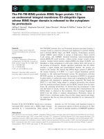
Figure 1 Induction of AtTSPO mRNA by abiotic stresses.(A) Quantitative real-time PCR analyses of AtTspO transcripts upon treatment of
different stresses, (a) 150 mM NaCl, (b) 1 μM ABA, (c) 250 mM mannitol and (d) 0.2 μM methyl viologen. Relative expression levels were
calculated and ACTIN (At3g18780) and 18S rRNA (At3g41768) here used as reference genes. (B) GUS expression in AtTSPO-437::GUS and LHCB::GUS
lines in 15-day-old transgenic Arabidopsis plants either untreated or treated with 150 mM NaCl.
Balsemão-Pires et al. BMC Plant Biology 2011, 11:108
/>Page 3 of 17
1 h, peaking by 3 h and then falling to basal levels
within before increasing between 12-24 h (Figure 1A-d).
To determine if AtTSPO accumulation was a transcrip-
tional response to NaCl stress, a construct, containing 437
bp ups tream the p utative translational sta rt site of the AtT-
SPO gene was fused to the uidA reporter gene (AtTSPO-
437::GUS), and transformed into plants, allowing in vivo
analysis of AtTSP O transcriptional r esponse to stress condi-
tions. AtTSPO-437::GUS was found to be induced by 150
mM NaC l within 3 h of treatment, w hich is similar to q RT-
PCR result s of the endogenous gene (Figure 1B). In control
experiments, 150 mM NaCl resulted in a small decrease of
expression of LHCB::GUS (Figure 1B). Together these
results s uggest that the 437 bp region of AtTSPO promoter
is s ufficient for transcriptional r egulation of T SPO.
Identification and characterization of AtTSPO mutants
To determine the function of AtTSPO in vivo,we
obtained a T-DNA insertional mutant (SALK_135023)
[25] in AtTSPO.Thisline(tspo-1)wasfoundtohave
two tandem T-DNA insertions, 123 bp upstream from
the translational initiation codon of the AtTSPO gene
(Figure 2A). Homozygous lines were then confirmed to
be knock-down mutants by quantitative real time PCR
(qRT-PCR) analysis. In this mutant, TSPO mRNA levels
are about 20% of wild type (Figure 2B).
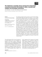
Figure 2 Phenotype of mutants with different levels of AtTSPO expression.(A) Schematic represen tation of isolated insertional mutant of
AtTSPO in Arabidopsis. Two copies of the T-DNA were inserted in tandem 123 bp upstream from the translational initiation codon of AtTSPO.(B)
Total RNA was isolated from 5 day-old seedlings, reverse-transcribed and subjected to qRT-PCR. Data shown represent mean values obtained
from independent amplification reactions (n = 3) and biological replicates (n = 2). Bars represent the standard error of biological replicates. (C)
Root lengths of at least 100 individual 7-day-old seedlings grown in 16 h photoperiods. (D) Chlorophyll concentrations in 14-day-old, in vitro-
grown plants of the indicated genotypes were determined spectrophotometrically. Values shown are means derived from three independent
samples, each sample containing 100 mg of fresh weight. Units are μg of chlorophyll a + b per g of fresh weight (fw).
Balsemão-Pires et al. BMC Plant Biology 2011, 11:108
/>Page 4 of 17
AtTSPO fused or not to the N-terminus of G FP was
constitutively overexpressed from the CaMV 35S pro-
moter in transgenic Arabidopsis lines (OxM1TSPO and
OxM1TSPO:eGFP). We obtained 10 over-expression
lines, but focused on the two homozygous lines that
exhibited ~500 fold over-expression of AtTSPO (Figure
2B). The tspo-1 , OxM1TSPO:eGFP and wild-type lines
were grown side-by-side on e ither Murashige & Skoog
(MS) agar medium or soil, and were monitored for pos-
sibl e abnormal phenotyp es. The knock-down plants had
longer roots compared to the wild type and the over-
expression lines (Figure 2C). Moreover, tspo-1 accumu-
lated ~30% less chlorophyll than either the wild type or
the overexpression lines in the presence of 150 mM
NaCl (Figure 2D).
The expression of stress-response genes is enhanced in
tspo-1
AtTSPO expression was previously shown to be regu-
lated by osmotic stress in germination and seedling
growth assays [23]. Because TSPO regulat es the expr es-
sion of photosynthetic genes in R. sphaeroides [16], we
hypothesized that tspo-1 or OxM1TSPO:eGFP mutants
might have an impaired salt stress response. We exam-
ined the expression of some well-known salt stress-regu-
lated genes (RAB18, E RD10 and DREB2A) [26]. As
expected, stress marker genes were in duced by 150 mM
NaCl in wild-type plants (Figure 3A, B and 3C). In AtT-
SPO over-expression line s, the levels of DREB2A and
RAB18 were lower but no significant change ERD10
expression was observed (Figure 3A, B and 3C).
In tspo-1 mutants, 3 h of 150 mM NaCl treatment
resulted in the increased expression of all three stress
marker genes (Figure 3A, B and 3C). Taken together
these results show that AtTSPO plays an important role
in regulating the expression of stress response genes.
Expression of light-regulated tetrapyrrole genes are
repressed in the tspo-1 knock-down mutant
Consistent with TSPO transporting tetrapyrroles
[17,19,27], tspo-1 plants accumulated less chlorophyll
than wild-type plants (Figure 2D). Because we found
that TSPO is involved in the sal t stress response and
because TSPO negatively regulates photosynthet ic genes
in R. sphaeroides [17]. We next analyzed the expression
of a few key chlorophyll biosynthesis genes in tspo-1
plants.
Initially, we determined the mRNA levels of most of
the key genes in the tetrapyrrole pathway (Additional
file 1) in tspo-1 an d gun5 mutants. GUN5 encodes the
H subunit of chloroplastic Mg-chelatase, which is
involved in the perception of altered levels of tetrapyrro-
lic intermediates [28]. All tetrapyrrole biosynthetic genes
known to be light-dependent [29] were found to be
down-regulated in tspo-1,aswellasingun5 mutants
[28] (Figure 4A, C, E, F, G and 4H), whereas the expres-
sion of the two light-ind ependent genes were unaffected
in wild-type and tspo-1 mutant (Figure 4B and 4D).
Correlation of tetrapyrrole pathway flux and AtTSPO
mRNA levels
tspo-1 mutants present reduced lev els of light- reg ulated
tetrapyrrole metabolism genes (Figure 4A, C, E, F, G,
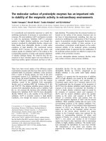
Figure 3 Stress-response genes are up-regulated in tspo-1
during salt stress.(A)-(C) Stress-induced gene expression in
OxM1TSPO:eGFP and tspo-1 lines compared to wild type plants, by
qPCR. 5-day-old seedlings grown under standard conditions and
transferred for 3 hours to plates containing 150 mM NaCl. (A)
DREB2A,(B) RAB18 and (C) ERD10 mRNA levels were determined by
quantitative qRT-PCR. Relative amounts were calculated and
normalized relative to Col-0 non-treated (100%). The ACTIN and 18S
rRNA were used as reference genes. ACTIN, At3g18780; 18S RNA,
At3g41768; RAB18, At5g66400; ERD10, At1g20450; DREB2A,
At5g05410. Data shown represent mean values obtained from
independent amplification reactions (n = 3) and biological replicates
(n = 2). Relative expression levels were calculated. Bars represent the
standard error of biological replicates.
Balsemão-Pires et al. BMC Plant Biology 2011, 11:108
/>Page 5 of 17
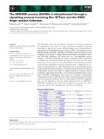
Figure 4 Expression of tetrapyrrole biosynthesis genes in tspo-1 mutant. qRT-PCR analyses of tetrapyrrole biosynthesis genes in Col-0, tspo-
1 and gun5 5-days-old seedlings grown in constant light. Relative amounts were calculated and normalized relative to Col-0 non-treated (100%).
With the exception of HEMA2 and FC1, all the genes have been show to be regulated by light. The data are presented following the enzymes
order in the tetrapyrrole biosynthesis. The ACTIN and 18S rRNA genes were used as control. ACTIN, At3g18780; 18S rRNA, At3g41768; (A) HEMA1
(Glutamyl-tRNA reductase 1 - At1g58290) (B) HEMA2 (Glutamyl-tRNA reductase 2 - At1g04490); (C) PPO (Protoporphyrinogen oxidase -
At4g01690); (D) FC1 (Ferrochelatase 1 - At5g26030); (E) FC2 (Ferrochelatase 2 - At2g30390); (F) GUN2 (Heme oxygenase 1 - At2g26670); (G) GUN4
(Regulator of Mg-porphyrin synthesis - At3g59400); (H) GUN5 (Mg-chelatase subunit H - At5g13630); (I) CAO (Chlorophyllide A oxygenase -
At1g44446); and (J) GUN1 (Pentatricopeptide repeat (PPR) protein - At2g31400). Data shown represent mean values obtained from independent
amplification reactions (n = 3) and biological replicates (n = 2). Relative expression levels were calculated. Bars represent the standard error of
biological replicates.
Balsemão-Pires et al. BMC Plant Biology 2011, 11:108
http://ww
w.biomedcentral.com/1471-2229/11/108
Page 6 of 17
and 4H) and also have low chlorophyll content (Figure
2D). In order to investigate if decreasing flux of tetra-
pyrrole intermediates would affect AtTSPO expression
in wild-type plants, we used two different drugs that
interfere with tetrapyrrole biosynthesis, Gabaculine and
Norflurazon. Gabaculine acts as a tetrapyrrole biosynt h-
esis inhibitor by blocking the glutamate-1-semi aldehyde
aminotransferase activity [30,31]. The herbicide
Norflurazon inhibits carotenoid biosynthesis and indir-
ectly affects enzymes in tetrapyrrole biosynthesis
[32-34]. AtTSPO mRNA levels increased 2-fold in plants
treated with 50 μM of gabaculine and up to 500-fold
after 500 nM norflurazon treatment (Figure 5A).
To explore if AtTSPO expression is affected by genetic
alterations of the tetrapyrrole biosynthesis pathway, we
analyzed the expression of AtTSPO in different mutant
Figure 5 Relationship between tetrapyrrole flux and AtTSPO expression.(A) AtTSPO expression in wild-type plants germinated in 50 μMof
gabaculine or 500 nM of norflurazon compared to untreated plants. (B) AtTSPO mRNA levels in different mutants of the tetrapyrrole pathway.
Relative amounts were calculated and normalized relative to Col-0 non-treated (100%). The ACTIN and 18S rRNA genes were used as control.
ACTIN, At3g18780; 18S RNA, At3g41768. Data shown represent mean values obtained from independent amplification reactions (n = 3) and
biological replicates (n = 2). Relative expression levels were calculated. Bars represent the standard error of biological replicates.
Balsemão-Pires et al. BMC Plant Biology 2011, 11:108
/>Page 7 of 17
backgrounds (Additional file 1). We found that AtTSPO
levels are differently altered in various tetrapyrrole path-
way mutants. AtTSPO steady-state levels were increased
in gun2 (allele of hy1 - required for phytochromobilin
synthesis from heme) [35], gun4 (mutant in the Proto-
porphyrin I X- and Mg-Protoporphyrin IX-binding pro-
tein) [36], fc1 (mutant in the ferrochelatase) [37],
hemA1hemA2 double mutant (mutant in b oth glutamyl-
tRNA reductases genes) [38] an d lin-2 (mutant in the
coproporphyrinogen III oxidase) [39] (Figure 5B). The
increased expression of AtTSPO in these mutants with
reduced tetrapyrrole levels is consistent with AtTSPO
transporting tetrapyrrolesforrolesinothercompart-
ments. The only biosynthetic mutant that resulted in
reduced AtTSPO levels was crd1 (mutant in the Mg-
protoporphyrin IX monomethyl ester cyclase) [40] (Fig-
ure 5B). All these mutations, in exception of crd1 [41],
inhibit s omehow ALA synthesis, suggesting that distur-
bances in tetrapyrrole biosynthesis or accumulation
affect AtTSPO mRNA expression.
AtTSPO localization depends on the translational start site
used
AtTSPO (At2g47770) encodes a protein with a predicted
molecular weight of 18 kDa. This protein has three pos-
sible in -frame ATG-start codons (M1, M21 and M42) in
its N-terminal extension region (Additional file 2) [19].
Since reports of plant TSPO localization have resulted
in different findings subcellular localization of plant
TSPO [19,21,23] we re-examined the subcellular loca-
tion of AtTSPO and evaluated the roles of the N-term-
inalextensionintargetingAtTSPO within the cell. Past
studies [20,23] have utilized N-terminal GFP fusions
that might block potential organellar targeting of AtT-
SPO, particularly mitochondrial or plastid localization.
To allow proper targeting of AtTSPO fusions to GFP,
AtTSPO was placed on the N-terminus of GFP. Three
constructs were made, representing each of the potential
start codons M1 (OxM1TSPO:eGFP), M21 (OxM21T-
SPO:eGFP) and M42 (OxM42TSPO:eGFP) and
expressed from the CaMV 35S promoter in Arabidopsis.
AtTSPO:eGFP subcellular localization was observed in
root, hypocotyls and cotyledons of these lines by conf o-
cal microscopy. Full-length AtTSPO:eGFP (OxM1TSPO:
eGFP) was found in the endoplasmic reticulum (ER) of
the root tip (Figure 6A) and cotyledons (Figure 6C) in
five day-old seedlings. However, in the hypocotyls of
these plants, the fusion protein was found in the ER and
in vesicles of unknown identity (Figure 6B). When M21
(OxM21TSPO:eGFP) or M42 (OxM42TSPO:eGFP) were
used, the fusion proteins always co-localized with mito-
tracker, indicating a mitochondrial localization (Figure
6D,E,F,G,Hand6I)(Additionalfile3).Theseresults
corroborate the previous obs ervations of mitochondrial
localization of TSPO in D. Lanata leaves by immuno-
gold staining and in Arabidopsis by western blot experi-
ments [19], as well as the endoplasmic reticulum located
protein [23], indica ting that the alternative use of three
initiation codons could be important for AtTSPO locali-
zation and its post-translational control.
OxM1TSPOeGFP becomes associated with plastids
following high salt stress
Having established a key role for AtTSPO in response to
abiotic stress, we next examined the localization of AtT-
SPO:e GFP fusion proteins in plants subjected to various
stress conditions. 5 day-old seedlings were treated with
250 mM mannitol, 1 μMABA,0.2μMMVand150
mM NaCl. After 18 hours of treatment, OxM1TSPO:
eGFP became localized to the plastid (Figure 7G, H, I, J,
K and 7L), while neither OxM21TSPO:eGFP nor
OxM42TSPO:eGFP had altered localization even with 5
day extended NaCl treatment (data not shown). AtT-
SPO:GFP localization did not change when plants were
treated with mannitol, ABA or MV (data not shown).
To verify the expression levels of AtTSPO during salt
stress, total protein from each lines was immunoblotted
with antibodies to GFP (Figure 8A). In all cases, AtT-
SPO:GFP protein was found to increase significantly
after 24h of salt treatment. Accumulation of AtTSPO:
GFP was dependent on the presence of AtTSPO because
empty vector controls using CaMV or Ubiquiti n 10 [42]
promoters to drive the expression of GFP did not
change in response to salt stress (Figure 8A and not
shown). These results indicate that AtTSPO accumula-
tion is regulated at the tr anscriptional, post-transcrip-
tional and post-translational levels.
To confirm th e location of AtTSPO we performed
protease prot ection assays on isolated chloroplasts from
OxM1TSPO:eGFP lines that were grown with or with-
out 150 mM NaCl treatment. Following chloroplast iso-
lation and protease protection, equal quantities of
chloroplasts were subjected to immunoblotting with
antibodies to GFP. AtTSPO was detected in chloroplast
fractions near its predicted monomeric molecular mass
(Additional file 4 and 5) in plants treated 18 hours with
150 mM NaCl, but not in untreated plants (Figure 8B).
Chloroplasts prepared from OxTSPO:eGFP lines occa-
sionally displayed a lower molecular mass band that is
approximately the mass of GFP. This band probably
results from proteolysis between AtTSPO and the GFP
tag during sample preparati on, although we cannot rule
out other possibi lities since we do not have a n antibody
to AtTSPO protein itself. Antibodies to RuBisCo and D1
confirmed the inte grity and presence of chloroplasts fol-
lowing protease protection. Antibodies to the cytosolic
protein UGPase also verified these fractions were free of
cytoplasmic con tamination (Additional file 5). These
Balsemão-Pires et al. BMC Plant Biology 2011, 11:108
/>Page 8 of 17
data together with confocal microscopy indicate th at the
region between the M1 and M21 is important for target-
ing AtTSPO to chloroplasts during salt stress. Since
AtTSPO was protected from trypsin digestion (Figure
8B), AtTSPO maybeintegraltothechloroplastouter
envelope.
Discussion
The localization of TSPO in both chloroplasts and mito-
chondria is consistent with its role in porphyrin traffick-
ing. Plant TSPO has been proposed to participate in the
interaction between plastid and mitochondrial tetrapyr-
role biosynthetic pathways [19]. In higher plants,
Figure 6 AtTSPO has different sub-cellular location depending on the translational start site used. Confocal images o f OxM1TSPO:eGFP
(A-C), OxM21TSPO:eGFP (D-F) and OxM42TSPO:eGFP (G-I) localization. OxM1TSPO:eGFP localizes in the ER and vesicles of unknown function in
the root (A), hypocotyl (B) and cotyledon (C). OxM21TSPO:eGFP localizes in the mitochondria of root (D), hypocotyl (E) and cotyledon (F).
OxM42TSPO:eGFP show mitochondria localization in root (G), hypocotyls (H) and cotyledons (I). GFP fluorescence is represented by green and
chlorophyll auto fluorescence in red. The samples were incubated with Mitotracker to identify mitochondria (see Additional file 3). Homozygous
transgenic plants harboring 35S-TSPO:eGFP in wild-type background were used for the analysis. Scale bars = 50 μm.
Balsemão-Pires et al. BMC Plant Biology 2011, 11:108
/>Page 9 of 17
tetrapyrroles are synthesized almost exclusively in plas-
tids, with the exception of the two last steps of heme
synthesis that may occur in bot h chloroplasts and mito-
chondria. If AtTSPO is involved in tetrapyrrole transport
[19],itisreasonabletoassumethatAtTSPO may trans-
locate tetrapyrrole i ntermediates across organellar mem-
branes, explaining why plants would need chloroplastic
and mitochondrial isoforms of TSPO.
Consistent with this hypothesis, the AtTSPO protein is
longer than its mammalian and bacterial counterparts.
The targeting det erminants for chloroplasts and mito-
chondria are usually located at the N-terminus of the
protein; therefore, a fusion protein with GFP fused to
the C-terminus of TSPO was made. Using this strategy
we demonstrated that AtTSPO had different sub-cellular
localization patterns depending on the translational start
Figure 7 OxM1TSPOeGFP localizes in chloroplasts upon salt stress.(A-F) Confocal analyses show OxM1TSPO:eGFP localization in the ER and
vesicles of unknown function in hypocotyls of 5-day-old seedlings grown in the standard conditions. (G-L) Confocal analyses show OxM1TSPO:
eGFP chloroplast localization in hypocotyls of 5-day-old seedlings grown in the presence of 150 mM NaCl. GFP fluorescence channel is
represented in green and chlorophyll auto fluorescence channel is represented in red. Homozygous transgenic plants harboring 35S-TSPO:eGFP
in wild-type background were used for the analysis. Scale bars = 50 μm.
Balsemão-Pires et al. BMC Plant Biology 2011, 11:108
/>Page 10 of 17
codon, tissue type or the abiotic stress to which t he
plant was subjected. Placement of the GFP fusion on
either the N- or C-terminus of TSPO probably explains
the inconsistencies in previous studies [19,21,23] com-
pared to those presented here. The construct used b y
Guillaumot [23] had the YFP fused to the N-terminus of
AtTSPO, which could potentially mask the transit pep-
tide that t argets TSPO to chloroplasts and mitochon-
dria. Indeed, a similar case was observed recently for the
Arabidopsis HEMERA protein (HMR). When HMR had
CFP on its C-terminus, it was localized exclusively in
chloroplasts, however, fusion of HMR to the C-terminus
Figure 8 AtTSPO accumulation and chloroplast localization upon salt stress.(A) Immunoblot analysis of OxAtTSPO:eGFP (OxM1TSPO:eGFP,
OxM21TSPO:eGFP and OxM42TSPO:eGFP) fusion proteins detected in plants with an antibody to GFP. Plants were untreated, or treated with 150
mM NaCl. As a control wild-type plants and plants over-expressing GFP (OxeGFP) seedlings were used. (B) Anti-GFP immunoblot of trypsinized
chloroplasts from Arabidopsis plants either untreated or treated with 150 mM NaCl. Control immunoblots were probed with antibodies
chloroplast proteins RuBisCo and D1; and to cytosolic UGPase. Each lane represents equal amounts of chloroplasts.
Balsemão-Pires et al. BMC Plant Biology 2011, 11:108
/>Page 11 of 17
of YFP (YFP-HMR) was localized to the nucleus and
cytoplasm but not chloroplasts [43].
Salt-stress of Arabidopsis results in movement of ER-
localized AtM1TSPO:eGFP to the chloroplast. We also
demonstrated that the different start codons within the
TSPO N-terminal extension could target the TSPO pro-
tein to different organelles. Other plant proteins such as
MDAR (Arabidopsis Monodehydroascorbate Reductase)
and tRNA nucleotidyltransferase [44,45] are also known
to be targeted to different organelles owing to alterna-
tive transcriptionsal start sites. Thus, it is tempt ing to
speculate that, cloroplastic AtTSPO may protect the
chloroplast from salt stress damage and the mitochon-
drial AtTSPOmaynormallyimportchloroplast-synthe-
sized porphyrins into the mitochondria. Alterations in
the sub-cellular localization of TSPO have been
observed in mammals. The mammalian TSPO localizes
to the mitochondrial outer membrane but during fast
cell proliferation, such as metastatic processes, it relo-
cates to the nuclear membrane, suggesting developmen-
tal control of its sub-cellular localization [46].
Our results suggest the existence of a chloroplast tar-
geting region in AtTSPO that operates during salt stress.
Constructs lacking the N-terminus of At TSPO (AtM2T-
SPO:eGFP and AtM3TSPO:eGFP) are not able to be tar-
get to this organelle. These results suggest that the first
twenty aminoacids of AtTSPO may be part of the chlor-
oplast targeting peptide of this protein. Further experi-
ments should be conducted to precisely characterize this
chloroplast targeting determinant.
TSPO localization in plant cells is complex, involving
a relocation of t he protein from ER and vesicles to
chloroplasts during salt-stress. In recent years, several
new mechanisms for import of proteins into chloro-
plasts have been proposed. For example, it is hypothe-
sized that close contacts between the envelopes of
chloroplasts, mitochondria and other organelle mem-
branes could allow protein movement between them
[47]. Such fusions have been observed between the
mitochondria and ER [48], where it was suggested that
vesicle associated membrane protein 1 (VAMP-1) might
be involved in the docking of mitochondria to target
membranes [49]. This, in turn, could facilitate a re-loca-
lization of proteins from mitochondria to other com-
partments. The recent discoveries of close intracellular
membrane contacts in plants, namely between chloro-
plasts and the ER [50], as well as between mitochondria
and the nucleus [51], corroborates this hypothesis. At
the present moment it is not clear which pathway is
used during AtTSPO relocat ion during salt-stress. How-
ever our data indicate that AtTSPO changes its localiza-
tion during stress, and that it is also possible that the
mitochondrial isoform observed previously by Frank et
al. [22]in P. patens and by Lindenman et a l.[19]inD.
lanata and Arabidopsis could be generated in Arabidop-
sis by the use of alternative translation start codons.
Transcriptional levels of AtTSPO in wild-type Arabi-
dopsis plants increase in response to salt, mannitol,
ABA and paraquat. The promoter region of AtTSPO
was also found to be sufficient for salt stress transcrip-
tional response. The induction of AtTSPO by salt stress
was also observed when constitutive promoters (35S and
UBI10) were used to express AtTSPO, suggesting that
the induction of AtTSPO occurs at both transcript ional
and post-transcriptional levels.
AtTSPO over-expression lines have decreased level s of
stress response genes (ERD10, DREB2A and RAB18),
while
tspo-1 mutants
over express these genes. This sug-
gests that AtTSPO expression and/or function is neces-
sary for the p roper regulation of these genes during
stress conditions. These results also imply that AtTSPO
is important for stress adaptation in Arabidopsis,and
this idea is consistent with results from P. patens [22].
Since Rhodobacter TSPO is a negative regulator of
photosynthetic genes [17], it is possible that AtTSPO
operates similarly in regulating stress responsive genes
in plants.
The precise function of AtTSPO in tetrapyrrole trans-
port during salt stress remains to be established. There
are, however, many reports suggesting that alterations in
tetrapyrrole flow can be involved in salt tolerance. Exo-
genous 5-Aminolevulinate (ALA) can improve salt toler-
ance in higher plants [51-56]. It has been also shown
that transgenic Arabidopsis, tobacco and rice that over-
produce ALA have improved salt tolerance [57,58].
Abdelkader et al.[59]assumedthathighsaltstress
inhibited chlorophyll accumulation mainly by reducing
the rate of porphyrin formation, and Zhang et al.[58]
showed that salt stress caused a significant decrease in
heme content. Thus in hig her plants, ALA and tetrapyr-
role synthesis is sensitive to salt stress.
Additionally, we demonstrated that AtTSPO is impor-
tant for tetrapyrrole flux and/or metabolism. The herbi-
cide Norflurazon, a non-competitive inhibitor of
phytoene desaturase, [31-33] and the neurotoxin Gaba-
culine, which inhibits tetrapyrrole biosynthesis by block-
ing glutamate-1-semi aldehyde aminotransferase activity
[29,30] were used in this study to decrease the flux
through the tetrapyrrole biosynthesis pathways. Our
data showed that mutations in tetrapyrrole biosynthesis
genes and the application of these two different drugs
that decrease flux of tetrapyrrole intermediates affect
AtTSPO expression. All mutations tested that inhibit the
synthesis of ALA increase AtTSPO mRNA steady-state
levels. The same was observed when the formation of
ALA is inhibited by the norflurazon and the gabaculine.
The only mutant tested with decreased AtTSPO expres-
sion is crd1, which accumulates Mg-Protoporphyrin
Balsemão-Pires et al. BMC Plant Biology 2011, 11:108
/>Page 12 of 17
monomethyl ester and this accumulation does not affect
the inhibition of ALA synthesis [40]. Finally, it is possi-
ble that AtTSPO could b e involved in the partitioning
of different tetrapyrrolic signal molecules within plant
cells. The steady state levels of several light-regulated
mRNAs of tetrapyrrole metabolism genes are down-
regulated in the tspo-1 mutant, suggesting that, AtTSPO
could act as a regulator of tetrapyrrole biosynthesis
similar to its bacterial counterpart [16].
Conclusions
TSPO has been shown to transport a number of small
molecules in multiple organisms, however its function in
plants is not known. Here we demonstrate that Arabidop-
sis TSPO is regulated at the transcriptional, post-transcrip-
tional and post-translational levels in response to abiotic
stress conditions such as salt stress. Our results suggest
that AtTSPO can localize to ER and mitochondria, but
when plant s are salt stressed AtTSPO is found in chloro-
plasts. Also our data suggest that under normal conditions
AtTSPO may be important for the import of chloroplastic
synthesized heme into the mitochondria. However, target-
ing AtTSPO to the chloroplast during salt stress may pro-
tect chloroplasts from damage. In addition, tetrapyrrole
intermediates has been suggested to operate in the chloro-
plast-to-nucleus retrograde signaling [35,60]. It is possible
that AtTSPO could be involved in the partitioning of dif-
ferent tetrapyrrole signal molecules within plant cells
depending on environmenta l conditions. AtTSPO may
play a role in re-directing tetrapyrrole intermediates dur-
ing salt stress or under conditions where tetrapyrrole
metabolism is compromised. This is suggested by our
finding that mutation or inhibition of the tetrapyrrole bio-
synthesis pathway increases AtTSPO expression. At the
same time, AtTSPO may directly contribute to the detoxi-
fication of highly reactive porphyrins in the cytoplasm. We
are currently investigating these possibilities.
Methods
Plant material and growth conditions
Arabidopsis thaliana seeds ecotype Col-0 were surface
sterilized and plated on MS
1/2
medium [61] with or
without 50 mM kanamycin. Seedlings were maintained
for t hree days at 4°C and than grown under 16/8 hours
light/dark cycles at 23°C in growth chambers. Root
length measurements were conducted using plants
grown on vertically oriented in standard conditions for
10 days. For abiotic stress treatment, 150 mM NaCl, 250
mM mannitol, 1 μM ABA (Sigma; St Louis, MO) or 0.2
μM paraquat was added to MS
1/2
agar plates, and the 5-
day-old seedlings were incubated under normal growth
condition. For Norflurazon or Gabaculine experiments
seeds were plated on MS
1/2
containing 1 or 2% sucrose
with or without 5 μMnorflurazon(Sandoz
Pharmaceuticals; Vienna, Austria) or 50 μM of gabacu-
line (Sigma, USA). All experiments were repeated three
times independently and the average was calculated.
RNA extraction and qRT-PCR analysis
Total RNA was isolated using Spectrum™ Plant Total
RNA Kit (Sigma #STRN250-1KT), according to manu-
facturer’ s instructions. One microgram of total RNA
was added to each cDNA synthesis reaction using the
First Strand cDNA Synthesis Kit (#K1611). For qRT-
PCR, DNA amplificatio n was performed in the presence
of SYBR
®
Green qPCR Detection (Invitrogen) in a
MyIQ™ Single Color Rea l-Time PCR Detection System
(BioRad), using the primer pairs at table 1. The cycle
Table 1 Primers used for quantitative real time PCR (qRT-
PCR)
PRIMER NAME FOR qPCR SEQUENCE
TSPO FWD ACAAAGGAAAACGCGATCAAA
TSPO RVS ACTTGAGACCACGTTTCGCC
GUN1 FWD GCGATTCTGAATGCTTGCAG
GUN1 RVS AGGAGCCATACATTCTCTCT
GUN2 FWD AGACTCCAATTTCCCAACTT
GUN2 RVS TTACCAGGACGTGTTGGTTC
GUN4 FWD GAAACCGCGACCATATTCGAC
GUN4 RVS CGGCTTCTCCGGATATCTGAA
GUN5 FWD CATCCACTTGCTCCAACCATG
GUN5 RVS CCGACAACCGTTGCATCTTT
HEMA1 FWD GCTTCCGCAGTCTTCAAACG
HEMA1 RVS CCAGCGCCAATTACACACATC
HEMA2 FWD AGCTCCTGCACGGTCCAAT
HEMA2 RVS TGCTATCGTTCCCATCGCAT
FC1 FWD ATACCAGAGTCGTGTTGGCCC
FC1 RVS TCATCGGTGTATGGCTTCAGC
FC2 FWD TGGTGCTATGGCTGTCTCAAAC
FC2 RVS AGCGGAACTAACGACTGTCGA
CAO FWD TGATGAGCCACCTGCACCTAT
CAO RVS AAGTAAACCGTGTTCCACCGG
PPO FWD GCTTCTTCCGTCGTTTTCGAA
PPO RVS TTGAAGATCCGACGGTTGGTC
DREB2A FWD CAGGCTTAAATCAGGACCGG
DREB2A RVS ATGAACCGTTGGCAACACTG
ERD10 FWD CACCGTTCCAGAGCAGGAGA
ERD10 RVS GCCGATGATTCCTCTGTTGC
RAB18 FWD AAGGAGAAGTTGCCAGGTCATC
RAB18 RVS CATCGCTTGAGCTTGACCAG
ACTIN 2/8 FWD TCTTGTTCCAGCCCTCGTTT
ACTIN 2/8 RVS TCTCGTGGATTCCAGCAGCT
18S RNA FWD TATAGGACTCCGCTGGCACC
18S RNA RVS CCCGGAACCCAAAAACTTTG
Balsemão-Pires et al. BMC Plant Biology 2011, 11:108
/>Page 13 of 17
use was: 95 C, 1 min and 30 sec; 40 × (95 C, 10 sec; 60
C, 1 min); 95 C, 1 min; 60 C, 1 min and 81 × (60 C, 10
sec). The relative mRNA levels were determined by nor-
malizing the PCR threshold cycle number with Actin
and 18S RNA. All experiments were repeated three
times independently and the average was calculated.
Verification of TSPO knock-out
The tspo-1 T-DNA mutant, SALK_135023, was obtained
from the Salk collection [24]. Homozygous mutants
were isolated by PCR-based genotyping using gene spe-
cific PCR primers AtTSPO-LP and AtTSPO-BP together
with LBa1 (Table 2). Only homozygous lines were used
for the phenotypic investigation.
Construction of AtTSPO GUS Fusion Vector and GUS
Assay
The 437 bp upstream of the translational star site of the
AtTSPO gene (At2g47770) was translational fused into
uidA gene in pKGWFS7 vector by Gateway
®
(Invitro-
gen™) [62] and introduced into Arabidop sis via Agro-
bacterium-mediated transformation [63]. For cloning
primers and constructs information see Tables 2 and 3,
respectively. For histochemical GUS expression plant
samples were soaked at 37°C for 16 hours in GUS assay
solution (1 mm 5-bromo-4-chloro-3-indolylglucronide,
0.5 mm K
3
Fe(CN)
6
,0.5mmK
4
Fe(CN)
6
, 0.3% (v/v) Tri-
ton X-100, 20% (v/v) methanol, and 50 mm i norganic
phosphate-buffered saline). The reaction was further
conducted at 37°C in the dark for a maximum of 16
hours.
Subcellular localization of AtTSPO fusion proteins
For the GFP fusion constructs, clones containing the
coding region of AtTSPO as well as fusions starting at
methionine 21 and 42 were generated and c loned into
pK7FWG2 [62] (Table 3) according to the manufac-
turer’s instructions (Invitrogen, CA, USA). Primers used
were: AtTSPO M1: TSPO NT1 and TSPO CT1; AtT-
SPO M2: TSPO NT2 and TSPO CT1; AtTSPO M3:
TSPO NT3 and TSPO CT1; AtTSPO 80aa: TSPO NT1
and TSPO CT80 (Table 2). Arabidopsis thaliana was
observed in a c onfocal laser sc anning microscope Leica
DM IRE2 (Leica microsystems). For the mitochondrial-
specific staining, Arabidopsis seedlings were incubated
in MitoTracker
®
Red CMXRos (Invitrogen, #M7512)
according to manufactures instructions. Excitation and
emission wavelengths were 488 and 505-530 nm (BP
505-530 filter) for GFP and, 543 and 56 0-615 nm (BP
560-615 filter) for MitoTracker
®
respectively. All images
were processed on Leica DM IRE2 Image Browser pro-
gram (Leica microsystems).
Determination of chlorophyll contents
Seedlings at 10 days after germination were we ighted,
frozen in liquid nitrogen, and ground in 80% (v/v) acet-
one. Ground tissue was centrifuged at 2,000 g for 5 min
to pellet any insoluble material. The absorbance of the
extracted chlorophyll at 645 and 663 nm was then
determined. Chlorophyll (a and b) contents of the sam-
ples were determined according to Lichtenthaler [64].
Chloroplast Isolation
Isolation of chloroplasts from plate-grown Arabidopsis
seedlingswasperformedasdescribed previously [65].
Final resuspension of chloroplast was in buffer (330 mM
sorbitol, 50 mM HEPES-KOH, pH 8.0) at a concentra-
tion of 1 mg chlorophyll ml
-1
.
Table 2 Primers used for cloning and genotyping
PRIMER NAME FOR GENOTYPING AND CLONING SEQUENCE
AtTSPO LP agagcaaatcgcatcagcgtc
AtTSPO RP ggaacgtaaccggatcccaaa
LBa1 tggttcacgtagtgggccatcg
TSPO NT1 aaaaagcaggctccatggattctcaggaca
TSPO NT2 aaaaagcaggctccatggccgagacagagagg
TSPO NT3 aaaaagcaggctccatggcgaaacgtggtctc
TSPO CT1 agaaagctgggtccgcgacagcaagctttaca
TSPO CT80 agaaagctgggtcggacttagctcgattcccgta
Table 3 Constructs information
CONSTRUCT NAME BINARY VECTOR RESISTANCE IN PLANT
UBQ10mCITRINE pB7m34GW basta
UBQ10M1TSPOmCITRINE pB7m34GW basta
UBQ10M2TSPOmCITRINE pB7m34GW basta
UBQ10M3TSPOmCITRINE pB7m34GW basta
OxeGFP pK7FWG2 kanamycin
OxMITSPO:eGFP pK7FWG2 kanamycin
OxM2TSPO:eGFP pK7FWG2 kanamycin
OxM3TSPO:eGFP pK7FWG2 kanamycin
AtTSPO-437::GUS pKGWFS7 kanamycin
Balsemão-Pires et al. BMC Plant Biology 2011, 11:108
/>Page 14 of 17
Immunoblotting
Total protein was extracted from 10-day-old seedlings
by adding protein extraction buffer (50 mM HEPES pH
7.9, 300 mM Sucrose, 150 mM NaCl, 10 mM Potassium
acetate, protease inhibitors cockta il - Roche, 1% (w/v)
Triton, 1 mM DTT). Ground tissue wa s centrifuged at
5,000×gfor5mintopelletthetissueandproteins
were quantify by Bradford assay [66]. Samples were
boiled for 5 min in 250 mM Tris-HCl, pH 6.8, 10% (w/
v) SDS, 30% (v/v) glycerol, 5% (v/v) b- mercaptoethanol
and 0.02% (w/v) bromophenol blue. SDS-PAGE was per-
formed using standard procedures. Chloroplast protein
samples were normalized loaded by equal amounts of
total chlorophyll. Following SDS-PAGE, the separated
proteins were transferred to a polyvinylidene difluoride
membrane (Bio-Rad). For immunodetection, membranes
were incubated with antibody against GFP (ROCHE,
#11814460001), UGPase (AGRISERA, #AS05086),
RuBisCo (AGRISERA, #AS03037) and D1(AGRISERA,
#AS05084). With the exception of GFP detection that
uses mouse secondary antibody, all the immunoreactive
proteins were detected by using rabbit secondary anti-
body. The immunoreaction was detected by chemilumi-
nescence kit (Thermo Scientific, #34076) according to
manufacturer’s instructions.
Additional material
Additional file 1: Schematic representation of tetrapyrrole
biosyntheses pathway in plants showing genes analyzed in this
study. In blue, are the genes already described for each step in the
pathway. The enzymes that correspond to these genes names and the
AGI code are: HEMA1 (Glutamyl-tRNA reductase 1, At1g58290); HEMA2
(Glutamyl-tRNA reductase 2, At1g09940); HEMA3(Glutamyl-tRNA reductase
3, At2g31250); FLU (Regulator of ALA synthesis, At3g14110); LIN2
(Coproporphyrinogen oxidase 1, At1g03475); GUN2 (Heme oxygenase 1,
At2g26670); GUN3 (Phytochromobilin synthase, At3g09150); GUN4
(Regulator of Mg-porphyrin synthesis, At3g59400); GUN5 (Mg-chelatase
subunit H, At5g13630); CHLI (Mg-chelatase subunit I, At4g18480 and
At5g45930); CHLD (Mg-chelatase subunit D, At1g08520); CHLM (Mg-
Protoporphyrin IX methyltransferase, At4g25080); CRD1 (Mg-
Protoporphyrin IX monomethylester cyclase, At3g56940); FC1
(Ferrochelatase 1, At5g26030); FC2 (Ferrochelatase 2, At2g303 90).
Additional file 2: Alignment of TSPO sequences from different
organisms. ClustalW sequence alignment of TSPO proteins from
Rhodobacter sphaeroides (AF195122.1), Rattus norvegicus (J05122) and
Arabidopsis TSPO (AtTSPO - At2g47770). The numbers in the left side
represent the amino acid position from the primary protein. In the
consensus line the conserved aminoacids are highlighted as (*), and as (.)
when one conserve position is observed. M1, M21 and M42 AtTSPO
isoforms are highlighted. The black arrow represents the first 80
aminoacids (AtTSPO80aa) of Arabidopsis TSPO.
Additional file 3: AtM42TSPO:eGFP co-localizes with mitotracker in
Arabidopsis thaliana. AtM42TSPO:eGFP 5-day-old seedlings transgenic
lines (A-C) were incubated with mitotracker to identify mitochondria. (A)
Image from GFP channel is shown in green. (B) Image from mitotracker
channel is shown in red. (C) Merge between GFP and mitotracker
channels shown in yellow. Scale bar = 50 μM.
Additional file 4: Immunoblot showing that AtTSPO:eGFP
accumulates during salt stress. Immunoblot analysis of protein level in
all three isoforms of OxAtTSPO:eGFP (OxM1TSPO:eGFP, OxM21TSPO:eGFP
and OxM42TSPO:eGFP) during salt stress show accumulation of the
protein. As a control wild-type plants and plants over-expressing GFP
(OxeGFP) were used. Anti-UGPase and Red-ponceau staining were used
as loading controls. Equal amounts of total protein were loaded.
Additional file 5: Immunoblot of chloroplasts prepared from
OxTSPO:eGFP plants. Arabidopsis chloroplasts were prepared from 10-
days-old seedlings either untreated or treated with 150 mM NaCl and
immunoblotted with antibodies to GFP, RuBisCo, D1 and UGPase. Equal
amounts of OxM1TSPO:eGFP chloroplast protein samples were loaded in
each lane. (TP) Total Protein; (Chl) Chloroplast protein; PP (Protease
Protection treatment).
Abbreviations
ABA: Abscisic acid; ALA: 5-Aminolevulinate; CFP: Cyan fluoresce nt protein;
D1: photosystem II reaction center D1 protein; HMR: Hemera protein; GFP:
Green fluorescent protein; GUS: β-Glucuronidase; Mg-Proto IX: Mg-
Protoporphyrin IX; MV: Methyl viologen; ROS: Reactive oxygen species;
RUBISCO: Ribulose-1,5-biphosphate carboxylase; TSPO: 18 kDa Translocator
protein;
UGPase: UDP-glucose pyrophosphorylase; VAMP: Vesicle associated
membrane protein; YFP: Yellow fluorescent protein.
Acknowledgements
We thank Jesse Woodson, Juan M. Perez-Ruiz, Ana Lucia Giannini, Amanda
Mangeon and Marcio Castro Silva-Filho for providing critical feedback on the
manuscript. The Salk Institute provided the insertion mutant lines and ABRC
for providing material. Pedro Paulo de Abreu Manso and Bernardo Miguel
de Oliveira Pascarelli from the Rede de Plataformas Tecnológicas da
Fundação Instituto Oswaldo Cruz (FioCruz) for technical support on the
confocal microscopy analysis. Luiza da Silva for technical support with plant
transformation. EBP was supported by PhD fellowship from CAPES
(Coordenação de Aperfeiçoamento de Pessoal de Nível Superior) and SWE
fellowship from CNPq (Conselho Nacional de Desenvolvimento Científi co e
Tecnológico), The Salk Institute and Balcoffee Trading Intermediações Ltda.
YJ is supported by a long-term fellowship from the European Molecular
Biology Organization (EMBO) and from the Marc and Eva Stern Foundation.
BJSCO is supported by fellowship F32GM086037 from the National Institutes
of Health and National Institute of General Medical Sciences. JGU is
supported by American Cancer Society grant RSG-05-196-01-CCG. This work
was supported by grants from DOE FG02-04ER15540 from the U.S.
Department of Energy to JC and the Conselho Nacional de
Desenvolvimento Científico e Tecnológico (CNPq) and the Fundação Carlos
Chagas Filho de Amparo à Pesquisa do Estado do Rio de Janeiro (FAPERJ) to
GSM.
Author details
1
Laboratório de Genômica Funcional e Transdução de Sinal, Departamento
de Genética, Universidade Federal do Rio de Janeiro, Rio de Janeiro, Brazil.
2
Plant Biology Laboratory, The Salk Institute, 10010 North Torrey Pines Road,
La Jolla, CA 92037, USA.
3
Howard Hughes Medical Institute 4000 Jones
Bridge RoadChevy Chase, MD 20815-6789, USA.
4
Laboratório de
Biomineralização, Instituto de Ciências Biomédicas, Universidade Federal do
Rio de Janeiro, Brasil.
Authors’ contributions
EBP, JC and GSM conceived and designed the experiments. EBP performed
all the experiments, analyzed the data and wrote the paper. YJ helped in
the confocal microscopy analyses. BJSCO helped in the fractionation
experiment. LRA and JGU gave technical support. JC and GSM were project
supervisors, participated in the discussion of all experiments from the project
and preparation of the manuscript. All authors read and approved the final
manuscript.
Received: 14 January 2011 Accepted: 20 June 2011
Published: 20 June 2011
References
1. Beale SI: Enzymes of chlorophyll biosynthesis. Photosynth Res 1999,
60:43-73.
Balsemão-Pires et al. BMC Plant Biology 2011, 11:108
/>Page 15 of 17
2. Papenbrock J, Grimm B: Regulatory network of tetrapyrrole biosynthesis -
studies of intracellular signaling involved in metabolic and
developmental control of plastids. Planta 2001, 213:667-681.
3. Vavilin DV, Vermaas WFJ: Regulation of the tetrapyrrole biosynthetic
pathway leading to heme and chlorophyll in plants and cyanobacteria.
Physiol Plant 2002, 115:9-24.
4. Reinbothe S, Reinbothe C: The regulation of enzymes involved in
chlorophyll biosynthesis. Eur J Biochem 1996, 237:323-343.
5. Grimm B: Novel insights in the control of tetrapyrrole metabolism of
higher plants. Curr Opin Plant Biol 1998, 1:245-250.
6. Papadopoulos V, Boujrad N, Ikonomovic MD, Ferrara P, Vidic B: Topography
of the Leydig cell mitochondrial peripheral-type benzodiazepine
receptor. Mol Cell Endocrinol 1994, 104:R5-R9.
7. McEnery MW, Snowman AM, Trifiletti RR, Snyder SH: Isolation of the
mitochondrial benzodiazepine receptor: association with the voltage-
dependent anion channel and the adenine nucleotide carrier. Proc Natl
Acad Sci USA 1992, 89:3170-3174.
8. Papadopoulos V, Baraldi M, Guilarte TR: Translocator protein (18 kDa): new
nomenclature for the peripheral-type benzodiazepine receptor based on
its structure and molecular function. Trends Pharmacol Sci 2006,
27:402-409.
9. Li H, Papadopoulos V: Peripheral-type benzodiazepine receptor function
in cholesterol transport. Identification of a putative cholesterol
recognition/interaction amino acid sequence and consensus pattern.
Endocrinology 1998, 139:4991-4997.
10. Papadopoulos V: Structure and function of the peripheraltype
benzodiazepine receptor in steroidogenic cells. Proc Soc Exp Biol Med
1998, 217:130-142.
11. Lacapere JJ, Papadopoulos V: Peripheral-type benzodiazepine receptor:
structure and function of a cholesterol-binding protein in steroid and
bile acid biosynthesis. Steroids 2003, 68:569-585.
12. O’Hara MF, Craig RC, Nemeth KR, Charlap JH, Knudsen TB: Mitochondrial
benzodiazepine receptors regulate oxygen homeostasis in the early
mouse embryo. Reprod Toxicol 2003, 17:365-75.
13. Galiegue S, Casellas P, Kramar A, Tinel N, Simony-Lafontaine J:
Immunohistochemical assessment of the peripheral benzodiazepine
receptor in breast cancer and its relationship with survival. Clin Cancer
Res 2004, 10:2058-2064.
14. Taketani S, Kohno H, Okuda M, Furukawa T, Tokunaga R: Induction of
peripheral-type benzodiazepine receptors during differentiation of
mouse erythroleukemia cells. A possible involvement of these receptors
in heme biosynthesis. J Biol Chem 1994, 269:7527-7531.
15. Yeliseev AA, Kaplan S: A sensory transducer homologous to the
mammalian peripheral-type benzodiazepine receptor regulates
photosynthetic membrane complex formation in Rhodobacter
sphaeroides 2.4.1. J
Biol Chem 1995, 270:21167-75.
16. Yeliseev AA, Kaplan S: A novel mechanism for the regulation of
photosynthesis gene expression by the TspO outer membrane protein
of Rhodobacter sphaeroides 2.4.1. J Biol Chem 1999, 274:21234-43.
17. Yeliseev AA, Krueger KE, Kaplan S: A mammalian mitochondrial drug
receptor functions as a bacterial ‘’oxygen’’ sensor. Proc Natl Acad Sci USA
1997, 94:5101-6.
18. Lindemann P, Koch A, Degenhardt B, Hause G, Grimm B, Papadopoulos V:
A novel Arabidopsis thaliana protein is a functional peripheral-type
benzodiazepine receptor. Plant Cell Physiol 2004, 45:723-733.
19. Vanhee C, Guillon S, Masquelier D, Degand H, Deleu M, Morsomme P,
Batoko H: A TSPO-related protein localizes to the early secretory
pathway in Arabidopsis, but is targeted to mitochondria when expressed
in yeast. Journal of Experimental Botany 2010, 16:1-12.
20. Corsi L, Avallone R, Geminiani E, Cosenza F, Venturini I, Baraldia M:
Peripheral benzodiazepine receptors in potatoes (Solanum tuberosum).
Biochemical and Biophysical Research Communications 2004, 313:62-66.
21. Frank W, Baar K-M, Qudeimat E, Woriedh M, Alawady A, Ratnadewi D,
Gremillon L, Grimm B, Reski R: A mitochondrial protein homologous to
the mammalian peripheral-type benzodiazepine receptor is essential for
stress adaptation in plants. The Plant Journal 2007, 51:1004-1018.
22. Guillaumot D, Guillon S, Deplanque T, Vanhee C, Gumy C, Masquelier D,
Morsomme P, Batoko H: The Arabidopsis TSPO-related protein is a stress
and abscisic acid-regulated, endoplasmic reticulum-Golgi-localized
membrane protein. The Plant Journal 2009, 60:242-256.
23. Kreps JA, Wu Y, Chang HS, Zhu T, Wang X, Harper JF: Transcriptome
changes for Arabidopsis in response to salt, osmotic, and cold stress.
Plant Physiol 2002, 130:2129-41.
24. Alonso JM, Stepanova AN, Leisse TJ, Kim CJ, Chen H, Shinn P,
Stevenson DK, Zimmerman J, Barajas P, Cheuk R, Gadrinab C, Heller C,
Jeske A, Koesema E, Meyers CC, Parker H, Prednis L, Ansari Y, Choy N,
Deen H, Geralt M, Hazari N, Hom E, Karnes M, Mulholland C, Ndubaku R,
Schmidt I, Guzman P, Aguilar-Henonin L, Schmid M, et al: Genome-wide
insertional mutagenesis of Arabidopsis thaliana. Science 2003, 301:653-7.
25. Chai M-F, Wei P-C, Chen Q-J, Rui A, Cheng J, Yang S, Wang X-C: NADK3, a
novel cytoplasmic source of NADPH, is required under conditions of
oxidative stress and modulates abscisic acid responses in Arabidopsis
.
The
Plant Journal 2006, 47:665-674.
26. Verma A, Nye JS, Snyder SH: Porphyrins are endogenous ligands for the
mitochondrial (peripheral-type) benzodiazepine receptor. Proc Natl Acad
Sci USA 1987, 84:2256-2260.
27. Mochizuki N, Brusslan JA, Larkin R, Nagatani A, Chory J: Arabidopsis
genomes uncoupled 5 (GUN5) mutant reveals the involvement of Mg-
chelatase H subunit in plastid-to-nucleus signal transduction. Proc Natl
Acad Sci USA 2001, 98:2053-2058.
28. Stephenson PG, Terry MJ: Light signalling pathways regulating the Mg-
chelatase branchpoint of chlorophyll synthesis during de-etiolation in
Arabidopsis thaliana. Photochem Photobiol Sci 2008, 10:1243-52.
29. Hill CM, Pearson SA, Smith AJ, Rogers LJ: Inhibition of chlorophyll
synthesis in Hordeum vulgare by 3-amino-2,3-dihydrobenzoicacid
(gabaculine). Biosci Rep 1985, 5:775-81.
30. Grimm B, Smith AJ, Kannangara CG, Smith M: Gabaculine-resistant
galutamate1-semialdehyde amino transferase of Synechococcus. J Biol
Chem 1991, 266:12496-501.
31. Mayfield SP, Taylor WC: Carotenoid-deficient maize seedlings fail to
accumulate light-harvesting chlorophyll a/b binding protein (LHCP)
mRNA. Eur J Biochem 1984, 144:79-84.
32. Oelmüller R, Mohr H: Photooxidative destruction of chloroplast and its
consequences for expression of nuclear genes. Planta 1986, 167:106-113.
33. Taylor WC: Regulatory interactions between nuclear and plastid
genomes. Ann Rev Plant Physiol Plant Mol Biol 1989, 40:211-233.
34. Susek RE, Ausubel FM, Chory J: Signal transduction mutants of Arabidopsis
uncouple nuclear CAB and RBCS gene expression from chloroplast
development. Cell 1993, 74:787-99.
35. Larkin RM, Alonso JM, Ecker JR, Chory J: GUN4, a regulator of chlorophyll
synthesis and intracellular signaling. Science 2003, 299:902-6.
36. Chow KS, Singh DP, Walker AR, Smith AG: Two different genes encode
ferrochelatase in Arabidopsis: mapping, expression and subcellular
targeting of the precursor proteins. Plant J 1998, 15:531-41.
37. Kumar AM, Söll D: Antisense HEMA1 RNA expression inhibits heme and
chlorophyll biosynthesis in Arabidopsis.
Plant Physiol 2000, 122:49-56.
38.
Ishikawa A, Okamoto H, Iwasaki Y, Asahi T: A deficiency of
coproporphyrinogen III oxidase causes lesion formation in Arabidopsis.
Plant J 2001, 27:89-99.
39. Tottey S, Block MA, Allen M, Westergren T, Albrieux C, Scheller HV,
Merchant S, Jensen PE: Arabidopsis CHL27, located in both envelope and
thylakoid membranes, is required for the synthesis of
protochlorophyllide. Proc Natl Acad Sci USA 2003, 100:16119-24.
40. Peter E, Rothbart M, Oelze M-L, Shalygo N, Dietz K-J, Grimm B: Mg-
protoporphyrin monomethylester cyclase deficiency and effects on the
tetrapyrrole metabolism in different light conditions. Plant and Cell Phys
2010, 51:1229-41.
41. Christensen AH, Quail PH: Ubiquitin promoter-based vectors for high-
level expression of selectable and/or screenable marker genes in
monocotyledonous plants. Transgenic Res 1996, 5:213-8.
42. Chen M, Galvão RM, Li M, Burger B, Buger J, Bolado J, Chory J: Arabidopsis
HEMERA/pTAC12 initiates photomorphogenesis by phytochromes. Cell
2010, 141:1230-1240.
43. Obara K, Sumi K, Fukuda H: The Use of Multiple Transcription Starts
Causes the Dual Targeting of Arabidopsis Putative
Monodehydroascorbate Reductase to Both Mitochondria and
Chloroplasts. Plant Cell Physiol 2002, 43(7):697-705.
44. von Braun SS, Sabetti A, Hanic-Joyce PJ, Gu J, Schleiff E, Joyce PBM: Dual
targeting of the tRNA nucleotidyltransferase in plants: not just the
signal. Journal of Experimental Botany 2007, 58(5/16):4083-4093.
Balsemão-Pires et al. BMC Plant Biology 2011, 11:108
/>Page 16 of 17
45. Hardwick M, Fertikh D, Culty M, Li H, Vidic B, Papadopoulos V: Peripheral-
type benzodiazepine receptor (PBR) in human breast cancer: Correlation
of breast cancer cell aggressive phenotype with PBR expression, nuclear
localization and PBR-mediated cell proliferation and nuclear transport of
cholesterol. Cancer Research 1999, 59:831-842.
46. Krause K, Krupinska K: Nuclear regulators with a second home in
organelles. Trends in Plant Science 2009, 14:194-199.
47. Soltys BJ, Gupta RS: Mitochondrial-matrix proteins at unexpected
locations: are they exported? Trends Biochem Sci 1999, 24:174-177.
48. Isenmann S, Khew-Goodall Y, Gamble J, Vadas M, Wattenberg BW: A splice-
isoform of vesicle-associated membrane protein-1 (VAMP-1) contains a
mitochondrial targeting signal. Mol Biol Cell 1998, 9:1649-1660.
49. Andersson MX, Goksör M, Sandelius AS: Optical manipulation reveals
strong attracting forces at membrane contact sites between
endoplasmic reticulum and chloroplasts. J Biol Chem 2007, 282:1170-1174.
50. Segui-Simarro JM, Coronado MJ, Staehelin LA: The mitochondrial cycle of
Arabidopsis shoot apical meristem and leaf primordium meristematic
cells is defined by a perinuclear tentaculate/cage-like mitochondrion.
Plant Physiol 2008, 148:1380-1393.
51. Watanabe K, Tanaka T, Hotta Y, Kuramochi H, Takeuchi Y: Improving salt
tolerance of cotton seedlings with 5-aminolevulinic acid. Plant Growth
Regul 2000, 32:99-103.
52. Nishihara H, Kizaka-Kondoh S, Insel PA, Eckmann L: Inhibition of apoptosis
in normal and transformed intestinal epithelial cells by cAMP through
induction of inhibitor of apoptosis protein (IAP)-2. Proc Natl Acad Sci USA
2003, 22:8921-6.
53. Watanabe K, Ryoji O, Rasid MM, Suliman A, Tohru T, Hitoshi K, Yasutomo T:
Effects of 5-aminolevulinic acid to recover salt damage on cotton,
tomato, and wheat seedlings in Saudi Arabia. J Arid Land Stud 2004,
14:105-113.
54. Wang LJ, Jiang WB, Liu H, Liu WQ, Kang L, Hou XL: Promotion of 5-
aminolevulinic acid on germination of pakchoi (Brassica campestris ssp.
chinensis var. communis Tsen et Lee) seeds under salt stress. J Integr
Plant Biol 2005, 9:1084-91.
55. Watanabe K, Ryoji O, Rasid MM, Suliman A, Tohru T, Hitoshi K, Zhang ZJ,
Li HZ, Zhou WJ, Takeuchi Y, Yoneyama K: Effect of 5-aminolevulinic acid
on development and salt tolerance of potato (Solanum tuberosum L.)
microtubers in vitro. Plant Growth Regul 2006, 49:27-34.
56. Youssef T, Awad MA: Mechanisms of enhancing photosynthetic gas
exchange in date palm seedlings (Phoenix dactylifera L.) under salinity
stress by a 5-aminolevulinic acid-based fertilizer. J Plant Growth Regul
2008, 27:1-9.
57. Jung S, Back K, Yang K, Kuk YI, Chon SU: Defence response produced
during photodynamic damage in transgenic rice overexpressing 5-
aminolevulinic acid synthase. Photosynthetica 2008,
46:3-9.
58. Zhang Z-P, Yao Q-H, Wang L-J: Expression of yeast Hem1 gene controlled
by Arabidopsis HemA1 promoter improves salt tolerance in Arabidopsis
plants. BMB reports 2010, 330-336.
59. Abdelkader AF, Aronsson H, Sundqvist C: High salt stress in wheat leaves
causes retardation of chlorophyll accumulation due to a limited rate of
protochlorophyllide formation. Physiol Plant 2007, 130:157-166.
60. Strand A, Asami T, Alonso J, Ecker JR, Chory J: Chloroplast to nucleus
communication triggered by accumulation of Mg-protoporphyrinIX.
Nature 2003, 421:79-83.
61. Murashige T, Skoog F: A revised medium for rapid growth and bioassays
with tobacco tissue cultures. Physiol Plant 1962, 15:473-497.
62. Karime M, Inzé D, Depicker A: GATEWAY™ vectors for Agrobacterium-
mediated plant transformation. Trends in Plant Science 2002, 5:193-5.
63. Clough SJ, Bent AF: Floral dip: a simplified method for Agrobacterium-
mediated transformation of Arabidopsis thaliana. Plant J 1998, 16:735-743.
64. Lichtenthaler HK: Chlorophyll and carotenoids: pigments of
photosynthetic biomembranes. Methods Enzymol 1987, 148:349-382.
65. Fitzpatrick LM, Keegstra K: A method for isolating a high yield of
Arabidopsis chloroplasts capable of efficient import of precursor
proteins. Plant J 2001, 27:59-65.
66. Bradford MM: A rapid and sensitive method for the quantitation of
microgram quantities of protein utilizing the principle of protein-dye
binding. Analyt Biochem 1976, 72:248-254.
doi:10.1186/1471-2229-11-108
Cite this article as: Balsemão-Pires et al.: The Arabidopsis translocator
protein (AtTSPO) is regulated at multiple levels in response to salt
stress and perturbations in tetrapyrrole metabolism. BMC Plant Biology
2011 11:108.
Submit your next manuscript to BioMed Central
and take full advantage of:
• Convenient online submission
• Thorough peer review
• No space constraints or color figure charges
• Immediate publication on acceptance
• Inclusion in PubMed, CAS, Scopus and Google Scholar
• Research which is freely available for redistribution
Submit your manuscript at
www.biomedcentral.com/submit
Balsemão-Pires et al. BMC Plant Biology 2011, 11:108
/>Page 17 of 17