báo cáo khoa học: "Localization of DIR1 at the tissue, cellular and subcellular levels during Systemic Acquired Resistance in Arabidopsis using DIR1:GUS and DIR1:EGFP reporters" ppsx
Bạn đang xem bản rút gọn của tài liệu. Xem và tải ngay bản đầy đủ của tài liệu tại đây (10.72 MB, 16 trang )
Champigny et al. BMC Plant Biology 2011, 11:125
/>
RESEARCH ARTICLE
Open Access
Localization of DIR1 at the tissue, cellular and
subcellular levels during Systemic Acquired
Resistance in Arabidopsis using DIR1:GUS and
DIR1:EGFP reporters
Marc J Champigny1,4, Heather Shearer1,4, Asif Mohammad1, Karen Haines1, Melody Neumann2, Roger Thilmony3,5,
Sheng Yang He3, Pierre Fobert4, Nancy Dengler2 and Robin K Cameron1*
Abstract
Background: Systemic Acquired Resistance (SAR) is an induced resistance response to pathogens, characterized by
the translocation of a long-distance signal from induced leaves to distant tissues to prime them for increased
resistance to future infection. DEFECTIVE in INDUCED RESISTANCE 1 (DIR1) has been hypothesized to chaperone a
small signaling molecule to distant tissues during SAR in Arabidopsis.
Results: DIR1 promoter:DIR1-GUS/dir1-1 lines were constructed to examine DIR1 expression. DIR1 is expressed in
seedlings, flowers and ubiquitously in untreated or mock-inoculated mature leaf cells, including phloem sieve
elements and companion cells. Inoculation of leaves with SAR-inducing avirulent or virulent Pseudomonas syringae
pv tomato (Pst) resulted in Type III Secretion System-dependent suppression of DIR1 expression in leaf cells.
Transient expression of fluorescent fusion proteins in tobacco and intercellular washing fluid experiments indicated
that DIR1’s ER signal sequence targets it for secretion to the cell wall. However, DIR1 expressed without a signal
sequence rescued the dir1-1 SAR defect, suggesting that a cytosolic pool of DIR1 is important for the SAR
response.
Conclusions: Although expression of DIR1 decreases during SAR induction, the protein localizes to all living cell
types of the vasculature, including companion cells and sieve elements, and therefore DIR1 is well situated to
participate in long-distance signaling during SAR.
Background
Acquired resistance, or “immunization” of plants was
originally documented more than seventy years ago in a
review published by Kenneth Chester in which varying
degrees of immunity were observed in plants that had
recovered from an initial pathogen attack [1]. The term
systemic acquired resistance (SAR) was originally used
by Ross to describe systemic resistance induced by
necrosis-causing viruses in tobacco [2] and is more generally defined as a defense mechanism induced by a
localized infection that results in broad-spectrum
* Correspondence:
1
Department of Biology, McMaster University, Hamilton, ON L8S 4K1 Canada
Full list of author information is available at the end of the article
resistance in distant tissues to normally virulent pathogens [3,4].
Research using tobacco, cucumber and, more recently,
Arabidopsis models indicates that SAR occurs in distinct
stages. The first, or induction, stage is initiated when a
necrosis-causing pathogen infects a leaf and results in
either the formation of a localized hypersensitive
response (HR) and local resistance, or in diseaseinduced necrosis [3]. A recent report demonstrated systemic immunity in the absence of necrotic cell death in
the induced leaf [5], highlighting the fact that the precise cellular mechanisms governing the initiation of SAR
are still unclear. Formation of the necrotic lesion results
in a 10 to 50-fold accumulation above basal levels of the
plant defense hormone, salicylic acid (SA),[6-11] and in
© 2011 Champigny et al; licensee BioMed Central Ltd. This is an Open Access article distributed under the terms of the Creative
Commons Attribution License ( which permits unrestricted use, distribution, and
reproduction in any medium, provided the original work is properly cited.
Champigny et al. BMC Plant Biology 2011, 11:125
/>
the expression of pathogenesis-related (PR) genes
[6,11,12]
During the initiation stage of SAR, a mobile signal or
signals is induced to travel and is later perceived in distant, uninfected tissues. Several lines of evidence indicate that the signal travels through the phloem,
including girdling experiments in tobacco that reduce
the translocation of molecules through phloem tissue.
Additionally, the pattern of sucrose transport from
source to sink leaves in Arabidopsis was similar to
transport of the SAR signal from induced leaves to protect upper leaves against Pseudomonas syringae pv
maculicola (Psm). Although these and other experiments [reviewed in 13] suggest the SAR signal is
phloem-mobile, cell-to-cell movement down the petiole,
or a combination of these two modes of transport cannot be ruled out.
The discovery that SA levels in the phloem rise dramatically in SAR-induced tobacco [9] and cucumber
[10] led to the hypothesis that SA itself may be a SAR
mobile signal [14]. SA was shown to be critically
involved in the SAR pathway because transgenic tobacco
plants expressing a salicylate hydroxylase gene (NahG)
were unable to accumulate SA or to manifest a SAR
response [14]. However, a number of experiments provide evidence that SA is not a SAR mobile signal.
Cucumber plants in which induced leaves were detached
prior to the accumulation of SA in their petioles still
manifested a SAR response in systemic tissue [15].
Furthermore, grafting experiments utilizing transgenic
NahG tobacco demonstrated that NahG-expressing
rootstocks blocked in the accumulation of SA were
nonetheless competent to translocate a mobile signal to
the scion [16].
The establishment phase of SAR involves the perception of the mobile signal(s) in distant tissue, resulting in
a modest accumulation of SA and expression of PR
genes in Arabidopsis and tobacco [7,8,11]. In the final,
or manifestation, stage of SAR, the plant responds to
normally virulent pathogens in a resistant manner [3].
Manifestation of SAR is associated with the expression
and activity of a set of SAR genes [17] including the
previously described PR genes. An earlier, more rapid or
more abundant accumulation of these SAR proteins
may be the molecular basis for systemic resistance. The
physiological function of many of these genes has not
been determined but increases in peroxidase activity in
induced cucumber [18], chitinase activity in Arabidopsis
and cucumber [19], as well as antifungal properties in
vitro [20] suggest that these proteins play a role in producing a resistant state.
Isolation and characterization of Arabidopsis mutants
has been a powerful approach to decipher the mechanism of SAR. By screening a collection of T-DNA tagged
Page 2 of 16
Arabidopsis lines for mutants that fail to develop SAR
following induction with avirulent Pseudomonas syringae
pv tomato (Pst), the defective in induced resistance 1-1
(dir1-1) mutant was identified [21]. The dir1-1 mutant
was not compromised in basal resistance and, interestingly, overexpression of DIR1 did not enhance disease
resistance or lead to a constitutive SAR response. Petiole
exudates, enriched for phloem sap, collected from SARinduced wild-type leaves were effective in inducing the
SAR marker gene PR-1 when infiltrated into wild-type
or dir1-1 plants, suggesting that the long-distance SAR
signal was present in these wild type petiole exudates
and that dir1-1 can perceive this signal. However, exudates similarly collected from dir1-1 leaves were incapable of inducing PR-1 expression in wild-type leaves,
suggesting that this mutant is defective either in the
synthesis of the SAR mobile signal or its transport to
distant leaves [21]. These data and the fact that DIR1
encodes a putative lipid transfer protein led to the
hypothesis that DIR1 is involved in long distance signaling and may chaperone a lipid signal to distant leaves
during SAR [21,13].
Lipid transfer proteins (LTPs) are ubiquitous in plants
and are associated with many developmental and stress
response processes [22]. The structure of a number of
LTPs has been determined revealing that they possess a
consensus motif of eight cysteine residues engaged in
four disulphide bridges forming a central hydrophobic
cavity which can bind long chain fatty acids [22]. Lascombe et al. [23] determined the structure and lipid
binding properties of DIR1 expressed in the yeast Pichia
pastoris using fluorescence and X-ray diffraction. DIR1
shares some structural and lipid binding properties with
the LTP2 family. In vitro, DIR1 can bind two monoacylated phospholipids and contains two proline-rich SH3
domains. SH3 domains participate in protein-protein
interactions in numerous proteins [23]. Lascombe et al.
postulate that the DIR1 SH3 domains may play a role in
interacting with the putative SAR signal receptor in distant leaves. A number of studies implicate glycerolipids
[24,25], methyl salicylate (MeSA) and azelaic acid (AA)
as SAR long distance signal candidates [26-28]. Overexpression/SAR studies in dir1-1 identified two tobacco
DIR1 orthologs indicating that DIR1 is important for
SAR in both Arabidopsis and tobacco [29]. A recent
paper by Chanda et al. [30] provides evidence suggesting
that glycerol-3-phosphate (G3P) may also be a SAR long
distance signal.
If DIR1 is chaperoning a signal(s) to distant leaves
during SAR, we hypothesize that DIR1 accesses sieve
elements for long distance movement. Therefore, DIR1
promoter transgenic lines were investigated to localize
DIR1 in leaves at the cellular and subcellular levels in
healthy untreated plants and during SAR. Our results
Champigny et al. BMC Plant Biology 2011, 11:125
/>
indicate that the DIR1 promoter directs constitutive
expression in seedlings and all leaf cell types. Moreover,
although DIR1 expression is reduced upon SAR induction, DIR1 is still expressed in all living cell types comprising the vascular tissue.
Results
Localization of DIR1 in leaves during SAR
Previous RNA and protein gel blot expression studies
indicated that DIR1 is expressed constitutively at low
levels in rosette leaves of 3 to 4 week old plants and its
expression is reduced after SAR induction [21]. If DIR1
is involved in the long distance signaling stage of the
SAR pathway, it is possible that DIR1 is expressed in
the phloem, specifically companion cells, providing it
direct access to the phloem for long distance movement.
Moreover, expression limited to the phloem would be
consistent with low DIR1 RNA and protein levels
observed in whole leaves [21]. DIR1 expression in leaves
was examined using the ß-glucuronidase (GUS) reporter
gene. The GUS reporter was chosen to amplify the weak
DIR1 expression signal and allow visualization of DIR1
expression in various tissues and at the cellular level.
Transgenic plant lines were created in which the DIR1
promoter region was placed upstream of GUS in wildtype (ecotype Ws) plants or upstream of a DIR1-GUS
fusion in the dir1-1 mutant background (see Methods
for details). A number of plant lines were examined at
four weeks post germination (wpg) for GUS activity
before and during SAR. DIR1pro:GUS in Ws lines 1, 11,
23 and DIR1pro:DIR1-GUS in dir1-1 lines 3, 15, 29
were mock-inoculated (10 mM MgCl2), inoculated with
SAR-inducing avirulent Pst (avrRpt2) or left untreated.
Similar results were observed in all plant lines (Figure 1
and Additional Files 1, 2) Inoculated leaves and uninoculated systemic leaves from the same plant were collected at 14 or 20 hours post inoculation (hpi), stained
for GUS activity and observed using light microscopy.
Under low magnification, abundant GUS activity was
observed in untreated and mock-inoculated leaves in the
vasculature and mesophyll cells in both the DIR1pro:
GUS-11 and DIR1pro:DIR1-GUS-29 lines. In contrast,
less intense GUS staining was observed in inoculated
and systemic leaves of both transgenic lines (11, 29)
inoculated with avirulent Pst (Figure 1A). Due to differences in cell density and vacuole size of cells in the midvein, secondary vein and mesophyll, it is not possible to
compare GUS activity levels between these tissues.
Therefore GUS activity was measured separately in each
of these tissues using a relative scale of 0 to 4, where 0
represents little to no GUS activity and 4 represents
intense GUS activity or staining (Figure 1B, C) to quantify the observed reduction in GUS activity observed in
Figure 1A. Intense staining occurred in the midvein and
Page 3 of 16
secondary veins in mock-inoculated or untreated leaves
of both the DIR1pro:GUS-11 and DIR1pro:DIR1-GUS29 lines, whereas the level of GUS activity was reduced
in inoculated and uninoculated systemic leaves of plants
inoculated with SAR-inducing Pst (avrRpt2). A similar
reduction in GUS activity was observed in mesophyll
cells of inoculated or systemic leaves collected from
plants induced for SAR compared to untreated or
mock-inoculated leaves (Figure 1). Comparable results
for DIR1pro:GUS-23 in Ws and DIR1pro:DIR1-GUS-3
in dir1-1 are presented as Additional Files 1,2 and 3.
These studies indicate that the DIR1 promoter region
initiates expression of GUS and DIR1-GUS throughout
the leaf and confirms previous RNA gel blot data [21]
that DIR1 expression is reduced after SAR induction
with Pst (avrRpt2). DIR1 expression in the vasculature
was examined in more detail to determine if DIR1 is
expressed in phloem cells using both DIR1pro:DIR1GUS-29/dir1-1 and DIR1pro:GUS-11/Ws lines. GUSstained leaf and petiole midveins from 4 week-old plants
were embedded, sectioned and viewed under high magnification. GUS activity was present in all living cell
types including the developing xylem tracheary elements, xylem parenchyma, phloem and phloem parenchyma in midveins of untreated, mock-inoculated,
inoculated and systemic leaves from plants induced for
SAR (Figure 2). DIR1 expression was reduced, but still
detectable in all cell types of the midvein in leaves
induced for SAR, including both companion cells and
sieve elements of the phloem (Figure 2 and Additional
File 4). DIR1-GUS activity was also observed in all cells
of untreated petiole midveins (see Additional file 5HI).
Therefore, DIR1 is expressed in the phloem before and
during SAR induction and may access the phloem for
long distance movement during SAR.
Expression of DIR1 in seedlings, roots and flowers was
also examined using the DIR1pro-DIR1-GUS-29/dir1-1
line. DIR1-GUS activity was observed throughout sevenday old seedlings including the roots, trichomes and in
flowers and flower bolts of mature plants (see Additional file 5A-G).
Reduction in DIR1 expression during SAR induction is Pstdependent
A number of studies have demonstrated that virulence
effectors delivered by the Type III Secretion System
(T3SS) of Pst are involved in suppressing Arabidopsis
cell wall-mediated basal resistance which includes the
formation of cell wall callose appositions near Pst colonies and the expression of a number of secreted proteins
including some LTPs [31-33]. We hypothesized that the
reduction in DIR1 expression after inoculation with Pst
observed in this and our previous study [21] could be
the result of T3SS delivery of virulence effectors into
Champigny et al. BMC Plant Biology 2011, 11:125
/>
Page 4 of 16
B
DIR1pro:DIR1-GUS-29/dir1-1 (14 hpi)
Midvein
2° vein
Mesophyll
Relative GUS activity
3
M
2.5
2
1.5
1
0.5
0
U M I
S
U M
I S
U M I
S
U
C
DIR1pro:GUS-11/Ws (20 hpi)
2° vein
Midvein
Mesophyll
Relative GUS activity
3
I
100 um
DIR1pro:DIR1GUS-29/dir1-1
S
DIR1pro:GUS
-11/Ws
2.5
2
1.5
1
0.5
0
U M
I
S
U M
I S
U M
I
S
Figure 1 DIR1 expression in leaves using the DIR1 promoter:GUS plant lines. (A) DIR1pro:DIR1-GUS-29/dir1-1 and DIR1pro:GUS-11/Ws
plants lines (3.5 wpg) were left untreated (U), mock-inoculated (M) or inoculated with 106 cfu ml-1 of SAR-inducing PstavrRpt2 (I) and harvested
at 14 hpi, 20, 40 hpi and subjected to histochemical GUS analysis. Staining pattern were similar at all time points, therefore 14 hpi is shown for
DIRpro:DIR1-GUS-29/dir1-1 and 20 hpi leaves for DIRpro:GUS-11/Ws. Systemic leaves were also collected from plants that were SAR induced (S).
Representative leaves from each line were photographed in a single sitting without adjusting microscope settings and two different leaves are
shown. The bar represents 100 μm. Measurement of relative GUS activity in (B) DIR1pro:DIR1-GUS-29/dir1-1 and (C) DIR1pro:GUS/Ws. Leaves from
the experiment presented in panel A were scored using a subjective relative scale of 0 to 4, with 0 representing little GUS staining and 4
representing intense GUS staining. U = uninoculated, M = mock-inoculated, I = inoculated leaf from SAR-induced plants, S = systemic leaf from
SAR-induced plants. The asterisk (*) denotes a significant difference (student’s t test) between mock-inoculated leaves and leaves induced for
SAR. This experiment was repeated once with similar results.
the plant cell. To test this hypothesis, DIR1 expression
was monitored in wild-type plants inoculated with either
virulent Pst or a hrpS Pst mutant. A high inoculum dose
was used (108 cfu ml-1) because nonpathogenic Pst hrp
mutants do not reliably induce host transcriptional
responses at the lower doses [34] typically used in Arabidopsis-Pst inoculation experiments. Leaves were collected at 3,6,9 and 18 hpi for RNA gel blot analysis. The
T3SS is not functional in hrpS mutants and therefore no
Pst-encoded virulence effectors would be delivered into
the plant cell [35,36]. DIR1 was expressed at low levels
in untreated leaves and its expression increased from 3
to 18 hpi after infection with hrpS Pst (Figure 3A). In
leaves inoculated with wild-type virulent Pst, DIR1
expression was reduced at 6 and 9 hpi, but this suppression was attenuated by 18 hpi (Figure 3A). These data
Champigny et al. BMC Plant Biology 2011, 11:125
/>
Page 5 of 16
Mock
Un
SARinduced
(inoculated
leaf)
SARinduced
(systemic
leaf)
DIR1pro:DIR1-GUS-29/dir1-1
DIR1pro:GUS-11/Ws
Figure 2 Cellular localization of DIR1 in leaves. Leaves from experiments presented in Figure 1 were collected and stained for GUS. Leaves
were embedded, sectioned and photographed. Representative sections through untreated (un), mock-inoculated (mock) and SAR-induced
(inoculated and systemic) leaf midveins of DIR1pro:DIR1-GUS-29/dir1-1 (14 hpi) and DIR1pro:GUS-11/Ws (20 hpi) plants are displayed.
demonstrate that reduction in DIR1 observed after
inoculation with Pst is not a response by the plant, but
rather a consequence of the delivery of Pst virulence
effectors into the plant cell.
To examine which cell types are affected by Pst virulence effectors, DIR1-GUS expression in the DIR1pro:
DIR1-GUS-29/dir1-1 line was monitored after inoculation with wild type Pst and a hrpA Pst mutant that does
Champigny et al. BMC Plant Biology 2011, 11:125
/>
Page 6 of 16
A
Pst hrpS
0
3
6
9
Pst
18
3
6
9
18
hpi
DIR1
B
hrpA Pst
Avir Pst
Midvein
not make the major pilus protein, HrpA and therefore
cannot form the T3SS Hrp pilus or deliver effectors into
the plant cell [36]. The hrpA mutant or wild-type virulent or avirulent Pst (avrRpt2) were inoculated (106 cfu
ml -1 dose) into DIR1pro:DIR1-GUS-29/dir1-1. Inoculated leaves were collected at 6 and 12 hpi, stained and
scored for GUS activity. Similar results were obtained at
both 6 and 12 hpi, therefore just the 12 hpi data is presented in Figure 3B and 3C. Mock-inoculated leaves and
leaves from plants inoculated with hrpA Pst displayed
high GUS activity in the midvein, secondary vein and
mesophyll cells compared to leaves inoculated with virulent (data not shown) or avirulent Pst (Figure 3B). These
visual results were corroborated by determining the relative GUS activity using the subjective GUS scale as
described above. GUS activity was reduced in the midvein, secondary vein and mesophyll cells in leaves inoculated with either avirulent or virulent Pst as compared
to leaves inoculated with hrpA Pst (Figure 3C). Therefore inoculation with virulent or avirulent Pst leads to
suppression of DIR1 expression in the midvein, secondary vein and mesophyll cells of leaves in a T3SS-dependent manner.
DIR1 is targeted to the cell wall
Mesophyll
and
secondary
veins
Midvein
2° vein
Mesophyll
Relative GUS Activity
C
M hA A V
M hA A V
M hA A V
Figure 3 Reduction in DIR1 expression during SAR is Pstdependent. (A) Plants were vacuum-infiltrated with 108 cfu ml-1
hrpS Pst or Pst, followed by RNA gel blot analysis of DIR1 expression
at 0,3,6,9 and 18 hpi. Total RNA before blotting is shown to indicate
equal RNA loading per well. This experiment was repeated once
with similar results. (B, C) DIR1pro:DIR1-GUS-29/dir1-1 plants were
inoculated with 106 cfu ml-1 PstavrRpt2 (Avir) or hrpA Pst. Inoculated
leaves were collected at 12 hpi and photographed (B) and relative
GUS activity was determined in midveins, secondary veins and
mesophyll cells using the 0-4 subjective GUS scale. The asterisk (*)
denotes a significant difference (student’s T- test) between mockinoculated (M) and leaves inoculated with hrpA (hA) or avirulent (A)
or virulent (V) Pst (C). This experiment was repeated once with
similar results.
Lipid transfer proteins enter the endoplasmic reticulum
(ER) and secretory pathway as preproteins under the
direction of a short, N-terminal ER entry peptide of 20
to 26 amino acids that is cleaved after entry into the ER.
The mature proteins are secreted outside the cell and
are typically associated with cell walls [37-39], although
several of these proteins have been discovered intracellularly within protein storage vacuoles or glyoxisomes
[40,41]. The functionality of the predicted DIR1 signal
sequence was examined by Agrobacterium-mediated
transient transformation with T-DNA encoding fulllength DIR1 fused to the EYFP (enhanced yellow fluorescent protein) reporter (35S:DIR1-EYFP), truncated
DIR1 lacking the putative signal sequence fused to EYFP
(35S:DIR1Δ1-25-EYFP) or 35S:EYFP into Nicotiana tobaccum followed by laser scanning confocal microscopy to
localize EYFP fusion proteins in tobacco leaf epidermal
cells.
Localization of DIR1Δ1-25-EYFP was identical to that
of the EYFP control, such that fluorescence was
observed in 60 of 60 cells at the cell periphery, in cytoplasmic strands and also within the nucleus (Figure 4A,
B). Detection of these proteins in the nucleus was likely
due to passive diffusion from the cytosol. The 27 kDa
EYFP protein, as well as the DIR1Δ1-25-EYFP fusion are
smaller than the 60 kDa exclusion limit of nuclear pores
[42] such that nuclear detection of cytosolic fluorescent
fusion proteins is commonly observed in plant cells
[43]. DIR1-EYFP exhibited two distinct patterns of
Champigny et al. BMC Plant Biology 2011, 11:125
/>
Page 7 of 16
G
0.9
Absorbance 683 nm
0.8
0.7
0.6
0.5
0.4
0.3
0.2
0.1
0
35S: DIR1 1-25
-GUS-5/dir1-1
DIR1pro:DIR1GUS-29/dir1-1
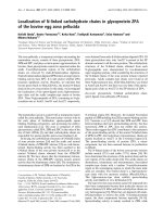
Figure 4 DIR1 ER signal sequence directs secretion of the protein to the apoplast. Fusion proteins consisting of full length DIR1 fused to
EYFP (DIR1-EYFP) and DIR1 lacking its signal sequence (DIR1Δ1-25 -EYFP) were expressed in Nicotiana tabaccum leaves via Agrobacteriummediated transient expression. Fluorescent proteins were visualized in epidermal cells after 48 hours using confocal microscopy. DIR1-EYFP
expression exhibited two distinct patterns. (A) Fluorescence in the region of the cortical ER and (D) the nuclear envelope and cell periphery.
Expression of DIR1Δ1-25-EYFP and EYFP is shown in (B) and (C), respectively. Propidium iodide staining of the plant cell wall is illustrated in (E),
and extensive colocalization of DIR1-EYFP with the propidium iodide signal is demonstrated in (F). Subcellular localization experiments were
performed three times with similar results. (G) IWFs were collected from untreated leaves of 35S:DIR1Δ1-25-GUS-5/dir1-1 and DIR1pro:DIR1-GUS-29/
dir1-1. GUS activity was determined by measuring the absorbance at 683 nm. This experiment was repeated 2 additional times with similar
results.
localization. In a small number of cells (5/60), DIR1EYFP was detected in a discrete network particularly
enriched near the plasma membrane (Figure 4C) coincident with the cortical ER. In a majority of cells, (55/60),
DIR1-EYFP was localized to the nuclear and cell periphery (Figure 4D). Tobacco epidermal cells have a large
central vacuole largely restricting the cytoplasm to a
thin layer near cell boundaries, making it difficult to distinguish between plasma membrane and cell wall localization. To confirm that DIR1-EYFP was secreted to the
cell wall, cells were counterstained with propidium
iodide, a dye which accumulates in the apoplast as it is
excluded by intact plasma membranes [44,45]. DIR1EYFP partially colocalized (Figure 4F) with the
Champigny et al. BMC Plant Biology 2011, 11:125
/>
A
35S:DIR1
1-25-GUS-5/dir1-1
Mesophyll
Midvein
U
M
I
S
100 μm
B
3
Relative GUS activity
propidium iodide signal (Figure 4E), demonstrating that
the signal sequence directed secretion of DIR1-EYFP
out of tobacco epidermal cells into the cell wall. Patches
of DIR1-EYFP signal did not colocalize with propidium
iodide, but rather with regions surrounding the nucleus
and the cell periphery indicating that some DIR1 molecules localize to the ER secretory system and perhaps
the cytosol.
Transgenic lines that express DIR1 lacking its signal
sequence in the dir1-1 mutant (35S:DIR1Δ1-25-GUS in
dir1-1) were constructed and used to demonstrate the
functionality of the DIR1 signal sequence in Arabidopsis.
A number of lines were characterized (see Methods) and
line 5 was chosen for further study. GUS activity in the
leaves of 35Spro: DIR1Δ1-25-GUS-5/dir1-1 line was monitored by inoculating leaves with 10 6 cfu ml -1 Pst
(avrRpt2) followed by GUS staining at 14 hpi. Similar to
DIR1 promoter-directed expression (Figures 1 and 2),
GUS activity in the 35Spro: DIR1Δ1-25-GUS-5/dir1-1 line
was higher in untreated and mock-inoculated leaves
compared to leaves inoculated with avirulent Pst (Figure
5A,B and Additional File 6). DIR1 Δ1-25 -GUS was
expressed in all cell types of the leaves similar to DIR1
promoter-driven expression of DIR1-GUS. These data
indicate that expression from the 35S promoter, like
that from the DIR1 promoter region, is reduced in
response to inoculation with Pst. However, unlike DIR1
promoter-directed expression, 35S promoter-directed
expression of DIR1Δ1-25-GUS in the midvein and secondary vein of systemic leaves of inoculated plants was
similar to untreated or mock-inoculated leaves
(Figure 5A, B and Additional File 6). Other researchers
have also observed a reduction in 35S promoter-driven
expression after pathogen inoculation. For example,
expression of GUS in 35S:GUS transgenic pear was significantly reduced following infection with Erwinia amylovora [46] and in Arabidopsis and tobacco roots
following infection with Heterodera and Globodera
nematodes [47].
To demonstrate that the DIR1 signal sequence does
target DIR1 to the cell wall in Arabidopsis, intercellular
washing fluids (IWFs) were collected from DIR1pro:
DIR1-GUS-29/dir1-1 and 35S:DIR1Δ1-25-GUS-5/dir1-1
untreated leaves from 4 week old plants. IWFs consist
of cell wall associated proteins and molecules and provide information about the soluble molecules associated
with plant cell walls [48,49]. IWFs collected from DIR1pro:DIR1-GUS-29/dir1-1 and 35S:DIR1 Δ1-25 -GUS-5/
dir1-1 leaves were assayed for GUS activity (see Methods). IWFs from DIR1 Δ1-25 -GUS plants displayed low
GUS activity while IWFs from DIR1-GUS plants displayed high GUS activity (Figure 4G). Therefore when
Page 8 of 16
Midvein
2º vein
Mesophyll
2.5
2
1.5
1
0.5
0
U M I
S
U M I
S U M I
S
Figure 5 Localization of DIR1 lacking its signal sequence. 35S:
DIR1Δ1-25-GUS-5/dir1-1 plants were left untreated (U), mockinoculated (M) or induced for SAR with PstavrRpt2 (106 cfu ml-1).
Inoculated (I) and systemic leaves (S) were collected from
inoculated plants at 14 hpi. Leaves were stained for GUS activity
and photographed in (A) or GUS activity levels were determined in
the midveins, secondary veins and mesophyll cells of untreated (U),
mock-inoculated (M) or leaves induced for SAR (I and S) in (B). The
asterisk (*) denotes a significant difference (student’s t test) between
untreated and inoculated leaves from SAR-induced plants (I). This
experiment was repeated once with similar results.
Champigny et al. BMC Plant Biology 2011, 11:125
/>
We hypothesize that DIR1 may be involved in long distance signaling during SAR and travel cytoplasmically
via the phloem and/or cell to cell. Evidence to date
indicates that proteins destined to travel in the phloem
in Arabidopsis are made in companion cells and enter
sieve elements via companion cell-sieve element plasmodesmata [50-52]. However, DIR1 is targeted to the
cell wall via the secretory system and according to current cell biology knowledge, DIR1 would have no
access to the cytosol and plasmodesmata. We hypothesize that DIR1’s targeting signal sequence is cleaved or
becomes nonfunctional upon SAR induction allowing it
to remain in the cytosol with access to plasmodesmata.
If this was true, then DIR1 without its signal sequence
may still function during SAR. To test this hypothesis,
SAR assays were performed with DIR1pro:DIR1-GUS29/dir1-1 and 35S:DIR1 Δ1-25 -GUS-5/dir1-1 lines plus
Ws and dir1-1. Plants were either induced for SAR
with 106 cfu ml-1 Pst (avrRpt2) or mock-inoculated on
two lower leaves, followed by challenge inoculation
with 105 cfu ml-1 virulent Pst in distant leaves two day
later. Bacterial densities were monitored in challenged
leaves at 3 dpi. Wild-type Ws plants were SAR-competent as demonstrated by the 10-fold reduction in Pst
levels in plants induced for SAR versus those that were
mock-inoculated, while the dir1-1 mutant displayed
high levels of Pst in plants that were or were not
induced for SAR (Figure 6A). Both transgenic lines
expressing either DIR1-GUS or DIR1 Δ1-25 -GUS were
SAR competent as demonstrated by the 6-fold and 4fold decrease, respectively, in Pst levels in induced versus mock-inoculated plants (Figure 6A). A replicate
experiment is shown in Figure 6B in which the transgenic lines displayed a 7- to 8-fold SAR response compared to 5-fold in Ws. Results similar to Figures 6A
and 6B were observed using additional transgenic lines
(DIR1pro;DIR1-GUS/dir1-1 lines 3, 15 and 35S:
DIR1Δ1-25-GUS/dir1-1 lines 17, 20) providing evidence
that expression of DIR1-GUS or DIR1 Δ1-25 -GUS
restores the SAR defect in the dir1-1 mutant. More
importantly, these data suggest that removal of the
DIR1 signal sequence has no deleterious effect on
DIR1’s ability to participate in SAR.
SAR-induced
A
Bacterial Density (cfu leaf disc-1)
Expression of DIR1-GUS or DIR1Δ1-25-GUS rescues the SAR
defect in dir1-1
Mock-induced
107
106
105
B
Bacterial Density (cfu leaf disc-1)
DIR1 possesses its native signal sequence, DIR1-GUS
activity is detected in IWFs which are enriched for soluble cell wall proteins. However, little GUS activity was
detected when the native DIR1 signal sequence was
removed. These results corroborate the tobacco immunofluorescence analysis demonstrating that the native
DIR1 signal sequence targets DIR1 to the cell wall in
Arabidopsis.
Page 9 of 16
107
106
nd
105
Ws
dir1-1
Figure 6 Expression of DIR1-GUS or DIR1Δ1-25-GUS rescues the
SAR defect in dir1-1. SAR assays were conducted on Ws, dir1-1,
DIR1pro:DIR1-GUS-29/dir1-1 and 35S: DIR1Δ1-25-GUS-5/dir1-1 by
inoculating with 10 mM MgCl2 (mock-induced) or inducing for SAR
with Pst-avrRpt2 (SAR-induced) in 1 to 2 lower leaves, followed by
challenge inoculation with virulent Pst in distant leaves 2 days later.
Bacterial density determination was performed in challenged leaves
3 dpi. Asterisks (*) denote a significant difference (student’s t-test) in
bacterial densities between challenged distant leaves of mock- and
SAR-induced plants. Representative results are presented in (A) and
(B) and these experiments have been repeated numerous times
with similar results (see text for details). nd = not determined.
Discussion
The non-specific lipid transfer proteins (LTPs) comprise
a large, multigene family present in numerous plant species [39]. LTPs are basic polypeptides of approximately
Champigny et al. BMC Plant Biology 2011, 11:125
/>
7-9 kD, whose key structural feature is the LTP fold
formed by four disulphide bridges between eight conserved cysteine residues [22]. The LTP fold forms a tunnel-like cavity and in vitro studies indicate it
accommodates various lipids, including phospholipids,
fatty acids, glycolipids, prostaglandin and jasmonic acid
[53-58]. Due to their ability to bind lipids in vitro, LTPs
were originally hypothesized to traffick lipids between
intracellular membranes [59]. However, this function
seems unlikely as a number of LTPs have been demonstrated to be synthesized as preproteins containing an
ER signal sequence such that the mature proteins are
secreted to the apoplast [37].
Although the biochemical mechanisms involved are
not clear, LTP proteins play important roles in plant
defense against pathogens. Several LTPs exhibit antimicrobial activity in vitro [60,61] and overexpression of
select LTP or LTP-like proteins leads to enhanced local
resistance against bacterial and fungal pathogens in Arabidopsis and tobacco [62,63]. DIR1 is the first LTP protein whose function in pathogen resistance is defined
genetically, as the dir1-1 Arabidopsis mutant is impaired
in systemic resistance to Pst (SAR) but local resistance
responses remain intact. Furthermore, 35S promotermediated overexpression of DIR1 does not lead to
enhanced basal resistance or a more robust SAR
response [21], strongly suggesting that DIR1 does not
participate directly in defense against Pst, but instead
plays a role in systemic disease signaling.
The overall goal of this study was to investigate the
signaling role of DIR1 during SAR by localizing it at
the tissue, cellular and subcellular levels. A number of
transgenic lines were created in which the DIR1 promoter region was placed upstream of GUS or a DIR1GUS fusion in Ws and dir1-1, respectively. Examination of these lines indicated that the DIR1 promoter
region initiated expression of GUS and DIR1:GUS in
seedlings, roots and floral tissues and in all living cells
including the veins and mesophyll cells of untreated
and mock-inoculated leaves. This was somewhat unexpected as we had hypothesized that DIR1 expression
might be limited to the vasculature which would
explain the constitutive, but low levels of DIR1 expression observed in leaves [21], while still providing DIR1
access to the phloem for movement during SAR. During SAR induction, DIR1-GUS expression was reduced
in mesophyll cells and vascular tissue of inoculated
and distant systemic leaves of plants induced with
SAR-inducing Pst (avrRpt2). RNA gel-blots [21]
revealed that DIR1 transcript levels declined following
SAR induction in leaves. Therefore reduced GUS activity observed in SAR-induced DIR1pro:GUS transgenic
plants is the result of decreased transcription driven by
the DIR1 promoter region.
Page 10 of 16
Reduction in DIR1 expression could be part of the
SAR response or could be due to Pst-derived effector
molecules delivered into plant cells. To test this hypothesis, expression of DIR1 in leaves inoculated with SARinducing avirulent Pst, virulent Pst, or with a Pst hrpS
mutant, was determined using RNA gel blot analysis.
DIR1 expression was reduced in leaves inoculated with
virulent Pst at 6 and 9 hpi, however by 18 hpi, DIR1
expression was no longer suppressed. Reduction in
DIR1 expression was not observed in leaves inoculated
with Pst hrpS. Instead, DIR1 transcripts accumulated
abundantly at 3, 6, 9 and 18 hpi. A high inoculum dose
was used (108 cfu ml-1) because nonpathogenic Pst hrp
mutants do not reliably induce host transcriptional
responses at the lower doses [34] typically used in Arabidopsis-Pst inoculation experiments. Similar experiments with the DIR1pro:GUS or DIR1pro:DIR1-GUS
plant lines using a lower inoculum level (106 cfu ml-1)
demonstrated that Pst Hrp-dependent suppression of
DIR1 expression occurs in the midvein, secondary veins
and mesophyll cells at 14 and 20 hpi. Numerous in
planta bacterial growth studies have demonstrated that
the infection process proceeds faster in high compared
to low dose experiments [64-68]. Therefore, we speculate that the difference in timing of suppression of DIR1
expression in these two experiments is due to the high
versus low inoculum doses used.
Collectively, these data suggest that suppression of
DIR1 expression occurs through the action of effector
molecules delivered through the Pst T3SS. This supports
numerous studies in which genes associated with Arabidopsis cell wall defense, including a number of LTPs,
are suppressed in a Pst Hrp-dependent manner [31-33].
Transcriptional mechanisms are involved in the Pstmediated downregulation of DIR1 expression in DIR1pro:DIR1-GUS and 35S:DIR1Δ1-25-GUS-5 lines, but it is
also possible that post-transcriptional mechanisms or
DIR1-GUS instability contribute to the observed expression patterns.
Hrp-dependent suppression of DIR1 occurs in all cell
types within inoculated leaves and in distant uninoculated leaves. Recently it was discovered that Pseudomonas syringae suppresses plant defenses not only in the
infected leaf but also in systemic tissues, rendering the
plant more susceptible to subsequent infection, a phenomenon known as Systemic Induced Susceptibility
(SIS) [64]. SIS observed after Pseudomonas infection of
Arabidopsis requires the bacterial toxin coronatine
[69,70], a structural and functional mimic of the defense
hormone jasmonic acid [71]. Interestingly, Pst hrp
mutants are deficient in the production of coronatine
[70]. Reduction of DIR1 expression in systemic tissue
may therefore involve the action of widely mobile, bacterially produced molecules such as coronatine.
Champigny et al. BMC Plant Biology 2011, 11:125
/>
DIR1 expression in the vasculature was examined in
more detail to determine if DIR1 has access to the
phloem and therefore the potential for movement to
distant leaves, a key characteristic of a SAR long distance signal. Microscopic examination of leaf and petiole
cross-sections demonstrated that DIR1-GUS expression
was observed in all living cell types including developing
xylem, xylem parenchyma, mesophyll, phloem parenchyma and phloem. Mature xylem tracheary elements are
dead, and as expected appeared empty with no detectable GUS activity. DIR1 expression was reduced but still
detectable after SAR induction in all living cell types
including companion cells and phloem sieve elements.
Therefore DIR1 is present at the right place (companion
cells) and the right time (during SAR induction) to participate in long distance signaling during SAR.
The subcellular localization of DIR1 and the functionality of DIR1’s predicted signal sequence were examined
by transiently expressing DIR1-EYFP fusion proteins in
tobacco epidermal cells followed by visualization using
confocal microscopy. As expected, EYFP alone and a
fusion construct lacking the predicted ER signal
sequence (DIR1Δ1-25-EYFP) localized to cytosolic strands
and diffused into the nucleus while intact DIR1-EYFP
localized to the ER, cell periphery and showed colocalization with propidium iodide, an apoplastic marker.
DIR1-GUS activity was detected in intercellular washing
fluids from plants expressing wild type DIR1, but GUS
activity was greatly reduced in IWFs collected from
plants expressing DIR1 lacking the signal sequence, corroborating the DIR1-EYFP tobacco localization experiments. Therefore, the DIR1 signal sequence does direct
secretion of DIR1 to the cell wall as has been previously
observed for other LTPs [37-39], Similar results were
also obtained in a recent paper in which DIR-GFP transiently expressed in Nicotiana benthamiana was
observed to localize to the ER [30]. It is difficult to distinguish the plasma membrane from the cell wall using
light microscopy [38] and this may explain why Chanda
et al. [30] concluded that DIR1 is not secreted to the
cell wall. We chose to examine intercellular washing
fluids for the presence of DIR1-GUS for two reasons: to
overcome the light-microscopy-associated problem of
distinguishing the plasma membrane from the cell wall
and to demonstrate that DIR1 is secreted to the cell
wall in both tobacco and Arabidopsis.
Evidence to date indicates that Arabidopsis proteins
destined to travel in the phloem are synthesized in companion cells and move into sieve elements through plasmodesmata [50-52]. Patches of intracellular DIR1:EYFP
were detected in this study, however it is difficult to distinguish the cytosol from the ER and the secretory system. Nevertheless, these data support the idea that some
DIR1 protein is present in the cytosol and therefore
Page 11 of 16
gains access to the phloem through the cytosol of companion cells. Alternatively, DIR1 could enter the cytosol
if the function of the signal sequence is disrupted during
SAR induction. It is also possible that pathogen-induced
cell membrane disruption during the HR (SAR induction) may allow cell wall proteins including DIR1 to
enter cells. In any case, we hypothesized that cytosolic
localization of a pool of DIR1 is required for translocation of the long-distance SAR signal. To address this
question, a transgenic line was created in which the signal sequence was deleted from DIR1 (35S:DIR1Δ1-25 GUS-5 in dir1-1 ). We chose to use the 35S promoter
in the signal sequence lines and the native DIR1 promoter in the DIR1pro:DIR1-GUS/dir1-1 lines because our
data indicated that DIR1 expression was very low in
wild type plants [21], and we wanted to increase the
chance of observing DIR1-GUS in at least one of our
lines. In retrospect, this was not necessary as DIR1
expression was observed in the DIR1pro:DIR1-GUS/
dir1-1 lines, however space and funding constraints
made it necessary to work with the 35S: DIR1 Δ1-25 GUS-5/dir1-1 lines. Little DIR1Δ1-25-GUS was detected
in IWFs collected from 35S:DIR1 Δ1-25 -GUS-5/dir1-1
plants. Additionally, removal of the signal sequence
restricted expression of a DIR1-EYFP fusion to the cytosol in tobacco cells. Expression of DIR1 without its ER
signal sequence rescued the SAR defect in dir1-1 to the
same extent as the entire protein (DIR1pro:DIR1-GUS/
dir1-1). These experiments suggest that restricting DIR1
to the cytosol does not impair SAR and supports the
idea that cytosolic localization of DIR1 is important during the induction stage of SAR. However, we can not
rule out the possibility that higher levels of DIR1Δ1-25GUS produced from the 35S promoter are responsible
for the SAR competent phenotype observed in 35S:
DIR1Δ1-25-GUS-5/dir1-1 lines.
Conclusions
DIR1, like a number of other Arabidopsis LTPs, is
expressed in seedlings, leaves, roots and flowers [72-74]
and contains a signal sequence that directs it to the cell
wall. Additionally, DIR1 is upregulated during the basal
resistance response to Pst hrp mutants in a manner
similarly observed in other plant-microbe systems [75].
Our results also confirm previous expression studies
that LTPs in Arabidopsis are the targets of Hrp-dependent suppression by Pseudomonas syringae [31-33].
Although DIR1 expression is suppressed by Pst, DIR1 is
still detected in companion cells during the SAR induction stage and restriction of DIR1 to the cytosol does
not impair SAR, suggesting that DIR1 gains access to
sieve elements for transport to distant leaves during
SAR. In other words, DIR1 is perfectly situated to participate in long distance signaling during SAR.
Champigny et al. BMC Plant Biology 2011, 11:125
/>
Methods
Plant growth conditions
Arabidopsis seeds from wild-type (ecotype Ws), dir1-1
and all transgenic Arabidopsis lines were surface sterilized, stratified for 2 days at 4 °C and germinated on
solid Murashige and Skoog (MS) medium for 5 to 7
days under continuous light. Seedlings were transferred
to soil (Sunshine Mix #1), hydrated with 1 g/L 20-20-20
fertilizer and grown for 3-4 weeks at 22°C, 9 h photoperiod at 150 μE m-2 s-1 light intensity and 65-85% relative humidity.
Pathogen culture and inoculation
Virulent (containing pVSP1) and avirulent (containing
pVSP1 + avrRpt2) Pseudomonas syringae pv. tomato
DC3000 strains are previously described [65]. SAR
experiments at McMaster were sometimes done using
the coronatine mutant Pseudomonas syringae pv maculicola ES4326 (Psm) containing avrRpt2 strain [69]. No
difference was found in terms of the ability to induce
SAR. Bacteria were cultured overnight in King’s B medium, diluted to either 10 5 or 10 6 cfu ml -1 in 10 mM
MgCl2 and pressure infiltrated into the abaxial side of a
leaf using a 1 ml syringe without needle. Quantification
of in planta bacterial levels was performed by dilution
plating essentially as described in [6].
SAR assays
Plant inoculations were initiated on 3.5 to 4 week old
plants (24 to 28 days post germination, dpg). SAR was
measured by comparing in planta growth of virulent
bacteria in plants induced for SAR with Pst-avrRpt2
(SAR-induced) with growth in plants inoculated with 10
mM MgCl 2 (mock-inoculated). Plants were SARinduced by inoculation of two lower leaves with avirulent Pst (106 cfu ml-1) or mock-inoculated, followed by
challenge inoculation of distant leaves with 105 cfu ml-1
virulent Pst and in planta bacterial level determination
3 dpi. Bacterial density measurements were measured in
triplicate for each genotype and treatment and were
plotted as the mean ± standard deviation. Pairwise statistical comparisons between SAR-induced plants and
the mock-inoculated control were conducted using a
Student’s T-test at a 0.05 level of significance.
Collection of intercellular washing fluids
Fully expanded leaves of 3 to 4 week old Arabidopsis
plants were vacuum infiltrated with sterile distilled
water for 30 min, blotted with absorbent paper to dry
the leaf surfaces, followed by intercellular washing fluid
(IWF) collection from leaves by centrifugation at 1000g
for 30 min at 4°C [76]. 50 leaves produced approximately 200-300 μl IWF, which is less than previously
reported [77] likely because leaves from 3-4 week old
Page 12 of 16
plants are smaller than those from 5-7 week old plants.
IWFs were sampled immediately for GUS activity.
Subcellular localization
Nicotiana tabaccum was grown under a 9 hour light
cycle with 150 μE m-2 s -1 light intensity and ambient
humidity. When plants were 5-6 weeks old, overnight
cultures of Agrobacterium tumefaciens grown in LB
medium were resuspended in freshly prepared infiltration buffer (50 mM MES, 2 mM sodium phosphate pH
5.6, 0.1 mM acetosyringone, 13.4 mM sucrose) at an O.
D. 600 of 0.1 then pressure infiltrated into the abaxial
side of a nearly fully-expanded leaf using a 1 ml syringe
without needle. 48 hours later, leaf epidermal cells were
imaged on a Zeiss Axiovert laser scanning confocal
microscope. In some experiments, a 1 μg/ml solution of
propidium iodide in water was pressure infiltrated as
described 15 minutes before imaging. Using an argon
laser, EYFP and propidium iodide were stimulated at
514 nm and 405 nm respectively, and detected with filter sets at 505-530 nm and 588-614 nm.
Preparation of tissue, GUS activity and light microscopy
Harvested leaves were washed three times in 50 mM
sodium phosphate pH 7.0 and vacuum infiltrated for
30 min with X-glucuronide staining solution consisting
of 1 mM X-glucuronide (Rose Scientific, Edmonton),
0.02% Silwet L-77, 20% methanol (v/v), 10 mM EDTA,
40 mM sodium phosphate pH 7.0 [78]. Staining was
developed by overnight incubation at room temperature. Stained leaves were fixed for 24-72 h in a solution containing 3.7% formaldehyde in 50 mM sodium
phosphate pH 7.0 then dehydrated and cleared by a
graded ethanol series. Whole leaves were wet mounted
in 70% ethanol and photographed with a Nikon
DXM1200F digital camera mounted on a Leica Labrolux 12 microscope using either PHACO2 25/0.5 or
Leitz Wetzlar EF 10/0.5 objective lenses. GUS activity
or staining intensity was scored on a relative scale of 0
to 4, with 0 representing no visible staining and 4
being extremely intense staining. Tissue to be
embedded was excised from the stained and partially
dehydrated leaf and further fixed for 2-24 h in a solution containing 1.85% formaldehyde, 5% glacial acetic
acid and 63% ethanol by volume. Tissue was then
completely dehydrated in a graded ethanol series and
embedded in Spurr’s resin (Marivac, Inc., Montreal).
Spurr’s resin blocks were sectioned into 1 μm sections
using a Reichert-Jung Ultracut ultramicrotome. Sections were fixed to a glass slide using heat and a portion of each slide was counterstained with an aqueous
solution of 0.1% saffranin-o then destained in water.
Slides were coverslipped in Permount® (Fisher Scientific, Hampton NH) and photographed using a Zeiss
Champigny et al. BMC Plant Biology 2011, 11:125
/>
Page 13 of 16
AXIO imager D1 microscope fitted with EC PlanNEOFLUOR 10/0.3 and 100/1.3 objective lenses.
type Ws plants via the Agrobacterium mediated floral
dip transformation technique [79].
Construction of DIR1-GUS transgenic lines
Characterization of DIR1-GUS transgenic lines
A 1266-bp fragment corresponding to the DIR1 promoter region upstream of the initiation codon was PCR
amplified, including engineered restriction sites, from
Arabidopsis Ws genomic DNA using forward primer 5’CTTCTGCAGCATTATGGTGTTTTCCTTTG and
reverse primer 5’-GTGGATCCTTGTGGTGTTGAAATGAATG. The engineered PstI and BamHI restriction
sites were then used to ligate the promoter fragment
into the respective sites of pCAMBIA1391Z binary vector (Cambia, Australia) upstream of the GUS reporter
gene, generating DIR1pro:GUS construct.
A 1613-bp fragment consisting of the native DIR1
promoter sequence immediately upstream of the start
codon and DIR1 coding sequence minus stop codon
was PCR amplified, including engineered restriction
sites, from Ws genomic DNA using forward primer 5’CTTCTGCAGCATTATGGTGTTTCCTTTG
and
reverse primer 5’-AGTGAATTCACAAGTTGGGG
CGTTG. The PCR product was digested with PstI
&EcoRI and ligated in-frame upstream of the GUS gene
of pCAMBIA1391Xa (Cambia), thus allowing translational fusion of DIR1/LTP to GUS and the resulting
vector construct was designated as DIR1pro:DIR1:GUS.
A 397-bp fragment consisting of a truncated DIR lacking signal sequence DIR1Δ1-25, and having an engineered
ATG, native stop codon, plus entire 3’ UTR was PCR
amplified from Ws genomic DNA using forward primer
5’-ATGGCGATAGATCTCTGCGGC and reverse primer 5’-TGTTTGGGCCTTGTGTAGTTTTC. The
blunt-end PCR fragment was ligated into the SmaI site
of pBI121 and its sequence was analyzed to be in correct orientation. The recombinant plasmid was digested
with PstI and EcoRI to release a 3.4-kb fragment, including CaMV35S promoter, and ligated into the PstI/EcoRI
sites of pCAMBIA1391Z, resulting in the vector construct designated as 35S: DIR1Δ1-25.
A 1114-bp fragment consisting of 35S promoter and a
truncated DIR1 Δ1-25 was PCR amplified from the
pMNDIR1-ssT15 vector (a recombinant pBI121 harbouring the DIR1 gene) using forward primer 5’-AGCGGATAACAATTTCACACAGG and reverse primer 5’AGTGAATTCACAAGTTGGGGCGTTG. The PCR
product was digested with HindIII and EcoRI and cloned
in-frame to the GUS gene of pCAMBIA1391Xa, resulting in the construct 35S: DIR1Δ1-25-GUS.
Each plasmid was sequenced prior to transferring it
into Agrobacterium tumefaciens GV3101 via electroporation. Each of these constructs was introduced into
dir1-1 mutant plants except for the DIR1 promoterGUS fusion construct which was introduced into wild-
Putative primary independent transformants for each
construct were selected in the T1 generation by plating
~1800 seeds (T1) on MS medium containing 15 mg/l
hygromycin, with a germination frequency of 1.2-1.6%.
After selection for 8-10 days, surviving hygromycinresistant T1 seedlings were transplanted into soil and
checked for the presence of the transgene by PCR using
GUS primers 32 GUS + (5’-GTCTGGTATCAGCGCGAAGT-3’) and 33 GUS - (5’-GGCACAGCACATCAAAGAGA-3’). Hygromycin-resistant and GUScontaining seedlings were allowed to self-fertilize in
order to obtain T2 seed. At least 5 - 10 independent
transformants for each of the four constructs were
screened for homozygous transgenic plants in the T2
generation based on segregation of the selectable marker
gene. Approximately 80 T2 seeds derived from each
independent T1 mother plant (Hyg+, GUS+) were grown
on hygromycin-containing MS medium and lines which
showed 100% survival were considered homozygous. At
least 10-15 T2 seedlings from the same T1 parents were
screened for the presence of the transgene using PCR
and primers that were specific for each construct, i.e.
GUS primers for the DIRpro:DIR1-GUS, 35Spro:
DIR1 Δ1-25 -GUS, and DIR1pro:GUS transgenics, and
DIR1 specific primers LTP-SSF1 (5’-ATGGCGATAGATCTCTGCGGC-3’) and LTP-SSR4 (5’-TGTTT
GGGCCTTGTGTAGTTTTC-3’) for the 35Spro:DIR1SS transgenic.
Histochemical assays of ß-glucuronidase (GUS) activity [78] were also performed on leaf samples from 3 to
5 T2 plants derived from at least 4 independently-transformed homozygous DIR1pro:GUS in Ws lines (L1, 4,
11, 23), DIR1pro:DIR1-GUS in dir1-1 lines (L1, 3, 4, 15,
14, 17, 29) and 35Spro: DIR1Δ1-25-GUS in dir1-1 lines
(L1, 5, 7, 9, 17). DIR1pro:GUS in Ws lines (1, 11, 23),
DIR1pro:DIR1-GUS in dir1-1 lines (1,3,29) and 35Spro:
DIR1-ss-GUS in dir1-1 lines (1, 5, 7) displayed a similar
intense uniform staining pattern and were determined
to be homozygous as described above and were used in
subsequent experiments.
Construction of Agrobacterium expressing EYFP fusion
proteins
Binary transformation vectors expressing C-terminal EYFP
fusion proteins under the control of the 35S promoter
were generated using Gateway® technology from Invitrogen. Sequence encompassing full-length DIR1 but lacking
a stop codon was PCR amplified from Arabidopsis Ws
genomic DNA using forward primer 5’-GGGGACAAGTTTGTACAAAAAAGCAGGCTTAATGGCGAG
Champigny et al. BMC Plant Biology 2011, 11:125
/>
CAAGAAAGCAGCT and reverse primer 5’-GGGGACCACTTTGTACAAGAAAGCTGGGTTACAAGTTG
GGGCGTTGGC DIR1 lacking its secretion signal
sequence but including an engineered start codon was
PCR amplified with forward primer 5’-GGGGACAAGTTTGTACAAAAAAGCAGGCTTAATGGCGATAGATCTCTGCGG and the reverse primer described
above. EYFP was PCR amplified from pEYFP-N1 (Clontech) with forward primer 5’-GGGGACAAGTTTGTACAAAAAAGCAGGCTTAATGGTGAGCAAGGGCGAGGA and reverse primer 5’-GGGGACCACTTTG
TACAAGAAAGCTGGGTTACTTGTACAGCTCGTC
CATGCC. PCR products were recombined into entry vector pDONR221 using a BP recombination reaction according to the manufacturer’s instructions. LR recombination
reactions were performed according to the manufacturer’s
instructions to introduce these coding sequences into
plant binary transformation vector p35S-NEYFP, which is
based on pMDC83 [80]. Resulting plasmids 35S:DIR1EYFP, 35S: DIR1 Δ1-25 -EYFP and 35S:EYFP were
sequenced, mobilized into Agrobacterium tumefaciens
strain GV3101/PmP90 by electroporation and transformed
bacteria were selected on 2YT medium containing rifampicin, gentamycin and spectinomycin.
RNA gel blot analysis
Total RNA was isolated from Arabidopsis leaves using
the RNAgents total RNA isolation system (Promega).
The RNA concentration was determined by absorbance
at 260 nm, and then separated on 2% formaldehyde
denaturing agarose gels. The RNA was transferred onto
Hybond N+ nylon membranes (Amersham, Piscataway,
NJ, USA) using 10X SSC, and UV-crosslinked using a
Stratalinker (Stratagene, La Jolla, CA, USA) with the
auto-crosslink setting. The DIR1 gene was amplified
from Col-0 genomic DNA using the following primers:
DIR1For 5’-AGCAATCCAATCTGGTTCAC-3’ and
DIR1Rev 5’-TAACATCCGATATTTAGAATAGGAG-3’.
The 491bp DIR1 fragment was cloned, reamplified using
vector primers and labeled with 32 P-dCTP using the
Stratagene Prime-It II random primer labeling kit. RNA
blot hybridization with the DIR1 probe was performed
using PerfectHyb Plus hybridization buffer (Sigma, St.
Louis, MO, USA) following the manufacturer’s protocol,
and then washed with 0.5 × SSC and exposed to film.
Additional material
Additional file 1: Supplementary Figure S1. GUS expression in
DIR1pro:GUS-23 leaves. DIR1pro:GUS-23 was left untreated, mock
inoculated or inoculated with 106 cfu ml-1 of virulent Pst or avirulent Pst
avrRpt2 and harvested for histochemical GUS analysis at 20 hpi.
Untreated, mock inoculated, inoculated and systemic leaves were
processed and photographed as in Figure 1.
Page 14 of 16
Additional file 2: Supplementary Figure S2. DIR1-GUS expression in
DIR1pro:DIR1-GUS-3/dir1-1 leaves. DIR1pro:DIR1-GUS-3 in dir1-1 was
left untreated, mock inoculated or inoculated with 106 cfu ml-1 of
virulent Pst or avirulent Pst avrRpt2 and harvested for histochemical GUS
analysis at 14 hpi. Untreated, mock inoculated, inoculated and systemic
leaves were processed and photographed as in Figure 1.
Additional file 3: Supplementary Figure S3. Relative GUS activity in
DIR1pro:DIR1-GUS and DIR1pro:GUS lines. Untreated, mock
inoculated, inoculated and systemic leaves from SAR-induced plants in
experiments presented in Figure 1 and Supplementary Figures 1 and 2
were scored using the scale described in Figure 1B. Asterisks denote a
significant difference between treatment and mock control.
Additional File 4: Supplementary Figure S4. GUS expression in
DIR1pro:GUS-11/dir1-1 and DIR1pro:DIR1-GUS-29/dir1-1 vasculature.
3.5 week-old DIR1pro:GUS-11/dir1-1 was mock inoculated and DIR1pro:
DIR1-GUS-29/dir1-1 was inoculated with 106 cfu ml-1 avirulent Pst avrRpt2.
Leaves were sampled 20 hpi and sectioned through the midvein.
Abbreviations: SE - sieve tube element; CC - companion cell (additional
SE/CC pairs are circled); Xi - immature xylem vessel; Xm - mature xylem
vessel; Xp - Xylem parenchyma.
Additional file 5: Supplementary Figure S5. Localization of DIR1 at
various developmental stages in DIR1 promoter- GUS lines. Various
tissues were stained for GUS and photographed. A. Ws seedling 7 dpg B.
DIR1pro:DIR1-GUS-29/dir1-1 seedling 7 dpg C. 35S:DIR1Δ1-25-GUS-5/dir1-1
seedling 7 dpg D. DIR1pro:DIR1-GUS-29/dir1-1 flower and E. flower bolt F.
DIR1pro:DIR1-GUS-29/dir1-1 seedling roots and G. root hairs H. Crosssection of DIR1pro:DIR1-GUS-29/dir1-1 untreated petiole. I. Cross-section
of 35S: DIR1Δ1-25-GUS-5/dir1-1 untreated petiole.
Additional file 6: Supplementary Figure S6. GUS expression in 35S:
DIR1Δ1-25-GUS-17/dir1-1 leaves. 35S:DIR1Δ1-25-GUS-17/dir1-1 was left
untreated, mock inoculated or inoculated with 106 cfu ml-1 of avirulent
Pst avrRpt2 and harvested for histochemical GUS analysis at 20 hpi.
Midveins and mesophyll cells of untreated, mock inoculated, inoculated
and systemic leaves were processed and photographed. Relative GUS
staining was scored according to the scale in Figure 1B.
Acknowledgements and Funding
We thank Yangdou Wei (University of Saskatchewan) for assistance with
confocal microscopy. This work was supported by grants to R. Cameron
(Natural Science and Engineering Research Council of Canada Discovery
Grant, Premier’s Research Excellence Award of Ontario) and Start-up funding
and growth chamber maintenance support from McMaster University as
well as an NSERC Discovery grant to P. Fobert and a U. S. Department of
Energy and National Science Foundation grant to S.Y. He.
Author details
1
Department of Biology, McMaster University, Hamilton, ON L8S 4K1 Canada.
2
Department of Cell and Systems Biology, University of Toronto, 25 Willcocks
Street, Toronto, ON, M5S 3B2, Canada. 3Department of Plant Biology,
Michigan State University, East Lansing MI, 48824 USA. 4Plant Biotechnology
Institute, 110 Gymnasium Place, Saskatoon, SK S7N 0W9 Canada. 5USDA-ARS,
Western Regional Research Center, Crop Improvement and Utilization
Research Unit, 800 Buchanan St., Albany, CA, 94710 USA.
Authors’ contributions
RC conceived of most of the experiments and she and her lab members
performed the majority of the experiments presented: MN created the
transgenic DIR1-GUS and DIR1Δ1-25-GUS Arabidopsis lines, AM and KH
molecularly characterized these lines, HS performed microscopy, including
quantifying relative GUS intensity of these lines. MC contributed significantly
to writing the manuscript, conceived of and subcellularly localized DIR1 in
tobacco epidermal cells, performed disease resistance assays and
constructed the Agrobacterium 35S:DIR1-EYFP, DIR1Δ1-25-EYFP and EYFP lines.
PF supported MC’s tobacco work. RT and SH performed the RNA gel blots
of DIR1 expression in response to hrpPst. ND provided plant cell biology
expertise. All authors read and approved the final manuscript.
Champigny et al. BMC Plant Biology 2011, 11:125
/>
Page 15 of 16
Received: 10 June 2011 Accepted: 6 September 2011
Published: 6 September 2011
24.
References
1. Chester KS: The problem of acquired physiological immunity in plants.
Quart Rev Biol 1933, 8:275-324.
2. Ross AF: Systemic acquired resistance induced by localized virus
infections in plants. Virology 1961, 14:340-358.
3. Kuc J: Induced immunity to plant disease. Bioscience 1982, 32:854-856.
4. Ross AF: Systemic effects of local lesion formation. In Viruses of plants.
Edited by: Beemster ABR, Dijkstra J. Amsterdam: North-Holland Publ. Comp.;
1966:127-150.
5. Mishina TE, Zeier J: Pathogen-associated molecular pattern recognition
rather than development of tissue necrosis contributes to bacterial
induction of systemic acquired resistance in Arabidopsis. Plant J 2007,
50(3):500-513.
6. Cameron RK, Paiva NL, Lamb CJ, Dixon RA: Accumulation of salicylic acid
and PR-1 gene transcripts in relation to the systemic acquired resistance
(SAR) response induced by Pseudomonas syringae pv. tomato in
Arabidopsis. Physiol Mol Plant P 1999, 55(2):121-130.
7. Delaney TP, Friedrich L, Ryals JA: Arabidopsis signal transduction mutant
defective in chemically and biologically induced disease resistance. Proc
Natl Acad Sci USA 1995, 92(14):6602-6606.
8. Lawton K, Weymann K, Friedrich L, Vernooij B, Uknes S, Ryals J: Systemic
acquired resistance in Arabidopsis requires salicylic acid but not
ethylene. Mol Plant Microbe In 1995, 8(6):863-870.
9. Malamy J, Klessig DF: Salicylic Acid and Plant Disease Resistance. Plant J
1992, 2(5):643-654.
10. Metraux JP, Signer H, Ryals J, Ward E, Wyss-Benz M, Gaudin J, Raschdorf K,
Schmid E, Blum W, Inverardi B: Increase in salicylic Acid at the onset of
systemic acquired resistance in cucumber. Science 1990,
250(4983):1004-1006.
11. Yalpani N, Silverman P, Wilson TM, Kleier DA, Raskin I: Salicylic acid is a
systemic signal and an inducer of pathogenesis-related proteins in virusinfected tobacco. Plant Cell 1991, 3(8):809-818.
12. Uknes S, Dincher S, Friedrich L, Negrotto D, Williams S, Thompsontaylor H,
Potter S, Ward E, Ryals J: Regulation of Pathogenesis-Related Protein-1a
Gene Expression in Tobacco. Plant Cell 1993, 5(2):159-169.
13. Champigny MJ, Cameron RK: Action at a Distance: Long-Distance Signals
in Induced Resistance. Plant Innate Immunity 2009, 51:123-171.
14. Gaffney T, Friedrich L, Vernooij B, Negrotto D, Nye G, Uknes S, Ward E,
Kessmann H, Ryals J: Requirement of Salicylic Acid for the Induction of
Systemic Acquired-Resistance. Science 1993, 261(5122):754-756.
15. Rasmussen JB, Hammerschmidt R, Zook MN: Systemic induction of
salicylic acid accumulation in cucumber after inoculation with
Pseudomonas syringae pv syringae. Plant Physiol 1991, 97:1342-1347.
16. Vernooij B, Friedrich L, Morse A, Reist R, Kolditzjawhar R, Ward E, Uknes S,
Kessmann H, Ryals J: Salicylic Acid Is Not the Translocated Signal
Responsible for Inducing Systemic Acquired Resistance but Is Required
in Signal Transduction. Plant Cell 1994, 6(7):959-965.
17. Ward ER, Uknes SJ, Williams SC, Dincher SS, Wiederhold DL, Alexander DC,
Ahlgoy P, Metraux JP, Ryals JA: Coordinate Gene Activity in Response to
Agents That Induce Systemic Acquired Resistance. Plant Cell 1991,
3(10):1085-1094.
18. Smith JA, Hammerschmidt R, Fullbright DW: Rapid induction of systemic
resistance in cucumber by Pseudomonas sysringae pv syringae. Physiol
Mol Plant P 1991, 33:255-261.
19. Siegrist J, Jeblick W, Kauss H: Defense Responses in Infected and Elicited
Cucumber (Cucumis sativus L) Hypocotyl Segments Exhibiting Acquired
Resistance. Plant Physiol 1994, 105(4):1365-1374.
20. Van Loon LC, Van Strien EA: The families of pathogenesis-related
proteins, their activities, and comparative analysis of PR-1 type proteins.
Physiol Mol Plant P 1999, 55(2):85-97.
21. Maldonado AM, Doerner P, Dixon RA, Lamb CJ, Cameron RK: A putative
lipid transfer protein involved in systemic resistance signalling in
Arabidopsis. Nature 2002, 419(6905):399-403.
22. Yeats TH, Rose JKC: The biochemistry and biology of extracellular plant
lipid-transfer proteins (LTPs). Protein Sci 2008, 17(2):191-198.
23. Lascombe MB, Bakan B, Buhot N, Marion D, Blein JP, Larue V, Lamb C,
Prange T: The structure of “defective in induced resistance’’ protein of
25.
26.
27.
28.
29.
30.
31.
32.
33.
34.
35.
36.
37.
38.
39.
40.
41.
42.
43.
44.
Arabidopsis thaliana, DIR1, reveals a new type of lipid transfer protein.
Protein Sci 2008, 17(9):1522-1530.
Nandi A, Welti R, Shah J: The Arabidopsis thaliana dihydroxyacetone
phosphate reductase gene SUPPRESSOR OF FATTY ACID DESATURASE
DEFICIENCY1 is required for glycerolipid metabolism and for the
activation of systemic acquired resistance. Plant Cell 2004, 16(2):465-477.
Chaturvedi R, Krothapalli K, Makandar R, Nandi A, Sparks AA, Roth MR,
Welti R, Shah J: Plastid omega 3-fatty acid desaturase-dependent
accumulation of a systemic acquired resistance inducing activity in
petiole exudates of Arabidopsis thaliana is independent of jasmonic
acid. Plant J 2008, 54(1):106-117.
Park SW, Kaimoyo E, Kumar D, Mosher S, Klessig DF: Methyl salicylate is a
critical mobile signal for plant systemic acquired resistance. Science 2007,
318(5847):113-116.
Vlot AC, Liu PP, Cameron RK, Park SW, Yang Y, Kumar D, Zhou F,
Padukkavidana T, Gustafsson C, Pichersky E, Klessig DF: Identification of
likely orthologs of tobacco salicylic acid-binding protein 2 and their role
in systemic acquired resistance in Arabidopsis thaliana. Plant J 2008,
56(3):445-456.
Jung HW, Tschaplinski TJ, Wang L, Glazebrook J, Greenberg JT: Priming in
Systemic Plant Immunity. Science 2009, 324(5923):89-91.
Liu PP, von Dahl CC, Park SW, Klessig DF: Interconnection between Methyl
Salicylate and Lipid-Based Long-Distance Signaling during the
Development of Systemic Acquired Resistance in Arabidopsis and
Tobacco. Plant Physiol 2011, 155(4):1762-1768.
Chanda B, Xia Y, Mandal MK, Yu K, Sekine KT, Gao QM, Selote D, Hu Y,
Stromberg A, Navarre D, Kachroo A, Kachroo P: Glycerol-3-phosphate is a
critical mobile inducer of systemic immunity in plants. Nat Genet 2011.
Underwood W, Zhang SQ, He SY: The Pseudomonas syringae type III
effector tyrosine phosphatase HopAO1 suppresses innate immunity in
Arabidopsis thaliana. Plant J 2007, 52(4):658-672.
Hauck P, Thilmony R, He SY: A Pseudomonas syringae type III effector
suppresses cell wall-based extracellular defense in susceptible
Arabidopsis plants. P Natl Acad Sci USA 2003, 100(14):8577-8582.
Chen ZY, Kloek AP, Boch J, Katagiri F, Kunkel BN: The Pseudomonas
syringae avrRpt2 gene product promotes pathogen virulence from
inside plant cells. Mol Plant Microbe In 2000, 13(12):1312-1321.
Thilmony R, Underwood W, He SY: Genome-wide transcriptional analysis
of the Arabidopsis thaliana interaction with the plant pathogen
Pseudomonas syringae pv. tomato DC3000 and the human pathogen
Escherichia coli O157 : H7. Plant J 2006, 46(1):34-53.
Hutcheson SW, Bretz J, Sussan T, Jin SM, Pak K: Enhancer-binding proteins
HrpR and HrpS interact to regulate hrp-encoded type III protein
secretion in Pseudomonas syringae strains. J Bacteriol 2001,
183(19):5589-5598.
Wei WS, Plovanich-Jones A, Deng WL, Jin QL, Collmer A, Huang HC, He SY:
The gene coding for the Hrp pilus structural protein is required for type
III secretion of Hrp and Avr proteins in Pseudomonas syringae pv.
tomato. P Natl Acad Sci USA 2000, 97(5):2247-2252.
Thoma S, Kaneko Y, Somerville C: A Nonspecific Lipid Transfer Protein
from Arabidopsis Is a Cell Wall Protein. Plant J 1993, 3(3):427-436.
Pyee J, Yu HS, Kolattukudy PE: Identification of a Lipid Transfer Protein as
the Major Protein in the Surface Wax of Broccoli (Brassica oleracea)
Leaves. Arch Biochem Biophys 1994, 311(2):460-468.
Kader JC: Lipid-transfer proteins in plants. Annu Rev Plant Phys 1996,
47:627-654.
Carvalho AD, Teodoro CED, Da Cunha M, Okorokova-Facanha AL,
Okorokov LA, Fernandes KVS, Gomes VM: Intracellular localization of a
lipid transfer protein in Vigna unguiculata seeds. Physiol Plantarum 2004,
122(3):328-336.
Yamada M: Lipid Transfer Proteins in Plants and Microorganisms. Plant
Cell Physiol 1992, 33(1):1-6.
Nigg EA: Nucleocytoplasmic transport: Signals, mechanisms and
regulation. Nature 1997, 386(6627):779-787.
Berg RH, Beachy RN: Fluorescent protein applications in plants. Method
Cell Biol 2008, 85:153-+.
Amano Y, Tsubouchi H, Shinohara H, Ogawa M, Matsubayashi Y: Tyrosinesulfated glycopeptide involved in cellular proliferation and expansion in
Arabidopsis. P Natl Acad Sci USA 2007, 104(46):18333-18338.
Champigny et al. BMC Plant Biology 2011, 11:125
/>
45. Takano J, Noguchi K, Yasumori M, Kobayashi M, Gajdos Z, Miwa K,
Hayashi H, Yoneyama T, Fujiwara T: Arabidopsis boron transporter for
xylem loading. Nature 2002, 420(6913):337-340.
46. Malnoy M, Venisse JS, Reynoird JP, Chevreau E: Activation of three
pathogen-inducible promoters of tobacco in transgenic pear (Pyrus
communis L.) after abiotic and biotic elicitation. Planta 2003,
216(5):802-814.
47. Goddijn OJM, Lindsey K, Vanderlee FM, Klap JC, Sijmons PC: Differential
Gene Expression in Nematode Induced Feeding Structures of Transgenic
Plants Harboring Promoter Gus-a Fusion Constructs. Plant J 1993,
4(5):863-873.
48. Flury T, Wagner E, Kreuz K: An inducible glutathione S-transferase in
soybean hypocotyl is localized in the apoplast. Plant Physiol 1996,
112(3):1185-1190.
49. Kristensen AK, Brunstedt J, Nielsen KK, Roepstorff P, Mikkelsen JD:
Characterization of a new antifungal non-specific lipid transfer protein
(nsLTP) from sugar beet leaves. Plant Sci 2000, 155(1):31-40.
50. Haritatos E, Medville R, Turgeon R: Minor vein structure and sugar
transport in Arabidopsis thaliana. Planta 2000, 211(1):105-111.
51. Imlau A, Truernit E, Sauer N: Cell-to-cell and long-distance trafficking of
the green fluorescent protein in the phloem and symplastic unloading
of the protein into sink tissues. Plant Cell 1999, 11(3):309-322.
52. Stadler R, Wright KM, Lauterbach C, Amon G, Gahrtz M, Feuerstein A,
Oparka KJ, Sauer N: Expression of GFP-fusions in Arabidopsis companion
cells reveals non-specific protein trafficking into sieve elements and
identifies a novel post-phloem domain in roots. Plant J 2005,
41(2):319-331.
53. Buhot N, Gomes E, Milat ML, Ponchet M, Marion D, Lequeu J, Delrot S,
Coutos-Thevenot P, Blein JP: Modulation of the biological activity of a
tobacco LTP1 by lipid complexation. Mol Biol Cell 2004, 15(11):5047-5052.
54. Douliez JP, Michon T, Marion D: Steady-state tyrosine fluorescence to
study the lipid-binding properties of a wheat non-specific lipid-transfer
protein (nsLTP1). Bba-Biomembranes 2000, 1467(1):65-72.
55. Douliez JP, Jegou S, Pato C, Molle D, Tran V, Marion D: Binding of two
mono-acylated lipid monomers by the barley lipid transfer protein,
LTP1, as viewed by fluorescence, isothermal titration calorimetry and
molecular modelling. Eur J Biochem 2001, 268(2):384-388.
56. Kader JC: Lipid-transfer proteins: A puzzling family of plant proteins.
Trends Plant Sci 1997, 2(2):66-70.
57. Tassin S, Broekaert WF, Marion D, Acland DP, Ptak M, Vovelle F, Sodano P:
Solution structure of Ace-AMP1, a potent antimicrobial protein extracted
from onion seeds. Structural analogies with plant nonspecific lipid
transfer proteins. Biochemistry-Us 1998, 37(11):3623-3637.
58. Tassin-Moindrot S, Caille A, Douliez JP, Marion D, Vovelle F: The wide
binding properties of a wheat nonspecific lipid transfer protein. Solution
structure of a complex with prostaglandin B2. Eur J Biochem 2000,
267(4):1117-1124.
59. Kader JC, Julienne M, Vergnolle C: Purification and characterization of a
spinach leaf protein capable of transferring phospholipids from
liposomes to mitochondria or chloroplasts. Eur J Biochem 1984,
139(2):411-416.
60. Terras FRG, Goderis IJ, Vanleuven F, Vanderleyden J, Cammue BPA,
Broekaert WF: In vitro Antifungal Activity of a Radish (Raphanus sativus L)
Seed Protein Homologous to Nonspecific Lipid Transfer Proteins. Plant
Physiol 1992, 100(2):1055-1058.
61. Cammue BP, Thevissen K, Hendriks M, Eggermont K, Goderis IJ, Proost P,
Van Damme J, Osborn RW, Guerbette F, Kader JC, et al: A potent
antimicrobial protein from onion seeds showing sequence homology to
plant lipid transfer proteins. Plant Physiol 1995, 109(2):445-455.
62. Molina A, GarciaOlmedo F: Enhanced tolerance to bacterial pathogens
caused by the transgenic expression of barley lipid transfer protein
LTP2. Plant J 1997, 12(3):669-675.
63. Jayaraj J, Punja ZK: Combined expression of chitinase and lipid transfer
protein genes in transgenic carrot plants enhances resistance to foliar
fungal pathogens. Plant Cell Rep 2007, 26(9):1539-1546.
64. Dong X, Mindrinos M, Davis KR, Ausubel FM: Induction of Arabidopsis
defense genes by virulent and avirulent Pseudomonas syringae strains
and by a cloned avirulence gene. Plant Cell 1991, 3(1):61-72.
65. Whalen MC, Innes RW, Bent AF, Staskawicz BJ: Identification of
Pseudomonas syringae pathogens of Arabidopsis and a bacterial locus
Page 16 of 16
66.
67.
68.
69.
70.
71.
72.
73.
74.
75.
76.
77.
78.
79.
80.
determining avirulence on both Arabidopsis and soybean. Plant Cell 1991,
3(1):49-59.
Guttman DS, Greenberg JT: Functional analysis of the type III effectors
AvrRpt2 and AvrRpm1 of Pseudomonas syringae with the use of a singlecopy genomic integration system. Mol Plant Microbe In 2001,
14(2):145-155.
Cameron RK, Dixon RA, Lamb CJ: Biologically Induced Systemic Acquired
Resistance in Arabidopsis thaliana. Plant J 1994, 5(5):715-725.
Varet A, Hause B, Hause G, Scheel D, Lee J: The Arabidopsis NHL3 gene
encodes a plasma membrane protein and its overexpression correlates
with increased resistance to Pseudomonas syringae pv. tomato DC3000.
Plant Physiol 2003, 132(4):2023-2033.
Cui J, Bahrami AK, Pringle EG, Hernandez-Guzman G, Bender CL, Pierce NE,
Ausubel FM: Pseudomonas syringae manipulates systemic plant defenses
against pathogens and herbivores. P Natl Acad Sci USA 2005,
102(5):1791-1796.
Zabala MD, Bennett MH, Truman WH, Grant MR: Antagonism between
salicylic and abscisic acid reflects early host-pathogen conflict and
moulds plant defence responses. Plant J 2009, 59(3):375-386.
Thines B, Katsir L, Melotto M, Niu Y, Mandaokar A, Liu GH, Nomura K, He SY,
Howe GA, Browse J: JAZ repressor proteins are targets of the SCFCO11
complex during jasmonate signalling. Nature 2007, 448(7154):661-U662.
Thoma S, Hecht U, Kippers A, Botella J, Devries S, Somerville C: TissueSpecific Expression of a Gene Encoding a Cell Wall-Localized Lipid
Transfer Protein from Arabidopsis. Plant Physiol 1994, 105(1):35-45.
Clark AM, Bohnert HJ: Cell-specific expression of genes of the lipid
transfer protein family from Arabidopsis thaliana. Plant Cell Physiol 1999,
40(1):69-76.
Arondel V, Vergnolle C, Cantrel C, Kader JC: Lipid transfer proteins are
encoded by a small multigene family in Arabidopsis thaliana. Plant Sci
2000, 157(1):1-12.
Garcia-Olmedo F, Molina A, Segura A, Moreno M: The defensive role of
nonspecific lipid-transfer proteins in plants. Trends Microbiol 1995,
3(2):72-74.
Kus JV, Zaton K, Sarkar R, Cameron RK: Age-related resistance in
Arabidopsis is a developmentally regulated defense response to
Pseudomonas syringae. Plant Cell 2002, 14(2):479-490.
Cameron RK, Zaton K: Intercellular salicylic acid accumulation is
important for age-related resistance in Arabidopsis to Pseudomonas
syringae. Physiol Mol Plant P 2004, 65(4):197-209.
Jefferson RA, Kavanagh TA, Bevan MW: Beta-Glucuronidase (Gus) as a
Sensitive and Versatile Gene Fusion Marker in Plants. J Cell Biochem 1987,
57-57.
Clough SJ, Bent AF: Floral dip: a simplified method for Agrobacteriummediated transformation of Arabidopsis thaliana. Plant J 1998,
16(6):735-743.
Curtis MD, Grossniklaus U: A gateway cloning vector set for highthroughput functional analysis of genes in planta. Plant Physiol 2003,
133(2):462-469.
doi:10.1186/1471-2229-11-125
Cite this article as: Champigny et al.: Localization of DIR1 at the tissue,
cellular and subcellular levels during Systemic Acquired Resistance in
Arabidopsis using DIR1:GUS and DIR1:EGFP reporters. BMC Plant Biology
2011 11:125.
Submit your next manuscript to BioMed Central
and take full advantage of:
• Convenient online submission
• Thorough peer review
• No space constraints or color figure charges
• Immediate publication on acceptance
• Inclusion in PubMed, CAS, Scopus and Google Scholar
• Research which is freely available for redistribution
Submit your manuscript at
www.biomedcentral.com/submit