báo cáo khoa học: " Dynamic distribution patterns of ribosomal DNA and chromosomal evolution in Paphiopedilum, a lady’s slipper orchid" doc
Bạn đang xem bản rút gọn của tài liệu. Xem và tải ngay bản đầy đủ của tài liệu tại đây (12.36 MB, 16 trang )
Dynamic distribution patterns of ribosomal DNA
and chromosomal evolution in Paphiopedilum,
a lady’s slipper orchid
Lan and Albert
Lan and Albert BMC Plant Biology 2011, 11:126
(12 September 2011)
RESEARCH ARTIC LE Open Access
Dynamic distribution patterns of ribosomal DNA
and chromosomal evolution in Paphiopedilum,
a lady’s slipper orchid
Tianying Lan and Victor A Albert
*
Abstract
Background: Paphiopedilum is a horticulturally and ecologically important genus of ca. 80 species of lady’s slipper
orchids native to Southeast Asia. These plants have long been of interest regarding their chromosomal evolution,
which involves a progressive aneuploid series based on either fission or fusion of centromeres. Chromosome
number is positively correlated with genome size, so rearrangement processes must include either insertion or
deletion of DNA segments. We have conducted Fluorescence In Situ Hybridization (FISH) studies using 5S and 25S
ribosomal DNA (rDNA) probes to survey for rearrangements, duplications, and phylogenetically-correlated variation
within Paphiopedilum. We further studied sequence variation of the non-transcribed spacers of 5S rDNA (5S-NTS) to
examine their complex duplication history, including the possibility that conce rted evolutionary forces may
homogenize diversity.
Results: 5S and 25S rDNA loci among Paphiopedilum species, representing all key phylogenetic lineages, exhibit a
considerable diversity that correlates well with recognized evolutionary groups. 25S rDNA signals range from 2
(representing 1 locus) to 9, the latter representing hemizygosity. 5S loci display extensive structural variation, and
show from 2 specific signals to many, both major and minor and highly dispersed. The dispersed signals mainly
occur at centromeric and subtelomeric positions, which are hotspots for chromosomal breakpoints. Phylogenetic
analysis of cloned 5S rDNA non-transcribed spacer (5S-NTS) sequences showed evidence for both ancient and
recent post-speciation duplication events, as well as interlocus and intralocus diversity.
Conclusions: Paphiopedilum species display many chromosomal rearrangements - for example, duplications,
translocations, and inversions - but only weak concerted evolutionary forces among highly duplicated 5S arrays,
which suggests that double-strand break repair processes are dynamic and ongoing. These results make the genus
a model system for the study of complex chromosomal evolution in plants.
Background
Paphiopedilum, a genus of approximately 80 species indi-
genous to tropical and s ubtropical Southeast Asia, is
among the most widely grown and hybridized of all orch-
ids. Species of Paphiopedilum are also ecologically impor-
tant narrow endemics in various mainland and island
habitats, which range from montane rainforest to seaside
cliffs [1]. Karyological studies of Paph iopedilum have
revealed considerable chromosomal variation, which ranges
from 2n = 26 to 2n = 42, in aneuploid increments sugges-
tive of centric fission [2]. Ba sic molecular phylogenetic
information on the genus is available [3]. Subgenus
Parvisepalum, which is sister to the rest of the genus, has
2n = 26 metacentric chromosomes, whereas the type sub-
genus Paphiopedilum includes both clades of 2n = 26 spe-
cies and two distinct lin eages of species that bear greater
than 26 chromosomes, with the number of telocentrics
equal to twice the number of metacentrics that ostensibly
split [3]. Hap loid genome size is extremely larg e in these
orchids, ranging from 16.1 to 35.1 megabases (Mb) [4].
Chromosome number has been shown to be positively cor-
related with genome size [4], so rearrangement processes
must include either insertion or deletion of DNA segments.
General issues in plant chromosomal evolution
include the contribution of rearrangements to genome
* Correspondence:
Department of Biological Sciences, University at Buffalo, Buffalo, NY 14260,
USA
Lan and Albert BMC Plant Biology 2011, 11:126
/>© 2011 Lan and Albert; licensee BioMed Central Ltd. This is an Open Access article distributed under the terms of the Creative
Commons Attribution License ( which permits unrestricted use, distribution, and
reprodu ction in any medium, provided the original work is properly cited.
structure and size. Rearrangement proc esses involve
double-strand break repair, which occurs frequently at
hotspots in pericentromeric and telomeric regions [5,6].
Gene duplications may be caused by unequal c rossing
over, retrotransposition, or genome duplication [7]. Tan-
dem repeats duplication or segmental duplication is one
of the possible outcomes of unequal crossing over [7,8].
These phenomena may be investigated empirically
through use of Fluorescence In Situ Hybridization
(FISH) on highly repetitive DNA loci subject to con-
certed evolution, such as the 18S-5.8S-25S (45S) and 5S
ribosomal DNA (rDNA) arrays, w hich may sho w dupli-
cation or evidence for rearrangement-producing hetero-
logous recombination [9]. Infrageneric comparative
rDNA FISH analyses, in which mobility and patterning
have been systematically investigated as species-specific
karyotype markers, are co mmon in the literature
[10-14]. We use such analyses here to document chro-
mosomal dynamics in Paphiopedilum. FISH has been
applied previously to Paphiopedilum, bu t in a limited
manner only, and especially in hybrids [15,16].
Both 45S and 5S rDNAs in plants are characterize d by
intergenic spacers. 5S rDNA non-tra nscribed spacer (5S-
NTS) sequences have seen some use as phylogenetic mar-
kers [17-21]. However, most studies of 5S-NTS to-date
have employed direc t sequencing of PCR products, and
thereisevidencethattheNTSbothwithinandamong
arrays can show polymorphism. We have cloned 5S-NTS
segments in Paphiopedilum in order to study pas t and
ongoing gene duplication events and the possibili ty of
gene conversion both within arrays and among duplicated
loci.
We briefly report distribution patterns of rDNA signals
from a phylogenetic systematic perspective [22] according
to accepted section -level classification. We do not aim to
provide complete karyotypic comparisons, nor a full cyto-
taxonomic treatment; rather, we concern ourselves with
demonstrable evidence for dynamic rearrangements dur-
ing the evolution of Paphiopedilum.5S-NTSsequence
data are also compared with a phylogenetic hypothesis in
order to ascertain duplication history of paralogs.
Results
Distribution patterns of ribosomal DNA by Fluorescence
In Situ Hybridization, according to phylogeny and
section-level classification
Section Parvisepalum
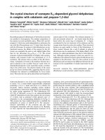
Section Parvisepalum is the sister group of all other
Paphiopedilum species (Figure 1). Two to four 25S
rDNA signals are apparent (Figure 2) among 2n = 26
chromosomes, with two signals most parsimoniously
interpretable as the basal condition since this state is
shared by the outgroup genera Mexipedium and Phra g-
mipedium (unpublished data; [23]). With 2 signals being
the inferred primi tive condition, rearrangement by dupli-
cation is observed in Paphiopedilum armeniacum,
P. emersonii and P. hangianum, which have more loci. 5S
rDNA patterns are stable, showing 2 subtelomeric signals
that are usually closely linked with one pair of 25S signals
(Table 1). In P. delenatii, translocation of either the 5S or
25S rDNA locus has occurred.Thisphenomenonisalso
seen in P. malipoense, with its two chromosomes that
show hemizygous 25S and 5S rDNA signals, respectively.
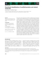
Section Concoloria
Species of section Concoloria show two 25S and 5S signals
(Table 1), each on separate chromosomes (2n = 26 total),
similarly to Paphiopedilum delenatii of section Parvisepa-
lum, except in that the 5S signals are interstitially instead
of subtelomerically placed (Figure 3).
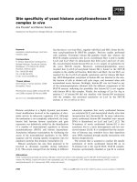
Section Cochlopetalum
Section Cochlopetalum displays an aneuploid number of
chromosomes, the telocentrics of which have been sug-
gested to descend via centric fission from 25 diploid
metacentrics [2]. According to phylogenetic relati onships
known at present (Figure 1), and the centric f ission
hypothesis, sections Cochlopetalum and Barbata (with
telocentrics descended from 26 diploid metacentrics)
have evolved aneuploid increa se independently. All four
species studied here have two telomeric 25S rDNA sig-
nals, and 4 major 5S rDNA signals (Figure 4; Table 1).
P
arv
i
sepa
l
um
Concoloria
Cochlopetalum
Paphiopedilum
Coryopedilum
Coryopedilum
Pardalopetalum
B
a
r
bata
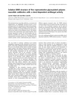
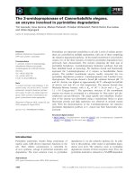
Figure 1 Section-level phylogenetic tree of genus Paphiopedilum.
Section-level phylogenetic tree based on rDNA ITS sequences
published b y Cox [3].
Lan and Albert BMC Plant Biology 2011, 11:126
/>Page 2 of 15
All 4 species have multiple dispersed 5S signals, rather
unlike species of sections Parviflora and Concoloria,and
these, like the major loci, are mostly subtelomeric, peri-
centromeric and centromeric in position. The 2 species
with 2n = 32 chromosomes, Paphiopedilum liemianum
(Figure 4C) and P. primulinum (Figure 4A), both have
two 5S bands localized on the same chromosomes as the
25S signals, whereas only a single 5S band is seen on the
Figure 2 FISH of 25S and 5S rDNA to metaphase chromosomes of Paphiopedilum section Parvisepa lum.(A)Paphiopedilum emersonii,(B)
P. delenatii, (C) P. malipoense, (D) P. hangianum, (E) P. armeniacum, (F) P. micranthum. 25S rDNA (green) and 5S rDNA (red) probes were
simultaneously detected in all Paphiopedilum species. Chromosomes were counterstained with DAPI. All scale bars = 10 μm.
Lan and Albert BMC Plant Biology 2011, 11:126
/>Page 3 of 15
Table 1 Paphiopedilum species studied, diploid chromosome numbers, rDNA FISH patterns, and 5S-NTS sequence
polymorphic sites
Number of rDNA sites Positions of rDNA sites
b
5S 25S+5S
Taxon 2n 25S major visible sites
a
Co-localization 5S 25S 5S-NTS Polymorphic sites
Paphiopedilum
Subg. Parvisepalum
Sect. Parvisepalum
armeniacum 26 4 2 2 2 st t 104
delenatii 26 2 2 2 0 st t 178
emersonii 26 4 2 2 2 st t 124
hangianum 26 4 2 2 2 st t 120
malipoense 26 2 2 2 1 st t 94
micranthum 26 4 2 2 2 st t 59
Subg. Paphiopedilum
Sect. Concoloria
bellatulum 26 2 2 2 0 i t 118
niveum 26 2 2 2 0 i t 198
Sect. Cochlopetalum
liemianum 32 2 4 22 2 st, i, p, c t 162
moquettianum 34 2 4 20 2 st, i, p, c t 225
primulinum 32 2 4 25 2 st, i, p, c t 71
victoria-regina 34 2 4 24 2 st, i, p, c t 137
Sect. Paphiopedilum
druryi 30 2 4 16 0 st, i, p, c t 184
fairrieanum 26 2 2 14 2 st, i, p, c t 146
henryanum 26 2 2 17 2 st, i, p, c t 180
hirsutissimum 26 2 6 21 2 st, i, p, c t 182
tigrinum 26 2 6 17 2 st, i, p, c t 141
Sect. Coryopedilum
adductum 26 9 4 28 6 st, i, p, c t, st 180
gigantifolium 26 6 6 32 6 st, i, p, c t 210
glanduliferum 26 4 4 26 4 st, i, p, c t 202
randsii 26 4 4 30 4 st, i, p, c t, st 187
sanderianum 26 2 4 16 0 st, i, p, c t 143
stonei 26 2 4 25 2 st, i, p, c t 114
supardii 26 9 4 26 7 st, i, p, c t 226
Sect. Pardalopetalum
dianthum 26 2 4 28 2 st, i, p, c t 251
haynaldianum 26 4 4 8 2 st, i, p, c t 110
lowii 26 6 4 28 4 st, i, p, c t, st 161
parishii 26 4 4 34 4 st, i, p, c t 189
Sect. Barbata
acmodontum 38 2 4 4 0 i t 169
curtisii 36 2 2 2 0 i t 164
dayanum 36 2 4 6 0 i, p t 169
hennisianum 34 2 2 6 0 i t 153
purpuratum 40 2 4 8 0 st, i t 138
sangii 38 2 4 18 0 st, i t 118
sukhakulii 40 2 2 13 0 st, i t 151
venustum 40 2 4 8 2 i t 109
wardii 42 2 4 4 0 i t 159
a
Minimum numbers of visible 5S rDNA FISH signals, including numbers of both major and visible dispersed sites.
b
st, subtelomeric; t, telomeric; i, interstitial; p, pericentromeric; c, centromeric
Lan and Albert BMC Plant Biology 2011, 11:126
/>Page 4 of 15
Figure 3 FISH of 25S and 5S rDNA to metaphase chromosomes of Paphiopedilum section Concoloria. (A) Paphiopedilum be llatulum,(B)P.
niveum.
Figure 4 FISH of 25S and 5S rDNA to metaphase chromosomes of Paphiopedilum section Cochlopetalum.(A)Paphiopedilum primulinum, (B) P.
moquettianum,(C)P. liemianum,(D)P. victoria-regina.
Lan and Albert BMC Plant Biology 2011, 11:126
/>Page 5 of 15
same chromosome in the 2n = 34 species P. moquettia-
num (Figure 4B) and P. victoria-regina (Figure 4D).
Section Paphiopedilum
All 5 species of section Paphiopedilum studied show two
25S signals in the telomeric region (Figure 5; Table 1). All
species, which are 2n = 26 except for P. druryi (Figure 5E)
at 2n = 30, show at least 2 specific 5S rDNA bands, as
many as 6, and numerous dispersed signals in the pericen-
tromeric and centromeric regions. In all but P. druryi the
major signals are closely linked with the 25S arrays. In
P. druryi, 4 of the major signals appear to be located on
different arms and on morphologically different chromo-
somes that may only be partly homologous (this condition
was observed in at least 4 cells).
Sections Coryopedilum and Pardalopetalum
In current phylogenetic results, section Pardalopetalum is
derived within section Coryopedilum (Figure 1); as such,
they will be discussed together here. Together, the Coryo-
pedilum/Pardalopetalum clade, all species having 2n = 26,
is the most dynamic in Paphiopedilum regarding chromo-
somal rearrangements (Figure 6, 7; Table 1). 25S signals
vary from 2 to 9, the latter showing hemizygosity. Signals
in all species except Paphiopedilum lowii (Figure 7A),
P. adductum (Figure 6E) and P. randsii (Figure 6F) are
telomeric. 1-4 subtelomeric 25S signals were observed
in P. lowii, P. adductum and P. randsii.InP. supardii
(Figure 6G), one hemizygous chromosome has telomeric
25S signals on each arm. P. addu ctum also shows 25S
hemizygosity, and both this spec ies and P. supardii show
the maximum number of signal s. Species of the Coryope-
dilum/Pardalopetalum groupshowatleast4major5S
rDNA signals (up to 8 in P. parishii (Figure 7B)) and mul-
tiple dispersed repeats in pericentromeric and centromeric
regions. In the Pardalopetalum group, all species show at
least 2 strong (up to 5) 5S bands located on one chromo-
some. Close linkage with 25S occurs throughout the
group, other than in P. sanderianum (Figure 6A), either
with major or minor 5S bands, and appearing in different
placements along chromosome arms.
Section Barbata
Species of section Barbata, which have 2n = 28-42 and the
largest genome sizes, show constancy in 25S rDNA distri-
bution, with 2 telomeric signals (Figure 8; Table 1). Major
5S signals number 2-4, and extremely few dispersed
Figure 5 FISH of 25S and 5S rDNA to metaphase chromosomes of Paphiopedilum section Paphiopedilum. (A) Paphiopedilum fairrieanum,
(B) P. hirsutissimum, (C) P. tigrinum, (D) P. henryanum, (E) P. druryi.
Lan and Albert BMC Plant Biology 2011, 11:126
/>Page 6 of 15
repeat s were observed. Most 5S loci are not centromeric ,
whereas telomeric, subtelomeric, pericentromeric, and
interstitial placements are observed. Only Paphiopedilum
curtisii (Figure 8G) and P. hennisianum(Figure 8B) have
two major 5S signals, and the first species shows no dis-
persed repeat s. P. sukhakulii (Figure 8C), P. venustum
(Figure 8F) and P. wardii (Figure 8A) show linked 5S sig-
nals. Only in P. venustum is close linkage of 25S and 5S
observed, and then only involving a minor 5S band.
Because Barbata is the most derived section in the genus
(Figure 1), either its species have lost 25S and 5S rDNA
loci, since Cochlopetalum, Paphiopedilum, Coryopedilum,
Figure 6 FISH of 25S and 5S rDNA to metaphase chromosomes of Paphiopedilum section Coryopedilum. (A) Paphiopedilum sanderianum,
(B) P. gigantifolium, (C) P. stonei, (D) P. glanduliferum, (E) P. adductum, (F) P. randsii, (G) P. supardii. Arrows indicate subtelomeric 25S rDNA signals.
Lan and Albert BMC Plant Biology 2011, 11:126
/>Page 7 of 15
and Pardalopetalum usually have more, or the species of
the latter sections have increased the number of rDNA
loci independently given the low number in sections Par-
visepalum and Concoloria.
Diversity of 5S ribosomal DNA non-transcribed spacer
sequences
We investigated duplication history correlated with the
dynamic rearrangements observed in 5S rDNA loci. In
order to survey sequence variation in 5S-NTS, random
clones, 7 (Paphiopedilum niveum)or8(allothers)per
species, were sequenced (Additional file 1). Only a few
clones were identical to each other (2 sequences from
P. acmodontum,2fromP. henryanum,2fromP. hirsutissi-
mum,2fromP. stonei,4fromP. dayanum,4from
P. malipoense,andonesequenceeachofP. stonei and
P. supardii). Sequences of 5S-NTS ranged from 283 bp
(P. micranthum 1) to 455 bp (P. bellatulum 5). Given
extensive sequence divergence of 5S-NTS and our desire
not to manually adjust alignment [24], an objective align-
ment was accomplished using MAFFT and default settings.
Numbers of polymorphic loci within species, and
Figure 7 FISH of 25S and 5S rDNA to metaphase chromosomes of Paphiopedilum section Pardalopetalum. (A) Paphio pedilum lowii, (B) P.
parishii, (C) P. dianthum, (D) P. haynaldianum. Arrows indicate subtelomeric 25S rDNA signals.
Lan and Albert BMC Plant Biology 2011, 11:126
/>Page 8 of 15
phylogen etic relationships, were assessed in order t o esti-
mate the strength of gene conversion and the extent of
paralogy, respectively. Numbers of polymorphic sites within
species positively correlated with minimum numbers of
visible 5S signals (P < 0.01, R^2 = 0.21; Figure 9), suggesting
that interlocus gene conversion is relatively weak. A phylo-
genetic tree outgroup-rooted using Phragmipedium besseae
showed 2 major groups of sequences: section Parvisepalum
Figure 8 FISH distribution pattern of 25S and 5S rDNA on metaphase chromosomes of Paphiopedilum section Barbata. (A) Paphiopedilum
wardii, (B) P. hennisianum, (C) P. sukhakulii, (D) P. purpuratum, (E) P. dayanum, (F) P. venustum, (G) P. curtisii, (H) P. acmodontum, (I) P. sangii.
Lan and Albert BMC Plant Biology 2011, 11:126
/>Page 9 of 15
versus the remainder of the genus. The single tree of maxi-
mum likelihood is shown ( as a phylogram, Additional
file 2), as is the majority-rule consensus tree based on
100 bootstrap replicates (Additional file 3). Some large spe-
cies-specific clades were observed, as well as some section-
specific clades. Overall, however, the phylogenetic tree was
poorly representative of phylog enetic relationships due to
extensive duplication of 5S l oci.
Discussion
Variation in numbers and chromosomal locations of rDNA
Variation in numbers and distribution patterns of rDNA
loci among related species is commonly observed in
many different plant genera, including Brassicaceae [10],
Cyperaceae [ 11], Asteraceae [25,26], Leguminosae [27],
Pinus [28], and Rosaceae [14]. Plants typically show
some degree of conservatism of rDNA repeat duplica-
tion, such that when mu ltiple loci do appear, species are
commonly polyploid relatives of diploids. There is no
evidence at all, however, for polyploidy in Paphiopedi-
lum, where the only chromosome number differences
are aneuploid, in a series reflective of centric fission or
fusion.
In general, FISH patterns of 25S rDNA loci are
reported to be more polymorphic th an those of the 5S
rDNA [12-14,26,28-32]. Conversely, in all sections of
Paphiopedilum, except for Parvisepalum and Concoloria,
5S rDNA sites showed much more variability both in
number and physical location than did 25S rDNA sites.
The most parsimonious ancestral number of 25S
rDNA sites in Paphiopedilum is two, based on outgroup
comparison to the genera Mexipedium an d Phragmipe-
dium (unpublished results; [3,22]). Duplication of 25S
rDNA sites was observed only in three of the seven sec-
tions of Paphiopedilum: Parvisep alum (2n = 26),
Coryopedilum (2n = 26) a nd Pardalopetalum (2n = 26)
(Table 1). The physical positions of 25S rDNA loci are
relatively conservative. In most Paphiopedilum species
we analyzed, 25S rDNA signals are located in terminal
chromosome positions. Variation w as only observed in
three species, Paphiopedilum adductum, P. randsii and
P. lowii, which showed 1-4 subtelomeric 25S rDNA sig-
nals (Figures 6E, F and 7A, respecti vely). The ancestral
number of 5S rD NA sites, again by outgroup compari-
son, is 2 (unpublished results from Mexipedium and
Phragmipedium), and is only observed in sections Parvi-
sepalum and Concoloria. Massive duplication and ampli-
fication of 5S rDNA loci, leading to large-scale
polymorphism of numbers, sizes and physical positions
of signals, was found prevalent in the remaining five
sections. The numbers and distribution of rDNA loci
vary widely among plants; however, usually less than
one-third of chromosomes display either 45S rDNA or
5S rDNA [13]. It is therefore noteworthy that in some
lineages of Paphiopedilum,upto24ofthe26chromo-
somes bear at least one rDNA locus, and a single chro-
mosome can bear up to five major 5S rDNA loci.
Apparently, there is no strong correlation between the
increase in the number of rDNA sites and the increase in
the number of chromosomes or genome size. A similar
situation has also been described in many other diploid
species, e.g. the diploid lineage of Brassicaceae [10],
Cyperaceae [11,12], Iris [13], and Rosacea e [14]. The
massive dupli cation of rDNA loci in Paphiopedilum sec-
tions Cochlopetalum, Paphiopedilum, Coryopedilum and
Pardalopetalum could partly contribute to the increase
of genome size. Perhaps paradoxically, species with the
smallest (P. exul; section Paphiopedilum)andlargest
(P. dianthum; section Pardalopetalum)haploidgenome
sizes are both members of groups that show considerable
25S and 5S locus duplication in our FISH experiments.
These two species differ more than two-fold in genome
size, 16.1 to 35.1 Mb, respectively [33]. If we assume that
the number of distinct genes among Paphiopedi lum spe-
cies is roughly constant, this would suggest that genome
size increase is primarily due to repetitive element ampli-
fication, but that since rDN A duplic ation is associated
with both smaller and larg er ge nom es in t he ge nus, s iz e
differences may be more logically traceable to other repe-
titive DNAs, such as mobile elements. However, a possi-
ble tendency for elimination of rDNA loci was found in
section Barbata, which has the greatest average genome
sizes and chrom osome numbers [4]. The number of 25S
rDNA loci in Barbata
remains two through all the spe-
cies
we studied, while the distribution pattern of 5S
rDNA is less dispersed than its sister group, Coryopedi-
lum plus Pardalop etalum. Due to the derived phyloge-
netic position of section Barbata (Figure 1), it is most
parsimonious to conclude that unique chromosomal
Figure 9 The relationship between polymorphism of 5S-NTS
sequences and numbers of observed 5S rDNA signals. Line
indicates trend derived from linear regression analysis based on 5S-
NTS within-species polymorphic sites and minimum numbers of
visible 5S signals (data from Table 1.). P < 0.01, R^2 = 0.21.
Lan and Albert BMC Plant Biology 2011, 11:126
/>Page 10 of 15
conditions seen in the group would be similarly deriv ed
(autapomorphic). As such, centric fission in Barbata
appears to be associated with loss of rDNA loci, while in
other systems, centric fission has led to rDNA gains [34].
Elimination of rDN A loci during chromosomal evolution
has been docum ented in, e.g., Brassicaceae and Rosaceae
[10,14]. The mechanism that accounts for such loss of
rDNA loci, however, remains unclear. A presumed evolu-
tionary loss of abundant terminal nucleolar organizing
regions (NOR) in Ar abidopsis has been hypothesized to
be the consequence of an ancient fusion event [35]. In
the case of section Barbata, additional traceable chromo-
some markers are needed to provide further evidence
that chromosomal rearrangements a re related to rDNA
loss.
A combination of different mechanisms causes high
mobility of rDNA
Different mechanisms have been postulated to account
for the mobility and polymorphism of numbers, sizes and
positions of rDNA sites, such as transposon-mediated
transposition al events [36-38], and chromosome rearran-
gements (translocation, inversion, duplication, deletion)
caused by homologous or non-homologous unequal
crossing-over and gene conversion [9,28,30,36]. These
processes could act alone or in combination, and they do
not necessarily imply changes in overall chromosome
morphology [31,34].
The great degree of 5S repeat dispersion seen in sections
Cochlopetalum, Paphiopedilum, Coryopedilum and Parda-
lopetalum has, to our knowledge, only been observed in
the monocots Alstroemeria, Tulipa,andIris [13,39,40].
The original seeding of rDNA repeats to ectopic locations
in the genome could be the result of transposable element
activity or perhaps incorporation of array segments into
breakpoints as part of non-homologous end joining during
DNA repair. Indeed, some of the signals we observed may
be pseudogenes transported within the genomes by retroe-
lements, t herefore leading to the false interpretation that
we are visualizing entire and active rDNA a rrays. Both
subtelomeric and pericentromeric regions are well known
as hot spots of breakpoints and are also enriched fo r TEs
[5,6]. Considering the abundant minor loci we observed in
these regions, a contribution of transpositions to the dis-
persed distribution pattern is tenable, and TEs containing
5S rDNA-derived sequences have in fact been observed in
many plants [41] and animals [42]. It is nonetheless possi-
ble t hat due to the si milarity of rDNA arrays, chromoso-
mal rearrangement could be induced via heterologous
recombination, and in turn, rearrangement could generate
repeated sequences through unequal crossovers. After
generation of a novel locus, in situ amplification cycles via
rearrangement could lead to the origin of FISH-detectable
loci. Furthermore, hemizygous 5S rDNA sites have been
widely observed in many Paphiopedilum species. A dou-
ble-strand break occurring in a hemizyg ous region would
increase the probability of causing other rearrangements,
owing to the absence of a homologous template for its
repair [5]. The lack of dispersed repeats in the basalmost
section Parvisepalum may reflect either a lack of seeding
events or slow amplification processes that do not yield
hybridization-visible arrays. However, in the case of 5S
rDNA, there is in fact strong evidence for NTS sequence
diversity, which could either be accounted for by the pre-
sence or small loci below the FISH detection limit or per-
haps by considerable within-array diversity. One future
experimental approach to determine whether considerable
intra-array diversity indeed exists would be to perform
FISH using 5S-NTS-specific probes.
Diversification of 25S rDNA distribution patterns is also
observed in Paphiopedilum, but the numbers of lo ci and
degree of dispersion is much lower than f or 5S rDNA.
Therefore, 5S rDNA might be more frequently seeded by
TEs via transpositional events, or, amplification or mainte-
nance of 5S rDNA loci via rearrangement could be more
effective and tolerated during t he chromosome evolution
process. The differe nt evolutiona ry tendencie s between
25S and 5S rDNA might be caused by their function and
sequence divergence or localization in distinct nuclear
compartments [43].
5S-NTS sequences highlight interlocus and intralocus
diversity and weak concerted evolutionary forces
Previous studies of other angio sperm species have sug-
gested that intralocus 5S rDNA diversity occurs. Within-
array 5S rDNA diversity appears very likely in Paphiopedi-
lum as well, since many species (e.g., all Parvisepalum and
Concoloria) have only one observable 5S locus. For exam-
ple, 6 species of section Parvisepalum are represented in
our phylogenetic analysis by 6-8 distinct sequence variants.
These 5S-NTS variants can be concluded to occur within
at least partial arrays, pseudogenized or not, since the
amplified piec es inc lude sections of 5S rDNA at their 5 ’
and 3’ ends. Recent within-species duplication events may
be indicated by single-species clades of 5S-NTS sequences,
such as P. dayanum, P. lowii, P. sangii,butthesecould
just as we ll indicate within-array variatio n, as single-spe-
cies clades of Parvisepalum (e.g., P. malipoense) and Con-
coloria ( P. bellatulum) most likely do. In many cases, it
can be readily seen that duplication of 5S loci has occurred
prior to speciation, for example, within Coryopedilum (a
large group of sequences representing P. sanderianum,
P. stonei, and P. supardii; similarly also wit hin a group of
P. adductum and P. randsi i sequences). In some cases,
ancient dupl ications must be much older than the major
phylogenetic groups of Paphiopedilum,since,forexample,
P. delenatii shares sequence variants similar to other
Parvisepalum species yet has at least one other variant
Lan and Albert BMC Plant Biology 2011, 11:126
/>Page 11 of 15
that i s more similar t o sequences from all other sections.
We investigated the possibility of contamination regarding
this finding, but discovered similar repeats across 8
distinct P. delenatii accessions (results not shown).
Another explanation fo r multi species clades, e.g., within
well-defined groups such as Parvisepalum could b e
ancient hybridization.
We observed that increasing within-species 5S NTS
sequence diversity correlates with increasing minimum
numbers of visibl e 5S rDNA loci in Paphiopedilum
(Figure 9); therefore we infer that interlocus concerted
evolution is weak within the genus. Our conclusion
concurs with previous findings in many plant genera,
such as Gossypium [17], Triticum [18], Chenopodium
[19], Nicotiana [20] and Pinus [27].Sofar,toour
knowledge, noticeable interlocus concerted evolution
of 5S rDNA arrays has not been demonstrated in
plants.
The best supported hypothesis to explain weak homo-
genization forces on 5S rDNA arrays is that the chromo-
somal location of rDNA arrays has a substantial impact
on interlocus concerted evolution [17,20,44-47]. Arrays
located in subtelomeric regions are thought to undergo
stronger interlocus homogenization forces than ones
located in proximal regions. Potential evidence was
observed in section Barbata, in which all of six species
studied possess two 5S loci. These six species can be
catego rized into two groups according to the locations of
5S loci. One group harboring proximal 5S loci includes
P. wardii, P. dayanum, P. venustum and P. acmodontum,
while the other group h arboring subtelomeric 5S loci
includes P. purpuratum and P. sangii (Figure 8; Table 1).
Considering that all six species are closely related and
possess the same number of l oci, it can be logically
assumed that the difference in sequence polymorphi sm
between the two groups is caused by the different loca-
tions of the 5S loci. The fact that the average number of
polymorphic sites in the proximal-loci group (151.5) is
18% more than that in the subtelome ric-loci group (128),
indicates that proximally located loci see m less homoge-
nized than the subtelomerically located loci.
Additionally, we found that not only interlocus but also
intralocus concerted evolution is also influenced by chro-
mosomal localization. In section Concoloria, two closely
related species, P. bellatulum and P. niveum, both have
one 5 S locus, but with different localizations. The differ-
ence in sequence polymorphism between the two species
may be caused by the different locations of the 5S loci.
P. niveum, which has a pericentromeric locus, showe d
1.68 - fo ld more polymorphic sites than P. bellatulum,
which has a subtelomeric locus (Table 1).
It is well-known that meiotic homologous recombina-
tion has b een largely suppressed in pericentromeric and
centromeric regions. Unequal crossovers between sister
chromatids and gene conversion documented in the cen-
tromeres of many organisms have been postulat ed as the
major homogenization force for tandem repeats located
in these areas [48-52]. A plausible explanation for this
has been proposed previously: if unequal crossover events
between rDNAs of tw o chromosomes occurred in the
proximal region to centromeres, this may result i n the
exchange of no t o nly a fraction of the rDNA but also the
centromeres themselves. Such an event is more likely to
have significantly greater negative consequences to the
organism than if the event occurred in the subtelomeric
region, which then might result in exchange of telomeres
[17,47]; loss of centromeres would prohibit cell division,
whereas loss of telomeres might not restrict mitosis or
meiosis. As such, centromerically-located rDNA arrays
are expe cted to show weaker homogeniza tion forces,
since fewer individuals with une qual crossovers in this
region are expected to survive. In contrast, the subtelo-
mericregionischaracterizedbyahigherrateofinter-
chromoso mal exc hange [5], thus stronger c oncerted
evolutionary forces could be expected in this region.
All s pecies of section Parvisepalum, as with P. bellatu-
lum, have subtelomeric 5S loci, some of which are closely
linked with 25S loci. If 5S localization c orrelates signifi-
cantly with homogenization, as with 25S, which is always
telomeric-subtelomerically located, we should expect sub-
telomeric 5S repeats to show decreased sequence diversity
due to stronger con certed evolutionary forces. However,
this is not the case, since variation in the number of poly-
morphic sites is not significantly different by section (with
or without Pardalopetalum included in Coryopedilum; sin-
gle factor ANOVA P = 0.06 and 0.1, respectively). We
therefore infer that localization of the 5S rDNA arrays only
partially contributes to the weak concerted evolution
observed in Paphiopedilum.
There are several other hypothesized mechanisms that
could lead to the weak concerted evolutionary force on 5S
rDNA arrays. For example, ongoing chromosomal rearran-
gement such as insertion, deletion, or transposition could
occur within arrays too frequently for interlocus concerted
evolution to be effective. Another possibility is that con-
certed evolutionary processes homogenize 5S rDNA arrays
at rates lower than the rate of speciation, thus novel muta-
tions cannot be fixed or removed and high levels of intralo-
cus polymorphism are expected within arrays [17].
Additionally, the base composition and secondary structure
of rDNA sequences may also affect the rate of concerted
ev
olution [53]. It is unknown whether weak concerted evo-
lutionary forces are shared by other Paphiopedilum tandem
repeats, or if this is characteristic of 5S rDNA arrays only.
This issue can be elucidated by further studies on other
tandem repeats, such as 25S rDNA arrays.
Lan and Albert BMC Plant Biology 2011, 11:126
/>Page 12 of 15
Conclusions
Paphiopedilum species display many chromosomal rear-
rangements - for example, duplications, translocations,
and inve rsions - but only we ak concerted evolutionary
forces among highly duplicat ed 5S arrays, which suggests
that double-strand break repair processes are dynamic and
ongoing. These results make the genus a model system for
the study of complex chromosomal evolution in plants.
Methods
Plant materials
Thirty-seven species of the Paphiopedilum genus covering
all seven sections were analyzed in this study. Information
on the species and sections is provided in Table 1. Actively
growing roots were u sed for chromosome preparation,
while leaves were used for genomic DNA extraction.
Chromosome preparation
Root tips were pre-treated with 0.004 M 8-hydroxyquino-
line for 4-6 h at 10°C, and fixed in freshly prepared fixative
(3:1 ethanol: acetic acid) for 48 h at 10°C. The fi xed r oot
tips were then rinsed thoroughly with tap water and
macerated in an enzyme mixture co ntaining 2% cellul ase
(Onozuka R-10, Rpi) and 1% pectolyase (Aspergillus japo-
nicus Y-23, MP) at 37°C for 30 min. After re-fixation in
fixative for 15 min, the merist ematic cells were squashed
in a d rop of 45% acetic acid under a coverslip (22 ×
22 mm) on a microscope slide. Slides were then dipped
into liquid nitrogen and air dried after the coverslips were
carefully removed by a blade.
Probe labelling and Fluorescence in situ hybridization (FISH)
25S rDNA, a 2.3-kb ClaI subclones of the 25S rDNA cod-
ing region o f Arabidopsis thaliana [54] and 5S rDNA
(pTa794) [55]were used as probes. 25S rDNA was labelled
with biotin-16- dUTP (Roche) and 5S rDNA was labelled
with digoxigenin-11-dUTP (Roche), all by nick translation
method using the kit from Roche. The hybridization buffer
consisted of 50% deionized formamide, 2 × SSC, 50 mM
sodium phosphate (pH 7.0), 10% dextran sulfate and
sheared salmon sperm DNA (Invitrogen) in 100 × excess
of labeled prob es. The 25S and 5S rDNA probes were
mixed to a final concentration of about 2 ng/μl and then
denatured at 94°C for 10 min bef ore being used. Slides
with metaphase spreads were treated with 70% deionized
formamide in 2 × SSC at 70°C for 2 min. Denatured
probes in hybridization buffer were then applied to the
slides, which were incubated at 37°C for 10 h in a humid
chamber. Post-hybridization washes and immunodetection
were carried out in an automated in situ hybridization
instrument, the InsituPro VSi (Intavis Bionanalytical
Instruments). The slides were washed in 2 × SSC at room
temperature for 5 min and twice in 2 ×SSC at 50°C for
10 min. Fluorescence signal was detected using anti-
Digoxigenin-Rhodamine conjugate (Roche) and streptavi-
din-fluorescein conjugate (Invitrogen). The pr eparations
were mounted and counterstained in Vectashield contain-
ing 1.5 μg/ml DAPI (4’ , 6-diamidino-2-phenylindole)
(Vector Laboratories). Images were taken by a Zeiss
AxioCam MRm black-and-white CCD cam era on a Zeiss
Imager. Z1 fluorescence microscope and then processed
unifo rmly using Zeiss AxioVision software. FISH signals
were false-colored, and DAPI fluorescence was left in
gray-tone.
PCR amplification, cloning and sequencing
Total genomic DNA was extracted from fresh leaves using
Qiagen DNeasy Plant Mini kit. The 5S-NTS region was
amplified by PCR using the univer sal degenerate primers:
5’-TGGGAAGTCCTYGTGTTGCA-3’ and 5’-KTMGYGC
TGGTATGATCGCA-3’ [56]. Touchdown amplification
was performed as follows: an initial step at 94°C for 5 min,
followed by 10 cycles of 94°C for 1 min, annealing for
1 min (start at 60°C, and decreased by 1°C per cycle), and
72°C for 1 min, then 35 cycles of 94°C for 1 min, 50°C for
1 min, a nd 72°C for 1 min, the final step at 72°C was
extended to 10 min. After gel purification using QIAquick
Gel Extraction Kit (Qiagen), PCR products were ligated
into pDrive Cloning vector and transformed into QIAGEN
EZ competent cells (Qiagen PCR Cloning kit) . Re combi-
nant clones were screened by colony direct PCR method
and were sequenced 7-8 clones per each species using T7
(5’-TAATACGACTCACTATAGGG-3’) primer.
Data analysis
Sequences were aligned using the MAFFT (Multiple
Alignment using Fast Fourier Transform) web server at
the European Bioinformatics Institute [57]. Default para-
meters were used: gap opening penalty = 1.53, gap
extension penalty = 0.123, tree rebuilding number = 1,
maxiterate = 0, and perfo rm FFTS = localpair. The
sequence alignment is available as a supplementary
FASTA file (Additional file 4).
Within-species 5S-NTS polymorphism was estimated,
based on the aforementioned multiple alignment, using
DnaSP version 5.10.01 [58]. The relationship between
numbers of polymorphic sites and minimum numbers
of visible 5S rDNA signals was investigated using linear
regression analysis (in Microsoft Excel).
Phylogenetic reconstruction was performed using max-
imum likelihood optimization available through the
RaxML BlackBox web server [59] running RaxML version
7.2.8 [60]. Default settings were used. The 8 Phragmipe-
dium besseae sequences were indicated as the outgroup.
RaxML was called using the following commands:
raxml -# 100 -n pasted -o bess1, bess2,
Lan and Albert BMC Plant Biology 2011, 11:126
/>Page 13 of 15
bess3, bess4, bess5, bess6, bess 7, bess8 -f
a -m GTRGAMMA -x 564547904 -p 5 64547904 - s
0VaDTW. All search information, as was output on the
web site, is included in Additional file 5.
Additional material
Additional file 1: GenBank data deposition information of 5S-NTS
sequences
Additional file 2: 5S-NTS sequences: the single tree of maximum
likelihood
Additional file 3: 5S-NTS sequences: the majority-rule consensus
tree based on 100 bootstrap replications
Additional file 4: The 5S-NTS sequence alignment using MAFFT,
provided in FASTA format
Additional file 5: Report from RAxML phylogenetic analysis of
5S-NTS sequences
Acknowledgements
We thank R. Hasterok and B. Liu for providing rDNA clones. This study was
supported by funds from the University at Buffalo.
Authors’ contributions
TL and VAA conceived of the study, TL performed all experiments, TL and
VAA analyzed data, and both TL and VAA prepared the manuscript. All
auhors read and approved the final manuscript.
Received: 1 July 2011 Accepted: 12 September 2011
Published: 12 September 2011
References
1. Cribb P: The genus Paphiopedilum. Natural History Publications. 2 edition.
Borneo; 1998.
2. Karasawa K: Karyomorphological studies in Paphiopedilum. Orchidaceae.
Bulletin of the Hiroshima Botanic Garden 1979, 2:1-149.
3. Cox A, Pridgeon AM, Albert VA, Chase MW: Phylogenetics of the slipper
orchids (Cypripedioideae, Orchidaceae). Nuclear rDNA ITS sequences Plant
Syst Evol 1997, 208(3-4):197-223.
4. Cox A, Abedelnour G, Bennett MD, Leitch IJ: Genome size and karyotype
evolution in the slipper orchids (Cypripedioideae: Orchidaceae). American
Journal of Botany 1998, 85(5):681-687.
5. Linardopoulou E, Williams EM, Fan Y, Friedman C, Young JM, Trask BJ:
Human subtelomeres are hot spots of interchromosomal recombination
and segmental duplication. Nature 2005, 437(7055):94-100.
6. Slotkin R, Martienssen R: Transposable elements and the epigenetic
regulation of the genome. Nat Rev Genet 2007, 8(4):272-285.
7. Zhang J: Evolution by gene duplcation: an update. Trends in Ecology and
Evolution 2003, 18(6):292-298.
8. Hurles M: Gene duplication: The genomic trade in spare parts. PLoS Biol
2004, 2(7):0900-0904.
9. Raskina O, Barber JC, Nevo E, Belyayev A: Repetitive DNA and
chromosomal rearrangements: speciation-related events in plant
genomes. Cytogenet Genome Res 2008, 120(3-4):351-357.
10. Hasterok R, Wolny E, Hosiawa M, Kowalczyk M, Kulak-Ksiazczyk S,
Ksiazczyk T, Heneen WK, Maluszynska J: Comparative analysis of rDNA
distribution in chromosomes of various species of Brassicaceae. Annals
of Botany 2006, 97(2):205-216.
11. Da Silva C, Quintas CC, Vanzela AL: Distribution of 45S and 5S rDNA sites
in 23 species of Eleocharis (Cyperaceae). Genetica 2010, 138(9-
10):951-957.
12. Sousa A, Barros e Siva AE, Cuadrado A, Loarce Y, Alves MV, Guerra M:
Distribution of 5S and 45S rDNA sites in plants with holokinetic
chromosomes and the “chromosome field” hypothesis. Micron 2011,
42(6):625-631.
13. Martinez J, Vargas P, Luceno M, Cuadrado A: Evolution of Iris subgenus
Xiphium based on chromosome numbers, FISH of nrDNA (5S, 45S) and
trnL-trnF sequence analysis. Plant Syst Evol 2010, 289:223-235.
14. Mishima M, Ohmido N, Fukui K, Yahara T: Trends in site-number change
of rDNA loci during polyploid evolution in Sanguisorba (Rosaceae).
Chromosoma 2002,
110(8):550-558.
15.
Lee Y, Chung MC: Identification of genome relationships among
Paphiopedilum species by genomic and fluorescent in situ hybridization.
Acta Horiticluturae 2008, 766:331-334.
16. Lee Y, Chang FC, Chung MC: Chromosome pairing affinities in
interspecific hybrids reflect phylogenetic distances among lady’s slipper
orchids (Paphiopedilum). Annals of Botany 2011, 108(1):113-121.
17. Cronn R, Zhao X, Paterson AH, Wendel JF: Polymorphism and concerted
evolution in a tandemly repeated gene family: 5S ribosomal DNA in
diploid and allopolyploid cottons. J Mol Evol 1996, 42(6):685-705.
18. Kellogg E, Appels R: Intraspecific and interspecific variation in 5S RNA
genes are decoupled in diploid wheat relatives. Genetics 1995,
140(1):325-343.
19. Maughan P, Kolano BA, Maluszynska J, Coles ND, Bonifacio A, Rojas J,
Coleman CE, Stevens MR, Fairbanks DJ, Parkinson SE, Jellen EN: Molecular
and cytological characterization of ribosomal RNA genes in
Chenopodium quinoa and Chenopodium berlandieri. Genome 2006,
49(7):825-839.
20. Fulnecek J, Lim KY, Leitch AR, Kovarík A, Matyásek R: Evolution and
structure of 5S rDNA loci in allotetraploid Nicotiana tabacum and its
putative parental species. Heredity 2002, 88(1):19-25.
21. Liu Z, Zhang DM, Wang XQ, Ma XF, Wang XR: Intragenomic and
interspecific 5S rDNA sequence variation in five Asian pines. American
Journal of Botany 2003, 90:17-24.
22. Albert V: Cladistic relationships of the slipper orchids (Cypripedioideae:
Orchidaceae) from congruent morphological and molecular data.
Lindleyana 1994, 9:115-132.
23. Anisimova M, Cannarozzi GM, Liberles DA: Finding the balance between
the mathematical and biological optima in multiple sequence
alignment. Trends in Evolutionary Biology 2010, 2(1):e7.
24. Garcia S, Panero JL, Siroky J, Kovarik A: Repeated reunions and splits
freature the highly dynamic evolution of 5S and 35S ribosomal RNA
genes (rDNA) in the Asteraceae family. BMC Plant Biology 2010, 10:176.
25. Malinska H, Tate JA, Matyasek R, Leitch AR, Soltis DE, Soltis PS, Kovarik A:
Similar patterns of rDNA evolution in synthetic and recently formed
natural populations of Tragopogon (Asteraceae) allotetraploids. BMC
Evolutionary Biology 2010, 10:291.
26. Moscone E, Klein F, Lambrow M, Fuchs J, Schweizer D: Quantitative
karyotyping and dual-color FISH mapping of 5S and 18S-25S rDNA
probes in the cultivated Phaseolus species (Leguminosae). Genome 1999,
42(6):1224-1233.
27. Cai Q, Zhang D, Liu Z-L, Wang X-R: Chromosomal localization of 5S and
18S rDNA in five species of subgenus Strobus and their implications for
genome evolution of Pinus. Annals of Botany 2006, 97(5):715-722.
28. Hanson R, Islam-Faridi MN, Percival EA, Crane CF, Ji Y, McKnight TD,
Stelly DM, Price HJ: Distribution
of 5S and 18S-28S rDNA loci in a
tetraploid cotton (Gossypium hirsutum L) and its putative diploid
ancestors. Chromosoma 1996, 105(1):55-61.
29. Linares C, Gonzalez J, Ferrer E, Fominaya A: The use of double
fluorescence in situ hybridization to physically map the positions of 5S
rDNA genes in relation to the chromosomal location of 18S-5. 8S-26S
rDNA and a C genome specific DNA sequence in the genus Avena.
Genome 1996, 39(3):535-542.
30. Thomas H, Harper JA, Meredith MR, Morgan WG, Thomas ID, Timms E,
King IP: Comparison of ribosomal DNA sites in Lolium species by
fluorescence in situ hybridization. Chromosome Research 1996,
4(7):486-490.
31. Datson P, Murray BG: Ribosomal DNA locus evolution in Nemesia:
transposition rather than structural rearrangement as the key
mechanism? Chromosome Research 2006, 14(8):845-857.
32. Ksiazczyk T, Taciak M, Zwierzykowski Z: Variability of ribosomal DNA sites
in Festuca pratensis, Lolium perenne, and their intergeneric hybrids,
revealed by FISH and GISH. J Appl Genet 2010, 51(4):449-460.
33. Plant DNA C-values database (release 5.0, Dec. 2010). [.
org/cvalues/].
Lan and Albert BMC Plant Biology 2011, 11:126
/>Page 14 of 15
34. Hall K, Parker JS: Stable chromosome fission associated with rDNA
mobility. Chromosome Research 1995, 3(7):417-422.
35. Lysak M, Berr A, Pecinka A, Schmidt R, McBreen K, Schubert I: Mechanisms
of chromosome number reduction in Arabidopsis thaliana and related
Brassicaceae species. Proc Natl Acad Sci USA 2006, 103(13):5224-5229.
36. Altinkut A, Raskina O, Nevo E, Belyayev A: En/Spm-like transposons in
Poaceae species: transposase sequence variability and chromosomal
distribution. Cellular & Molecular Biology Letters 2006, 11(2):214-230.
37. Schubert I, Wobus U: In situ hybridization confirms jumping nucleolus
organizing regions in Allium. Chromosoma 1985, 92:143-148.
38. Raskina O, Belyayev A, Nevo E: Activity of the En/Spm-like transposons in
meiosis as a base for chromosome repatterning in a small, isolated,
peripheral population of Aegilops speltoides Tausch. Chromosome
Research 2004, 12(2):153.
39. Baeza C, Schrader O, Budahn H: Characterization of geographically
isolated accessions in five Alstroemeria L. species (Chile) using FISH of
tandemly repeated DNA sequences and RAPD analysis. Plant Syst Evol
2007, 269(1-2):1-14.
40. Mizuochi H, Marasek A, Okazaki K: Molecular cloning of Tulipa fosteriana
rDNA and subsequent FISH analysis yields cytogenetic organization of
5S rDNA and 45S rDNA in T. gesneriana and T. fosteriana. Euphytica
2007, 155(1-2):235-248.
41. Kalendar R, Tanskanen J, Chang W, Antonius K, Sela H, Peleg O,
Schulman AH: Cassandra retrotransposons carry independently
transcribed 5S RNA. Proc Natl Acad Sci USA 2008, 105(15):5833-5838.
42. Kapitonov V, Jurka J: A novel class of SINE elements derived from 5S
rRNA. Mol Biol Evol 2003, 20(5):694-702.
43. Mantovani M, Abel LD, Moreira-Filho O: Conserved 5S and variable 45S
rDNA chromosomal localisation revealed by FISH in Astyanax
scabripinnis (Pisces, Characidae). Genetica 2005, 123(3):211-216.
44. Wendel J, Schnabel A, Seelanan T: Bidirectional interlocus concerted
evolution following allopolyploid speciation in cotton (Gossypium). Proc
Natl Acad Sci USA 1995, 92(1):280-284.
45. Zhang D, Sang T: Physical mapping of ribosomal RNA genes in peonies
(Paeonia, Paeoniaceae) by fluorescent in situ hybridization: implications
for phylogeny and concerted evolution. Am J Bot 1999, 86(5):735-740.
46. Fukushima K, Imamura K, Nagano K, Hoshi Y: Contrasting patterns of the
5S and 45S rDNA evolutions in the Byblis liniflora complex
(Byblidaceae). J Plant Res 2011, 124(2):231-244.
47. Eickbush T, Eickbush DG: Finely orchestrated movements: evolution of
the ribosomal RNA genes. Genetics 2007,
175(2):477-485.
48. Talbert P, Henikoff S: Centromeres convert but don’t cross. PLoS Biol 2010,
8(3):e1000326.
49. Ma J, Wing RA, Bennetzen JL, Jackson SA: Plant centromere organization:
a dynamic structure with conserved functions. Trends Genet 2007,
23(3):134-139.
50. Shi J, Wolf SE, Burke JM, Presting GG, Ross-Ibarra J, Dawe RK: Widespread
gene conversion in centromere cores. PLoS Biol 2010, 8(3):e1000327.
51. Schindelhauer D, Schwarz T: Evidence for a fast, intrachromosomal
conversion mechanism from mapping of nucleotide variants within a
homogeneous alpha-satellite DNA array. Genome Res 2002,
12(12):1815-1826.
52. Roizès G: Human centromeric alphoid domains are periodically
homogenized so that they vary substantially between homologues:
mechanism and implications for centromere functioning. Nucleic Acids
Res 2006, 34(6):1912-1924.
53. Escobar J, Glémin S, Galtier N: GC-biased gene conversion impacts
ribosomal DNA evolution in vertebrates, angiosperms and other
eukaryotes. Mol Biol Evol 2011.
54. Unfried I, Gruendler P: Nucleotide sequence of the 5.8S and 25S rRNA
genes and of the internal transcribed spacers from Arabidopsis thaliana.
Nucleic Acids Res 1990, 18(13):4011.
55. Gerlach W, Dyer TA: Sequence organization of the repeating units in the
nucleus of wheat which contain 5S rRNA genes. Nucleic Acids Res 1980,
8(21):4851-4865.
56. Cox A, Bennett MD, Dyer TA: Use of the polymerase chain reaction to
detect spacer size heterogeneity in plant 5S-rRNA gene clusters and to
locate such clusters in wheat (Triticum aestivum L). Theor Appl Genet
1992, 83(6-7):684-690.
57. MAFFT web server at the European Bioinformatics Institute. [http://www.
ebi.ac.uk/Tools/msa/mafft/].
58. Librado P, Rozas J: DnaSP v5: a software for comprehensive analysis of
DNA polymorphism data. Bioinformatics 2009, 25(11):1451-1452.
59. RaxML BlackBox web server. [ />php].
60. Stamatakis A, Hoover P, Rougemont J: A rapid bootstrap algorithm for the
RAxML web servers. Syst Biol 2008, 57(5):758-771.
doi:10.1186/1471-2229-11-126
Cite this article as: Lan and Albert: Dynamic distribution patterns of
ribosomal DNA and chromosomal evolution in Paphiopedilum, a lady’s
slipper orchid. BMC Plant Biology 2011 11:126.
Submit your next manuscript to BioMed Central
and take full advantage of:
• Convenient online submission
• Thorough peer review
• No space constraints or color figure charges
• Immediate publication on acceptance
• Inclusion in PubMed, CAS, Scopus and Google Scholar
• Research which is freely available for redistribution
Submit your manuscript at
www.biomedcentral.com/submit
Lan and Albert BMC Plant Biology 2011, 11:126
/>Page 15 of 15