báo cáo khoa học: " Tic20 forms a channel independent of Tic110 in chloroplasts" pot
Bạn đang xem bản rút gọn của tài liệu. Xem và tải ngay bản đầy đủ của tài liệu tại đây (1.6 MB, 16 trang )
RESEARCH ARTICLE Open Access
Tic20 forms a channel independent of Tic110 in
chloroplasts
Erika Kovács-Bogdán
1,2†
, J Philipp Benz
1,2,3†
, Jürgen Soll
1,2
and Bettina Bölter
1,2*
Abstract
Background: The Tic complex (Translocon at the inner envelope membrane of chloroplasts) mediates the
translocation of nuclear encoded chloroplast proteins across the inner envelope membrane. Tic110 forms one
prominent protein translocation channel. Additionally, Tic20, another subunit of the complex, was proposed to
form a protein import channel - either together with or independent of Tic110. However, no experimental
evidence for Tic20 channel activity has been provided so far.
Results: We performed a comprehensive biochemical and electrophysiological study to characterize Tic20 in more
detail and to gain a deeper insight into its potential role in protein import into chloropl asts. Firstly, we compared
transcript and protein levels of Tic20 and Tic110 in both Pisum sativum and Arabidopsis thaliana. We found the
Tic20 protein to be generally less abundant, which was particularly pronounced in Arabidopsis. Secondly, we
demonstrated that Tic20 forms a complex larger than 700 kilodalton in the inner envelope membrane, which is
clearly separate from Tic110, migrating as a dimer at about 250 kilodalton. Thirdly, we defined the topology of
Tic20 in the inner envelope, and found its N- and C-termini to be oriented towards the stromal side. Finally, we
successfully reconstituted overexpressed and purified full-length Tic20 into liposomes. Using these Tic20-
proteoliposomes, we could demonstrate for the first time that Tic20 can independently form a cation selective
channel in vitro.
Conclusions: The presented data provide first biochemical evidence to the notion that Tic20 can act as a channel
protein within the chloroplast import translocon complex. However, the very low abundance of Tic20 in the inner
envelope membranes indicates that it cannot form a major protein translocation channel. Furthermore, the
independent complex formation of Tic20 and Tic110 argues against a joint channel formation. Thus, based on the
observed channel activity of Tic20 in proteoliposomes, we speculate that the chloroplast inner envelope contains
multiple (at least two) translocation channels: Tic110 as the general translocation pore, whereas Tic20 could be
responsible for translocation of a special subset of proteins.
Background
Plastids originate from a single endosymbiontic event
involving a cyanobacterium-related or ganism [1,2]. In
the course of endosymbiosis a massive gene transfer
occurred, during wh ich most plastidic genes were trans-
ferred to the host cell nucleus. Consequently, today the
majority of plastidic proteins must be post-translation-
ally imported back into the organelle. So far, two pro-
tein translocation complexes have been characterized in
the outer and inner envelope (IE) membrane: Toc and
Tic (
Translocon at the outer/inner envelope membrane
of
chloroplasts) [3,4]. After passing the outer membrane
via the Toc translocon, the Tic complex catalyses
import across the IE membrane. So far, seven compo-
nents have been unambiguously described as Tic subu-
nits: Tic110, Tic62, Tic55, Tic40, Tic32, Tic22 and
Tic20 (for a detailed review see [5,6] and references
therein).
Tic110 is the largest, most abun dant [7-9] and best
studied Tic component. It contains two hydrophobic
trans membrane-helices at its N-terminus, anchoring the
protein in the membrane [8,10], and four amphipathic
a-helices in the large C-terminal domain that are
* Correspondence:
† Contributed equally
1
Ludwig-Maximilians-Universität München, Department Biologie I, Plant
Biochemistry, Grosshaderner Str. 2-4, D-82152 Planegg-Martinsried, Germany
Full list of author information is available at the end of the article
Kovács-Bogdán et al. BMC Plant Biology 2011, 11:133
/>© 2011 Kovács-Bogdán et al; licensee BioMed Central Ltd. This is an Open Access article distributed under the terms of the Creative
Commons Attribution License ( which permits unrestricted use, distribution, and
reproductio n in any me dium, pr ovided the original work is properly cited.
responsible for channel formation [11,12]. At the inter-
memb rane space side, Tic110 contacts the Toc machin-
ery and recognizes preproteins [8,13,14]. M oreover,
loops facing the stroma provide a transit peptide dock-
ing site and recruit chaperones such as Cpn60, Hsp93
and Hsp70 [13-17].
Tic110 is expressed in flowers, leaves, stems and root
tissues, indicating a role in import in all types of plastids
[14,18]. It is essential for chloroplast biogenesis and
embryo development [14]. Heterozygous knockout
plants are clearly affected: they have a pale green pheno-
type, exhibit defects in plant growth, display strongly
reduced amounts of thylakoid m embranes and starch
granules in chloroplasts, coupled with impaired protein
translocation across the IE membrane.
Tic20 is a second candidate within the Tic complex
that was proposed to constitute a protein translocation
channel [19-22]. For instance, Tic20 was detected in a
cross-link with the Toc complex after in vitro import
experiments in pea [21]. In a more recent study, Tic20
was found to form a complex of one megadalton con-
taining a preprotein en route into the plastid after mild
solubilization of pea and Arabidopsis chloroplasts [20],
also suggesting its involvement in protein import.
Tic20 is predicted to have four a-helical transmem-
brane domains, and is thus structurally related to mito-
chondrial inner membrane transloco n proteins, namely
Tim17 and Tim23 (TMHMM Server [23] and [21]). Dis-
tant sequence similarity was also reported b etween
Tic20 and two prokaryotic branched-chain amino acid
transporters [24]. Computational predictions place the
N- and C-termini in the stroma (TMHMM Server [23]
and [25]), however, there is no experimental evidence
for the proposed topology in higher plants. The only
indication for a N
in
-C
in
topology is a result of a C-term-
inal GFP-fusion to a highly divergent member of the
Tic20 protein family from Toxoplasma gondii [22]. In
the same study, tgtic20 mutants were analysed for pro-
tein import into apicoplasts, a plastid type originating
from secondary endosymbiosis, and it was found that
also this distant homolog of Tic20 is important, albeit
probably as an accessory component.
The Arabidopsis thaliana genome encodes four Tic20
homologs: AtTic20-I, -II, -IV and -V. AtTic20-I shows
the closest homology to Pisum sativ um Tic20 (PsTic20).
It is present in all plant tissues, and its expression is
highest during rapid leaf growth [19]. AtTic20-I anti-
sense plants exhibit a severe pale phenotype, growth
def ects and deficiency in plastid functio n, such as smal-
ler plastids, reduced thylakoids, decreased content of
plastidic proteins, and altered import rates of prepro-
teins [19,26]. Knockouts of AtTic20-I are albino even in
the youngest parts of the seedlings [27]. The presence of
another closely related Tic20 homolog (AtTic20-IV) may
prevent attic20-I plants from lethality, since Tic20-IV is
upregulated in the mutants [26,27]. However, additional
overexpression of AtTic20-IV can only compensate the
observed defects to a very low extent indicating that
AtTic20-IV cannot f ully substitute for the function of
AtTic20-I [26]. Two m ore distantly related homologs
are also present in Arabidopsis (AtTic20-II and AtTic20-
V). However, their closest orthologs are cyanobacterial
proteins [11], and even though a chloroplast transit pep-
tide is weakly predicted [28], their localization (and
function) in the cell remain unknown [29].
Based on stru ctural similarity to channel-forming pro-
teins, cross-links to imported preprotein and protein
import defects detectable in the knockdown mut ant s, it
was hypothesized that Tic20 forms a protein transloca-
tion ch annel in the IE membrane [21,24]. Furthe rmore,
a cross-link of a minor fraction of Tic110 to Tic20 in a
Toc-Tic supercomplex [19] indicates an asso ciation of
the two proteins. Therefore, it was proposed that the
two proteins possibly cooperate in chann el formation.
However, the re was no cross-link detected between the
two proteins in the absence of the Toc complex, making
a direct or permanent interaction unlikely [21]. Recently,
Tic20 was demonstrated to be a component of a one
megadalton translocation complex detected on BN-
PAGE after in vitro import into pea and Arabido psis
chloroplasts [20]. Tic110 could not be observed in this
translocation complex, it formed a different, several
hundred kilodalto n smaller complex, suppor ting the
idea that the two proteins do not asso ciate. However,
the expected channel activity of Tic20 has not been
demonstrated experimentally yet.
In this work we explored the role of Tic20 in relation
to Tic110 in more detail. We analysed the expression of
Tic20 in Pisum sativum (PsTic20) and Arabidopsis thali-
ana (focusing on AtTic20-I and AtTic20-IV)byquanti-
tative RT-PCR, and compared it directly with the
expression of Tic110 in both organisms. Furthermore,
semi-quantitative immunoblot analyses revealed the
absolute amounts of Tic20 and Tic110 in chloroplast
envelopes. Moreover, we showed that Tic20 and Tic110
are not part of a mutual complex in isolated pea IE.
After the successful expression and purification of Tic20
we were able to experimentally verify its predicted a-
helical structure and N
in
-C
in
topology. Finally, we report
for the first time that Tic20 forms a cation selective
channel when reconstituted into liposomes.
Results and Disc ussion
Tic20 and Tic110 display a differential expression pattern
Due to errors in the annotation of AtTic20-I,currently
available Affymetrix micro-arrays do not contain specific
oligonucleotides for this isoform and therefo re cannot
be used to investigate the expression levels of AtTic 20-I
Kovács-Bogdán et al. BMC Plant Biology 2011, 11:133
/>Page 2 of 16
[27]. We designed specific primers for Tic20 and Tic110
in pea and Arabidopsis and performed a quantitative
RT-PCR (qRT-PCR) analysis to obtain comprehensive
and more reliable quantitative data about the expression
of Tic20 than those available from semi-quantitative
analysis and the Massively Parallel Signature Sequencing
database [19,26,27].
For the analysis, RNA was isolated from leaves and
roots of two-week-old pea seedlings as well as four-
week-old Arabidopsis plants. Arabidopsis was grown
hydroponically to provide easy access to root tissue. In
all samples, expression of Tic20 was analysed in direct
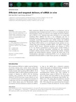
comparison to Tic110 (Figure 1).
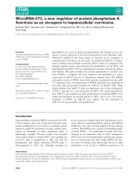
In pea, expression of both genes was found to be
lower in root tissue as compared to leaves. In roots,
PsTic110 RNA is 40% more abundant, while in leaves
the expression levels of PsTic20 and PsTic110 seem to
be in a similar range. In Arabidopsis, AtTic20-I and
AtTic110 are expressed to a lower extent in roots than
in leaves, similar to pea (Figure 1B). These results see-
mingly contradict those of Hirabayashi et al. [26], who
concluded a comparable expression level of Tic20-I in
shoots and roots. However, they used a non-quantifiable
approach in contrast to our quantitative analysis.
Furthermore, in our experiments the o verall expression
of AtTic20-I and AtTic110 diff ers notably from that in
pea, AtTic110 RNA being about 3.5 and 6 times more
abundant than AtTic20-I in leaves and roots,
respectively.
We also designed specific primers for the second
Tic20 homolog in Arabidopsis, AtTic20-IV,andour
quantitative method was s ufficiently sensitive to pre-
cisely define its RNA levels in Arabidopsi s leaves and
roots, allowing direct comparison with the expression of
AtTic20-I and At Tic110 (Figure 1B). Transcription of
AtTic20-IV had al so been investigated in parallel to
AtTic110 by Teng et al. [27], who observed a differential
ratio of expression using two different methods, of
which one was not even sensitive enough to detect
AtTic20-IV. A very recent study [26] also investigated
the expression of AtTic20-IV, however, without any
quantification of their data.
Our data show that AtTic20-IV is present in leaves
and roots with transcript levels similar to AtTi c20-I,but
less abundant than AtTic110. Interestingly, and in accor-
dance with the data presented by Hirabayashi et al. [26],
transcript levels of AtTic20-IV in roots are higher than
those of AtTic20-I , while the opposite is tr ue in leaf tis-
sue.
It can be speculated that the observed expression
pattern reflects tissue-specific differentiation of both
genes. AtTic20-IV may still partially complement for the
function of AtTic20-I, as becomes evident from the via-
bility of attic20-I knockout plants and the yellowish
phenotype of attic20-I mutants overexpressing AtTic20-
IV [26,27]. However, the seve re phenotype of attic20-I
plants, in conjunction with the observed differential
expression pattern, clearly indicates specific functions of
the two homologs. Furthermore, a higher AtTic110
expression rate as observed in antisense attic20-I lines
might indicate another possible compensatory effect
[19].
The expression pattern of the three investigated genes
was found to be similar in Arabidopsis growing hydro-
ponically with or without sucrose (Figure 1B) or on soil
(data not shown) . However, gene expression was gener-
ally higher in plants growing without sucrose.
Tic20 protein is much less abundant than Tic110 in
envelope membranes
Semi-quantitative analysis of Tic20 and Tic110 on pro-
tein level was performed using immunoblots of envelope
membranes isolated from two-week-old pea and four-
week-old Arabidopsis plants. In parallel, calibration
curves were generated using a series of known conc en-
trations of overexpressed and purified proteins (Figure
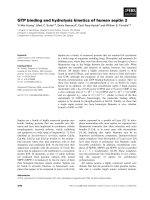
2A, B, D and 2E). After quantification of immunoblots
from envelopes, amounts of PsTic20, PsTic110, AtTic20
and AtTic110 were determined using the corres ponding
calibration curve. The amount of PsTic110 in IE was
found to be almost eight times higher than that of
PsTic20 (Figure 2C), which differs strikingly from the
similar transcript levels of the two genes detected in
leaves (Figure 1A), indicating profound differences in
posttranslational processes such as translation rate and
protein turnover. In Arabidopsis, the absolute amount of
AtTic110 is nearly the same as in pea (Fig ure 2F), how-
ever, Arabidopsis envelope s represent a mixture, con-
taining both outer and IE vesicles. Thus, the relative
amount of AtTic110 is possibly higher than in pea . Sur-
prisingly, the amount of AtTic20 is more than 100
times lower than that of AtTic110, showing an even
greater difference in comparison to the observed RNA
expression levels (Figure 2F). Taking the different mole-
cular size of Tic110 an d Tic20 into account (~5:1), we
still observe 20 times more AtTic110 t han AtTic20 pro-
tein. In pea, we found 1.4 times more Tic110 RNA than
Tic20, whereas in Arabidopsis the ratio of Tic110 to
Tic20 is 20.3. The number of channel forming units
must even be more different, since Tic110 was shown to
form dimers [11], whereas Tic20 builds very large com-
plexes between 700 kDa (this study) and 1 MDa [20].
Thus, two Tic110 molecules would be necessary to form
a channel in contrast to Tic20, which would require
many more molecules to form the pore. Though we
cannot exclude that Tic20 might be subject to degrada-
tion by an unknown protease in vivo, protease treat-
ments with thermolysin of right-side out IE vesicles in
vitro c learly shows that Tic20 is very protease resistant,
Kovács-Bogdán et al. BMC Plant Biology 2011, 11:133
/>Page 3 of 16
even in the presence of detergent. In cont rast, Tic110 is
easily degraded already without addition of detergent
(Additional file 1). This argues against more rapid
degradation of Tic20 compared to Tic110 during pre-
paration of IE. The difference in Tic110 to Tic20 ratios
both on the RNA and protein level between pea and
Arabidopsis maybeduetothedifferentageofthe
plants or the different n eeds under the given growth
condit ions, and suggests that there is no st rict stoichio-
metry between the two proteins. Moreover, the low
abundance of Tic20 in comparison to Tic110 in envel-
opes (see also additional file 2) clearly demonstrates that
0
2
4
6
8
10
12
14
AtTic20-I
AtTic20-IV
AtTic110
RNA expression level
Leaves + suc
Leaves - suc
Roots + suc
Roots - suc
0
5
10
15
20
25
PsTic20
PsTic110
RNA expression level
Leaves
Roots
A
B
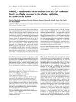
Figure 1 RNA expression levels of Tic20 and Tic110. RNA expression levels of (A) PsTic20, PsTic110 and (B) AtTic20-I, AtTic20-IV and AtTic110 in
leaves and roots of two-week-old Pisum sativum (Ps) and four-week-old Arabidopsis thaliana (At) plants as determined by quantitative RT-PCR
using gene-specific primers. Pea plants were grown on soil and Arabidopsis plants were cultured hydroponically, the latter in the presence and
absence of 1% sucrose (+/- suc). Presented data are the average of at least three measurements.
Kovács-Bogdán et al. BMC Plant Biology 2011, 11:133
/>Page 4 of 16
0
3
6
9
0.00 0.10 0.20 0.30
0.01 0.025 0.05 0.1 0.25 μg
0
2
4
6
8
10
0.00 0.10 0.20 0.30 0.40 0.50
0.025 0.05 0.1 0.25 0.5 μg
0.01 0.025 0.05 0.075 0.1 μg
D
Amount of PsTic20 (μg)
0
10
20
30
40
0.00 0.05
0.10
Signal intensity
Amount of AtTic20 (μg)
0.01 0.025 0.05 0.075 0.1 μg
A
B
D
E
C
F
Signal Intensity
Amount of AtTic110(μg)
0
5
10
15
0.00 0.20 0.40
0.025 0.05 0.1 0.25 0.5 μg
0.5 1 2 5 μg
αPsTic110
αPsTic20
13.6
129.7
0
60
120
180
PsTic20
PsTic110
0.76
111
0
40
80
120
160
AtTic20
AtTic110
αAtTic110
αAtTic20
0.5 1 2 5 μg
Amount of PsTic110(μg)
Signal Intensity
Ng protein/μg IE
Signal Intensity
Ng protein/μg IE
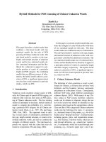
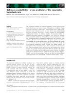
Figure 2 Protein levels of Tic20 and Tic110 in envelope membranes. Semi-quantitative analysis of Tic20 and Tic110 protein levels in (A-C)
Pisum sativum (Ps) and (D-F) Arabidopsis thaliana (At). A dilution series of purified PsTic20, PsTic110, AtTic20 and AtTic110 was quantified after
immunodetection with specific antibodies (A, B, D and E in inset). Calibration curves were calculated using known concentrations of proteins
plotted against the quantified data (A, B, D and E). These curves were used to determine the amount of Tic20 and Tic110 in (C) pea and (F)
Arabidopsis envelope samples. Insets in (C) and (F) show dilution series of corresponding envelopes after immunodetection with the indicated
antibody. Presented data are the average of two independent experiments; a representative result is depicted.
Kovács-Bogdán et al. BMC Plant Biology 2011, 11:133
/>Page 5 of 16
Tic20 cannot be the main channel of the Tic translocon
as previously suggested [21,24], since it cannot possibly
support the required import rates of some highly abun-
dant preproteins that are needed in the chloroplast.
Tic20 forms high molecular weight complexes separately
from Tic110
Experimental data suggested a common complex
between Tic110 and Tic20 in chloroplast envelope
membranes using a cross-linking approach [21]. How-
ever, the interaction was not visible in the absence of
Toc components, making a stable association unlikely.
Furthermore, no evidence for a common complex was
found by Kikuchi et al. [20] using solubilized chloro-
plasts of pea and Arabidopsis for two-dimensional blue
native/SDS-PAGE (2D BN/SDS-PAGE) analysis. Like-
wise, the difference in Tic110 to Tic20 ratios both on
the RNA and protein le vel between pea and Arabidopsis
indicates that a common complex, in which both p ro-
teins cooperate in translocation channel formation in a
reasonable stoichiometry, is improbable.
To clarify this issue, we addressed these partly con-
flicting results by using IE vesicles, which should mini-
mize the possible influence of the interactio n with Toc
component s on complex formation. Pea IE v esicles were
solubilized in 5% digitonin and subjected to 2D BN/
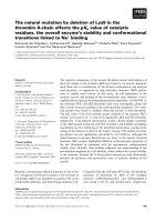
SDS-PAGE. Immunoblots revealed that both Tic20 and
Tic110 are present in distinct high molecula r weight
complexes (Figure 3A): Tic110-containing complexes
migrate at a size of ~ 200-300 kDa, whereas Tic20 dis-
plays a much slower mobility in BN-PAGE and is pre-
sent in complexes exceeding 700 kDa, in line with the
results from Kikuchi et al. [20]. However, at a similar
molecular weight of 250 kDa on BN-PAGE not only
Tic1 10 but also Hsp93, Tic62 and Tic55 were described
[30]. The molecular weight o f a complex containing all
of these components would be much higher. Therefore,
components of the Tic complex might associate with
Tic110 very dynamically resulting in different composi-
tions under different conditions, or alternatively, there
are different complexes present at the same molecular
weight.
An open question to date is the identity of possible
interaction partners of Tic20 in the complex. Tic22, the
only Tic c omponent located in the intermembrane
space, is a potential candidate, since both proteins were
identified together in cross-linking experiments [21].
However, only minor amounts of Tic20 and Tic22 were
shown to co-localize after gel filtration of solubilized
envelope membranes [21]. A second candidate for com-
mon complex formatio n is PIC1/Tic21: Kikuchi et al.
[20] demonstrated that a one-megadalton complex of
Tic20containsPIC1/Tic21asaminorsubunit.PIC1/
Tic21 was proposed to form a protein translocation
channel in the Tic complex, mainly based on protein
import defects of knockout mutants and on structural
similarities to amino acid transporters and sugar per-
meases [27]. An independent study by Duy et al. [31]
4% 13%
1
st
dimension BN-PAGE
PsTic110
PsTic20
~670 kDa
~140 kDa
AtTic20
B
A
2
n
d
dimension
SDS-PAGE
2
nd
dimension
SDS-PAGE
4% 13%
1
st
dimension BN-PAGE
pea inner envelope vesicles
Tic20 proteoliposomes
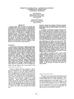
Figure 3 Complex formation of Tic20 in inner envelope membranes and proteoliposomes. Two-dimensional BN/SDS-PAGE of (A) inner
envelope vesicles of Pisum sativum (Ps, 100 μg protein) and (B) AtTic20-proteoliposomes (20-30 μg protein). Samples were solubilized in 5%
digitonin and separated by 4-13% BN-PAGE followed by 12.5% SDS-PAGE. Indicated specific antibodies were used for immunodetection.
Representative results are depicted. At - Arabidopsis thaliana.
Kovács-Bogdán et al. BMC Plant Biology 2011, 11:133
/>Page 6 of 16
favours the hypothesis that PIC1/Tic21 forms a metal
permease in the IE of chloroplasts, rendering the
import-related role question able. This discrepancy will
have to be addressed in the future.
To test the complex formation of Tic20 in vitro with-
outtheinvolvementofotherproteins,weusedTic20-
proteoliposomes for 2D BN/SDS-PAGE analysis, simi-
larly to IE vesicles (Figure 3B). The migration behaviour
of the protein resembles that observed in IE: the major-
ity of the protein localizes in high molecular weight
range, however, the signal appears more widespread and
a portion is also detected at lower molecular weights,
possibly as monomers. This observation reveals that
Tic20 has the inherent ability to homo-oligomerize in
the presence of a lipid bilayer. The less distinct signal
could be due to different solubilization of Tic20 by digi-
tonin in IE vesicles vs. liposomes, or could be an indica-
tion that addi tional subunits stab ilize the endogenous
Tic20 complexes, which are not present after the recon-
stitution. However, we interpret these observations as
support for the hypothesis that the major component of
the one megadalton complex in IE are homo-oligomers
composed of Tic20.
The N- and C-termini of Tic20 face the stromal side
In silico ana lysis of Tic20 predicts the presence of four
hydrophobic transmembrane helices positioning both
N- and C-termini to one side of the membrane
(TMHMM Server [23] and [21,25]). Accordi ng to these
predictions, three cysteins (Cys) in PsTic20 face the
same side, while the fourth would be located in the
plane of the membrane. We used pea IE vesicles pre-
pared in a right-side-out orientation [32] to determine
the topology of Tic20 empl oying a Cys-labelling techni -
que. To this end, the IE vesicles were incubated with a
membrane-impermeable, Cys-reactive agent (metoxypo-
lyethylenglycol-maleimide, PEG-Mal) that adds a mole-
cular weight of 5,000 Da to the target protein for each
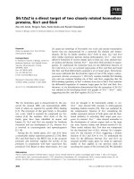
reactive Cys residue. In our experiments PEG-Mal did
not strongly label any Cys residues of Tic20 under the
conditions applied (Figure 4A), indicating the absence
of accessible Cys residues on the outside of the mem-
brane. Only one faint additional band of higher molecu-
lar weight was detectable (Figure 4A, marked with
asterisk), possibly due to a partially accessible Cys
located within the membrane. In the presenc e of 1%
SDS, however, all four Cys residues present in PsTic20
are rapidly PEGylated, as demonstrated by the appear-
ance of four intense additional bands after only five
minutes of incubation. The observed gain in molecular
weight per modification is bigger than the expected 5
kDa for each Cys, but this can be attributed to an aber-
rant mobility of the modified protein in t he Bis-Tris/
SDS-PAGE used in the assay.
Our results support a four transmembrane helix topol-
ogy in which both the C- and N-termini are facing t he
stromal side of the membrane (Figure 4B), with no Cys
residues oriented towards the intermembrane space.
Cys
108
is most likely located in helix one, Cys
227
and
Cys
230
are oriented to the stromal side of helix four and
Cys
243
is located in the stroma. This topology is also in
line with green fluorescent protein-labelling studies by
van Dooren et al . [22] indicating that t he N- and C-ter-
mini also of the Toxoplasma gondii homolog of Tic20
face the stromal side of the inner apicoplast membrane.
Tic20 is mainly a-helical
Tic20 was identified more than a decade ago but since
then no heterologous expression and purification proce-
dure has been reported, which could successfully
synthesize folded full-length Tic20. Here, we report two
efficient Escherichia coli (E. coli) based systems for
Tic20 expression and purification from both pea and
Arabidopsis: codon optimized PsTic20 (Additional file 3)
was overexpressed in a S12 cell lysate in presence of
deter gents, and AtTic20 overexpression was successfully
accomplished by adaptation of a special induction sys -
tem [33]. Following these steps, both pea and Arabidop -
sis proteins could be purified to homogeneity by metal
affinity purification (Figure 4C).
Using the purified pr otein, we performed structural
characterization studies of Tic20 by subjecting it to cir-
cular dichroism (CD) spectroscopy (Figure 4D). The
recorded spectra of PsTic20, displaying two minima at
210 and 222 nm and a large peak of positive ellipticity
centered at 193 nm, are highly characteristic of a-helical
proteins, and thus demonstrate that the protein exists in
a folded state after purification in the presence of deter-
gent. The secondary structure of Tic20 was estimated by
fitting spectra to reference data sets (DichroWeb server
[34,35]) resulting in an a-helical content of approxi-
mately 78%, confirming in silico predictions [21,25].
Purified Tic20 protein inserts firmly into liposomes
To better characterize Tic20 in a membrane-mimicking
environment, heterologously expressed and purified
AtTic20 was reconstituted into liposom es in vitro.Initi-
ally, flotation experiments were performed to verify a
stable insertion. In the presence or a bsence of lipo-
somes, Tic20 was placed at the bottom of a gradient
ranging from 1.6 M (bot tom) to 0.1 M (top) sucrose. In
the presence of liposomes, Tic20 migrated to the middle
of the gradient, indicating a change in its density caused
by interaction with liposomes. In contrast, the protein
alone remained at the bottom of the gradient (Figure
5A). Proteoliposomes were also treated with various buf-
fers before flotation (for 30 min at 4°C), to test whether
the protein is firmly insert ed into the liposomal
Kovács-Bogdán et al. BMC Plant Biology 2011, 11:133
/>Page 7 of 16
membrane or just loosely bound to the vesicle surface.
None of the applied conditions (control: 10 mM MOPS/
Tris, pH 7; high ionic strength: 1 M MOPS/Tris, pH 7;
high pH: 10 mM Na
2
CO
3
, pH 11; denaturing: 6 M urea
in 10 mM MOPS/Tris, pH 7) changed the migration
behaviour of Tic20 in the gradient (Figure 5B), indicat-
ing that T ic20 was deeply inserted into the liposomal
membrane. Thus, proteoliposomes represent a suitable
in vitro system for the analysis of Tic20 channel activity.
Tic20 forms a channel in liposomes
Even though Tic20 has long been suggested to form a
channel in the IE membrane, this notion was solely
based on structural analogy to ot her four-transmem-
brane helix proteins [21,24], and no experimental evi-
dence has been provided so far. To investigate whether
Tic20 can indeed form an ion channel, Tic20-proteoli-
posomes were s ubjected to swelling assays (Figure 5C).
Changes in the size of liposomes in the presence of high
salt concentrations, as revealed by changes in the optical
density, can be used to detect the presence of a pore-
forming protein [36]. After addition of 300 mM KCl to
liposomes and Tic20-proteoliposomes, their optic al den-
sities dropped initially, due to shrinkage caused by the
increased salt concentration [37]. However, the optical
density of protein-free liposomes remained at this low
level, showing no change in their size; wher eas in the
case of Tic20-proteoliposomes the optical density
increased constantly w ith time. The increase in optical
density ( and therefore size) strongly supports the pre-
sence of a channel in Tic20-proteoliposomes that is
permeable for ions, thereby creating an equilibrium
A
Cys
243
IE
stroma
IMS
Cys
227
Cys
230
Cys
108
B
C
66
45
36
29
24
20
14
D
Figure 4 Topology and secondary structure of Tic20. (A) PEG-Mal labelling of Pisum sativum (Ps) inner envelope (IE) vesicles in the presence
or absence of 1% SDS for the indicated times using a specific antibody against PsTic20 for immunodetection. Asterisks indicate a weak band
most likely representing Tic20 with one labelled cystein (Cys) within the transmembrane region. A representative result of three repetitions is
shown. (B) Topological model of Tic20 - indicating the position of Cys residues in PsTic20 - considering the PEGylation assay in (A) (based on
structural prediction of TMHMM Server [23] and [25]). Boxes symbolise a-helical transmembrane domains (TM 1-4). IMS - intermembrane space.
(C) The mature parts of Tic20 from Pisum sativum (PsTic20, amino acids 83-253) and Arabidopsis thaliana (AtTic20 amino acids 59-274) were
overexpressed in an E.coli cell lysate system and in E.coli BL21 cells, respectively. Both proteins were purified by Ni
2+
-affinity chromatography.
Coomassie-stained gels of representative purifications are shown. (D) Circular dichroism spectrum of overexpressed and purified PsTic20 in 20
mM Na-phosphate buffer (pH 8.0), 150 mM NaF, 0.8% Brij-35. The presented chromatogram is the average of three independent experiments.
Secondary structure elements were quantified using the CDSSTR method from the DichroWeb server and results are presented in the inset.
Kovács-Bogdán et al. BMC Plant Biology 2011, 11:133
/>Page 8 of 16
between the inner compartment of the proteoliposomes
and the surrounding buffer.
To exclude the possible effects of (i) contaminating
channel-forming proteins derived from the bacterial
membrane and (ii) a protein inserted into the liposomes
(but not forming a channel), a fur ther negative control
was set up: Tic110 containing only the first three trans-
membrane helices (NtTic110) was purified similarly to
Tic20 and reconstituted into liposomes. We chose this
construct, since NtTic110 inserts into the membrane
during in vitro prot ein import experiments [10].
Furthermore, as the full length and N-terminally trun-
cated Tic110 possess very similar channel activities
[11,12], it is unlikely that the N-terminal part alone
forms a channe l. The insertion of NtTic110 into lipo-
somes was confir med by incubation under different buf-
fer conditions (high salt concentratio n, high pH and 6
M urea) followed by flotation experiments, similarly to
Tic20 (data not shown). However, these NtTic110-pro-
teoliposomes behaved similarly to the empty liposomes
during swelling assays: after addition of salt, the optical
density decreased, and except for a small initial increase,
it remained at a constant level (Figure 5C). This makes
it unlikely that a contamination from E. coli or simply
the insertion of a protein into the liposomes caused the
observed effect in the optical density of Tic20-
proteoliposomes.
To further characterize the channel a ctivity of Tic20,
electrophysiological measurements were performed.
After the fusion of Tic20-proteo liposomes with a lipid
bilayer, ion channel activity was observed (Figure 6A, B).
The total conductance under symmetrical buffer condi-
tions (10 mM MOPS/Tris (pH 7.0), 250 mM KCl) was
dependent on the direction of the applied potential:
1260 pS (± 70 pS) and 1010 pS (± 50 pS) under negative
and positive voltage values, respectively. The channel
was mostly in the completely open state, however, indi-
vidual single gating events were also frequently
observed, varying in a broad range between 25 pS to
600 pS ( Figure 6A-D). All detec ted gating events were
depicted in two histograms (Figure 6C, D for negative
and positive voltages, respectively). Two conductance
classes (I and II) were defined both at negative and posi-
tive voltage values with thresholds of 220 pS and 180
pS, respectively (Figure 6A-E). Note that gating events
belonging to the smaller conductance cl asses (I)
occurred more frequently. The observed pore seems to
be asymmetric, since higher conductance classes notably
differ under positive and negative voltages. This is prob-
ably due to interactions of the permeating ions with the
channel, which presumably exhibits an asymmetric
potential profile along the pore. Since small and large
opening events were simultaneously observed in all
experiments, it is very unlikely that they belong to two
different pores.
The selectivity of Tic20 was investigated under asym-
metric salt conditions (10 mM MOPS/Tris (pH 7.0),
250/20 mM KCl). Similarly to the conductance v alues,
the channel is intrinsically rectifying (behaving differ-
ently under negative and positive voltage values),
A
C
B
0.088
0.090
0.092
0.094
0.096
0.098
0.100
0.102
0 5 10 15 20
25
OD
500nm
Time (min)
Liposomes
Tic20-proteoliposomes
NtTic110-proteoliposomes
+ KCl
Figure 5 Tic20 insertion into liposomes and channel formation. (A) Flotation experiments of Tic20-proteoliposomes and Tic20 without
vesicles in a sucrose gradient. Samples containing 1.6 M sucrose were loaded at the bottom of a sucrose step gradient and centrifuged to
equilibrium (100,000 g, 19 h, 4°C). Fractions were analysed by silver-staining. (B) Flotation experiments of Tic20-proteoliposomes (similar to (A))
incubated under the indicated buffer conditions for 30 min at 4°C before centrifugation. (C) Swelling assay of liposomes, Tic20-proteoliposomes
and NtTic110-proteoliposomes containing 20 mM Tris-HCl (pH 8.0), 100 mM NaCl. Change in optical density was measured at 500 nm (OD
500 nm
)
of 1 ml solutions every minute. Arrow indicates the addition of 300 mM KCl. Presented results are the average of at least five repetitions;
standard deviations were within 1.5-3%.
Kovács-Bogdán et al. BMC Plant Biology 2011, 11:133
/>Page 9 of 16
-100 mV 250/250 mM KCl
Current (pA)
A
I
II
II
I
+100 mV 250/250 mM KCl
Current (pA)
I
II
III
B
I
II
I
II
C
D
E
F
Negative voltage
Positive voltage
Figure 6 Electrophysiological characterization of Tic20. (A) and (B) Current traces of a Tic20 channel in lipid bilayer at -100 mV and +100
mV, respectively. Dotted lines indicate thresholds of each conductance class (I and II). Lower panels show representative gating events
belonging to each class. (C) and (D) Conductance histograms of all gating events of Tic20 at negative and positive voltages, respectively. Colours
represent different conductance classes (I and II). (E) Current-voltage relationship diagram of all analysed gating events ordered in the four
indicated conductance classes using the same colour code as in (C) and (D). Indicated conductance values correspond to the slope of fitted
linears in each class. (F) A representative voltage ramp of Tic20 demonstrating the cation selectivity of the channel with a positive reverse
potential (E
rev
). Measurements were performed under symmetrical (A)-(E) and asymmetrical (F) buffer conditions (20 mM MOPS/Tris (pH 7.0), 250
mM and 20/250 mM KCl, respectively). Presented data derive from two independent fusions accounting for more than 4500 gating events and
16 voltage ramps.
Kovács-Bogdán et al. BMC Plant Biology 2011, 11:133
/>Page 10 of 16
supporting asymmetric channel properties. The obtained
reverse potential is 37.0 ± 1.4 mV (Figure 6D). Accord-
ing to the Goldman-Hodgkin-Katz approach, this corre-
sponds to a selectivity o f 6.5:1 for K
+
:Cl
-
-ions, thus
indicating cation selectivity similar to Tic110 [11].
To determine the channel’ s orientation within the
bilayer, two side-specific characteristics were taken into
acco unt: the highes t total conductance under symmetri-
cal buffer conditions wa s measured under negative vol-
tage values, and the channel rectifies in the same
direction under asymmetrical buffer conditions (see vol-
tage ramp, Figure 6D). Therefore, it seems that the pro-
tein is randomly inserted into the bilayer.
The pore size was roughly estimated according to
Hille et al. [38]. Considering the highest conductance
class (350 pS), a channel l ength of 1-5 nm and a resis-
tivity of 247.5 Ω cm for a solution containing 250 mM
KCl, taking into account that the conductivity of the
electrolyte solution within the pore is ~5 times lo wer
than in the bulk solution [39], the pore size was esti-
mated to vary between 7.8-14.1 Å. This is in good
agreement with the size of protein translocation chan-
nels such as Toc75 (14-26Å, [40]) in the outer envelope
membrane and Tic110 (15-31 Å, [12]) in the IE. Thus,
thesizeoftheTic20porewouldbesufficientforthe
translocation of precursor proteins through the
membrane.
NtTic110, as a negativ e control, did not show any
channel activity during electrophysiological measure-
ments, indicating that the measured channel is not the
result of a possible bacterial contamination (data not
shown).
Considering our data presented here and those pub-
lished in previous studies, we can concl ude that the Tic
translocon consists of distinct (at least two) transloca-
tion channels: On the one hand, Tic110 forms the main
translocation pore and therefore facilitates import o f
most of the chloroplast-targeted preproteins; on the
other hand, Tic20 might facilitat e the translocation of a
subset of proteins. This scenario would match the one
found in the inner mitochondrial membrane, where spe-
cific translocases exist for defined groups of precursor
proteins: the import pathway of mitochondrial carrier
proteins being clearly separated from that of matrix tar-
geted preproteins [41]. The situation in chloroplasts
does not seem as clear-cut, but an analogous separation
determined by the final dest ination and/or intrinsic
properties of translocated proteins is feasible.
The severe phenotype of attic20-I mutants prompts us
to hypothesize that Tic20 might be specifically required
for the translocation of some essential proteins. Accord-
ing to cross-linking results [21], Tic20 is connected to
Toc translocon components. Therefore, after ent ering
theintermembranespaceviatheToccomplex,some
preproteins might be transported through the IE via
Tic20. On the contrary, Kikuchi et al. [20] presented
that Tic20 migrates on BN-PAGE at the same molecular
weight as the imported precursor of the small subunit
of Rubisco (pSSU) and that tic20-I mutants display a
reduced rate of the artificial precursor protein RbcS-nt:
GFP. The authors interpreted these results in a way that
Tic20 might function at an intermediate step between
the Toc translocon and the channel of Tic110. However,
being a substantial part of the general import pathway
seems unlikely due to the very low abundance of Tic20.
It is feasible to speculate that such abundant proteins as
pSSU, which are imported at a very high rate, may inter-
act incidentally with nearby proteins or indifferently use
all available i mport channels. To clarify this question,
substrate proteins and interaction partners of Tic20
should be a matter of further investigation.
Additionally, a very recent study [26] suggested
AtTic20-IV as an import channel working side by side
with AtTic20-I. However, detailed characterization of
the protein (e.g. localization, topology) and experimental
evidence for channel activity are still missing.
Conclusions
In this study we could clearly demonstrate that Tic20
and Tic110 function separately from each other, based
on their different stoichiometry and their independent
complex formation in IE vesicles. We furthermore pre-
sent the first experimental evidence for Tic20 channel
function. The very low abundance of Tic20 compared to
Tic110 argues against Tic20 forming a major protein
translocation channel, which would import the large
number of preproteins that are needed in the chloro-
plast. Therefore, our data favour t he idea that the Tic
translocon comprise s at least two translocation chan-
nels: Tic110, constituti ng the main import channel [11],
and Tic20, which might import a special subset of pre-
proteins (a hypothetical model of the two Tic transloca-
tion channels is depicted in Figure 7). A similar system
exists in the inner membrane of mitochondria, where
the TIM22 and TIM23 complexes mediate the import
of different sets of pro teins [41]. Unfortunat ely, due to
the lethality of tic110 and the very severe phenoty pe of
tic20-I homozygous knockout mutants, their separate
mode of action will be very difficult to investigate in
vivo.
Methods
Plant growth conditions
Pea plants (Pisum sativum var. Arvica) were grow n
under a 14-h light/10-h dark regime at 20°C/ 15°C. Ara-
bidopsis thaliana plants (ecotype Columbia) were grown
either on s oil under the conditions described in Benz et
al. [42] or hydroponically. T he latter were cultured
Kovács-Bogdán et al. BMC Plant Biology 2011, 11:133
/>Page 11 of 16
under non-sterile conditions as modified from [43].
Briefly, after expansion of their first true leaves, seed-
lings were transferred from plates (containing 0.5 × MS
with 1% sucrose) into a GA-7 Magenta vessel (Sigma-
Aldrich) o nto a one cm thick cut sponge. The nutrient
solution contained 1 mM KH
2
PO
4
,0.5mMMgSO
4
,
0.25 mM CaSO
4
,20μMFe-EDTA,25μMH
3
BO
3
,2
μMZnSO
4
,2μMMnSO
4
,0.5μMCuSO
4
,0.5μM
(NH
4
)
6
Mo
7
O
24
and 0.5 mM NH
4
NO
3
in the presence or
absence of 1% sucrose. The growth chambers were kept
under 16-h light/8-h dark regime at 21°C/16°C for four
weeks. Plants were either harvested from the dark or
before noon from growth light. Material was usually
used immediately and fresh. If not possible, leaf material
was shock-frozen in liquid nitrogen and stored at -80°C
until use.
qRT-PCR
RNAisolation,cDNApreparation,qRT-PCRanddata
analysis were performed essenti ally as described in [44].
Gene-specific primers were constructed for PsTic20
[GenBank: AF095285. 1], PsTic110 [GenBank: Z68506.1],
AtTic20-I [TAIR: At1g04940.1], AtTic20-IV [TAIR:
At4g03320.1] and AtTic110 [TAIR: At1g06950.1] (Table
1). All reactions were performed in quadruplicates.
Isolation of envelope vesicles
Membrane fractions enriched in right-side-out IE vesi-
cles of pea chloroplast membranes w ere isolated from
intact chloroplasts of 10 to 12-day-old pea plants as
described previously [32]. For Arabidopsis envelope
To c
IE
OE
Tic110
Tic20
Figure 7 Two independent channels at the Tic translocon. Hypothetical model of Tic20 and Tic110 channels in the inner envelope (IE) of
chloroplasts. After passing the Toc complex in the outer envelope (OE), preproteins are imported either via Tic110 (red) or Tic20 (blue) through
the IE. Hypothetical precursors that might use both channel proteins are depicted in yellow. Tic110 is thought to form a homodimer with a total
of eight amphipathic transmembrane helices forming the translocation channel and four hydrophobic a-helices involved in the insertion into
the membrane (according to [8] and [11]). The proposed Tic20 channel is depicted as a homo-oligomer (only three molecules are shown,
however, it is > 700 kDa) and, based on the low overall abundance, might be responsible for the import of a subset of preproteins, indicated by
a smaller number of depicted precursors waiting at the entrance of the channel.
Table 1 Primers for qRT-PCR analysis
primer forward reverse
PsTic20 CCTAGATGGTCTCTCATAGC GCAGTAGTCCAGAAATGC
PsTic110 CAAGGAAACTGCTCTGTC CTCCTTTGATGTCCTCTACC
Ps18SrRNA CCAGGTCCAGACATAGTAAG GAGGGTTACCTCCACATAG
AtTic20-I AGGTTATAGGGACCGTTAGC CTTAGTCGTACGGAATCTGG
AtTic20-IV CTATGTCCAACCTTTTCTCG CTGTTTCAAGAAGCATACCC
AtTic110 CTAAAGGAGTGGTCTTGTCG GCAGAAGATAATGCTCCATC
At18SrRNA AACTCGACGGATCGCATGG ACTACCTCCCCGTGTCAGG
Gene-specific primers generated for PsTic20, PsTic110, Ps18SrRNA, AtTic20-I,
AtTic20-IV, AtTic110 and At18SrRNA applied in the qRT-PCR analysis.
Kovács-Bogdán et al. BMC Plant Biology 2011, 11:133
/>Page 12 of 16
preparation, chloroplasts were first isolated from 4-
week-old soil-grown plants from the dark as described
by Seigneurin-Berny et al. [45]. Chloroplasts were subse-
quently resuspended in 15 ml of 10 mM HEPES-KOH,
pH 7.6, 5 mM MgCl
2
, and lysed using 50 strokes in a
small (15 ml) Dounce tissue grinder (Wheaton Science
Products, Millville, NJ, USA). Further separation into
stroma, thylakoids and envelopes was performed accord-
ing to Li et al. [46].
Protein expression and purification
The sequence coding for t he mature part of Tic20 from
Pisum sativum (PsTic20, amino acids 83-253) was
codon optimized wit h the Leto 1.0 program by Entele-
chon (Regensburg, Germany) (Additional file 3). The
optimized gene was then synthesized by Entelechon and
finally cloned into pIVEX2.3 (Roche, Germany). The
mature part of Arabidopsis thaliana Tic20-I (AtTic20,
amino acids 59-274) was cloned into pCOLDII (Takara-
Bio, Kyoto, Japan). The mature part of Tic110 from
Pisum sativum without the N-terminal hydrophobic
domain (PsTic110, amino acids 122-996) and a similar
construct for the homologous part from Arabidopsis
thalia na Tic110 (AtTic110, amino acids 141-1016) were
cloned into pET21d [11]. The same expression vector
was used for the cloning of the N-terminal part of
mature Tic110 (NtTic110, amino acids 76-258) from
Arabidopsis thaliana.
The codon optimized PsTic20 was overexpressed in a
self-made E.coli cell-free lysate system (S12) which was
prepared essentially as de scribed by Kim et al. [47].
Shortly, expression of soluble PsTic20 was carried out at
30°C for 1-2 h with constant roll ing in 100-200 μl reac-
tion mixture (57 mM Hepes-KOH (pH 8.2), 1.2 mM
ATP, 0.65 mM cAMP, 0.85 mM each of CTP, GTP and
UTP, 2 mM DTT, 90 mM potassium glutamate, 80 mM
ammonium acetate, 15 mM magnesium acetate, 34 μg/
ml
L
-5-formyl-5,6,7,8-tetrahydrofolic acid, 0.75 mM each
of 20 amino acids, 2% polyethylene glycol 8000, 100
mM creatine phosphate, 0.27 mg/ml creatine kinase,
0.17 mg/ml E. coli total tRNA mixture (from strain
MRE600), 10 μg/ ml plasmid DNA, 25% BL21 (DE3 ) and
2% BL21 (DE3) RIL-pAR1219 cell extract and 0.8% Brij-
35). After removing insoluble material (10,000 g, 4°C, 10
min) the supernatant was diluted 1:3 with 50 mM
NaH
2
PO
4
-NaOH (pH 8.0), 300 mM NaCl, 0.8% Brij-35,
20 mM imidazole and purified using Ni-NTA-Sepharose
(GE Healthcare, Munich, Germany).
ForexpressionandpurificationofAtTic20/pCOLDII,
transformed BL21 (DE3) cells (Novagen/Merck) w ere
grown at 37°C in M9ZB medium to an OD
600
of 0.4
and then shifted to 15°C for 30 min. After induction
with 1 mM isopropyl-1-thio-b-D-galactopyranoside, cells
were further grown at 15°C overnight. The harvested
cells were resuspended in 50 mM Tris-HCl (pH 8.0),
150mMNaCl,5mMdithiothreitol(DTT),lysed(M-
110L Microfluidizer Processor, Microfluidics, Newton,
MA, USA), pelleted (20,000 g, 4°C, 20 min) and solubi-
lized in the presence of 1% n-lauroylsarcosine (N-LS)
for 1 h at 4°C. Purification was carried out in the pre-
sence of 0.3% N-LS using Ni-NTA-Sep harose (GE
Healthcare, Munich, Germany). PsTic110/pET21d was
ove rexpressed and purified as described previously [11].
AtTic110/pET21d overexpression and purification was
performed similarly to PsTic110. NtTic110 was overex-
pressed similarly to PsTic110, whereas its purification
was performed similarly to AtTic20 except that in all
buffers 300 mM NaCl was present.
Immunoblotting
Immunoblotting was performed using polyclonal anti-
sera from rabbits raised against heterologously overex-
pressed proteins, followed by incubation with
monoclonal rabbit secondary antibody, visualize d by
alkaline phosphatase or by a chemiluminescence detec-
tion system (Pierce, Rockford, IL, USA). The antisera
against atTic110 and psTic20 were purified against a
poly-histidine matrix. To this end, Poly-L-Histidine was
coupled to CNBr-activated sepharose (GE Healthcare)
according to the manufacturer’s recommendations. Anti-
sera were diluted 3 times and incubated over night at 4°
C with the Poly-His matrix. Sepharose beads were sedi-
mented and the supernatant was used as purified serum
in the immunoblots (see additional file 4).
Semi-quantitative protein analysis
For semi-quantitative protein analysis, a dilution series
of purified PsTic20, PsTic110, AtTic20 and AtTic110
was loaded on SDS-PAGE in parallel to a dilution series
of pea IE vesicles and Arabidopsis mixed envelope mem-
branes. After immunodetection with specific antibodies,
the intensity of the resulting bands was quantified
(AIDA Software). The band intensity of the pur ified
proteins was first plotted against the known prot ein
amount. This c alibration curve was then applied to
determine the amount of Tic20 and Tic110 present in
the membrane samples. The analysis was repeated two
times with different envelope preparations.
Two dimensional BN/SDS-PAGE
BN-PAGE was performed essentially as described by
Schaegger and von Jagow [48] and Küchler et al. [30]
with minor modifications. IE membranes (50-200 μg
protein) or Tic20-proteoliposomes (30 μgprotein)were
solubilized in 50 mM Bis-Tris/HCl (pH 7.0), 750 mM 6-
aminocaproic acid and 5% digitonin. After incubation
onice(IE)oratroomtemperature(liposomes)for15
min, samples were centrifuged at 256,000 g for 10 min
Kovács-Bogdán et al. BMC Plant Biology 2011, 11:133
/>Page 13 of 16
at 4°C. The supernatant was supplemented w ith 0.1
volume of a Coo massie Blue G solution (5% Coomassie
Brilliant Blue G-250, 750 mM 6-aminocaproic acid) and
loaded on a polyacrylamide gradient gel. Following the
first dimension, lanes were incubated sequentially in 1%
SDS, 1 mM b-mercaptoethanol (b-ME), in 1% SDS with-
out b-ME and in SDS-PAGE running buffer (25 mM
Tris, 192 mM glycine, and 0.1% SDS) at ro om tempera-
ture for 15 min each and then horizontally subjected to
a second dimension SDS-PAGE. After separation,
immunodetection was performed.
CD-spectroscopy
Purified PsTic20 was dialysed against 20 mM Na
2
HPO
4
/
NaH
2
PO
4
buffer (pH 8.0), 150 mM NaF, 0.8% Brij-35
prior to CD analysis. Experiments were carried out at
20°C using a J-810 spectropolarimeter ( Jasco, Grob-
Umstadt, Germany) flushed with N
2
. Spectra were col-
lected from 260 to 190 nm using a 1 mm path length of
a cylindrical quar tz cell. Each spectrum was the average
of three scans taken at a scan rate of 20 nm/min with a
spectral bandwidth of 1 nm. The experiment was
repeated three times in a concentration range of 0.02 to
0.284 mg/ml protein. For the f inal representation, the
baseline was subtracted from the spectrum. The analysis
was performed using the CDSSTR method from the
DichroWeb server [34,35].
PEGylation assay
IE vesicles were treated with 10 mM metoxypolyethylen-
glycol-maleimide 5000 Da (PEG-Mal, Laysan Bio, Arab,
AL, USA) in a buffer containing 100 mM Tris-HCl (pH
7.0), 1 mM EDTA, for the in dicated times at room tem-
perature in the dark in absence or presence of 1% SDS.
The PEGylation reaction was stopped by addition of 100
mM DTT and SDS-PAGE sample buffer. NuPAGE Bis-
Tris gels (10% acrylamide) were employed using a MES
running buffer. Tic20 was detected by immunoblotting.
Liposome preparation and flotation assay
Proteoliposomes of AtTic20 and NtTic110 were pre-
pared as described previously [11]. To prepare unilamel-
lar liposome vesicles, samples were extruded 21 times
through a 200 nm polycarbonate filter (Liposofast, Aves-
tin, Ottawa, Canada). Puri fied AtTic20 (in 20 mM Tris-
HCl pH 8.0, 150 mM NaCl, 0.3% N-LS) (or NtTic110 or
buffer as controls) was mixed with liposomes and i ncu-
bated for 1.5 h at 4°C. The samples were dialysed for 16
hat4°Cagainstabufferwithoutdetergent(20mM
Tris-HCl pH 8.0, 100 mM NaCl) and the remaining
detergent was removed during 2 h incubation a t 4°C
with Bio-Beads SM-2 (Bio-Rad Laboratories, Hercules,
USA). Liposome-associated and liposome-free proteins
were separated by flotation through a sucrose gradient,
similar to Balsera et al. [11]: Samples were adjusted to a
sucrose concentration of 1.6 M (1 ml, bottom) and over-
laid with 3 ml of step sucrose gradient (0.8, 0.4 and 0.1
M, top). After centrifugation (100,000 g, 19 h, 4°C) 0.5
ml fractions were collected and precipitated with tri-
chloroacetic acid (TCA). The samples were resuspended
in Laemmli-buffer, separated by SDS-PAGE and
detected by silver-staining.
Swelling assay
Freshly prepared liposomes and proteoliposomes
(AtTic20 and NtTic110) were diluted to 1 ml to a star t-
ing optical density of appro ximately 0.1 at 500 nm. The
optical density of the samples was measured with a Shi-
madzu UV-2401PC Spectrophotometer (Columbia,
USA) for the indicated time. At the beginning of the
measurements 300 mM KCl was added to the samples.
Experiments were repeated at least five times.
Electrophysiological measurements
Electrophysiological measurements were performed
using the IonoVation Bilayer Explorer (Osnabrück, Ger-
many) according to suppliers instructions. Proteolipo-
somes were fused with the bilayer by applying an
osmotic gradient of 250/20 mM KCl between the two
chambers separated by the bilayer (the sam ple was
added to the cis chamber). Conductance was measured
under symmetric buffer conditions (10 mM MOPS/Tris
(pH 7.0), 250 mM KCl) at 15 different voltage values in
a step gradient from -140 mV to +140 mV applying
each voltage value for 5 min. Selectivity was tested
under asymmetric buffer conditions (10 mM MOPS/
Tris (pH 7.0), 250/20 mM KCl) wi th voltage values
changing in a linear gradient from -100 mV to +100
mV and vice versa, eight times for each fusion.
For analysis, AxoScope 10.2 (Axon Instruments, Union
City, USA), Ephys 5.0 (made by Thomas Steinkamp,
University of Osnabrück) in combination with Origin
7.0 (OriginLab Corporation, Northampton , MA, U SA)
and Microsoft Excel 2007 softwares were used. Pre-
sented data are derived from two independent fusion
events.
Additional material
Additional file 1: Thermolysin treatment of inner envelope vesicles.
Right side-out IE vesicles were treated for the indicated times with the
protease thermolysin (1 μg/10 μg inner envelope). 1% Tx100 indicates
the presence of 1% Triton X100 during the treatment. Proteolysis was
terminated by EDTA and the samples analyzed by immunodetectio n
with antibodies against Tic110, Tic62 and Tic20.
Additional file 2: Coomassie-stained samples of inner and outer
envelope vesicles.20μgofPisum sativum outer and inner envelope
vesicles, respectively, were loaded onto a 12.5% SDS-PAGE gel and
stained with Coomassie Blue. Tic110 and Toc75 are indicated by asterisks.
The region where Tic20 should be located is marked by a bracket.
Kovács-Bogdán et al. BMC Plant Biology 2011, 11:133
/>Page 14 of 16
Additional file 3: Sequence alignment of PsTic20 with its codon-
optimized form. Sequence alignment was performed with the cDNA
sequence of the mature Tic20 from Pisum sativum (PsmTic20, amino acids
83-253) and the codon-optimized form (PsmTic20-opt) obtained from
Entelechon (Regensburg, Germany). Identical nucleotides are shaded by
black boxes.
Additional file 4: Test of antibodies against the His-tag. (A) Indicated
amounts of purified His-FNRL1 and 10 μgofPisum sativum inner
envelope vesicles were loaded onto SDS-PAGEs and blotted on
nitrocellulose. Immunodetection with the indicated antisera revealed
unspecific detection of the His-moiety by anti-PsTic20, anti-PsTic110 and
anti-AtTic110. (B) anti-PsTic20 and anti-AtTic110 were purified against
CNBr-coupled Poly-His and again tested for reactivity. (C) Indicated
amounts of purified N-terminally His-tagged proteins A and B as well as
10 μg and 15 μg of AtEnv were loaded onto SDS-PAGEs and blotted on
nitrocellulose. Immunodetection was performed with antiserum against
AtTic20. The endogenous AtTic20 protein is indicated by an arrow.
Acknowledgements
We thank Eike Petersen for excellent technical assistance and Carsten Studte,
Anke Harsman and Tom-Alexander Götze for the help with the
electrophysiological measurements and their evaluation. We furthermore
thank Dr. Christoph Schwartz and Dr. Hüseyin Besir from the Dept. of
Membrane Biochemistry, AG Prof. Dr. Oesterhelt at the MPI for Biochemistry
(Martinsried) for help with the set-up of the cell-free E.coli protein expression
system. This work was supported by Deutsche Forschungsgemeinschaft
(SFB594), Bayerisches Hochschulzentrum für Mittel-, Ost- und Südosteuropa
(EKB) and International Max-Planck Research School for Life Sciences (EKB).
JPB acknowledges support by the Elitenetzwerk Bayern.
Author details
1
Ludwig-Maximilians-Universität München, Department Biologie I, Plant
Biochemistry, Grosshaderner Str. 2-4, D-82152 Planegg-Martinsried, Germany.
2
Munich Center for Integrated Protein Science CiPS, Feodor-Lynen-Strasse 25,
D-81377 Munich, Germany.
3
Energy Biosciences Institute, University of
California Berkeley, Berkeley, CA 94720, USA.
Authors’ contributions
JPB developed the expression and purification procedures for Tic20, carried
out the qRT-PCR and semi-quantitative immunoblot analyses, performed the
two-dimensional BN/SDS-PAGE, as well as the topological characterization of
Tic20 by PEGylation and CD-spectroscopy. EKB carried out all
proteoliposome assays including the electrophysiology of Tic20 and drafted
the manuscript. JS conceived of the study and participated in its design and
coordination. BB participated in the design and coordination of the study.
All authors read and approved the final manuscript.
Received: 16 February 2011 Accepted: 30 September 2011
Published: 30 September 2011
References
1. Gould SB, Waller RF, McFadden GI: Plastid evolution. Annu Rev Plant Biol
2008, 59:491-517.
2. Martin W, Herrmann RG: Gene transfer from organelles to the nucleus:
how much, what happens, and Why? Plant Physiol 1998, 118:9-17.
3. Balsera M, Soll J, Bölter B: Protein import machineries in endosymbiotic
organelles. Cell Mol Life Sci 2009, 66:1903-1923.
4. Inaba T, Schnell DJ: Protein trafficking to plastids: one theme, many
variations. Biochem J 2008, 413:15-28.
5. Kovács-Bogdán E, Soll J, Bölter B: Protein import into chloroplasts: The Tic
complex and its regulation. Biochim Biophys Acta 2010, 1803:740-7.
6. Li HM, Chiu CC: Protein transport into chloroplasts. Annu Rev Plant Biol
2010, 61:157-180.
7. Block MA, Dorne AJ, Joyard J, Douce R: Preparation and characterization
of membrane fractions enriched in outer and inner envelope
membranes from spinach chloroplasts. II. Biochemical characterization. J
Biol Chem 1983, 258:13281-13286.
8. Lübeck J, Soll J, Akita M, Nielsen E, Keegstra K: Topology of IEP110, a
component of the chloroplastic protein import machinery present in the
inner envelope membrane. EMBO J 1996, 15:4230-4238.
9. Schnell DJ, Kessler F, Blobel G: Isolation of components of the chloroplast
protein import machinery. Science 1994, 266:1007-1012.
10. Lübeck J, Heins L, Soll J: A nuclear-coded chloroplastic inner envelope
membrane protein uses a soluble sorting intermediate upon import into
the organelle. J Cell Biol 1997, 137:1279-1286.
11. Balsera M, Goetze TA, Kovács-Bogdán E, Schürmann P, Wagner R,
Buchanan BB, Soll J, Bölter B: Characterization of Tic110, a channel-
forming protein at the inner envelope membrane of chloroplasts,
unveils a response to Ca(2+) and a stromal regulatory disulfide bridge. J
Biol Chem 2009, 284:2603-2616.
12. Heins L, Mehrle A, Hemmler R, Wagner R, Küchler M, Hörmann F,
Sveshnikov D, Soll J: The preprotein conducting channel at the inner
envelope membrane of plastids. EMBO J 2002, 21:2616-2625.
13. Inaba T, Li M, Alvarez-Huerta M, Kessler F, Schnell DJ: atTic110 functions as
a scaffold for coordinating the stromal events of protein import into
chloroplasts. J Biol Chem 2003, 278:38617-38627.
14. Inaba T, Alvarez-Huerta M, Li M, Bauer J, Ewers C, Kessler F, Schnell DJ:
Arabidopsis tic110 is essential for the assembly and function of the
protein import machinery of plastids. Plant Cell 2005, 17:1482-1496.
15. Jackson DT, Froehlich JE, Keegstra K: The
hydrophilic domain of Tic110, an
inner envelope membrane component of the chloroplastic protein
translocation apparatus, faces the stromal compartment. J Biol Chem
1998, 273:16583-16588.
16. Kessler F, Blobel G: Interaction of the protein import and folding
machineries of the chloroplast. Proc Natl Acad Sci USA 1996, 93:7684-7689.
17. Su PH, Li HM: Stromal Hsp70 is important for protein translocation into
pea and Arabidopsis chloroplasts. Plant Cell 2010, 22:1516-1531.
18. Dávila-Aponte JA, Inoue K, Keegstra K: Two chloroplastic protein
translocation components, Tic110 and Toc75, are conserved in different
plastid types from multiple plant species. Plant Mol Biol 2003, 51:175-181.
19. Chen X, Smith MD, Fitzpatrick L, Schnell DJ: In vivo analysis of the role of
atTic20 in protein import into chloroplasts. Plant Cell 2002, 14:641-654.
20. Kikuchi S, Oishi M, Hirabayashi Y, Lee DW, Hwang I, Nakai M: A1-
megadalton translocation complex containing Tic20 and Tic21 mediates
chloroplast protein import at the inner envelope membrane. Plant Cell
2009, 21:1781-1797.
21. Kouranov A, Chen X, Fuks B, Schnell DJ: Tic20 and Tic22 are new
components of the protein import apparatus at the chloroplast inner
envelope membrane. J Cell Biol 1998, 143:991-1002.
22. van Dooren GG, Tomova C, Agrawal S, Humbel BM, Striepen B: Toxoplasma
gondii Tic20 is essential for apicoplast protein import. Proc Natl Acad Sci
USA 2008, 105:13574-13579.
23. TMHMM Server. [ />24. Reumann S, Keegstra K: The endosymbiotic origin of the protein import
machinery of chloroplastic envelope membranes. Trends Plant Sci 1999,
4:302-307.
25. Balsera M, Soll J, Buchanan BB: Protein Import in Chloroplasts: An
Emerging Regulatory Role for Redox. Advances in Botanical Research 2009,
52:277-332.
26. Hirabayashi Y, Kikuchi S, Oishi M, Nakai M: In Vivo Studies on The Roles of
Two Closely Related Arabidopsis Tic20 Proteins, AtTic20-I and AtTic20-IV.
Plant Cell Physiol 2011, 52:469-478.
27. Teng YS, Su YS, Chen LJ, Lee YJ, Hwang I, Li HM: Tic21 is an essential
translocon component for protein translocation across the chloroplast
inner envelope membrane. Plant Cell 2006, 18:2247-2257.
28. Aramemnon: Plant Membrane Protein Database. [http://aramemnon.
botanik.uni-koeln.de/].
29. Reumann S, Inoue K, Keegstra K: Evolution of the general protein import
pathway of plastids (review). Mol Membr Biol 2005, 22:73-86.
30. Küchler M, Decker S, Hörmann F, Soll J, Heins L: Protein
import into
chloroplasts involves redox-regulated proteins. EMBO J 2002,
21:6136-6145.
31. Duy D, Wanner G, Meda AR, von Wiren N, Soll J, Philippar K: PIC1, an
ancient permease in Arabidopsis chloroplasts, mediates iron transport.
Plant Cell 2007, 19:986-1006.
32. Waegemann K, Soll J: Characterization and isolation of the chloroplast
protein import machinery. Methods Cell Biol 1995, 50:255-267.
Kovács-Bogdán et al. BMC Plant Biology 2011, 11:133
/>Page 15 of 16
33. Qing G, Ma LC, Khorchid A, Swapna GV, Mal TK, Takayama MM, Xia B,
Phadtare S, Ke H, Acton T, et al: Cold-shock induced high-yield protein
production in Escherichia coli. Nat Biotechnol 2004, 22:877-882.
34. Whitmore L, Wallace BA: DICHROWEB, an online server for protein
secondary structure analyses from circular dichroism spectroscopic data.
Nucleic Acids Res 2004, 32:W668-W673.
35. Whitmore L, Wallace BA: Protein secondary structure analyses from
circular dichroism spectroscopy: methods and reference databases.
Biopolymers 2008, 89:392-400.
36. Ertel F, Mirus O, Bredemeier R, Moslavac S, Becker T, Schleiff E: The
evolutionarily related beta-barrel polypeptide transporters from Pisum
sativum and Nostoc PCC7120 contain two distinct functional domains. J
Biol Chem 2005, 280:28281-28289.
37. Pencer J, White GF, Hallett FR: Osmotically induced shape changes of
large unilamellar vesicles measured by dynamic light scattering. Biophys
J 2001, 81:2716-2728.
38. Hille B: Ionic channels of excitable membranes Sunderland, Massachusetts:
Sinauer Associates Inc.; 1992.
39. Smart OS, Breed J, Smith GR, Sansom MS: A novel method for structure-
based prediction of ion channel conductance properties. Biophys J 1997,
72:1109-1126.
40. Hinnah SC, Wagner R, Sveshnikova N, Harrer R, Soll J: The chloroplast
protein import channel Toc75: pore properties and interaction with
transit peptides. Biophys J 2002, 83:899-911.
41. Mokranjac D, Neupert W: The many faces of the mitochondrial TIM23
complex. Biochim Biophys Acta 2010, 1797:1045-1054.
42. Benz JP, Stengel A, Lintala M, Lee YH, Weber A, Philippar K, Gügel IL,
Kaieda S, Ikegami T, Mulo P, et al: Arabidopsis Tic62 and ferredoxin-NADP
(H) oxidoreductase form light-regulated complexes that are integrated
into the chloroplast redox poise. Plant Cell 2009, 21:3965-3983.
43. Okamoto M, Vidmar JJ, Glass AD: Regulation of NRT1 and NRT2 gene
families of Arabidopsis thaliana: responses to nitrate provision. Plant Cell
Physiol 2003, 44:304-317.
44. Benz M, Bals T, Gugel IL, Piotrowski M, Kuhn A, Schunemann D, Soll J,
Ankele E: Alb4 of Arabidopsis Promotes Assembly and Stabilization of a
Non Chlorophyll-Binding Photosynthetic Complex, the CF1CF0-ATP
Synthase. Mol Plant 2009, 2:1410-1424.
45. Seigneurin-Berny D, Salvi D, Dorne AJ, Joyard J, Rolland N: Percoll-purified
and photosynthetically active chloroplasts from Arabidopsis thaliana
leaves. Plant Physiol Biochem 2008, 46:951-955.
46. Li HM, Moore T, Keegstra K: Targeting of proteins to the outer envelope
membrane uses a different pathway than transport into chloroplasts.
Plant Cell 1991, 3:709-717.
47. Kim TW, Keum JW, Oh IS, Choi CY, Park CG, Kim DM: Simple procedures
for the construction of a robust and cost-effective cell-free protein
synthesis system. J Biotechnol 2006, 126:554-561.
48. Schaegger H, von Jagow G: Blue native electrophoresis for isolation of
membrane protein complexes in enzymatically active form. Anal Biochem
1991, 199:223-231.
doi:10.1186/1471-2229-11-133
Cite this article as: Kovács-Bogdán et al.: Tic20 forms a channel
independent of Tic110 in chloroplasts. BMC Plant Biology 2011 11:133.
Submit your next manuscript to BioMed Central
and take full advantage of:
• Convenient online submission
• Thorough peer review
• No space constraints or color figure charges
• Immediate publication on acceptance
• Inclusion in PubMed, CAS, Scopus and Google Scholar
• Research which is freely available for redistribution
Submit your manuscript at
www.biomedcentral.com/submit
Kovács-Bogdán et al. BMC Plant Biology 2011, 11:133
/>Page 16 of 16