báo cáo khoa học: " Conjugated polymer nanoparticles for effective siRNA delivery to tobacco BY-2 protoplasts" potx
Bạn đang xem bản rút gọn của tài liệu. Xem và tải ngay bản đầy đủ của tài liệu tại đây (1.71 MB, 14 trang )
METH O D O LOG Y AR T I C LE Open Access
Conjugated polymer nanoparticles for effective
siRNA delivery to tobacco BY-2 protoplasts
Asitha T Silva
1
, Alien Nguyen
2
, Changming Ye
1
, Jeanmarie Verchot
1*
, Joong Ho Moon
2*
Abstract
Background: Post transcriptional gene silencing (PTGS) is a mechanism harnessed by plant biologists to knock
down gene expression. siRNAs contribute to PTGS that are synthesized from mRNAs or viral RNAs and function to
guide cellular endoribonucleases to target mRNAs for degradation. Plant biologists have employed electroporation
to deliver artificial siRNAs to plant protoplasts to study gene expression mechanisms at the single cell level. One
drawback of electroporation is the extensive loss of viable protoplasts that occurs as a result of the transfection
technology.
Results: We employed fluorescent conjugated polymer nanoparticles (CPNs) to deliver siRNAs and knockdown a
target gene in plant protoplasts. CPNs are non toxic to protoplasts, having little impact on viability over a 72 h
period. Microscopy and flow cytometry reveal that CPNs can penetrate protoplasts within 2 h of delivery. Cellular
uptake of CPNs/siRNA complexes were easily monitored using epifluorescence microscopy. We also demon strate
that CPNs can deliver siRNAs targeting specific genes in the cellulose biosynthesis pathway (NtCesA-1a and NtCesA-
1b).
Conclusions: While prior wo rk showed that NtCesA-1 is a factor involved in cell wall synthesis in whole plants, we
demonstrate that the same gene plays an essential role in cell wall regeneration in isolated protoplasts. Cell wall
biosynthesis is central to cell elongation, plant growth and development. The experiments presented here shows
that NtCesA is also a factor in cell viability. We show that CPNs are valuable vehicles for delivering siRNAs to plant
protoplasts to study vital cellular pathways at the single cell level.
Background
Post transcriptional gene silencing (PTGS) is a cellular
mechanism t hat regulates gene expression in the cyto-
plasm [1,2]. In this mechanism, mRNA is r everse tran-
scribed to prod uce long double-stranded RNA which is
then digested by the Dicer enzyme to produce smaller
fragments of discrete sizes. There are two classes of
silencing RNAs, known as microRNA (miRNA) and
small interfering RNA (siRNA) [2-4]. miRNAs are endo-
genous noncoding small RNAs that are 18 to 25 nucleo-
tide (nt) long and function to repress mRNA translation
or target mRNA for degradation [5]. miRNAs contribute
to the regulation of gene expression for development,
responses to external stress ors, and cell cycle contro l [6].
siRNAs are 21 t o 24 nt long and derive from mRNAs or
viral RNAs [7]. Endoribonuclease- containing complex es,
known as RNA-induced silencing complexes (RISCs),
incorporate the miRNAs and siRNAs which act to guide
the RISCs to homologous cellular mRNAs, targeting
them for degradation [8-10]. PTGS acts to prevent trans-
lation of targeted gene products and effectively knock out
gene expression.
PTGS has been harnessed by plan t biolo gists as a tool
to knock down expression of essential genes during
investigations of their role in metabolism in whole
plants and protoplasts [11]. Viral vectors are commonly
used for delivery of siRNAs or miRNAs into plants.
Viral vectors offer the advantage of transiently and
directly expressing the siRNA without relying on plant
transformation. The most widely used vector for delivery
of siRNAs is the bipartite Tobacco rattle virus (TRV)
[12,13]. The Cabbage leaf curl virus (CbLCV) vector
was recently develope d for expressing synthetic and
* Correspondence: ;
1
Department of Entomology and Plant Pathology, Oklahoma State
University, Stillwater, OK, USA 74078
2
Department of Chemistry and Biochemistry, Florida International University,
Miami, FL 33199, USA
Full list of author information is available at the end of the article
Silva et al. BMC Plant Biology 2010, 10:291
/>© 2010 Silva et al; licensee BioMed Central Ltd. This is an Open Access article distribut ed under the terms of the Creative Co mmons
Attribution License ( which perm its unrestricted use, distribut ion, and reproduction in
any medium, provided the original work is p roperly cited.
endogenous miRNAs in plants [15]. For TRV and
CbLCV vectors, the genomic cDNA was introduced into
T-DNA vectors and used for Agrobacterium tumefaciens
delivery by infiltrat ion into leaves [12,14,15]. Entire or
partial gene sequences are expressed from the TRV vec-
tor while artificial miRNA precursors have been
expressed from the CbLCV genom e which share homol-
ogy to the host gene targeted for silencing. As the virus
spreads systemically, virus-derived siRNAs or miRNAs
guide the RISC complex to degrade target transcripts.
Because most viruses are limited in their host range,
additional viral vectors are being developed for small
RNA delivery to diverse plant species. Another draw-
back of viral vectors is that they do not uniformly infect
all tissues, although they mig ht spread systemically. In
addition, the phenotype attributed to PTGS is mixed
with the onset of virus symp toms which include mosaic
pattern of disease and mild leaf curling.
Protoplasts are isolated from plant suspension cells or
intact tissues by treating them with cell wall degrading
enzymes. Protoplasts have inta ct plasma membranes but
are fragile because of the loss of the cell wall. P roto-
plasts are typically employed in cell culture assays for
physiological, biochemical, and molecular studies of
plant ce ll functions. They can survive for up to 72 h in
culture with some loss of viability, but in suitable media,
cultured protoplasts can regenerate cell walls, undergo
cell division, and even regenerate plants [16-18]. Gene
transfer or siRNA delivery into protoplasts is typically
achieved using electroporation which involves applying
an electric field to protoplasts held in a cuvette. Electro-
poration increases the plasma membrane permeability
and enables nucleic acid penetration [19,20]. One
important drawback is the significant loss of viable pro-
toplasts during electroporation. Depending on the
source of protoplasts (i.e. plant species as well as the
source tissues such as leaf, cotyledons, young shoots,
suspension cells) and the voltage applied, losses of 50%
viable protoplasts can occur [19,21].
CPNs are intrinsically fluorescent nanoparticles fabri-
cated by ultrafiltration of amine-containing conjugated
polymers (CPs) treated with an organic acid in aqueous
phases [22,23]. These organic nanoparticles are stably
suspended in water (without evidence of precipitation)
for several months under ambient storag e condition.
The spectral properties of CPN were previous described
and the absorption maximum is centered at 438 nm and
emission maximum is at 483 nm [23]. Dynamic light
scattering measurements revealed the hydrodynamic
radius of CPNs is around 60-80 nm, depending on
molecular weights and organic acid treatments. CPNs are
positively charged, and exhibit affinity with negatively
charged biological substan ces such as nucleic acids [22].
CPNs have the potential to act as a protective, efficient
and self-tracking transfection agent for RNA interference
experiments. The compl exation of siRNA with CPNs can
improve RNA stability by protecting them from RNAse
degradation, as reported for other polymeric siRNA
delivery systems [ 24]. In addition, various m ammalian
cells take up CPNs without toxic effects. Given their abil-
ity to traverse cellular membranes we postulated that
CPNs can be used to visually monitor siRNA internaliza-
tion using a simple complexation between CPNs and
siRNAs.
In this study, we examine the delivery of siRNA to pro-
toplasts using CPNs. We employed siGLO Red siRNA,
which is a commercially available, red fluorescent
dye-labeled siRNA. We found that CPN is a potent trans-
fection agent that can be used to deliver and visually
monitor the uptake of abundant siRNAs to plant pr oto-
plasts. We also demonstrate that CPNs can deliver siR-
NAs targeting specific genes in the ce llulose biosynthesis
pathway (NtCesA-1a and NtCesA-1b). Cellulose synthase
is a multigene family that is not fully characterized in
tobacco. NtCesA-1a and NtCesA-1b are related acces-
sions but NtCesA-2 is a distinct gene with 80% homology
to NtCesA-1a. In a prior report, a PVX vector containing
CesA-1a gene fragments were delivered to intact plants.
The outcomes of CesA-1a silencing included reduced cel-
lulose content of the plant cell walls, but this was also
accompanied by an increase in homogalacturonan and
decreased es terification of pectic poly saccharides in
silenced plants [25]. Here, we show that CPNs de liver
NtCesA-1 siRNAs that effectively knockdown cell wall
biosynthesis during the early stages of synthesis in proto-
plasts indicating that NtCesA-1 is crucial. Therefore,
CPNs provide an attractive alternative for siRNA delivery
and gene knockout in cultured protoplasts.
Results
CPNs are taken up by BY-2 protoplast but not by intact
cells
BY-2 protoplasts were incubated with v arious concentra -
tions of CPNs (5, 10, 15, and 25 μM) for either 2 or 24 h
followed by counting cells to determine the proportion of
green fluorescing cells under the microscope (n = 400).
At 2 h following the delivery of 5 μMCPNstothecul-
ture medium, 35% of protoplasts sh owed green f luores-
cence, while 60-75% of protoplasts treated with 10-25
μM CPNs showed fluorescence. At 24 h, the propo rtion
of green fluor escent protoplasts increased to 50% follow-
ing treatment with 5 μM CPNs and 79-90% following
treatment with 10-25 μM CPNs (Figure 1A, B). Impo r-
tan tly, untreated samples did not fluoresce green (Figure
1C, D ). Optical sections obtained by laser-powered con-
focal microscopy confirming internal localization of
CPNs (data not shown). Fluorescence was mainly cytoso-
lic, and did not appear to be nuclear (Figure 1A, B).
Silva et al. BMC Plant Biology 2010, 10:291
/>Page 2 of 14
We treated intact BY-2 suspension cells with 5, 10, 15,
and 20 μM CPNs (Figure 1E, F) and the plant cell wall
was a barrier to uptake. Optical sections obtained by
laser-powered confocal microscopy of CPN-treated BY-2
cells showed the fluoresce nce remained bound to the
cell surface even after 24 h of incubation (Figure 1I, J).
Untreated samples showed no green fluorescence
(Figure 1G, H).
Uptake of CPNs is reminiscent of endocytic pathway
In a recent study, positively charged nanogold particles
were transferred at the plasma membrane to the early
endosome and then into larger peripheral vesicles [26].
The role of the large peripheral vesicles and the destina-
tion beyond these vesicles in plant cells has not been
described, althou gh there is some speculation that these
are prevacuolar vesicles [26]. In protoplasts, the CPNs
often occur in cytoplas mic granules and we hypothe-
sized that these are either aggregates of nanoparticles,
endocytic vesicles, or both. Given that CPNs have posi-
tive charge, they might enter the endocytic pathwa y,
similar to the charged nanogold particles, and then be
released into either the cytoplasm or another membrane
bound compartment. Therefore we employed FM4-64,
which is a membrane-staining dye for live cell imaging,
to track endocytic vesicles budding from the plasma
membrane in CPN-tr eated protoplasts [27]. U ntreated
protoplasts stained with FM4-64 for 10 min, s howed
uniform red fluorescence in the plasma membrane, and
bright spots where vesicles begin to form (Figure 2A,
arrowheads). Few internal vesicles appear. Following
staining for 20 min, the red fluorescence occurred in
prevacuolar and vacuolar membranes (Figure 2B).
Protoplasts were incubated with 10 μMofCPNsfor24
h followed by incubation with FM4-64 for 10 -30 min.
Green and red fluorescence co-localized in vesicles at the
cell margin and internally. There was a profusion of red
fluorescent vesicles, which was not seen in control sam-
ples (not treated with CPNs) . The exogenous applica tion
of CPNs stimulated either the production o f endosomes
by the cell or dye uptake by an alternative route (com-
pare Figure 2A, B, and 2D). We followed the transition of
FM4-64 dye over time. After 10 min of stain ing, green
and red fluorescence appeared in granules along the
plasma membrane (Figure 2C, D, arrowheads). Green
and red fluorescence then co localized in large peripheral
vesicles around 30 min later (Figure 2E-G). The larger
peripheral bodies (Figure 2E-G) resembled prevacuolar
vesicles (such as multi-vesiculate bodies). Given that pro-
teins taken up by the early endosome can be transported
either to the Golgi apparatus or prevacuolar vesicles, the
pattern of FM4-64 staining is expected. Figure 2 shows a
pattern of CPNs transitioning from small granules at the
cell surface t o larger vesicles, argues that CPNs follow
the same uptake pathway as FM4-64. While further high
resolution experiments are needed to define the various
internal compartments, the pattern of CPN-uptake sug-
gests a membrane mediated route rather than diffusion
across the plasma membrane.
5-25 μM CPNs are nontoxic to BY-2 protoplasts
Reports indicate that cadmium-based nanoparticles
have the potential to be cytotoxic to mammalian cells.
The cytotoxic potential can be influenced by the par-
ticle sizes and concentrati ons, distribution to different
regions of the cell, or liberation of Cd
2+
from the
nanoparticle lattice [28,29]. Although CPNs are
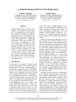
Figure 1 CPN-treated BY-2 cells and protoplasts.Panelsshow
bright field and fluorescence images. (A,B) BY-2 protoplasts were
incubated for 2 h with 10 μM CPNs or (C, D) left untreated. (E, F)
Chains of attached BY-2 cells treated with 10 μM CPNs for 2 h.
Green fluorescence is greatest at the cross walls suggesting that
CPNs attach to the cell walls and do not penetrate the interior.
(G, H) Images of untreated BY-2 cells at 2 h. (I, J) Confocal images
of intact BY-2 cells treated with 10 μM CPNs at 24 h. Experiments
were repeated with similar results. Single optical section through
the center of the cell shows fluorescence along the cell wall and
does not penetrate the interior. Scale bars equal 50 μm.
Silva et al. BMC Plant Biology 2010, 10:291
/>Page 3 of 14
polymers that do not contain Cd
2+
and are distributed
inthecytoplasm,wecannotruleoutthepossibilityof
a concentration dependent cytotoxicity. Therefore,
propidium iodide staining was employed to measure
the cytotoxic impact of var ious concentrations (0, 5,
10, 25, 50, 100, 250, and 500 μM) of CPNs following
treatments for 0, 2, 5, 8, 16, 24, and 48 h (Figure 3).
The a verage percent of viable protoplasts at each time
point was calculated for three replicate experiments.
Concentrat ions of 5-25 μM are non-toxic to proto-
plast and do not significantly reduce their viability.
Typically, following preparation of BY-2 protoplasts
from intact cells, w e noted 90- 96% viable protoplasts
during the first 8 h of culture. This declines to 85% at
24 h and then 60% at 48 h (Table 1). 80-95% of BY-2
protoplasts were viable during the first 8 h of culture
followin g treatment with 5, 10, and 25 μM of CPNs (log
molar concentrations of -4.3 to -5.3), which is compar-
able to untreated protoplasts. Protoplast viability follow-
ing CPN treatment further declined to 81-86% at 24 h
and 53-60% at 48 h. Thus, the average percent viability
is similar over time among the CPN-treated (5-25 μM)
and untreated protoplasts.
Concentrations over 50 μM (log molar concentrations of
-3.3 to 4.0) cause the p roportion of viable protoplasts to
decline profoundly after 8 h (Figure 3; Table 1). The aver-
age percentage of intact protoplasts treated with 50 μM
CPNs was 68% at 24 h and 32% at 48 h. For concentrations
of 100- 5 00 μM the average percent viability was 20 to 32%
at 24 h and 14 to 22% at 48 h (Figure 3; Table 1). These
data show that the concentrations of CPNs (5-25 μM) used
in the pri or experiments for visualizing uptake are essen-
tially non-toxic to BY-2 protoplasts but excessive amounts
of polymer can be detrimental.
CPNs deliver both siGLO Red and NtCes1-A siRNAs to
protoplasts
Commercially available siGLO Red are fluorescently
labeled RNA duplexes which were combined with CPNs
and delivered to BY-2 protoplast culture medium to
assess protopl ast transfection. Both green and red fluor-
escence, which corresponds to CPNs and siGLO Red,
respectively, were seen inside BY-2 protoplasts within
2 h of delivery (Figure 4). FACS methods were
employed to: a) measure the fluorescence intensity in
each cell b) detect and count the number of fluorescent-
protoplasts in large populations (10,000 protoplasts).
A set of untreated BY-2 protoplasts without CPN treat-
ment were gated to represent the major non-fluorescent
population (Figure 5A). FACS demonstrated that 10 or
25 μM CPNs penetrated BY-2 protoplasts following
treatment for 2 or 24 h. Clearly, not all protoplasts
showed CPN-uptake. However comparing treatments at
2 and 24 h, there was undoubtedly an increase in CPN-
uptake by protoplasts that was concentration and time
dependent (Figure 5B).
Cytometric analysis also showed that siGLO Red failed
to penetrate protoplasts in the absence of CPNs (Figure
5A). However, combining 10 μMor25μMCPNswith
200 nM siGLO Red, there is a significant and positive
shift in the number of events containing red fluores-
cence (Figure 5A, C). The combined e pifluorescence
microscopic and cytometric analyses (Figures 4 and 5)
indicate that CPNs were responsible for siGLO Red
uptake.
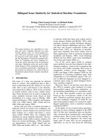
Figure 2 CPN and FM4-64 treated BY-2 protoplasts examined
using confocal microscopy. (A) Protoplast that was treated with
medium (no CPNs) and then FM4-64 for 10 min or (B) more than
20 min. FM4-64 fluorescence is in the plasma membrane and
vesicles budding from the plasma membrane at 10 min. Following
a 20 min or longer incubation, red fluorescence is in the plasma
membrane, perinuclear membranes, and intracellular vesicles. Arrow
heads point to vesicles budding at the plasma membrane and in
the cortical region. (C, D) Green and red fluorescent images of
protoplasts treated with CPNs and then FM4-64 for 10 min. Arrow
heads point to vesicles along the plasma membrane that contain
both green and red fluorescence. There are a greater number of red
than green fluorescent vesicles. However, most green granules also
contain red fluorescence. (E, F, G) Protoplasts were treated with
CPNs and then FM4-64 for 20 min showed green and red
fluorescence in vesicles along the periphery of the cell. Repeated
experiments showed similar outcomes. Arrows point to examples
where green and red fluorescence overlap. Scale bars equal 20 μm.
Silva et al. BMC Plant Biology 2010, 10:291
/>Page 4 of 14
The ability of CPNs to deliver siRNAs targeting an
end ogenous gene was also examined. siRNAs were gen-
erated to the plant cellulose synthase gene, NtCesA-1,
and CPN-siRNA complexes were delivered to proto-
plasts. Under suitable media conditions, BY-2 proto-
plasts can regener ate their cell w alls duri ng 3 d of
culture [30,31]. NtCesA-1 is a central factor in cell wall
deposition in plants and therefore we hypothesized that
knocking down NtCesA-1 expression would block cell
wall regeneration in protoplasts. Given that the flow
cytometry data shows ~ 40% uptake of siGLO Red
accompanied by CPNs, it is possible that a similar popu-
lation of protoplasts received CesA-1 siRNAs. We
employed calcofluor white M2R staining [32] to assess
cell wall regeneration following silencing NtCesA-1a and
propidium iodide staining to monitor viability [33].
Calcofluor white M2R staining conducted upon the
completion of cell wall digestion (T = 0 h) confirms that
there was no residual cell wall material remaining along
protoplast surfaces (Figure 6A). At 72 h, greater
amounts of calcofluor white M2R fluorescence was
observed at the margins of untreated protoplasts indi-
cating that the culture conditions were suitable for cell
wall regeneration (Figure 6C). For siRNA and CPN trea-
ted protoplasts, calcofluor staining was significantly
reduced to a level that was barely visible. Rare, minor
patches of cellulose occurred along the plasma mem-
brane of some protoplasts at 72 h (Figure 6B). Interest-
ingly, we tested CPN-siRNA complexe s formed by
0.0
20.0
40.0
60.0
80.0
100.0
-6 -5 -4 -3
% Viability
Log [CPN](M)
48 hrs
24 hrs
16 hrs
8 hrs
5 hrs
2 hrs
0 hrs
% Viable Protoplasts
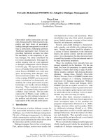
Figure 3 Protoplast viability wa s determined following specific incu bation with vario us concentrations of CPNs. Protoplasts were
cultured for various times between 0 and 48 h following treatment with the following CPN concentrations: 5, 10, 25, 50, 100, 250 and 500 μM.
The percentage of viable protoplasts was determined using propidium iodide staining at 0, 2,5,8,16,24 and 48 h. The data were expressed as the
average % viability at each time point for log molar concentration of CPNs taken from three independent experiments.
Table 1 Effect of various concentrations of CPNs on
viability of BY-2 protoplasts at 0 and 24 h
Conc (μM) 0 h 24 h
0 95.7 ± 2.5 85.7 ± 3.2
5 93.3 ± 3.0 84.3 ± 3.5
10 96.0 ± 2.0 84.0 ± 2.7
25 95.7 ± 3.0 81.0 ± 2.7
50 94.0 ± 2.0 67.7 ± 4.2
100 94.3 ± 1.5 32.0 ± 4.4
250 94.7 ± 3.5 22.0 ± 5.2
500 96.0 ± 2.0 20.0 ± 2.7
Low concentrations of (5-25 μM) CPNs have minimal impact on protoplast
viability at 24 h.
Concentrations ≥ 50 μM CPNs cause significant loss of BY-2 viability at 24 h.
Silva et al. BMC Plant Biology 2010, 10:291
/>Page 5 of 14
mixing for 3 h and overnight at 4°C and we noted that
the outcome of calcofluor staining was significantly
reduced using CPN-siRNA complexes that were pre-
pared by overnight incubation (data not shown). It is
worth speculating that the overnight incubation led to
maximal incorporation of siRNAs into complexes.
To determine the effectiveness of NtCesA-1 siRNA-
delivery in blocking cell wall regeneration, we recorded
the average percentage of calcofluor positive protoplasts
relative to the total number of living protoplasts counted
(n = 400 protoplasts) in two replicate experiments. Proto-
plast viability was confirmed using propidium iodide (see
below). For protoplasts that were untreated (no siRNA or
CPN) or treated only with NtCesA-1 siRNAs, 51-54%
were calcofluor positively by 72 h. Treating pro toplasts
with CPN or siRNA alone had no effect on cell wall
regeneration until the time period between 48-72 h.
There was a steady increase in the proportion of
untreated or siRNA-treated protoplasts that stained posi-
tive over time (Figure 6D). For protoplasts treated with
CPN alone, there is a slight plateau between 48 and 72 h
(19-23%) and only a few f aint patches of newly synthe-
sized cell walls were seen in those protoplasts (Figure 6B
and 6 D) [33]. However, this is contrasted by protoplasts
treat ed with 10 μM CPNs and 200 nM NtCesA-1 siRNAs
which showed no change in cell wall regeneration
between 0 and 24 h foll owed by a slow increase in calco-
fluor staining until 48 h. There appeared to be a plateau
between 48 and 72 h where cell wall regeneration did not
continue in a manner similar to untreated protoplasts
(Figure 6C). Calcofluor staining was seen in 33-38% of
protoplasts at 48 and 72 h, suggesting that CPN tr eat-
ment could hamper growth at later times.
Propidium iodide was used to determine the p ercent
viable protoplasts harvested at 0, 24, 48, and 72 h (Fig-
ure 6E). Propidium iodide stains nonviable protoplasts and
cells, shows absorption/emission maximum at 536/617
nm, and was employed to measure the impact of CPNs on
cell viability. We counted populations of 100-250 proto-
plas ts to determine the proportions that were propidium
iodide positive and/or contained CPNs. Untreated proto-
plasts show a slow decline in viability from 90% at 0 h to
~ 60% at 72 h. Protoplasts that were treated with only
CPNs or only siRNAs showed comparable levels of
decline. However, protoplasts treated with both CPNs and
siRNAs showed a significant drop in via bility between 24
and 72 h with ~ 35% of protoplasts remaining alive (Figure
6E). These data suggest that cellulose synthase activity is
essential for extending the lifetime of protoplast. The
effect of CPN plus siRNAs on cell viability and deposition
of cellulose on the cell surface [33] indicates that CPNs
were effective vehicle for siRNA delivery and targeted
downregulation of NtCesA-1expression.
Knockdown of NtCesA-1a transcript accumulation was
confirmed by semi-quantitative PCR (Figure 6F).
NtCesA-1a silenced protoplasts were harvested at 48 h
post delivery of CPNs alone and 200 nM siRNAs plus
CPNs. The messages were reduced >17% and >76%
compared with untreated control samples. Ubiquitin
mRNA served as an internal control for RNA qua lity
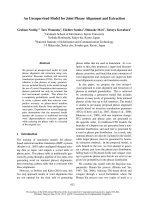
Figure 4 CPNs deliver siGLO Red small RNAs to protoplasts. Bright field and epifluorescence images of protoplasts treated with: (A, B, C) 25 μM
CPNs and 200 nM siGLO Red small RNAs (red); (D, E) only 200 nM siGLO Red small RNAs; (F, G) untreated protoplasts (negative controls). (E) Image
shows no uptake of small RNAs in the absence of CPNs. (G) Image shows no green fluorescence, as expected. Experiments were repeated 2-3 times.
Scale bars equal 20 μm.
Silva et al. BMC Plant Biology 2010, 10:291
/>Page 6 of 14
and RT-PCR amplification, and the mRNA levels were
similar (Figure 6F).
Discussion
CPNs are fluorescent conjugated polymer nanoparticles
and are valuable for live plant cell imaging. Their inher-
ent photophysical properties include high fluorescence
qua ntum yield, large extinction coeffi cient, and efficient
optical signal transduction making them a superior
choice for biological imaging [22,23]. Furthermore, we
demonstrate that CPNs are an effective transfection
vehicle for delivery of siRNAs into plant protoplast.
Other transfection methods that are widely employed
for delivery of nucleic acids to plant protoplasts incl ude
electroporation, polyethylene glycol, and lipofectamine
[34-36], and only electroporation and polyethylene gly-
col has been used for direct delivery of siRNAs [37,3 8].
With respect to electroporation, the electric pulse can
cause an immediate loss of up to 40% viable protoplast
[35,39]. This much greater loss than following the use
of CPN-delivery, which causes only 5-20% lo ss of viabi-
lity within the first 24 h of delivery (Table 1). Unlike the
CPN d elivery method, the optimal conditions for deliv-
ery of siRNAs by electroporation require extensive opti-
mization [34] of the voltage and pulse time to ensure
high transfection rates. Therefore CPNs are attractive
Figure 5 Presence of CPN and siGLO Red small RNAs in protoplasts. (A) Dot plots of BY-2 protoplasts cultured with medium only, 200 nM
siGLO Red, 10 μM CPNs, 10 μM CPNs + 200 nM siGLO Red, 25 μM CPNs, or 25 μM CPNs+200 nM siGLO Red. CPN fluorescence is detected with
FITC filter (x axis) and siGLO Red fluorescence (y axis) is detected with PE filter in protoplasts cultured for 24 h. Each dot represents a single event
with emissions frequency that is the combination of the fluorophores. The gated population in the lower left quadrant represents the majority of
nonfluorescent cells. The upper right quadrant represents the majority of events that contain both green and red fluorescence due to CPNs and
siGLO Red small RNAs. The upper left quadrant represent events that are positive only for siGLO Red small RNAs and the lower right quadrant
represent events that are positive for only CPNs. Highly fluorescent protoplasts are located furthest along the x- and y- axes. (B) Bar graph reports
the average of 10 replicate experiments using FACs to record the number of green fluorescent events inside protoplasts, as an indication of the
internalization of CPNs (lower right quadrant of dot plots). Samples were treated with 0, 10, or 25 μM CPNs and then incubated for 2 and 24 h.
Between 18-37% of protoplasts produce positive events via cytometric analyses. (C) Bar graph reports the average and stand deviations of 10
replicate experiments, recording the number of events reporting internalization of both CPNs and siGLO Red (upper right quadrant of the dot
plots). Between 27 and 42% of recorded events are positive for both CPNs and siGLO Red RNAs when they are co-delivered to protoplasts.
Silva et al. BMC Plant Biology 2010, 10:291
/>Page 7 of 14
and facile choice for efficient siRNA delivery into proto-
plasts without compromising cell viability. Furthermore,
the intrinsic fluorescence enables r eal time detection of
CPN-uptake by pro toplasts and offer s the opp ortunity
to monitor the rate of cellular responses following
siRNA uptake in synchronously treated cells.
Flow cytometry for studying physiological events or
dye uptake in plant protop lasts has been used in recent
years. Protoplast morphology and the distribution of
light-scattering intensities can vary widely for different
species and cell cultures and this can impact the quality
of the results. We examined green and red fluorescence
Figure 6 CPN delivery of CesA-1 siRNAs suppress cell wall regeneration. (A) Protoplasts were harvested and immediately stained with
calcofluor white (T = 0 h) to verify complete digestion and elimination of cell walls. Image shows no calcofluor fluorescence. (B) Protoplasts at 72 h
treated with 10 μM CPNs and 200 nM NtCesA-1 siRNAs show few faint patches of blue fluorescence at the plasma membrane. (C) Untreated
protoplasts at 72 h show significant deposition of cellulose at the cell surface. Experiments were repeated five times. Scale bars represent 10 μm.
(D) The average percent of protoplasts from two experiments that showed calcofluor staining at 0, 24, 48, and 72 h following treatment with CPNs
and NtCesA-1 siRNAs. (E) Propidium iodide was used to determine the percent viable protoplasts at 0, 24, 48, and 72 h following treatment with
CPNs and NtCesA-1 siRNAs. Averages were determined for three replicate experiments. (F) Ethidium bromide- stained 1% agarose gels containing
semi quantitative RT-PCR products detecting NtCesA-1 or ubiquitin (Ubi) gene expression. The treatments with siRNAs and CPNs are indicated
above each panel and the numbers of PCR cycles from 30-45 are indicated below each lane. Lane “L” indicates DNA ladder at the bottom of the
gel and size (bp) markers are indicated on the left. As a control, semi-quantitative PCR shows ubiquitin gene expression.
Silva et al. BMC Plant Biology 2010, 10:291
/>Page 8 of 14
(FL1-H and FL4-H) and expected to detect a minor
population of dots that would result from cell debris or
dying protoplasts. Notably the FACS resul ts (Figure 5B)
and manual counting of CPN-uptake by protoplasts pro-
duced somewhat different quantitative outcomes. A
maximum of 35% of protoplasts were green fluorescent
following treatment with 25 μM CPNs for 2 hrs as mea-
sure by FACS, but under the microscope we noted
60-75% of protoplasts were gr een fluorescent. One
explanation is that the larger population that was sorted
by FACS led to a broader and unbiased assessment.
Another possibility is that the CPN fluorescence inside
some cells might be low and might overlap with the
autofluorescence of the gated population. Thus fewer
CPN-positive protoplasts may be detected by FACS than
by manual counting. A third explanation for the low
counts by FACS is that the method of mixing proto-
plasts with CPNs may require further optimization to
ensure a broader population is exposed to CPNs. Per-
haps placing the culture on a rotary shaker at low speed
would enhance mixing of protoplasts and CPNs and
could increase the percentage of t ransfected protoplasts.
We also observed CPNs binding to debris in the cell
cultures. The nature of the debris is a mixture o f lysed
cell constituents and cell wall materials. We know from
examining CPN-treated BY-2 cells that CPNs bind easily
to plant cell walls. Therefore, the presence of cell wall
material in the cultures likely depletes CPNs that could
transfect protoplasts. Perhaps improved handling of pro-
toplast preparations through further filtration or wash-
ing could reduce cell wall debris and enhance the
percentage of transfected protoplasts. Further experi-
ments are needed to determine the conditions for
improved CPN internalization.
Cell wall biosynthesis is central to cell elongation,
plant growth and development and new methodologies
to modify the cellulose and lignin content could be
employed for generating genetically improved plants
[40]. Researchers are working on agronomically impor-
tant crops to increase cellulose content and decrease lig-
nin cont ent to improve forage digestibility and improve
the use of crops as biofuels for ethanol production. The
biosynthetic pathways leading to cell wall deposition
(cellulose and lignin) and assembly into a functional
wall are not well described [40]. Significant advances
have been made in recent years with the identification
of CesA genes that encode the catalytic subunits for ce l-
lulose synthase. In Arabidopsis thaliana,thereare10
members of the AtCESA gene family and only three
genes AtCESA1, AtCESA3 and AtCESA6 are known to
be important for primary cell wall synthesis [41]. CesA
subunits assemble in rosettes and these rosettes produce
glucan chains. The rosettes assemble into microfibrils
along the plasma membrane [40]. Patches of cellulose
occurring along the plasma membrane can be seen by
calcofluor staining [33]. In our exper iments, we no ted
that at 72 h in untreated BY-2 protoplasts, there were
patches of cellulose which coalesced to form larger
areas of deposited primary cell wall (Figure 6C). Chemi-
cals such as ancymidol and isoxaben are known to inhi-
bit cell wall production and have been employed to
increase our knowledge of t he mechanisms controlling
cellulose biosynthesis and deposition a long the plasma
membrane. One drawback of using a chemical approach
to knockdown cellulose synthase is that they can have
additional impacts on other subcellular functions [42].
For example, isoxaben disrupts microtubule organization
as well as inhibits the synthesis of cellulose microfibrils
[43]. Therefore, siRNA delivery targeting specific genes
in the cellulose biosynthesis pathway is preferred to
examine the role of each gene in cell wall deposition.
Recently, siRNA delivery via viral vectors (Potato virus
X, Ba rley stripe mosaic virus)tointactN. benthami ana
and barley plants has provided valuable evidence that
RNAi technology can be employed to knockdown CesA-
1, C esA-2, and CesA-6 gene expression [25,44]. By silen-
cing individual members of the CesA gene family
researchers were able to determine which genes are pri-
mary contributors to cell wall formation. In addition,
plants showed compensatory change s in polysaccharide
composition of the cell walls and this demonstrated that
an RNAi approach created further opportunities to
explore the relati onships between the cellulose synthase
genes and pectin biosynthesis [25,44]. Importantly, using
viral vectors to deliver siRNAs into protoplasts may not
be advisable because the viruses themselves may impact
protoplast viability and cell wall regeneration. I n addi-
tion, viral delivery relies on electroporation or PEG
transfection methods which may also impact protoplast
viability. In this study, we employed a CPN-siRNA deliv-
ery method that is taken up by protop lasts in a manner
that does not hamper viability and had no obvious effect
on cell wall regeneration during the first 48 h. CPN
delivery of CesA-1 siRNAs was effective for silencing
cellulose synthase in protoplasts showing that this tech-
nology can be used to monitor the early stages of cellu-
lose deposition. When we compare Figure 6D and 6E,
we note that there is reduced protoplast viability along
with hampered cellulose synthesis. Protoplasts left
untreated, treated with siRNAs only (no CPNs), or
CPNs only (no siRNAs) showed comparable levels of
cell wall regeneration and viability. Only the addition of
10 μM CPNs plus 200 nM siRNAs caused a significant
loss in viability and cell wall regeneration. Calcofluor
staining showed no obvious buildup of cellulosic patches
along the plasma membrane, while untreated samples
showed greater regeneration. We also realize that there
is significant sequence similarity among tobacco CesA
Silva et al. BMC Plant Biology 2010, 10:291
/>Page 9 of 14
genes and that the long p iece of CesA used to generate
the siRNAs might directly knock down multiple CesA
genes. The correlation suggests that silencing CesA
genes reduced protoplast viability, indicating that cellu-
lose synthesis is an important housekeeping function.
Events that slow regeneration or hamper cell expansion
could lead to loss of viability. Complete and speedy
regeneration of the primary cell wall might be important
for protoplasts to respond to the osmotic pressure of the
medium and remain alive. Future research is needed to
understand the link between CesA gene function and
cell viability. These outcomes show that CPN delivery
methods may be valuable for such studies in protoplasts.
Animal cells take up extracellular materials by at least
four different routes: receptor-mediated endocytosis,
non-ionic diffusion, carrier mediated uptake, and facili-
tated diffusion. T hese routes are l argely unexplored in
plants, and little is known about the compartments carry-
ing endocytic cargo from the plant plasma membrane
[45]. Several approaches have been used to identify speci-
fic compartments in the endomembrane system. First is
the use of immune electron microscopy and antibodies
recognizing known markers to identify the compartment.
Second is the use of organelle reporters such as GFP
fused to organellar targeted protein domains [46-48].
Thi rd is the use of FM4-64 fluorescent styryl dyes which
are taken up by endocytic vesicles and are used to follow
the vesicle trafficking network to Golgi and prevacuolar
vesicles [49,50]. Such tools have led researchers to de ter-
mine that the pathway from the early endosome to preva-
cuolar/multivesiculate bodies in animals and plants differ.
In animals, the multivesiculate bodies send cargo to the
lysosome while in plants they deliver their cargo to the
vacuole. The plant prevacuolar vesicles can deliver cargo
to storage vac uoles as well as the lytic compartment [51].
The recycling endosome is a compartment that returns
cargo to the plasma membrane and is well described in
animal cells but is relatively unknown in pla nt cells,
although FM4-64 staining suggests plants likely have
these types of vesicles [50,27]. Rec ently, nanogold p arti-
cles have be en used to probe the plant endocytic path-
ways [26]. Detailed electron microscopic analysis of BY-2
protoplasts treated with positively and negatively charged
nanogold particles revealed the presence of clathrin-
dependent and -independent pathways that lead to
degradation or recycling [26]. In Figure 2 we report the
pattern of CPN localizati on in granules near the cell sur-
face followed by accumulation in larger vesicles, and this
coincides with the pattern of FM4-64 staining. The FM4-
64 staining was more pronounced in CPN-treated cells
than in nontreated cells suggesting that dye uptake was
stimulated by CPNs. Further research using confocal
microscopy will be needed to follow the true path of
CPN internalization, and to learn if this pathway involves
receptor mediated endocytosis or an alternative carrier
mediated uptake that coincidently leads to increased
internal staining by FM4-64. In addition, the possibility
of CPNs entering the prevacuolar compartment is intri-
guing, given that we show in later experiments CPN-
siRNA complexes are effective for PTGS. Based on these
data, it is worth speculating that if CPNs-siRNAs com-
plexes are endocytosed, that they are then released into
the cytoplasm. Perhaps there are cell ular conditions that
enable siRNAs to dissociate from CPNs and to become
active in the silencing pathw ay. Therefore the route for
CPNs to enter prevacuolar vesicles from the endoso me
or cytoplasm is not made obvious by these experiments.
If CPN uptake is via the endosome, then one explanation
is that siRNAs exit the endosome while CPNs remain
and are transported to the prevacuolar compartment.
Perhaps a shift in the pH of the endocytic compartment
causes dissociation of the complex. An alternative route
is that CPN-siRNA complexes exit the endosome, dis-
sociate in the cytoplasm, and then CPNs enter the preva-
cuolar compartment by an undefined mechanism.
In total, CPNs present a promising tool for live cell
imaging of the endocytic trafficking pathways in plants.
This study shows that we can deliver positively charged
CPNs to protoplast culture medium and because of
their intrinsic fluorescence we can visually monitor the
route of uptake. Future research will examine the possi-
bility of recording the trafficking of CPNs from the
plasma membrane to and within the endocytic pathway
alongside fluorophores fused to endocytic markers. This
is an advance over the use of fusion proteins which are
often transgenically expressed. One limitation to
employing transgenic plants is that the protein fusions
are synthesized within the cell and their pathway to the
plasma membrane potentially overlaps with their path-
way of recycling from the plasma membrane back into
the cell. On the other hand CPNs can be delivered into
the culture medium and their intrinsic fluorescence can
be relied on to visually monitor the trafficking of mole-
cules within the endocytic pathway to obtain new infor-
mation about these pathways.
Conclusions
CPNs represent a significant advance in technology for
the delivery of siRNAs to plant protoplasts. Other meth-
ods of siRNA delivery include the use of viral vectors,
electroporation, and polyethylene glycol. For example,
one advantage of CPNs-siRNA complexes over t he use
of viral vectors is the ability to deliver siRNAs that
knockdown expression of genes that are vital cellular
functions without concern for viral pathology affecting
experimental outcom es. With respect to elec troporation
and polyethylene glycol, it is possible that CPNs may
have a lower impact on protoplasts viability, although
Silva et al. BMC Plant Biology 2010, 10:291
/>Page 10 of 14
further experiments are needed to compare CPN-
mediated transfer of siRNAs with these other methods
to determine which are less disruptive to cell functions.
Because CPNs are nontoxic to protoplasts and are easily
added to culture medium, this technology could be
adapted to high throughput applications. It would be
straightforward to synthesize siRNAs targeting various
regulatory steps in a pathway, deliver CPN-siRNA com-
plexes to protoplasts, and then monitor the outcomes of
suppressing housekeeping genes on cellular functions.
We also learned that CPNs could be a valuable ima-
ging tool for plant biology. The endocytic pathway is
not as well explored in plants as in vertebrate systems.
Live cell imaging recording trafficking of CPNs via the
endocytic pathway could yield valuable new information
about membrane transport in plants.
Methods
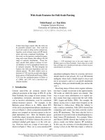
Synthesis of CPNs
The fabrication of CPNs was previously desc ribed [23]
(Figure 7). Briefly, an amine-containing poly(phenylene
ethynylene) (PPE) was synthesized by polymerizing
13,13’-(2,5-diethynyl-1,4-phenyle ne)bis(oxy)bis(2,5,8,11-
tetraoxatridecane) and 2,2’(2,2’-(2,5-dibromo-1,4-pheny-
lene)bis(ethane-2,1-diyl))bis(oxy)diethanamine in a
mixed solvent of N-methyl pyrrolido ne and morpholine
using palladium/copper catalysts. The PPE solution was
treated with excess amounts of glacial acetic acid fol-
lowed by dialysis (10,000 MWCO) against dH
2
O. Final
PPE- dH
2
O solution was filtered using a syringe filter
(0.45 μm) and stored at room temperature.
Compound (1) is 13,13’-(2,5-dibromo-1,4-phenylene)bis
(oxy)bis(2,5,8,11- tetr aoxatridecane) and was prepared by
incubating at 80°C overnight a suspension of 2-{2-[2-(2-
methoxyethoxy)ethoxy]ethoxy}thyl 4-methylbenzenesulfo-
nate
1
(20.6 mM; 5.9 g), 2,5-dibromohydroquinone (10.3
mMl; 2.76 g) and K
2
CO
3
(103 mM; 14 g) in 30 mL of
dimethyl formamide (DMF). The mixture was concen-
trated in vacuo and diluted with 50 mL of dichloro-
methane. The solution was washed with three times with
20 mL dH
2
O, dried over Na
2
SO
4
, and evaporated in
vacuo. The crude product was purified by column chro-
matography (silica gel, ethyl acetate/hexane (3:1, v/v).
Yield : 3.7 g (55%).
1
H NMR(600 MHz) : δ = 7.15 (s, 1H,
Ar-H, J = 6), 4.12 (t, 4H, Ar-OC
H
2
, J = 6), 3.87 (t, 4H,
OCH
2
, J = 6), 3.76 (t, 4H, OCH
2
, J = 6 ), 3.69-3.63 (m, 16H,
OCH
2
), 3.55 (t, 4H, O CH
2
, J = 6), 3.37 (s, 6H, CH
3
); 13C
NMR(150 MHz) : δ = 150.5, 119.4, 111.6, 72.1, 71.3, 70.9,
70.8, 70.72, 70.4, 69.8, 59.2.
Compound (2) is (2,5-bis(2,5,8,11-t etraoxatridecan-13-
yloxy)-1,4-phenylene)bis(ethyne-2,1-diyl)bis(trimethylsi-
lane) and was prepared by adding Compound (1) (5 g,
7.7 mM) to a s hrink flask fitted with a stir bar and Pd
(PPh
3
)
2
Cl
2
(0.54 g, 0.77 mM) and CuI (0.073 g, 0.39
mM). Thirty mL of a 2:1 mixture of tetrahydrofuran and
diisopropylamine was added to the reaction. Following
addition of trimethylsilylacetylene (4.4 mL, 31 mM), the
reaction mixture was heated to 60°C for 12 h. After eva-
porating solvent, the crude mixture was dissolved in
methylenechlorid e and washed twice with 30 mL satu-
rated ammonium chloride, followed by drying over
anhydrous MgSO
4
. The solvent was evaporated to pro-
duce dark brown oil, which was purified by column
chromatography (silica gel, ethyl acetate/hexane (4:1, v/
v)). Yield : 5 g (95%).
1
H NMR(600 MHz) : δ =6.91(s,
1H,Ar-H),4.12(t,4H,Ar-OC
H
2
, J = 6), 3.87 (t, 4H,
OCH
2
, J = 6), 3.78 (t, 4H, OCH
2
, J = 6), 3.68-3.66 (m,
12H, OCH
2
), 3.64 (t, 4H, OCH
2
, J = 6), 3.54 (t, 4H,
OCH
2
, J = 6), 3.38 (s, 6H, CH
3
), 0.25 (s, 9H, SiMe
3
);
13C NMR(150 MHz) : δ = 154.0, 117.9, 114.3, 100.9,
100.4, 72.0, 71.2, 70.8, 70.7, 70.6, 69.7, 69.6, 59.0, 0.0.
Compound (3) is 13,13’ -(2,5-diethynyl-1,4-phenylene)
bis(oxy)bis(2,5,8,11-tetraoxatridecane). A 100 mL round-
flask was charged with the compound (2) (2.5 g, 3.7
mM), 50 mL tetrahydrofuran, and 10 mL MeOH. Then
5 mL of 1 M KOH (aq) solution was added to the reac-
tion mix ture and stirred for 2 h. The solvent was evapo-
rated and the reaction mixture was purified by column
chromatography (silica gel, ethyl acetate). Yield : 1.1 g
(90%).
1
H NMR(400 MHz) : δ = 7.00 (s, 1H, Ar-H), 4.15
(t, 4H, Ar-OC
H
2
, J = 6), 3.87 (t, 4H, OCH
2
, J = 6), 3.76
(dd, 4H, OCH
2
, J = 6), 3. 73-3.63 (m, 16H, OCH
2
, J =6),
3.55 (dd, 4H, OCH
2
, J = 6), 3.38 (s, 6H, CH
3
, J =6),
3.35 (s, 2H, CH); 13C NMR(150 MHz) : δ = 154.1,
118.3, 113.6, 82.8, 79.6, 71.9, 71.1, 70.7, 70.6, 70.5, 69.6,
69.5,59.0.
Preparation of siRNAs for cellulose synthases, NtCesA-1a
and NtCesA-1b
TRIzol® reagent was used to extract total RNA from
tobacco (N. tabacum) leaves (Invitrogen Corp, Carlsbad,
CA). cDNA of the putative cellulose synthase (AF233892),
NtCesA-1a and NtCesA-1b was prepared using Superscript
III reverse transcriptase (Life Technologies) and then a
fragment of the gene was PCR amplified (640 base pairs
(bp) using forw ard (5’ - AGTGTATGTGGGTACCG-
GATG- 3’) and reverse (5’- CCATATGGGACA ATGCC-
TAC - 3’) primers. Following a 5-min denaturation at 94°
C, PCR was performed for 34 cycles of 94°C for 2 min, 94°
Figure 7 Fabrication of compounds 1, 2, and 3.
Silva et al. BMC Plant Biology 2010, 10:291
/>Page 11 of 14
C for 15 s, 50°C for 30 s, and 68°C for 45 s, followed by
final 5-min extension at 68°C [25]. The 640 bp PCR pro-
duct was purified using PCR Preps DNA Purification sys-
tem (Promega, Madison,WI) and cloned into the pGEM-T
Easy vector (Promega) according to manufacturer’ s
instructions. The nucleotide sequence of this cDNA frag-
ment was confirmed as 100% and 98% identical for
NtCesA-1a and NtCesA-1b, respectively. To generate
sense and anti sense RNA, pGEM-T:NtCe sA was linear-
ized using NcoIorSalI and in vitro transcription was per-
formed (RiboMAX, Promega) with SP6 and T7 RNA
polymerases. Transcription products were purified using
MEGAclear Kit (Ambion, Austin, TX). Sense and anti
sense RNAs were annealed in annealing buffer (100 mM,
KOAc, 4 mM MgCl
2
and 60 mM HEPES- KOH, pH 7.4),
boiled for 5 min, and incubated overnight at 37°C [37].
The resulting double strand RNAs were precipitated using
ethanol and then dissolved in nuclease-free double dis-
tilled (dd) H
2
O. siRNAs were generated by treating double
stranded NtCesA-1 with recombinant Dicer enzyme
according to the manufacturer’s instructions (Gene Ther-
apy Systems, San Diego, CA). The reaction was stopped by
adding the Dicer stop solution and 22 bp products were
detected using 3% agarose gel electrophoresis [37]. The
final siRNA products were purified using RNA purification
column 1(Gene Therapy Systems) and dissolved in nucle-
ase-free ddH
2
O.
BY-2 protoplast preparation
Typically, BY-2 cells are grown are subcultured from a 4
d-old liquid culture by transferring 10 mL BY-2 cells to
40 mL fresh BY-2 culture medium (Murashige and
Skoog salts pH 5.6 (Sigma-Aldrich Co, St. Louis, MO),
30 g.L
-1
sucrose, 256 mg.L
-1
KH
2
PO
4
, 100 mg.L
-1
myo-
inositol, 1 mg.L
-1
thiamine, and 0.2 mg.L
-1
2,4-dichloro-
phenoxyacetic acid). Cells are grown on a rotary shaker
that is maintained in the dark at 120 rpm at 28°C.
Protoplasts were prepared from 3 d old to bacco BY-2
suspension cells using standard methods [52,53]. Ten ml
of packed BY-2 cells (sedimented by centrifugation at
100-g for 5 min) were resuspended in 100 mL o f
enzyme solution (1.5% cellulase “ Onozuka RS” (Yakult
Pharmaceutical Ind. Co. Ltd., Tokyo, Japan), 0.2%
macerase (Calbiochem-Novabiochem Corp., La Jolla,
CA), 0.5 M mannitol, and 3.6 mM 2-(N-morpholino)
ethanesulfonic acid (pH 5.5) ) in a 1 L flask and incu-
bated for 3-4 h at 28°C on a rotary shaker at 100 rpm.
Protoplasts were recovered by filtration through 41 μm
nylo n mesh (Spectrum Laboratories, Inc., Ranch o Dom-
inguez,CA),andwashedtwice with Protoplast Wash
Solution (0.5 M mannitol, 3.6 mM 2-(N-morpholino)
ethanesulfonic acid (pH 5.5) at 59 g for 5 min. Proto-
plasts were resuspended in Protoplast Resuspension
Solution (BY-2 c ulture media plus 0.45 M mannit ol) to
a density about 1 × 10
6
protoplasts mL
-1
. Protoplast via-
bility was measured using 0.1% fluorescein diacetate pre-
pared in 1 ml of 50 mM phosphate buffer (pH 7.4). For
siRNA delivery experiments, protoplasts were cultured
in cell wall regeneration medium.
siRNA delivery to protoplasts and intact BY-2 cells
Protoplasts (1 × 10
6
mL
-1
) or intact BY-2 cells (1 × 10
6
mL
-
1
) were mixed with CPNs in Protoplast Resuspension
Solution to a final concentration of 0, 5, 10, 15 and 20 μM
and added to 6-well culture plates (Corning Inc., Corning,
NY). Each well of the culture plate was lined with Proto-
plast Resuspension Solutio n plus 1.0% agarose (pH 5.6).
Protoplasts and BY-2 cells were cultured at 28°C. Samples
were harvested at 2, 5, 10 and 24 h and the proportion of
CPN containing protoplasts and intact BY-2 cells were
determined using a haemocytometer. In addition, the pro-
portion of protoplasts or cells for which fluorescence was
seen to be associating with the cell wall, plasma mem-
brane, nucleus, and/or cytoplasm was recorded.
Protoplasts were resuspended in Protoplast Resuspen-
sion Solution (BY-2 culture medi a plus 0.45 M mannitol)
to a density about 1 × 10
5
proto plasts.mL
-1
. CPNs (10 or
25 μM) were incubated with 200 nM siGLO Red for 3 h
(Thermo Fisher Scientific, Pittsburgh, PA) and the c om-
plex was delivered to protoplasts. CPNs were incubated
with 200 nM NtCesA-1 siRNAs overnight and then deliv-
ered to protoplasts. We found that mixing CPNs and
siGLO Red for 3 h was sufficient to demonstrate transfec-
tion of CPN-siRNA complexes, but overnight mixing of
CPNs and NtCesA-1 siRNAs improved complexation and
improved siRNA delivery to plant protoplasts resulting in
a measura ble phenotype. Protoplast cultures were main-
tainedinthedarkat28°Cfor2hand24h.Controls
included treating protoplasts with Protoplast Resuspension
Solution, 200 nM siGLO Red only, NtCesA-1 siRNAs or
CPNs only. The protoplast containing both CPNs and
siGLO Red were counted using a haemocytometer.
Propidium iodide and FM4-64 dye treatment
Propidium iodide, contained in the Plant Cell Viability
Assay Kit (Sigma-Aldrich Co), was solubilized according
to manufacturer’s instructions. FM4-64 (Invitrogen Corp)
staini ng w as carried out to monitor CPN uptake, as pre-
viously described [54]. Protoplasts (1 × 10
5
.mL
-1
)were
incubated with 10 μM CPNs for 24 h at 28°C. 20,000 pro-
toplasts (which were previously treated with medium or
10 μM CPNs and incubat ed for 24 h at 28°C) wer e incu-
bated with 10 μM FM4-64 at room tempe rature for 10
min and then monitored using confocal microscopy.
Epifluorescence and confocal microscopy
A Nikon E600 (Nikon Corp., Tokyo, Japan) epifluores-
cence microscope with a B2A filter cube (470- to 490-nm
Silva et al. BMC Plant Biology 2010, 10:291
/>Page 12 of 14
excitation filter), a DM505 dichroic mirror, and a BA520
barrier filter was used to monitor FDA staining following
enzymatic digestio n of BY-2 cells and to study uptake of
CPNs protoplasts and intact BY-2 cells. Propidium iodide
was detected in protoplasts using a UV filter cube. siGLO
Red fluorescence (absorption/emission maximum at 557
nm/570 nm) was viewed using a Y-2E/C TX red filter
cube containing a 540- to 580-nm excitation filter, a
DM595 dichroic m irror, and a BA600-660 barrier filter.
Images were captured using the Optronics Magnafire
camera (Optronics Inc., Goleta, CA) and were edited
using Adobe Photoshop software (Adobe Systems Inc.,
San Jose, CA). Haemocytometer observations were
recorded using Microsoft Excel software.
A Leica TCS SP2 (Leica Microsystems, Bannockburn,
IL) confocal imaging system attached to a Leica DME
14 upright micr oscope equipped wit h Ar/Kr lasers were
used to study BY-2 cells treated with CPNs and FM4-64
staining protoplasts. Serial images were collected using
0.3 μm steps and 3-D images of 100 μm t hick sections
were compiled.
Fluorescence activated cell sorting (FACS) flow-cytometry
of treated BY-2 protoplasts
A Becton Dickinson FACS Calibur flow cytometer (Bec-
ton Dickinson, Franklin Lakes, NJ) equipped with an Ar
laser (excitation of 488 nm) was used to assess CPN-
uptake by protoplasts. Protoplasts (1 × 10
6
.mL
-1
)were
mixed with 10 μMand20μMCPNsinProtoplast
Resuspension Solution andaddedto6-wellculture
plates (Corning Inc., Corning, NY) containing Protoplast
Resuspension Solution plus 1.0% agarose (pH 5.7). Pro-
toplasts treated with buffer, or a 1:1 mixture of
untreated plus CPN-treated protoplasts were used as
controls. Protoplasts were cultured at 28°C and FACS
was performed at 2 h and 24 h of incubation.
The sorting capability of 10000 cells.s
-1
and fluores-
cence emission (FL1-H, FL2-H) was detected using a
530 nm and 585 nm band pass filters. The percentages
of fluorescence-emitting protoplasts w ere assessed as
evidence of CPNs and siGLO Red uptake by protoplasts.
Data were analyzed on a Macintosh computer equipped
with BD CellQuest Pro program (Becton Dickinson) and
were presente d as two dimensional dot plots whic h
represent CPN fluorescence emissions on the X-axis
and siGLO Red fluorescence emissions on Y- axis. Data
was compiled using Adobe Photoshop software.
Semi-quantitative RT-PCR of silenced protoplasts
Semi-quantitative RT-PCR was utilized to monitor
NtCesA transcriptional levels following siRNA delivery.
Extraction of total RNA from BY-2 protoplasts was car-
ried out using SV Total RNA Isolation System (Pro-
mega,Madison,WI).ThefirststrandcDNAwas
synthesized using SuperScript III reverse transcriptase
(Invitrogen Corp), 1 μg total RNA and oligo(dT) pri-
mers. PCR was performed using NtCesA-1 specific for-
ward primer (5’-AGTGTA T GTGGGTACCGG ATG-3’ )
and NtCesA reverse (5’ -CCATATGGGACAATGCC-
TAC-3’) primer that also shares homology with NtCesA-
2.Forward(5’-GCCT CCGTGGTGGTG CTAAG- 3’ ),
and reverse (5’-TCAATCGGCACC GGCCTT G-3’) p ri-
mers were used to a mplify ubiquiti n (AY912494) cDNA
(261 bp) as the internal control. Following 10-min dena-
turation at 95°C, PCR was performed for 30, 35, 40, 45
cycles of 95° C for 15 s and 60°C for 60 s. PCR products
were anal yzed using ethidium bromide stained 1% ag ar-
ose gel. Gels were scanned using Alpha Image system
(Alpha Innotech, San Leandro, CA) and the images
were recorded. Densito metry was perf ormed by Alp ha
Ease FC software (Alpha Innotech).
Abbreviations
CPN: conjugated polymer nanoparticles; PTGS: post-transcriptional gene
silencing; ddH
2
O: double distilled water; miRNA: microRNA; siRNA: small
interfering RNA; TRV: Tobacco rattle virus; T-DNA: Agrobacterium tumefaciens
based plasmid that can transform plant tissues; dsRNA: double stranded
RNA; CbLCV: Cabbage leaf curl virus; FDA: Fluorescein diacetate
Acknowledgements
We gratefully thank Dr. Changming Ye for assistance with primer design and
semi-quantitative PCR. This research was supported by ICx Technologies,
Oklahoma Center for Advancement of Science and Technology (OCAST)
ONAP08-018. JM acknowledges the Florida International University for
Faculty Startup Funds.
Author details
1
Department of Entomology and Plant Pathology, Oklahoma State
University, Stillwater, OK, USA 74078.
2
Department of Chemistry and
Biochemistry, Florida International University, Miami, FL 33199, USA.
Authors’ contributions
AS carried out plant protoplast experiments. JV and JM oversaw the analysis,
design and implementation. AN carried out CPN preparations. JV and JM
drafted the manuscript. All authors read and approved the final manuscript.
Received: 11 August 2010 Accepted: 30 December 2010
Published: 30 December 2010
References
1. Carrington JC: RNA silencing. Moving targets. Nature 2000,
408(6809):150-151.
2. Baulcombe D: RNA silencing. Trends Biochem Sci 2005, 30(6):290-293.
3. Yang Z, Ebright YW, Yu B, Chen X: HEN1 recognizes 21-24 nt small RNA
duplexes and deposits a methyl group onto the 2’ OH of the 3’ terminal
nucleotide. Nucleic Acids Res 2006, 34(2):667-675.
4. Dunoyer P, Lecellier CH, Parizotto EA, Himber C, Voinnet O: Probing the
microRNA and small interfering RNA pathways with virus-encoded
suppressors of RNA silencing. Plant Cell 2004, 16(5):1235-1250.
5. Pfeffer S, Voinnet O: Viruses, microRNAs and cancer. Oncogene 2006,
25(46):6211-6219.
6. Yu B, Chapman EJ, Yang Z, Carrington JC, Chen X: Transgenically
expressed viral RNA silencing suppressors interfere with microRNA
methylation in Arabidopsis. FEBS Lett 2006, 580(13):3117-3120.
7. Hamilton AJ, Baulcombe DC: A species of small antisense RNA in
posttranscriptional gene silencing in plants. Science 1999, 286(5441):950-952.
8. Tseng YH, Hsu HT, Chou YL, Hu CC, Lin NS, Hsu YH, Chang BY: The two
conserved cysteine residues of the triple gene block protein 2 are
Silva et al. BMC Plant Biology 2010, 10:291
/>Page 13 of 14
critical for both cell-to-cell and systemic movement of Bamboo mosaic
virus. Mol Plant Microbe Interact 2009, 22(11):1379-1388.
9. Hammond SM, Boettcher S, Caudy AA, Kobayashi R, Hannon GJ:
Argonaute2, a link between genetic and biochemical analyses of RNAi.
Science 2001, 293(5532):1146-1150.
10. Baumberger N, Baulcombe DC: Arabidopsis ARGONAUTE1 is an RNA
Slicer that selectively recruits microRNAs and short interfering RNAs.
Proc Natl Acad Sci USA 2005, 102(33):11928-11933.
11. Johansen LK, Carrington JC: Silencing on the spot. Induction and
suppression of RNA silencing in the Agrobacterium-mediated transient
expression system. Plant Physiol 2001, 126(3):930-938.
12. Dinesh-Kumar SP, Anandalakshmi R, Marathe R, Schiff M, Liu Y: Virus-
induced gene silencing. Methods Mol Biol 2003, 236:287-294.
13. Ratcliff F, Martin-Hernandez AM, Baulcombe DC: Technical Advance.
Tobacco rattle virus as a vector for analysis of gene function by
silencing. Plant J 2001, 25(2):237-245.
14. Matzke MA, Matzke AJ, Pruss GJ, Vance VB: RNA-based silencing strategies
in plants. Curr Opin Genet Dev 2001, 11(2):221-227.
15. Tang Y, Wang F, Zhao J, Xie K, Hong Y, Liu Y: Virus-based microRNA
expression for gene functional analysis in plants. Plant Physiol 2010,
153(2):632-641.
16. Du L, Bao M: Plant regeneration from protoplasts isolated from
embryogenic suspension cultured cells of Cinnamomum camphora L.
Plant Cell Rep 2005, 24(8):462-467.
17. Gandhi R, Khurana P: Regeneration from leaf protoplasts of Arabidopsis
thaliana ecotype estland. Indian J Exp Biol 2001, 39(7):705-709.
18. Kamo KK, Griesbach RJ: Evaluation of DAPI as a fluorescent probe for
DNA in viable Petunia protoplasts. Biotech Histochem 1993, 68(6):350-359.
19. Fisk HJ, Dandekar AM: Electroporation: introduction and expression of
transgenes in plant protoplasts. Methods Mol Biol 2005, 286:79-90.
20. Davey MR, Anthony P, Power JB, Lowe KC: Plant protoplasts: status and
biotechnological perspectivesw. Biotechnol Adv 2005, 23:131-171.
21. Chadegani M, Brink JJ, Shehata A, Ahmadjian V: Optimization of protoplast
formation, regeneration, and viability in Microsporum gypseum.
Mycopathologia 1989, 107(1):33-50.
22. Moon JH, Deans R, Krueger E, Hancock LF: Capture and detection of a
quencher labeled oligonucleotide by poly(phenylene ethynylene)
particles. Chem Commun (Camb) 2003, , 1: 104-105.
23. Moon JH, McDaniel W, Maclean P, Hancock LF:
Live-cell-permeable poly (p-
phenylene ethynylene). Angew Chem Int Ed Engl 2007, 46(43):8223-8225.
24. de Martimprey H, Vauthier C, Malvy C, Couvreur P: Polymer nanocarriers
for the delivery of small fragments of nucleic acids: Oligonucleotides
and siRNA. European Journal of Pharmaceutics and Biopharmaceutics 2009,
71:490-504.
25. Burton RA, Gibeaut DM, Bacic A, Findlay K, Roberts K, Hamilton A,
Baulcombe DC, Fincher GB: Virus-induced silencing of a plant cellulose
synthase gene. Plant Cell 2000, 12(5):691-706.
26. Onelli E, Prescianotto-Baschong C, Caccianiga M, Moscatelli A: Clathrin-
dependent and independent endocytic pathways in tobacco protoplasts
revealed by labelling with charged nanogold. J Exp Bot 2008,
59(11):3051-3068.
27. Bolte S, Talbot C, Boutte Y, Catrice O, Read ND, Satiat-Jeunemaitre B: FM-
dyes as experimental probes for dissecting vesicle trafficking in living
plant cells. J Microsc 2004, 214(Pt 2):159-173.
28. Braydich-Stolle L, Hussain S, Schlager JJ, Hofmann MC: In vitro cytotoxicity
of nanoparticles in mammalian germline stem cells. Toxicol Sci 2005,
88(2):412-419.
29. Chan WH, Shiao NH, Lu PZ: CdSe quantum dots induce apoptosis in
human neuroblastoma cells via mitochondrial-dependent pathways and
inhibition of survival signals. Toxicol Lett 2006, 167(3):191-200.
30. Nagata T, Takebe I: Cell wall regeneration and cell division in isolated
tobacco mesophyll protoplasts. Planta 1970, 92:301-308.
31. Nagata T, Takebe I: Plating of isolated tobacco mesophyll protoplasts on
agar medium. Planta 1971, 99:12-20.
32. Mishima T, Teranishi K, yamada T, Hisamatsu M: Adhesion of
polysaccharides to intact cells and protopalsts of Nicotiana tabacum BY-
2 and its stimulative effect on protoplast growth. J Bioscience and
Bioengineering 1999, 87:56-60.
33. Hofmannova J, Schwarzerova K, Havelkova L, Borikova P, Petrasek J,
Opatrny Z: A novel, cellulose synthesis inhibitory action of ancymidol
impairs plant cell expansion. J Exp Bot 2008, 59(14):3963-3974.
34. Fraley RT, Dellaporta SL, Papahadjopoulos D: Liposome-mediated delivery
of tobacco mosaic virus RNA into tobacco protoplasts: A sensitive assay
for monitoring liposome-protoplast interactions. Proc Natl Acad Sci USA
1982, 79(6):1859-1863.
35. Fromm M, Taylor LP, Walbot V: Expression of genes transferred into
monocot and dicot plant cells by electroporation. Proc Natl Acad Sci USA
1985, 82(17):5824-5828.
36. Datta K, Datta SK: Transformation of rice via PEG-mediated DNA uptake
into protoplasts. Methods Mol Biol 1999, 111:335-347.
37. Qi Y, Zhong X, Itaya A, Ding B:
Dissecting RNA silencing in protoplasts
uncovers novel effects of viral suppressors on the silencing pathway at
the cellular level. Nucleic Acids Res 2004, 32(22):e179.
38. Vanitharani R, Chellappan P, Fauquet CM: Short interfering RNA-mediated
interference of gene expression and viral DNA accumulation in cultured
plant cells. Proc Natl Acad Sci USA 2003, 100(16):9632-9636.
39. Guerche P, Bellini C, Le Moullec JM, Caboche M: Use of a transient
expression assay for the optimization of direct gene transfer into
tobacco mesophyll protoplasts by electroporation. Biochimie 1987, 69(6-
7):621-628.
40. Doblin MS, Kurek I, Jacob-Wilk D, Delmer DP: Cellulose biosynthesis in
plants: from genes to rosettes. Plant Cell Physiol 2002, 43(12):1407-1420.
41. Desprez T, Juraniec M, Crowell EF, Jouy H, Pochylova Z, Parcy F, Hofte H,
Gonneau M, Vernhettes S: Organization of cellulose synthase complexes
involved in primary cell wall synthesis in Arabidopsis thaliana. Proc Natl
Acad Sci USA 2007, 104(39):15572-15577.
42. Mutwil M, Debolt S, Persson S: Cellulose synthesis: a complex complex.
Curr Opin Plant Biol 2008, 11(3):252-257.
43. Fisher DD, Cyr RJ: Extending the Microtubule/Microfibril paradigm.
Cellulose synthesis is required for normal cortical microtubule alignment
in elongating cells. Plant Physiol 1998, 116(3):1043-1051.
44. Held MA, Penning B, Brandt AS, Kessans SA, Yong W, Scofield SR,
Carpita NC: Small-interfering RNAs from natural antisense transcripts
derived from a cellulose synthase gene modulate cell wall biosynthesis
in barley. Proc Natl Acad Sci USA 2008, 105(51):20534-20539.
45. Murphy AS, Bandyopadhyay A, Holstein SE, Peer WA: Endocytic cycling of
PM proteins. Annual Review of Plant Biology 2005, 56(1):221-251.
46. Viotti C, Bubeck J, Stierhof YD, Krebs M, Langhans M, van den Berg W, van
Dongen W, Richter S, Geldner N, Takano J, et al: Endocytic and secretory
traffic in Arabidopsis merge in the trans-Golgi network/early endosome,
an independent and highly dynamic organelle. Plant Cell 2010,
22(4):1344-1357.
47. Yamada K, Fuji K, Shimada T, Nishimura M, Hara-Nishimura I: Endosomal
proteases facilitate the fusion of endosomes with vacuoles at the final
step of the endocytotic pathway. Plant J 2005, 41(6):888-898.
48. Tanaka H, Kitakura S, De Rycke R, De Groodt R, Friml J: Fluorescence
imaging-based screen identifies ARF GEF component of early
endosomal trafficking. Curr Biol 2009, 19(5):391-397.
49. Lam SK, Siu CL, Hillmer S, Jang S, An G, Robinson DG, Jiang L: Rice
SCAMP1 defines clathrin-coated, trans-golgi-located tubular-vesicular
structures as an early endosome in tobacco BY-2 cells. Plant Cell 2007,
19(1):296-319.
50. van Gisbergen PA, Esseling-Ozdoba A, Vos JW: Microinjecting FM4-64
validates it as a marker of the endocytic pathway in plants. J Microsc
2008, 231(2):284-290.
51. Jiang L, Rogers JC: Integral membrane protein sorting to vacuoles in
plant cells: evidence for two pathways. J Cell Biol 1998, 143(5):1183-1199.
52. Nagata T, Nemoto Y, Hasezawa S: Tobacco BY-2 cell line as the “Hela” cell
in the cell biology of higher plants. Internat Rev of Cytol 1992, 132:1-31.
53. Ju HJ, Samuels TD, Wang YS, Blancaflor E, Payton M, Mitra R,
Krishnamurthy K, Nelson RS, Verchot-Lubicz J: The potato virus X TGBp2
movement protein associates with endoplasmic reticulum-derived
vesicles during virus infection. Plant Physiol 2005, 138(4):1877-1895.
54. Ju HJ, Brown JE, Ye CM, Verchot-Lubicz J: Mutations in the central domain
of Potato Virus X TGBp2 eliminate granular vesicles and virus cell-to-cell
trafficking. J Virol 2007, 81(4):1899-1911.
doi:10.1186/1471-2229-10-291
Cite this article as: Silva et al.: Conjugated polymer nanoparticles for
effective siRNA delivery to tobacco BY-2 proto plasts. BMC Plant Biology
2010 10:291.
Silva et al. BMC Plant Biology 2010, 10:291
/>Page 14 of 14