báo cáo khoa học: " Two novel types of hexokinases in the moss Physcomitrella patens" ppsx
Bạn đang xem bản rút gọn của tài liệu. Xem và tải ngay bản đầy đủ của tài liệu tại đây (5.27 MB, 15 trang )
RESEARCH ARTIC LE Open Access
Two novel types of hexokinases in the moss
Physcomitrella patens
Anders Nilsson
1†
, Tina Olsson
2†
, Mikael Ulfstedt
1
, Mattias Thelander
2
, Hans Ronne
1*
Abstract
Background: Hexokinase catalyzes the phosphorylation of glucose and fructose, but it is also involved in sugar
sensing in both fungi and plants. We have previously described two types of hexokinases in the moss
Physcomitrella. Type A, exemplified by PpHxk1, the major hexokinase in Physcomitrella, is a soluble protein that
localizes to the chloroplast stroma. Type B, exemplified by PpHxk2, has an N-terminal membrane anchor. Both
types are found also in vascular plants, and localize to the chloroplast stroma and mitochondrial membranes,
respectively.
Results: We have now characterized all 11 hexokinase encoding genes in Physcomitrella. Based on their N-terminal
sequences and intracellular localizations, three of the encoded proteins are type A hexokinases and four are type B
hexokinases. One of the type B hexokinases has a splice variant without a membrane anchor, that localizes to the
cytosol and the nucleus. However, we also found two new types of hexokinases with no obvious orthologs in
vascular plants. Type C, encoded by a single gene, has neither transit peptide nor membrane anchor, and is found
in the cytosol and in the nucleus. Type D hexokinases, encoded by three genes, have membrane anchors and
localize to mitochondrial membranes, but their sequences differ from those of the type B hexokinases.
Interestingly, all moss hexokinases are more similar to each other in overall sequence than to hexokinases from
other plants, even though characteristic sequence motifs such as the membrane anchor of the type B hexokinases
are highly conserved between moss and vascular plants, indicating a common origin for hexokinases of the same
type.
Conclusions: We conclude that the hexokinase gene family is more diverse in Physcomitrella, encoding two
additional types of hexokinases that are absent in vascular plants. In particular, the presence of a cytosolic and
nuclear hexokinase (type C) sets Physcomitrella apart from vascular plants, and instead resembles yeast, where all
hexokinases localize to the cytosol. The fact that all moss hexokinases are more similar to each other than to
hexokinases from vascular plants, even though both type A and type B hexokinases are present in all plants, further
suggests that the hexokinase gene family in Physcomitrella has undergone concerted evolution.
Background
Hexokinases catalyze the first step in hexose metabo-
lism, the phosphorylation of glucose and fructose. Hexo-
kinases that show a higher specificity for glucose than
for fructose are somet imes called glucokinases. The
yeast Saccharomyces thus has a glucokinase, ScGlk1, and
two dual specificity hexokinases, ScHxk1 and ScHxk2.
The eukaryotic hexokinases are all related to each other,
but are unrelated to prokaryotic glucokinases and hexo-
kinases. Plants also have a fructokinase which is unre-
lated to the hexokinases [1-3].
Hexokinases are found in several different intracellular
locations. The three yeast hexokinases are cytosolic, but
ScHxk2 can also enter the nucleus [4]. Animal type I
and II hexokinases have hydrophobic N-termini that tar-
get them to the outer mitochondrial membrane, whereas
type III and IV hexokinases are cytosolic, but the latter
can also e nter the nucleus [3]. We have previously
described two types of plant hexokinases [5]. Type A is
exemplified by the Physcomitrella hexokinase PpHxk1, a
soluble protein with a transit peptide [6] that localizes
* Correspondence:
† Contributed equally
1
Department of Microbiology, Swedish University of Agricultural Sciences,
Box 7025, SE-750 07 Uppsala, Sweden
Full list of author information is available at the end of the article
Nilsson et al. BMC Plant Biology 2011, 11:32
/>© 2011 Nilsson et al; licensee BioMed Central Ltd. This is an Open Access article distributed under the terms of the Creative Commons
Attribution License ( which permits unrestricted use, distribution, and reproduction in
any medium, provided the original work is properly cited.
to the chloroplast stroma. Type B hexokinases exempli-
fied by PpHxk2, have N-terminal membrane anchors.
Both types are present also in vascular plants, where
they localize to the chloroplast stroma and to the outer
mitochondrial membrane, respectively [7-14].
In addition to their metabolic roles, eukaryotic hexoki-
nases have also b een implicated in signal transduction.
Mitochondria-associated hexokinases have thus been
shown to negatively affect programmed cell death in
both animals and plants, by preventing the release of
cytochrome c from mitochondria [14-17]. It should be
noted, however, that this does not prove that a signal is
transmitted by hexokinase, which could have a constitu-
tive inhibitory effect on cytochrome c release. A more
direct role for he xokinases in signal transduction is sug-
gested by studies of the response to glucose in several
organisms. Thus, early work in yeast showed that
ScHxk2 is required for glucose repression [18,19], but
the molecular mechanism has resisted anal ysis for more
than 30 years [20-23].
An import ant question is where the enzyme exerts its
signaling function. Early work in yeast focused on the
cytosol, since the yeast hexokinases are cytosolic. How-
ever, further studies have shown that ScHxk2 also can
translocate into the nucleus, where it forms a complex
with the Mig1 repressor [4,24]. Similarly, evidence from
Arabidopsis [25] and rice [26,27] suggest that plant type
B hexokinases may enter the nucleus and p articipate in
gene regulation.
The moss Physcomitrella patens is unique among
plants in that gene targeting by homologous recombina-
tion works in it with frequencies comparable to yeast
[28]. This has made Physcomitrella a pow erful model
system for studies of plant gene function [29,30]. The
recent sequencing of the Physcomitrella genome has
further strengthened it as a model plant [31]. We have
previously characterized the Physcomitrella hexokinase
PpHxk1, which by gene targeting was shown to account
for 80% of the glucose phosphorylating activity in proto-
nemal tissue [5]. Further studies of a PpHxk1 knockout
mutant revealed a number of interesting phenotypes,
but no conclusive evidence was obtained as to the possi-
bleroleofthishexokinaseinsignaling[32].Partofthe
problem is that Physcomitrella like other plants pos-
sesses several hexokinases, which makes it difficult to
draw conclusions about gene function from the knock-
out of a single gene.
We here report the characterization of all eleven genes
encoding putative hexokin ase proteins in the Physcomi-
trella genome. Seven of the genes predict proteins that
clearly belong to the previously described types A and B
[5]. However, the re maining four genes encode two
novel types of hexokinases, which we call C and D. The
type C hexokinase PpHxk4 is a soluble protein which
lacks both organelle targeting peptide and m embrane
anchor. The three type D hexokinases PpHxk9,
PpHxk10 and PpHxk11 resemble the type B hexokinases
in that they possess hydrophobic membrane anchors,
but differ in sequence from the latter. The type D hexo-
kinases also have a similar localization as the type B
hexokinases, being found in the outer mitochondrial
membrane, and to some extent in the chloroplast
envelope.
Methods
Plant material and growth conditions
The growth condi tions used were growth at 25°C under
constant light in a Sanyo MLR-350 light chamber with
irradiation from the sides. Light was supplied from
fluorescent tubes (FL40SS W/37, Toshiba) at 30 μmol
m
-2
s
-1
. Subculturing of Physcomitrella patens protone-
mal tissue was done on cellophane overlaid 0.8% agar
plates containing BCD media (1 mM MgSO
4
,1.85mM
KH
2
PO
4
,10mMKNO
3
,45μMFeSO
4
, 1 mM CaCl
2
,
and trace elements [33]), supplemented with 5 mM
ammonium tartrate.
Cloning of hexokinase cDNAs and genomic sequences
In the same degenerative polymerase chain reac tion
(PCR) where we isolated PpHXK1 we also found several
other hexokinase encoding sequenc es [5]. From these,
we could design primers to amplify f ull length cDNAs
and genes of PpHXK2 and PpHXK3 (Additional files 1
and 2: Tables S1 and S2). The sequences of PpHXK1
and PpHXK2 were then used to search the PHYSCO-
base EST da ta base [34] for more hexokinase sequences.
Based on the sequences found, primers were designed
to amplify the PpHXK4 gene and cDNA and the
PpHXK5 gene. Several of the partially sequenced EST
clones identified in the PHYSCObase were also order ed
from the RIKEN bioresource center and fully sequenced
(Additional file 1: Table S1). When t he sequence of the
Physcomitrella patens genome became available [31],
we searched it for additional hexokinase encoding
sequences. This revealed six more putative hexokinase
genes: PpHXK6-PpHXK11. Primers were designed to
amplify genes and cDNAs of these hexokinases (Addi-
tional files 1 and 2: Tables S1 and S2).
GFP fusions and localization studies
In our localization studies we used the Green Fluores-
cent Protein (GFP) from the vector psmRS-GFP, a
pUC118 based plasmid with the 35S promoter in front
of a soluble modified red shifted GFP followed by the
NOS1 terminator [35]. Primers ending with BamHI or
BglII sites were designed to facilitate sticky end ligation
of PCR products into the BamHI site between the 35S
promoter and the rsGFP coding region (Additional file
Nilsson et al. BMC Plant Biology 2011, 11:32
/>Page 2 of 15
2: Table S2). GFP fusions were made for all eleven Phys-
comitrella hexokinases. For PpHXK2, 3, 4, and 7 the full
length cDNAs were fused in frame to GFP, but for
PpHXK5, 8, 9, 10 and 11 partial cDNAs were used since
no full length cDNAs were available. No cDNA was
available for PpHXK6, so the first exon amplified from
the genomic DNA was used to construct a GFP fusion
in that case. For all hexokinases two differen t versions
of the hexokinase-GFP fusions were made: one contain-
ing the N-terminal membrane anchor or chloroplast
transit peptide and one in which the membrane anchor
or chloroplast transit peptide had been deleted
(Additional file 3: Table S3). For PpHxk10 a hexokinase-
GFP fusion was also made where the membrane anchor
of PpHxk10 was fused directly to GFP.
GFP fusion constructs were transiently expressed in
wild type protoplasts after PEG-mediated transformation
[36]. The transformed protoplasts were analyzed after
one to two days of incubation in the dark in a Zeiss
Axioskop 2 mot fl uorescence light microscope equipped
with either a HR o r MRm AxioCam camera from Zeiss.
The GFP signal was detected using a FITC filter (excita-
tion 480 nm, emission 535 nm, dichronic beamsplitter
505 nm) while chloroplast autofluorescence was detected
using a TRITC filter (excitation 535 nm, emission
620 nm, dichronic beamsplitter 565 nm). The mitochon-
dria specific dye MitoTracker
®
Orange was detected with
Zeiss filter set number 20 (excitation 546/12 nm, emis-
sion 575-640 nm, dichronic beamsplitter 560 nm). The
nucleic acid stain 4’ ,6-diamidino-2-phenylindole dihy-
drochloride (DAPI) was used to visualize the nucleus and
detected using a D API/Hoechst filter (excitation 360 nm,
emission 460 nm, dichronic beamsplitter 400 nm).
Yeast complementation experiments
A y east strains with triple knockouts of the HXK1,
HXK2 and GLK1 genes in the W303-1A background
[37] was kindly provided by Stefan Hohmann [20]. Hex-
oki nase-encoding cDNA sequen ces from Physcomitrella
were cloned into the high copy number 2 μm URA3
shuttle vector pFL61 [38], which expresses inserts in
yeast from the constitutive PGK promoter (Additional
files2and3:TablesS2andS3).Transformantswere
selected on synthetic media lacking uracil, with 2%
galactose as carbon source in order to permit hexoki-
nase deficient strains to grow. Colonies were picked to
synthetic galactose plates lacking uracil, and the result-
ing grids were replicated to synthetic media lacking ura-
cil and containing different carbon sources. Growth was
scored after 6 days at 30°C.
Sequence analysis
The Vector NTI software package with ContigExpress
(Invitrogen) was used for sequence editing, sequence
analysis and building of contigs. The sequence of
PpHxk1 differs in one position (leucine-55) from the
published sequence [5] due to a sequence error that has
now been corrected in GenBank. For the tree-building,
we used the Neighbour-Joining method [39] as pre-
viously described [40].
Results
The Physcomitrella genome encodes eleven putative
hexokinases
We have previously shown that the major hexokinase
in Physcomitrella, PpHxk1, is responsible for most of
the hexokinase activity in protonemal tissue extracts.
Thus, 80% of the total glucose phosphorylating activ-
ity, including almost all of the activity in the chloro-
plast stroma, disappears when the PpHXK1 gene is
disrupted [5]. However, the same experiment also
showed that a minor glucose phosphorylating activity
which is associated with chloroplast membranes is
unaffected by the PpHXK1 disruption [5]. We th ere-
fore expected that other hexokinases would be respon-
sible for the residual enzymatic activity that is
independent of PpHxk1, and in particular for the
activity that is associated with the membrane fraction.
Consistent with this the genome sequence [31]
revealed that there are no less than eleven hexokinase
genes in Physcomitrella and we found that they can be
grouped into four different types that show some var-
iation in their exon-intron organization (Figure 1).
This exceeds the number of genes in both Arabidopsis
(six) and rice (ten). It has previously been noted that
metabolic enzymes are overrepresented in Physcomi-
trella, possibly reflecting a more diverse metabolism in
mossesthaninseedplants[41].
The well-conserved protein sequences and the pre-
sence of c DNAs for most of the gene s among our PCR
products and in public EST data bases [34,42] suggest
that they encode functional products which are
expressed in protonemal tissue. The only possible
exception is PpHXK6, for which no transcript has been
found. However, for four of the genes, PpHXK5,
PpHXK9, PpHXK10 and PpHXK11,onlyaberrantly
spliced transcripts causing premature termination have
been sequenced. It should be noted that two other
genes, PpHXK3 and PpHXK7, had both correctly and
incorrectly spliced transcripts. This suggests that alter-
native splicing is common, and that correctly spliced
products therefore could exist also for the four aber-
rantly spliced genes. A sequence analysis of the genes
does not suggest that any of them is a pseudogene,
since both predicted protein sequences and other
important features such as consensus sites for splicing
are well conserved. The only possible exception is
PpHxk11 which has a few amino acid substitutions in
Nilsson et al. BMC Plant Biology 2011, 11:32
/>Page 3 of 15
positions suggested to be important for ca talytic activity
(see below).
Two novel types of hexokinases, types C and D, are
present in Physcomitrella
We have previously classified plant hexokinases into two
types [5] depending on their N-terminal sequences
which contain either chloroplast transit peptides (type
A) or hydrophobic membrane anchors (type B). Further-
more, the membrane anchors of the type B hexokinases
are highly conserved between different plant species,
suggesting a common evolutionary origin for this
sequence [5]. Most of the Physcomitre lla hexokinases
belong to the two previously described types. Thus, in
addition to PpHxk1, two more type A hexokinases are
encoded by PpHXK5 and PpHXK6.Basedonthe
sequences, PpHxk6 appears to be more closely related
to PpHxk1 than PpHxk5. Four of the predicted Physco-
mitrella hexokinases, PpHxk2, PpHxk3, PpHxk 7 and
PpHxk8 have N-te rminal membrane anchors similar to
the N-termini of type B hexokinases from other plants.
However, some o f the Physcomitrella hexokinases do
not conform to the cri teria that we used to define types
A and B. One hexokinase, PpHxk4, has a truncated
N-terminus without either a membrane anchor or an
organelle import peptide. We will refer to this novel
type as a type C hexokinase. Interestingly, no hexokinase
with a truncated N-terminus is encoded by the Arabi-
dopsis genome. The rice genome predicts two hexoki-
nases with truncated N-termini, the OsHXK7 and
OsHXK8 gene products [7], but their N-terminal
sequences do not resemble PpHxk4. Instead, they look
like truncated type B hexokinase membrane anchors,
with most of the twelve first amino acid residues being
alanines or valines.
The Physcomitrella genome also predicts three addi-
tional hexokinases, PpHxk9, PpHxk10, and PpHxk11,
which we will refer to as type D. Like the type B hexoki-
nases, they possess N-terminal membrane anchors, but
the se anchors differ in sequence from the type B hexoki-
nases (Additional file 4: Table S4). Thus, the N-termini
ofthetypeBhexokinasesfromArabidopsis,riceand
Physcomitrella are more similar to each other than to the
N-termini of the type D hexokinases (Figure 2). As dis-
cussed below, several other diagnostic mo tifs, the overall
sequence similarity (Figure 3), and the exon-intron struc-
ture (Figure 1) also distinguish the type D proteins from
the previously described type B hexokinases.
Conserved motifs and amino acid residues in the
Physcomitrella hexokinases
The N-termini of the Physcomitrella hexokinases were
further analyzed using prediction software. As shown in
Table1,TMHMM2.0[43]foundasingleN-terminal
transmembrane helix in all four type B hexokinases and all
three type D hexokinases, but no helix in any type A or C
protein. Consist ent with this, TargetP 1.1 [44] predi cts a
“secretory pathway” location for all type B and D proteins.
As previously noted [5], proteins with N-terminal mem-
brane anchors tend to be classified as secretory pathway
proteins, since secreted proteins have a hydrophobic signal
peptide. As expected, TargetP also predicts that two of the
three type A hexokinases (PpHxk1 and PpHxk6) localize
to chloroplasts, and the type C hexokinase (PpHxk4) was
classified as “ other”, consi stent with a cytosolic location
(Table 1). The only unexpected result was that the type A
hexokinase PpHxk5 was predicted to localize to mitochon-
dria rather than to chloroplasts, which is inconsistent with
our GFP fusion data (see below).
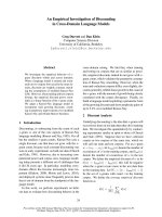
Figure 1 Overview of the hexoki nase genes in Physco mitrella.
Exons are shown as gray boxes and introns as solid black lines. The
predicted exon/intron organization is based on existing cDNA
sequences and, if cDNA sequences were missing or aberrantly
spliced, on the known splice pattern of other plant hexokinase
genes, provided that the consensus donor and acceptor splice sites
are conserved. The predicted transit peptides in the type A
hexokinases and the membrane anchors in the type B and D
hexokinases are shown as small boxes under exon 1.
Nilsson et al. BMC Plant Biology 2011, 11:32
/>Page 4 of 15
A number of conserved sequence motifs and structu-
rally or funct ionally important amino acid residues have
been identified by x-ray crystallography and compari-
sons of hexokinases from different organisms. Bork
et al. [45,46] described seven conserved regions in hexo-
kinases which they named phosphate 1, sugar binding,
connect 1, phosphate 2, helix, adenosine and connect 2,
based on the known or suspected functions of these
regions. Kuser et al. ([47] Table II) identified 20 amino
acid residues that are highly conserved in 317 hexoki-
nases. Mutational and structural studies have shown
that the catalytic residue is an aspartic acid (D211 in the
yeast hexokinase ScHxk2) whereas four other residues
(S158, K176, E269 and E302 in ScHxk2) contribute to
hexose binding [48].
First, we note that the catalytic aspartic acid is strictly
conserv ed in all eleven Physcomitrella sequences, as are
allbutonehexosebindingresidue.Theonlyexception
is the K176 in ScHxk2, which is replaced by a glutamic
acid in PpHxk11. As for the 20 most conserved residues
[48], we note that 19 of them are strongly conserved in
all plant hexokinases (the exception is C268 in ScHxk2).
Interestingly, these 19 residues are strictly conserved in
all Physcomitrella sequences except PpHxk11, which has
four substitutions (Additional file 5: Figure S1). For
comparison, we note that the highly divergent catalyti-
cally inactive AtHkl3 protein [13] has 12 substitutions
in these 19 positions. This includes the catalytic aspartic
acid, which is an asparagine in AtHkl3, and two of the
hexose binding residues. The less divergent AtHkl1 and
AtHkl2 proteins, also thought to be catalytically inactive,
have two and three substitutions, respectively, in the 19
conserved residues, none of w hich involve the cat alytic
or hexose binding residues.
An inspection of the seven regions described by Bork
et al. [46]showsthattheyallarewellconservedinthe
Physcomitrella proteins (Additional file 5: Figure S1).
There are ho wever, some noteworthy exceptions. First,
the type D hexokinases share several substitutions in the
conserved regions which are not found in any other
hexokinases.Thus,theyhaveacysteinefollowedbya
leucine in the phosphate 1 motif where most other hex-
okinases have a vali ne followed by a glutamine. Further-
more, a phenylalanine in the sugar binding motif, which
is strictly conserved in all other hexokinases, is replaced
byaleucineinthethreetype D proteins. Finally, the
latter also share a deleti on of two residues at the end of
the phosphate 2 motif which is not found in any other
hexokinases. None of these changes involve residues
shown to be critical for catalytic activity, but it is still
possible that they could affect the activity and/or sub-
strate specificity of the type D proteins. In addition to
these changes, PpHxk11 has several more substitutions
in the conserved regions, consistent with its generally
more divergent sequence. Finally, we note that all
Physcomitrella hexokinases have an insertion in the ade-
nosine motif, which is found also in other plant hexoki-
nases [7,13].
The Physcomitrella hexokinases show evidence of
concerted evolution
In order to gain a better understanding of how the dif-
ferent hexokinases are related to each other, we used
the predicted sequences of the Arabidopsis,riceand
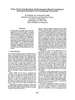
Figure 2 Comparison of the N-terminal sequences of type B and D hexokinases. The sequences shown are the N-terminal ends of the
proteins. Type B hexokinases from rice, Arabidopsis and Physcomitrella are shown at the top, and the three Physcomitrella type D hexokinases at
the bottom. The colour coding used is: L, V, I, M, A - yellow; K, H, R - blue; E, D - red; W, F, Y - magenta; T, S - green; N, Q - pink; G - gray; P -
violet; C - orange.
Nilsson et al. BMC Plant Biology 2011, 11:32
/>Page 5 of 15
Physcomitrella hexokinases to construct an evolutionary
tree. We limited the analysis to these three plant species
since their genome sequences have been completed and
since the rice and Arabidopsis hexokinases already have
been fairly well studied [7,13,49]. The variable N-termini
and C-termini were excluded from the analysis in order
to avoid ambiguities in the sequence alignment, and to
ensure that the result would be independent of the
N-termini, thus making it possible to assess to what
extent the latter have co-evolved with the rest o f the
proteins (Additional file 5: Figure S1).
The resulting tree is shown in Figure 3. Surprisingly,
we found that all eleven Physcomitrella hexokinases are
more closely related to each other than to other plant
hexokinases, thus forming a single branch within the
tree. This was unexpected since the Arabidopsis and
rice sequences do not cluster in this way, but instead
are interspersed (Figure 3). This is particularly evident
in the case of the type A hexokinases, where the single
proteins present in Arabidopsis (AtHxk3) and rice
(OsHxk4) are more similar to each other than to the
other Arabidopsis and rice hexokinases (Figure 3). In
contrast, the three type A hexokinases in Physcomitrella,
PpHxk1, PpHxk5 and PpHxk6, are more similar to the
other Physcomitrella hexokinases than to their orthologs
AtHxk3 and OsHxk4. We conclude from this that the
Physcomitrella hexokinases show evidence of concerted
evolution, unlike the Arabidopsis and rice proteins.
It should further be noted that within the Physcomi-
trella sequences, the four above described hexokinase
types form well-defined branches suggesting a distinct
origin for each type. Thus, the three type D hexokinases
are clearly more closely related to each other than to
thefourtypeBhexokinases,andvice versa.Thissug-
gests that each type of hexokinases arose f rom a single
ancestral gene, which subsequently underwent duplica-
tions. This interpretation is further confirmed by the
fact that the moss type B hexokinases have lost intron 2,
which is present in the other moss hexokinases, includ-
ing the type D hexokinases (Figure 1). Finally, we note
that the sequence of the type C hexokinase, PpHxk4, is
more distantly related to the other Physcomitrella hexo-
kinasesthantheyaretoeachother.Thissuggeststhat
the type C hexokinase may represent an early branch on
the tree, which has been lost in seed plants.
Intracellular localization of the Physcomitrella hexokinases
We proceeded to study the intracel lular locations of the
moss hexokinases. Sequences from the new hexokinases,
expressed from the 35S promoter, were fused in frame
to GFP. These constructs were transiently expressed in
Physcomitrella protoplasts and the GFP fluorescence
was monitored (Figure 4). Based on the sequence simi-
larity of t he N-terminal membrane anchors in PpHxk2,
PpHxk3, PpHxk7 and PpHxk8 to those found in
AtHxk2 (Figure 2) we expected that they would localize
to the outer mitochondrial membrane, as shown
for AtHxk2 and several other type B hexokinases
[7-9,12,13,49]. Consistent with this, we found that the
Physcomitrella type B hexokinases tested also localize to
small ring-like membrane structures (Figure 4) which
were identified as mitochondrial membranes by
co-staining with MitoTracker
®
(Figure 5). In contrast,
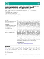
Figure 3 Phylogenetic tree of plant hexokinases. The sequences
included in the comparison were those predicted by the ten
hexokinase-encoding genes in the rice genome, the six hexokinase
and hexokinase-like genes in the Arabidopsis genome and the
eleven Physcomitrella hexokinases discussed in the present work.
Aligned amino acid sequences corresponding to residues 69-439 in
PpHxk1, which excludes the divergent N- and C-termini, were used
to calculate a phylogenetic tree as described in Methods. The
alignment is shown in additional file 5: Figure S1. Hexokinase
sequences from the budding yeast S. cerevisiae (ScHxk2), the fission
yeast S. pombe, the nematode C. elegans, and human glucokinase
(hexokinase IV) were included to root the tree. The subdivisions of
the hexokinases into types A, B, C and D and their intracellular
localisation, if known, are also shown. BX stands for seed plant
proteins that cluster with the type B hexokinases, but whose
N-termini are less conserved. The bar represents a PAM value
(percent accepted point mutations) of 10%. The numbers at the
branch points are bootstrap values derived from 1000 randomized
sequences.
Nilsson et al. BMC Plant Biology 2011, 11:32
/>Page 6 of 15
truncated GFP fusions which lacked the membrane
anchors showed a diffuse localization throughout the
cell (Additional file 6: Figure S2). We conclude that the
N-terminal membrane anchors target the proteins to
the mitochondria. We further note that the mitochon-
dria often formed aggregates (Figure 5). This may be an
artefact caused by protein overexpression, as shown for
other membrane-anchored GFP fusions expressed in
plants [50]. A similar aggregation of mitochondria was
also seen when several of the Arabidopsis hexokinase
GFP fusions were overexpressed [13].
Surprisingly, the type B hexokinase-GFP f usions also
showed fluorescence that was associated with the chlor-
oplast envelope (Figures 4 and 6). This fluorescence was
weaker than that being associated with the mitochon-
dria, but it was seen for all four type B hexokinases.
This is intriguing since the spinach type B hexokinase
SoHxK1 originally was thought to localize to chloroplast
envelopes [51]. This finding was, however, challenged by
Damar i-Weiss ler et al. [9] who reported that SoHxK1 is
found only in the outer mitochondrial membrane, with
no evidence of a chloroplast localisation. It is conceiva-
ble that the hydrophobic anchors in these hexokinases
might cause them to adhere non-specifically also to
chloroplast membranes. However, we do not think that
this is likely since the type D hexokinase PpHxk9 did
not show any fluorescence associated with chloroplasts,
despite having a membrane anchor and being localized
to mitochondria (see below). This suggests that the
chloroplast membrane association of some hexokinases
is specific. Furthermore, we note that our previous sub-
cellular fractionation revealed that some hexokinase
activity is associated with chloroplast membranes, and
that this activity, unlike that in the chloroplast stroma,
is unaffected by a knockout of PpHXK1 [5]. We note
that som e proteins that are known to target to the
chloroplast outer membrane contain N-terminal mem-
brane anchors similar to those found in the type B hex-
okinases [52].
The three type D hexokinases PpHxk9 , PpHxk10 and
PpHxk11 also possess membrane anchors and show a
similar, though more restricted localisation as the type B
proteins. Thus, both PpHxk9 and PpHxk11 localize to
the outer mitochondrial membrane, but only PpHxk11
is also associated with the chloroplast envelope, like the
type B hexokinases (Figures 4, 5, 6). For PpHxk10, we
were unable to clone a PpHXK10 full length transcript
that was correctly spliced. We therefore made two
incomplete PpHxk10-GFP fusions: one containing
the entire region encoded by the first exon including
the membrane anchor (Figure 4) and one containing the
membrane anchor alone. Both fusions localized through-
out the cytosol. It is, however, possible that these partial
fusions are incorrectly folded due to the hydrophobic
nature of the membrane anchor, and that the targeting
signal is thus not functional. We cannot therefore rule
out that a full-length fusion of PpHxk10 to G FP would
localize to the outer mitochondrial membrane, similar
to PpHxk9 and PpHxk11.
In contrast to the above findings, the PpHxk4-GFP
fusion shows a diffuse fluorescence throughout the cell,
indicating a cytosolic locali zation (Figure 4) but
co-staining with DAPI revealed that it is also enriched
in the nucleus (Figure 7). This is similar to what is seen
for GFP alone (Figure 4; s ee also [53] ) and is consistent
with the absence of either a membrane anchor or a target-
ing peptide in the N-terminus of PpHxk4. Similar to GFP
expressed alone, PpHxk4-GFP is also clearly excluded
from the chloroplasts. A likely explanation for this result is
that in the absence of specific targeting signals, PpHxk4 is
Table 1 Predicted intracellular locations and transmembrane helices of moss hexokinases
Protein Type cTP
a
mTP
a
SP
a
other
a
Loc
a
RC
a
TPlen
a
TMH
b
TMhelix
b
PpHxk1 A 0.800 0.207 0.007 0.046 C 3 37 0 -
PpHxk2 B 0.040 0.129 0.724 0.013 S 3 22 1 aa 7-26
PpHxk3 B 0.043 0.091 0.777 0.016 S 2 22 1 aa 7-26
PpHxk4 C 0.337 0.196 0.080 0.452 -5- 0 -
PpHxk5 A 0.120 0.373 0.008 0.129 M 4 14 0 -
PpHxk6 A 0.685 0.110 0.010 0.043 C 3 44 0 -
PpHxk7 B 0.164 0.043 0.540 0.032 S 4 22 1 aa 7-26
PpHxk8 B 0.082 0.062 0.771 0.022 S 2 22 1 aa 7-26
PpHxk9 D 0.052 0.067 0.350 0.182 S 5 32 1 aa 7-29
PpHxk10 D 0.048 0.031 0.527 0.298 S 4 28 1 aa 7-29
PpHxk11 D 0.010 0.064 0.877 0.108 S 2 20 1 aa 5-24
a
Intracellular locations and target peptides predicted by TargetP 1.1 [44]. Abbreviations: cTP, chloroplast transit peptide; mTP, mitochondrial targeting peptide; SP,
secretory pathway signal peptide; other, any other location; Loc, predicted subcellular localization (C, chloroplasts; S, secretory pathway; M, mitochondria); RC,
Reliability Class (1 is the most reliable prediction and 5 the weakest); TPlen, predicted target peptide length. For each protein, the predicted location with the
highest score is shown in bold style.
b
Transmembrane helices predicted by TMHMM 2.0 [43]. Abbreviations: TMH, predicted number of N-terminal transmembrane helices; TMhelix, amino acids
predicted to be part of a transmembrane helix.
Nilsson et al. BMC Plant Biology 2011, 11:32
/>Page 7 of 15
distributed throughout the cytosolic and nuclear compart-
ments. Our finding t hat Physcomitrella possesses a novel
type of soluble hexokinase might explain earlier reports of
cytosolic hexokinase activities in different plants [54-59].
However, such activities could also be derived from disso-
ciated or alternatively spliced membrane bound hexoki-
nases (see below). That cytosolic hexokinases are likely to
exist also in other plants is further suggested by the fact
that the glucose which is exported from the chloroplasts
after starch degradation would require phosphorylation to
be further metabolized [60].
The PpHxk5-GFP and PpHxk6-GFP fusions, finally,
had localizations resembling that of PpHxk1 [5]. Thus,
we found that they are imported into the chloroplast
stroma (Figures 4 and 6). Truncated versions of
PpHxk5-GFP and PpHxk6-GFP lacking the transit pep-
tide were evenly distributed in the cytosol, similar to
GFP expressed alone (Additional file 6: Figure S2). We
conclude that chloroplast import of PpHxk5 and
PpHxk6 is dependent of their N-terminal transit pep-
tides, similar to PpHxk1 [5]. Interestingly, a PpHxk5-
GFP fusion with a shorter N-terminal truncation of
amino acid residues 1-18 is still imported into the chlor-
oplasts, so the targeting information is not immediately
adjacent to the N-terminal end of PpHxk5 (Additional
file 6: Figure S2).
PpHxk3 but not PpHxk1 can complement a hexokinase-
deficient yeast strain
Several plant hexokinases were cloned by their ability to
complement hexokinase-deficient yeast strains [61-63].
We previously found that PpHxk1 fails to complement a
hxk1 hxk2 glk1 triple mutant yeast strain. We noted that
PpHxk1 is a type A hexokinase, while all those that had
been shown to work in yeast at that time were type B
hexokinases [5]. This prompted us to test if a type B
hexokinase from Physcomitrella would work in yeast. To
this end, we cloned a cDNA encoding PpHxk3 into the
Figure 4 Intracellular localization of Physcomitrella hexokinase-
GFP fusions. Fluorescence microscopy pictures of wild type
protoplasts transiently expressing different GFP fusions. GFP
fluorescence is shown in green, with the chlorophyll auto-
fluorescence in red as a chloroplast marker. Protoplasts expressing
GFP alone were also included as a control. The white bars represent
5 μm.
Figure 5 Localization of Physcomitrella hexokinases to
mitochondria. GFP fluorescence is show in green and the
mitochondria specific dye MitoTracker
®
in orange. The white bars
represent 1 μm.
Nilsson et al. BMC Plant Biology 2011, 11:32
/>Page 8 of 15
yeast shuttle vector pFL61 where the inserts are
expressed from the PGK promoter. The plasmid was
transformed into the hxk1 hxk2 glk1 yeast strain and
tested for ability to support growth on different carbon
sources. As shown in Figure 8, we found that PpHxk3
complements the hexokinase-deficient yeast strain for
growth on glucose, which shows that PpHxk3 is
expressed and active in yeast. We further found that
PpHxk3 can support growth on raffinose, which requires
fructokinase activity (Figure 8). This shows that PpHxk3
has a dual specificity for glucose and fructose, similar to
PpHxk1 [5]. In contrast, PpHxk1 failed to complement
the hxk1 hxk2 glk1 triple mutant when expressed from
the same vector (Figure 8). To test if this is due to the
presence of the chloroplast transit peptide, which might
interfere with its function in yeast, we tested a truncated
PpHxk1 which lacks residues 1-38. This is the same
truncation that causes the PpHxk1-GFP fusion to loca-
lize to the cytosol instead of to the chloroplasts [5].
However, the truncated PpHxk1 was still unable to
complement the hexokinase-deficient yeast strain
(Figure 8). This is in contrast to the type A hexokinases
OsHxk4 and LeHxk4 which could complement a hexo-
kinase-deficient yeast strain when their chloroplast tran-
sit peptides were deleted [7,12].
A recent microsatellite mutation in the PpHXK3 gene
During the sequencing of the cDNA and genomic clones
we found a polymorphism in an AG microsatellite
repeat in the 5’ -untra nslated region of the PpHXK3
gene.ThetwocDNAsthatweresequenceddifferby
one AG (Additional file 7: Figure S3a), with the shorter
variant being present in our genomic clone. We first
considered the possibility that two duplicated genes
might exist which differ onlyinthisrepeat.However,
we saw no evidence of this, and only one PpHXK3 gene
is found in the genome sequence [31]. Interestingly, this
gene has the longer variant, unlike our genomic clone.
This made us consider the possibility that loss of one
AG may have occurred recently in our moss line, which
would still be heterogeneous for this mutat ion, thus
explaining the two cDNAs. To test this we cloned two
new PCR fragments from the 5’-untranslated region of
the PpHXK3 gene.Significantly,wefoundthatonehas
the extra AG and one does not, thus confirming the
presence of a polymorphism in our genomic DNA. Two
polymorphisms involving microsatellite repeats were
also seen in PpHXK2, though we did not investigate
these as carefully as the mutation in PpHXK3.Wecon-
clude that sequence evolution by acquisition or loss of
microsatellite repeats seems to occur very rapidly in
Physcomitrella. This could be a consequence of the high
frequency of homologous recombination, since unequal
sister chromatid exchange and gene conversion, both of
which depend on homologous recombination, can gen-
erate this kind of polymorphisms.
Alternative splicing produces a type B hexokinase
without a membrane anchor
We found at least one cDNA for ten of the eleven hexo-
kinases in Physcomitrella, the only exception being
PpHXK6. When t he cDNA clones were sequenced and
compared to other plant hexokinases we found several
unexpected splice variants (Additional file 8: Table S5).
Thus, we found both intron retention and exon skipping
but the most frequent mode of alternative splicing was
the use of alternative donor and/or acceptor sites. Most
of these aberrantly spliced cDNA sequences would not
encode functional hexokinases due to premature termi-
nation. The most interesting exception is the PpHXK7
cDNA clone pdp03464 that was obtained from the
Figure 6 Localization of Physcomitrella hexokinases to
chloroplasts. GFP fluorescence is shown in green and chlorophyll
autofluorescence in red. The white bars represent 1 μm.
Nilsson et al. BMC Plant Biology 2011, 11:32
/>Page 9 of 15
RIKEN bioresource center [34]. PpHxk7 is a type B hexo-
kinase with an N-terminal membrane anchor, but the
anchor is not encoded by the alternatively spliced
pdp03464 clone (Additional file 7: Figure S3b). In the
resulting transcript, the predicted protein instead starts
with the methionine codon at position 64. This truncated
protein is likely to be functional since the deletion does
not affec t the phospha te, sugar or a denosine binding
domains. Interestingly, we also cloned a normally spliced
cDNA from PpHXK7 (Additional file 9: Table S6) which
encodes a protein with an N-termina l membrane anchor
(Additional file 7: Figure S3b). It thus appears that alter-
native splicing produces two PpHxk7 proteins, one with
a membrane anchor and one without it.
Significantly, we found that the splice variant without
a membrane anchor, PpHxk7a, localizes to the cytosol
and in particular to the nucleus (Figure 7), whereas
PpHxk7b localizes to mitochondrial membranes (Figure
7), consistent with the presence of a membra ne anchor
in that splice variant. It is therefore possible that the
PpHxk7a splice variant could be involved in gene
regulation. In this context, it should be noted that an
artificial deletion of the mem brane anchor in the two
rice type B hexokinases OsHxk5 and OsHxk6 changed
their localization to the nucleus, due to the presence of
a cryptic nuclear local ization sequence in these proteins
[27]. No obvious nuclear localization signal was found
in PpHxk7a, but its nuclear localization could be the
result of passive diffusion, as is seen also for GFP alone
[53]. Our finding suggests the interesting possibility that
similar splice variants may exist for type B hexokinases
in other plants, and that alternative splicing could pro-
vide a general mechanism by which type B hexokinases
may enter the nucleus and affect gene expression.
Discussion
We have previously reported that the major hexokinase
in Physcomitrella, PpHxk1, which accounts for 80% of
the glucose phosphorylating activity, is a novel type of
plant hexokinase that is targeted to the chloroplast
stroma [5]. We have now extended our study of the
hexokinase gene family in Physcomitrella by the cloning
Figure 7 Cytosolic and nuclear localization of PpHxk4 and PpHxk7a. Fluorescenc e microscopy pictures of wild type mo ss protoplasts
transiently expressing PpHxk4, the PpHxk7a splice variant, or the PpHxk7b splice variant fused to GFP. GFP fluorescence is shown in green, with
the chlorophyll auto-fluorescence in red as a chloroplast marker. The nucleus is visualized in blue by the fluorescent DNA binding dye DAPI. The
white bars represent 5 μm.
Nilsson et al. BMC Plant Biology 2011, 11:32
/>Page 10 of 15
and characterization of ten new putative hexokinase-
encoding genes (Figure 1). An inspection of the encoded
protein sequences, in particular the N-termini (Figure 2),
suggests that they represent several different types of
hexokinases which are targeted to different intracel lular
compartments.
Four of the moss hexokinases, PpHxk2, PpHxk3,
PpHxk7 and PpHxk8, are clearly type B hexokinases
since they have N-terminal membrane anchors tha t are
similar in sequence to those found in other type B hexo-
kinases [5]. We therefore expected that GFP fusions to
these proteins would localize to mitochondria, as do
several type B hexokinases in seed plants [7,8,10,13]. We
found that t hey indeed localize to the outer mitochon-
drial membrane, but surprisingly, also t o the chloroplast
envelope (Figs. 5 and 6). A similar dual localization was
also seen for one type D hexokinase (PpHxk11). We
note that subcellular fractionation data suggested that
the spinach type B hexokinase SoHxK1 localizes to the
chloroplast envelope [51], but more recent results with
GFP fusions suggested that this is not the case [9]. Dual
targeting of proteins to mitochondria and chloroplasts
has been described in Physcomitrella and several other
plants, but only for proteins that are targeted to the
interior of the organelles [64-66].
PpHxk5 and PpHxk6 are type A hexokinases as evi-
denced both from the sequence of their N-termini,
which resemble o rganelle import peptides and from the
fact that they are closely related to PpHxk1 (Figure 3).
Consistent with this, we found that both the PpHxk5-
GFP and PpHxk6-GFP fusions localize to the chloroplast
stroma (Figure 6). This was surprising in view of our
previous finding that a knockout of the PpHXK1 gene
eliminates all glucose phosphorylating activity in the
chloroplast stromal fractio n [5]. One possible explana-
tion could be that PpHxk5 and PpHxk6 are minor hexo-
kinases, which contribute only a small part of the total
activity, or that they preferentially phosphorylate fruc-
tose, an activity which was not abolishe d in the absence
of PpHXK1 [5]. An alternative explanation could be that
neither PpHxk5 nor PpHxk6 is expressed in the young
protonemal tissue used for the subcellular fractionation
experiments [5]. Yet another possibility could be that
PpHxk5 and PpHxk6 lack hexokinase activity and
instead have some other function, as has been suggested
to be the case for the Hkl1-3 proteins in Arabidopsis
[13,67]. Finally, it is possible that PpHXK5 and PpHXK6
are pseudogenes, since we have not been able to clone
any cDNA from PpHXK6 and the t wo cDNAs from
PpHXK5 that we sequenced were not correctly spliced.
However, we think this is unlikely, since PpHXK5 and
PpHXK6 show no other signs of being pseudogenes.
PpHxk4 represents a novel type of hexokinase, distinct
from both types A and B, which we call type C. We
base this distinction on two facts. First, the sequence of
PpHxk4 shows that it is more distantly related to the
type A and B hexokinases in Physcomitrella than the lat-
ter are to each other. PpHxk4 is thus clearly a separate
type of hexokinase. In a ddition, the truncated N-
terminus of PpHxk4 differs both from the membrane
anchors found in type B hexokinases and from the orga-
nelle targeting peptides found in type A hexokinases.
The predicted location of PpHxk4 is in the cytosol,
since this is where a protein will end up in the absence
of a targeting peptide. Consistent with this, we found
that a PpHxk4-GFP fusion shows a diffuse fluorescence
throughout the cell, indicating a primarily cytoso lic, but
also nuclear localization (Figure 7). We further note that
PpHxk4 occupies a basal position among the Physcomi-
trella hexokinases, being the most divergent member of
the protein family (Figure 3). This suggests that it may
represent an ancient type of hexokinase that has been
lost in seed plants. Interestingly, while no orthologue of
PpHxk4 appears to be present in seed plants, the
two rice hexokinases OsHxk7 and OsHxk8 also have
truncated N-termini, and OsHxk7 was shown to have a
cytosolic localisation [7]. However, the N-termini of
OsHxk7 and OsHxk8 look more like truncated versions
of the type B hexokinase membrane anchor, with most
Figure 8 PpHxk3 can complement a hexokinase-deficient yeast
strain. The picture shows growth of the hxk1 hxk2 glk1 triple
disrupted yeast strain containing the pFL61 vector with different
inserts on plates containing 2% galactose, 2% glucose or 3%
raffinose as carbon source. Growth on glucose requires glucokinase
activity and growth on raffinose fructokinase activity. The inserts
from left to right are: PpHXK3 cDNA; PpHXK1 cDNA encoding an
N-terminally truncated protein; PpHXK1 cDNA; no insert.
Nilsson et al. BMC Plant Biology 2011, 11:32
/>Page 11 of 15
of the twelve first amino acid residues being hydropho-
bic. Furthermore, OsHxk7 and OsHxk8 also group
together with the type B hexokinases in the phylogenetic
tree (Figure 3). Thus, they seem to be divergent type B
hexokinases rather than orthologs of the Physcomitrella
type C hexokinase. Still, it is conceivable that OsHxk7
and OsHxk8 may have a function in rice which is analo-
gous to that of PpHxk4 in moss.
Our finding of a new type of cytosolic hexokinase in
Physcomitrella is interesting in view of the proposed
role of plant hexokinases in glucose sensing and signal-
ing [21,61,68 -70]. These discussions have so far focused
on membrane integrated type B hexokinases such as
AtHxk1 and AtHxk2, which was the only type of plant
hexokinase that had been studied pri or to the disco very
of the type A hexokinases [5]. It has recently been
shown that some type B hexokinases can translocate
into the nucleus and affect gene expression [25,27].
However, it is not clear how these membrane anchored
hexokinases are released from their m embrane associa-
tion and translocated into the nucleus. In c ontrast, the
type C hexokinase PpHxk4, which lacks membrane
anchor and is a soluble protein, could more easily move
into the nucleus.
In this context, we note that we found evidence that
moss type B hexokina ses also may translocate to the
nucleus. Thus, PpHXK7 encodes two differently spliced
cDNAs, one of which is missing the membrane anchor
(Additional file 7: Figure S3b). The intracellular localiza-
tion of these two proteins is also very different as seen
from the expression of the translational fusions with
GFP. In protoplasts expressing the PpHxk7a splice var-
iant that lacks the membrane anchor, the fluorescence is
thus localized throughout the cytosol but is also asso-
ciated with the nucleus (Figure 7). This suggests that
alternative splicing could be a molecular mechanism
whereby membrane bound type B hexokinases, p erhaps
also in other plants, may become soluble and thus exert
a function inside the nucleus.
Theothernewtypeofplanthexokinase,typeD,
appears to have a similar localization as t he type B hex-
okinases, i.e. in the outer mitochondrial membrane and
also to some extent in the chloroplast envelope. How-
ever, they differ from the type B hexokinases in the
sequences of their membrane anchors (Figure 2), and
form a distinct clade in the evolutionary tree (Figure 3).
Furthermore, they do not share the fusion of exons 2
and 3 that is found in all moss type B hexokinases (Fig-
ure 1). Still, the overlapping localizations, and the fact
that the type D hexokinases also have membrane
anchors, suggests that the type B and D hexokinases
may have similar functions. In this context it should be
noted that the type B hexokinases is a large and diverse
groupinseedplants,andthatsomemembershave
N-termini that are less well conserved (labelled BX in
Figure 3). Thus, while no obvious orthologues of the
type D hexokinases exist in seed plants, it is conceivable
that the more divergent members of the type B group
may perform an analogous function as the type D hexo-
kinases do in moss.
It has been proposed that some hexokinas es may lack
catalytic activity but still have other functions, based on
data in fungi [71], flies [72] and plants [13,67]. In
particular, three of the six predicted hexokinases in
Arabidopsis, AtHkl1-AtHkl3, appe ar to lack glucose
phosphorylating activity but are conserved between A.
thaliana and A. lyrata , which suggests that they still are
under selecti on [13]. On the other hand, all ten hexoki-
nases in rice could complement a hexokinase-deficient
yeast strain, indicating that they are catalytically active
[7]. This raises the question whether non-enzymatic
hexokinases are peculiar to Arabidopsis or more wide-
spread in plants. The knockout phenotypes and enzy-
matic activities of PpHxk2-PpHxk11 remain to be
determined, but we note that PpHxk2-PpHxk10 are as
strongly conserved as PpHxk1, which i s an active
enzyme [5], suggesting that they also may be active.
Consistent with this, we found that PpHxk3 can com-
plement a hexokinase deficient yeast strain (Figure 7). In
contrast, PpHxk11 has several substitutions which could
affect its activity. With four substitutions in the 19 most
conserv ed residues it is not as divergent as AtHkl3 (12/
19), but instead resembles AtHkl1 (3/19) and AtHkl2
(2/19). This is also evident from the t ree in Figure 3
where AtHkl3 has a very long branch, whereas AtHkl1,
AtHkl2 and PpHxk11 have much shorter branches. It is
therefore conceivable that PpHxk11 could have a non-
enzymatic function, perhaps in regulation or signaling,
as has been suggested for the AtHkl proteins [13].
Surprisingly, we found that the eleven Physcomitrella
hexokinases are more closely related to each other than
to any othe r plant hexokinase , desp ite the fact that they
represent different types of hexokinases, some of which
are found also in seed plants (Figure 3). This is in con-
trast to the situation in seed plants, where hexokinases
of the same type from different plants typically are more
closely related to each other than hexokinases of differ-
ent types from the s ame plant (ref. [5] and Figure 3).
There are at least two possible explanations for this.
One is that the different hexokinases in Physcomitrella
originated by gene duplications after the separation of
mosses from seed plants. Thisisthemoststraightfor-
ward interpretation of the tree in Figure 3 , but we do
not think that this is a l ikely explanation since the
sequence of the membrane anchor in the type B hexoki-
nases is highly conserved between seed plants and Phys-
comitrella (Figure 2). This suggests a common origin for
the latter, since it is unlikely that this unique sequence
Nilsson et al. BMC Plant Biology 2011, 11:32
/>Page 12 of 15
would have been created twice in evolution just by
chance.
A more likely explanation is therefore that several
genes encoding different types of hexo kinases were pre-
sent already in the common ancestor of mosses and
seed plants, a nd that these genes co-evolved in Physco-
mitrella by gene conversion [73], making them appear
to be more closely related to each other than they really
are. It has already been noted that tandemly arrayed
genes in Physcomitrella are highly similar in sequence,
which suggests that they may undergo concerted evolu-
tion by gene conversion [74]. The hexokinase genes are
not tandemly arrayed, in fact they are all located on dif-
ferent scaffolds in the draft sequence of the Physcomi-
trella genome [31], and those scaffolds that could be
linked to the genetic map [75] were all in different link-
age groups. However, work in yeast has shown that
gene conversion also can be ectopic, i. e. take place
between related genes on different chromosomes [76].
Such ectopic gene conversion could have provided a
mechanism by which the moss hexokinases co-evolved.
A testable prediction of this hypothesis is that other dis-
persed gene families also will show evidence of co-evo-
lution in Physcomitrella.
Conclusions
We have characterized all 11 hexokinase encoding genes
in the moss Physcomitrella and classified them into dif-
ferent types based on se quence motifs and intracellular
localization. We found that the hexokinase gene family
is more diverse in Physcomitrella than in other plants
studied so far, encoding two novel types of hexokinases,
types C and D. The presence of a cytoplasmic and
nuclear hexokinase (type C) sets Physcomitrella apart
from vascular plants, and instead resembles yeast, where
all hexokinases localize to the cytosol. The fact that all
moss hexokinases are more similar to each other than
to hexokinases from vascular plants, even though both
type A and type B hexokinases are present in all plants,
further suggests that the hexokinases in Physcomitrella
have undergone concerted evolution.
Additional material
Additional file 1: Genomic and cDNA clones encoding hexokinases.
Physcomitrella hexokinase genes and cDNA clones and the primers used
for cloning them into the pCR
®
®2.1-TOPO vector.
Additional file 2: Oligonucleotide primers. Oligonucleotide primers
used. Most of the primers are named after the gene to be amplified,
whether it binds to the 3’ or 5’ part, and whether it is followed by a
BamHI, BglII or SmaI site. Primers whose names end with a T were used
to clone inserts where the membrane anchor or chloroplast transit
peptide was removed. The primer combinations used in the various
cases are listed in Tables S1 and S3.
Additional file 3: Hexokinase-GFP fusion and yeast expression
plasmids. The first column lists the plasmids used for intracellular
localization and yeast complementation studies. The primers and
templates used to make these plasmids are listed in the last two
columns. The amino acid residues of the different hexokinases that are
predicted to be expressed after cloning into the vectors psmRS-GFP (GFP
fusions) and pFL61 (yeast complementation) are also listed.
Additional file 4: Sequence Identity matrix for the N-terminal region
of type B and D hexokinases. Comparison of N-terminal regions
containing the membrane anchor of type B and D hexokinases,
illustrated as a two-way sequence identity matrix. The two or three most
similar hexokinases in each comparison are in highlighted in bold.
Additional file 5: Alignment of the hexokinases and hexokinase-like
proteins that are predicted by the Arabidopsis, rice, and
Physcomitrella genomes. The protein sequences shown are those
predicted by the annotated genomes. The most common residues in
each position are enclosed within boxes. The 20 most conserved
residues identified by Kuser et al. [47] are marked with asterisks and the
seven conserved regions defined by Bo rk et al. [45,46] are also indicated.
The core of the alignment, corresponding to amino acid residues 69-439
in PpHxk1, was used to compute the evolutionary tree in Figure 3. Four
non-plant hexokinase sequences, from the budding yeast S. cerevisiae,
the fission yeast S. pombe, the nematode C. elegans and human
hexokinase IV, were included as an outgroup in order to root the tree.
Additional file 6: Intracellular localization of truncated hexokinase-
GFP fusions. Fluorescence microscopy pictures of wild type moss
protoplasts transiently expressing different truncated versions of the
Physcomitrella hexokinases fused to GFP. The hexokinase codons that
were fused in frame to GFP are indicated for each hexokinase. GFP
fluorescence is shown in green, with chlorophyll auto-fluorescence in red
serving as a chloroplast marker. Protoplasts expressing GFP alone were
also included as a control.
Additional file 7: Sequence polymorphism in the PpHXK3 promoter
and alternative splicing of the PpHXK7 transcript. a. Microsatellite
repeat in the PpHXK3 promoter that shows evidence of rapid evolution.
The two sequence variants are shown.b. The two splice variants of
PpHXK7 with (PpHXK7b) and without (PpHXK7a) an N-terminal membrane
anchor. The nucleotide sequences and predicted encoded peptide
sequences of the two splice variants are shown. The splice sites and the
start codons are underlined, and the methionines are highlighted by a
black background.
Additional file 8: Alternative splicing of Physcomitrella hexokinase
transcripts. The different Physcomitrella hexokinase transcripts showing
alternative splicing. The plasmids names and the type of alternative
splicing within these transcripts are listed together with the predicted
effect on the expressed protein.
Additional file 9: Accession numbers for Physcomitrella hexokinases.
The accession numbers of the PpHXK2-pPHXK11 transcripts and the
corresponding GeneIDs are listed.
Acknowledgements
We thank Stefan Hohmann for providing us with the hexokinase deficient
yeast strain. This work was supported by grants from Formas, the Swedish
Research Council for Environment, Agricultural Sciences and Spatial Planning,
and from SSF, the Swedish Strategic Research Foundation.
Author details
1
Department of Microbiology, Swedish University of Agricultural Sciences,
Box 7025, SE-750 07 Uppsala, Sweden.
2
Department of Plant Biology and
Forest Genetics, Swedish University of Agricultural Sciences, Box 7080, SE-750
07 Uppsala, Sweden.
Authors’ contributions
AN, TO, MU and MT carried out the experimental work. TO performed the
yeast complementation study. All authors were involved in the sequencing
Nilsson et al. BMC Plant Biology 2011, 11:32
/>Page 13 of 15
and in the phylogenetic analysis. AN, TO, MU and MT cloned the
Physcomitrella hexokinases and constructed various plasmids. AN, TO, MU
and MT transformed Physcomitrella protoplasts and analyzed the GFP
expression. AN and MT analyzed the transformed Physcomitrella protoplasts
treated with the mitochondria specific dye. All authors participated in the
design and coordination of the study. All authors have read and approved
the final manuscript.
Received: 19 October 2010 Accepted: 14 February 2011
Published: 14 February 2011
References
1. Cardenas ML, Cornish-Bowden A, Ureta T: Evolution and regulatory role of
hexokinases. Biocim Biophys Acta 1998, 1401:242-264.
2. Claeyssen E, Rivoal J: Isozymes of plant hexokinase: Occurrence,
properties and functions. Phytochemistry 2007, 68:709-731.
3. Wilson JE: Isozymes of mammalian hexokinase: structure, subcellular
localization and metabolic function. J Exp Biol 2003, 206:2049-2057.
4. Moreno F, Ahuatzi D, Palomino CA, Herrero P: Glucose sensing through
the Hxk2-dependent signaling pathway. Biochem Soc Trans 2005,
33:265-268.
5. Olsson T, Thelander M, Ronne H: A novel type of chloroplast stromal
hexokinase is the major glucose phosphorylating enzyme in the moss
Physcomitrella patens. J Biol Chem 2003, 278:44439-44447.
6. Reumann S, Inoue K, Keegstra K: Evolution of the general protein import
pathway of plastids (Review). Mol Membr Biol 2005, 22:73-86.
7. Cho J-I, Ryoo N, Ko S, Lee S-K, Lee J, Jung K-H, Lee Y-H, Bhoo SH,
Winderickx J, An G, Hahn T-R, Jeon J-S: Structure, expression, and
functional analysis of the hexokinase gene family in rice (Oryza sativa
L.). Planta 2006, 224:598-611.
8. Damari-Weissler H, Kandel-Kfir M, Gidoni D, Mett A, Belausov E, Granot D:
Evidence for intracellular spatial separation of hexokinases and
fructokinases in tomato plants. Planta 2006, 224:1495-1502.
9. Damari-Weissler H, Ginzburg A, Gidoni D, Mett A, Krassovskaya I, Weber AP,
Belausov E, Granot D: Spinach SoHXK1 is a mitochondria-associated
hexokinase. Planta 2007, 226:1053-1058.
10. Giegé P, Heazlewood JL, Roessner-Tunali U, Millar AH, Fernie AR, Leaver CJ,
Sweetlove LJ: Enzymes of glycolysis are functionally associated with the
mitochondrion in Arabidopsis cells. The Plant Cell 2003, 15:2140-2151.
11. Giese JO, Herbers K, Hoffmann M, Klösgen RB, Sonnewald U: Isolation and
functional characterization of a novel plastidic hexokinase from
Nicotiana tabacum. FEBS Letters 2005, 579:827-831.
12. Kandel-Kfir M, Damari-Weissler H, German MA, Gidoni D, Mett A, Belausov E,
Petreikov M, Adir N, Granot D: Two new identified membrane-associated
and plastidic tomato HXK:s characteristics, predicted structure and
intracellular localization. Planta 2006, 224:1341-1352.
13. Karve A, Rauh LB, Xia X, Kandasamy M, Meagher BR, Sheen J, Moore DB:
Expression and evolutionary features of the hexokinase gene family in
Arabidopsis. Planta 2008, 228:411-425.
14. Kim M, Lim J-H, Ahn CS, Park K, Kim GT, Kim WT, Pai H-S: Mitochondria-
associated hexokinases play a role in the control of programmed cell
death in Nicotiana benthamiana. Plant Cell 2006,
18:2341-2355.
15.
Danial NN, Gramm CF, Scorrano L, Zhang C-Y, Krauss S, Ranger AM,
Datta SR, Greenberg ME, Licklider LJ, Lowell BB, Gygi SP, Korsmeyer SJ: BAD
and glucokinase reside in a mitochondrial complex that integrates
glycolysis and apoptosis. Nature 2003, 424:952-956.
16. Majewski N, Noquiera V, Bhaskar P, Coy PE, Skeen JE, Gottlob K, Chandel NS,
Thompson CB, Robey RB, Hay N: Hexokinase-mitochondria interaction
mediated by Akt is required to inhibit apoptosis in the presence or
absence of Bax and Bak. Mol Cell 2004, 16:819-830.
17. Pastorino JG, Shulga N, Hoek JB: Mitochondrial binding of hexokinase II
inhibits Bax-induced cytochrome c release and apoptosis. J Biol Chem
2002, 277:7610-7618.
18. Entian K-D, Zimmermann FK, Scheel I: A partial defect in carbon catabolite
repression in mutants of Saccharomyces cerevisiae with reduced hexose
phosphorylation. Mol Gen Genet 1977, 156:99-105.
19. Entian K-D: Genetic and biochemical evidence for hexokinase PII as a
key enzyme involved in carbon catabolite repression in yeast. Mol Gen
Genet 1980, 178:633-637.
20. Hohmann S, Winderickx J, de Winde JH, Valckx D, Cobbaert P, Luyten K, de
Meirsman C, Ramos J, Thevelein JM: Novel alleles of yeast hexokinase PII
with distinct effects on catalytic activity and catabolite repression of
SUC2. Microbiology 1999, 145:703-714.
21. Rolland F, Winderickx J, Thevelein JM: Glucose-sensing mechanisms in
eukaryotic cells. Trends Biochem Sci 2001, 26:310-317.
22. Rolland F, Winderickx J, Thevelein JM: Glucose-sensing and -signaling
mechanisms in yeast. FEMS Yeast Res 2002, 2:183-201.
23. Santangelo GM: Glucose signaling in Saccharomyces cerevisiae. Microbiol
Mol Biol Rev 2006, 70:253-282.
24. Ahuatzi D, Herrero P, de la Cera T, Moreno F: The glucose-regulated
nuclear localization of hexokinase 2 in Saccharomyces cerevisiae is
Mig1-dependent. J Biol Chem 2004, 279:14440-14446.
25. Cho Y-H, Yoo S-D, Sheen J: Regulatory functions of nuclear hexokinase1
complex in glucose signaling. Cell 2006, 127:579-589.
26. Aki T, Yanagisawa S: Application of rice nuclear proteome analysis to the
identification of evolutionarily conserved and glucose-responsive
nuclear proteins. J Proteome Res 2009, 8:3912-3924.
27. Cho J-H, Ryoo N, Eom Lee D-W, Kim H-B, Jeong S-W, Lee Y-H, Kwon Y-K,
Cho M-H, Bhoo SH, Hahn T-R, Park Y-I, Hwang I, Sheen J, Jeon J-S: Role of
the rice hexokinases OsHXK5 and OsHXK6 as glucose sensors. Plant
Physiol 2009, 149:745-759.
28. Schaefer D: A
new moss genetics: Targeted mutagenesis in
Physcomitrella patens. Annu Rev Cell Biol 2002, 53:477-501.
29. Cove D: The moss Physcomitrella patens. Annu Rev Genet 2005,
39:339-358.
30. Cove D, Benzanilla M, Harries P, Quatrano R: Mosses as model systems for
the study of metabolism and development. Annu Rev Plant Biol 2006,
57:497-520.
31. Rensing SA, et al: The Physcomitrella genome reveals evolutionary
insights into the conquest of land by plants. Science 2008, 319:64-69.
32. Thelander M, Olsson T, Ronne H: Effect of the energy supply on
filamentous growth and development in Physcomitrella patens. J Exp Bot
2005, 56:653-662.
33. Nishiyama T, Hiwatashi Y, Sakakibara K, Kato M, Hasebe M: Tagged
mutagenesis and gene-trap in the moss Physcomitrella patens by
shuttle mutagenesis. DNA Res 2000, 7:9-17.
34. Nishiyama T, Fujita T, Shin-I T, Seki M, Nishide H, Uchiyama I, Kamiya A,
Carninci P, Hayashizaki Y, Shinozaki K, Kohara Y, Hasebe M: Comparative
genomics of Physcomitrella patens gemetophytic transcriptome and
Arabidopsis thaliana: Implication for land plant evolution. Proc Natl Acad
Sci USA 2003, 100:8007-8012.
35. Davis SJ, Vierstra RD: Soluble derivatives of green fluorescent protein
(GFP) for use in Arabidopsis thaliana. Weeds World 1996, 3:43-48.
36. Schaefer D, Zrÿd J-P, Knight C, Cove D: Stable transformation of the moss
Phycomitrella patens. Mol Gen Genet 1991, 226:418-424.
37. Thomas BJ, Rothstein RJ: Elevated recombination rates in transcriptionally
active DNA. Cell 1989, 56:619-630.
38. Minet M, Dufour ME, Lacroute F: Complementation of Saccharomyces
cerevisiae auxotrophic mutants by Arabidopsis thaliana cDNAs. Plant J
1992, 2:417-422.
39. Saitou N, Nei M: The nieghbor-joining method: a new method for
reconstructing phylogenetic trees. Mol Biol Evol 1987, 4:406-425.
40. Thelander M, Olsson T, Ronne H: Snf1-related protein kinase 1 is
needed for growth in a normal day-night light cycle. EMBO J 2004 ,
23:1900-1910.
41. Lang D, Eisinger J, Reski R, Rensing SA: Representation and high-quality
annotation of the Physcomitrella patens transcriptome demonstrates a
high proportion of proteins involved in metabolism in mosses. Plant Biol
(Stuttg)
2005, 7:238-250.
42.
Machuka J, Bashiardes S, Ruben E, Spooner K, Cuming A, Knight C, Cove D:
Sequence analysis of expressed sequence tags from an ABA-treated
cDNA library identifies stress response genes in the moss Physcomitrella
patens. Plant Cell Physiol 1999, 40:378-387.
43. Krogh A, Larsson B, von Heijne G, Sonnhammer EL: Predicting
transmembrane protein topology with a hidden Markov model:
Application to complete genomes. J Mol Biol 2001, 305:567-580.
44. Emanuelsson O, Nielsen H, Brunak S, von Heijne G: Predicting subcellular
localization of proteins based on their N-terminal amino acid sequence.
J Mol Biol 2000, 300:1005-1016.
45. Bork P, Sander C, Valencia A: An ATPase domain common to prokaryotic
cell cycle proteins, sugar kinases, actin, and hsp70 heat shock proteins.
Proc Natl Acad Sci USA 1992, 89:7290-7294.
Nilsson et al. BMC Plant Biology 2011, 11:32
/>Page 14 of 15
46. Bork P, Sander C, Valencia A: Convergent evolution of similar enzymatic
function on different protein folds: the hexokinase, ribokinase, and
galactokinase families of sugar kinases. Protein Sci 1993, 2:31-40.
47. Kuser P, Cupri F, Bleicher L, Polikarpov I: Crystal structure of yeast
hexokinase PI in complex with glucose: A classical “induced fit” example
revised. Proteins 2008, 72:731-740.
48. Kuser PR, Krauchenco S, Antunes OA, Polikarpov I: The high resolution
crystal structure of yeast hexokinase PII with the correct primary
sequence provides new insights into its mechanism of action. J Biol
Chem 2000, 275:20814-20821.
49. Karve R, Lauria M, Virnig A, Xia X, Rqauh BL, Moore BD: Evolutionary
lineages and functional diversification of plant hexokinases. Mol Plant
2010, 3:334-346.
50. Lisenbee CS, Karnik SK, Trelease RN: Overexpression and mislocalization of
a tail-anchored GFP redefines the identity of peroxisomal ER. Traffic
2003, 4:491-501.
51. Wiese A, Gröner F, Sonnewald U, Deppner H, Lerchl J, Hebbeker U,
Flügge U, Weber A: Spinach hexokinase I is located in the outer
envelope membrane of plastids. FEBS Lett 1999, 461:13-18.
52. Hofmann NR, Theg SM: Chloroplast outer membrane protein targeting
and insertion. Trends Plant Sci 2005, 10:450-457.
53. Thelander M, Nilsson A, Olsson T, Johansson M, Girrod P-A, Schaefer DG,
Zrÿd J-P, Ronne H: The moss genens PpSKI1 and PpSKI2 encode nuclear
SnRK1 interacting proteins with homologues in vascular plants. Plant Mol
Biol 2007, 64:559-573.
54. Baldus B, Grahame KJ, Latzko E: Hexokinases of spinach leaves.
Phytochemistry 1981, 20:1811-1814.
55. Copeland L, Morell M: Hexose kinases from the plant cytosolic fraction of
soybean nodules. Plant Physiol 1985, 79:114-117.
56. Galina A, Reis M, Albuquerque MC, Puyou AG, Puyou MG, Meis L: Different
properties of the mitochondrial and cytosolic hexokinases in maize
roots. Biochem J 1995, 309:105-112.
57. Miernyk JA, Dennis DT: Mitochondrial, plastid, and cytosolic isozymes of
hexokinase from developing endosperm of Ricinus communis. Arch
Biochem Biophys 1983, 226:458-468.
58. Schnarrenberger C: Characterization and compartmentation, in green
leaves, of hexokinases with different specificities for glucose, fructose,
and mannose and for nucleoside triphosphates. Planta 1990, 181
:249-255.
59. da-Silva WS, Rezende GL, Galina A: Subcellular distribution and kinetic
properties of cytosolic and non-cytosolic hexokinase in maize seedling
roots: implications for hexose phosphorylation. J Exp Bot 2001,
52:1191-1201.
60. Granot D: Putting plant hexokinases in their proper place. Phytochemistry
2008, 69:2649-2654.
61. Jang JC, Léon P, Zhou L, Sheen J: Hexokinase as a sugar sensor in higher
plants. The Plant Cell 1997, 9:5-19.
62. Veramendi J, Roessner U, Renz A, Willmitzer L, Trethewey RN: Antisense
repression of hexokinase 1 leads to an overaccumulation of starch in
leaves of transgenic potato plants but not to significant changes in
tuber carbohydrate metabolism. Plant Physiol 1999, 121:123-133.
63. Veramendi J, Fernie AR, Leisse A, Willmitzer L, Trethewey RN: Potato
hexokinase 2 complements transgenic Arabidopsis plants deficient in
hexokinase 1 but does not play a key role in tuber carbohydrate
metabolism. Plant Mol Biol 2002, 49:491-501.
64. Millar AH, Whelan J, Small I: Recent surprises in protein targeting to
mitochondria and plastids. Curr Opin Plant Biol 2006, 9:610-615.
65. Carrie C, Giraud E, Whelan J: Protein transport in organelles: Dual
targeting of proteins to mitochondria and chloroplasts. FEBS J 2009,
276:1187-1195.
66. Mitschke J, Fuss J, Blum T, Höglund A, Reski R, Kohlbacher O, Rensing SA:
Prediction of dual protein targeting to plant organelles. New Phytol 2009,
183:224-236.
67. Karve A, Moore B: Function of Arabidopsis hexokinase-like1 as a negative
regulator of plant growth. J Exp Bot 2009, 60:4137-4149.
68. Frommer WB, Schultze WX, Lalonde S: Hexokinase, jack-of-all-trades.
Science 2003, 300:261-263.
69. Moore B, Zhou L, Rolland F, Hall Q, Cheng WH, Liu YX, Hwang I, Jones T,
Sheen J: Role of the Arabidopsis glucose sensor HXK1 in nutrient, light,
and hormonal signaling. Science 2003, 300:332-336.
70. Rolland F, Baena-Gonzalez E, Sheen J: Sugar sensing and signaling in
plants: conserved and novel mechanisms. Annu Rev Plant Biol 2006,
57:675-709.
71. Bernardo SMH, Gray K-A, Todd RB, Cheetham BF, Katz ME: Characterization
of regulatory non-catalytic hexokinases in Aspergillus nidulans. Mol
Genet Genomics 2007, 277:519-532.
72. Kulkarni GV, Deobagkar DN, Deobagkar DD: Hexokinase like protein from
Drosophila melanogaster. Dros Inf Serv 2002, 85:65-68.
73. Ronne H, Widmark E, Rask L, Peterson PA: Intron sequences reveal
evolutionary relationships among major histocompatibility complex
class I genes. Proc Natl Acad Sci USA 1985, 82:5860-5864.
74. Lang D, Zimmer AD, Rensing SA, Reski R: Exploring plant biodiversity: the
Physcomitrella genome and beyond. Trends Plant Sci 2008, 13:542-549.
75. Kamisugi Y, von Stackelberg M, Lang D, Care M, Reski R, Rensing SA,
Cuming AC: A sequence-anchored genetic linkage map for the moss,
Physcomitrella patens. The Plant J 2008, 56:855-866.
76. Lichten M, Borts RH, Haber JE: Meiotic gene conversion and crossing over
between dispersed homologous sequences occurs frequently in
Saccharomyces cerevisiae. Genetics 1987, 115:233-246.
doi:10.1186/1471-2229-11-32
Cite this article as: Nilsson et al.: Two novel types of hexokinases in the
moss Physcomitrella patens. BMC Plant Biology 2011 11:32.
Submit your next manuscript to BioMed Central
and take full advantage of:
• Convenient online submission
• Thorough peer review
• No space constraints or color figure charges
• Immediate publication on acceptance
• Inclusion in PubMed, CAS, Scopus and Google Scholar
• Research which is freely available for redistribution
Submit your manuscript at
www.biomedcentral.com/submit
Nilsson et al. BMC Plant Biology 2011, 11:32
/>Page 15 of 15