A Lange Medical Book Pediatrics on call - part 4 ppsx
Bạn đang xem bản rút gọn của tài liệu. Xem và tải ngay bản đầy đủ của tài liệu tại đây (742.57 KB, 82 trang )
45. HYPERTENSION 217
5. Enalaprilat. Effective in 5–10 mcg/kg doses q8–24h. Because
neonates have a more active renin-angiotensin system, they
are more sensitive to drug than older children and should be
given dose in lower range. Closely monitor renal function and
serum potassium level.
6. Hydralazine. Old but trustworthy drug given at 0.1–0.5 mg/kg
as a bolus. Maximum dose per bolus is 20 mg. Can be repeat-
ed q3–4h. Monitor heart rate and hold doses if significant
tachycardia. Watch for resistance to BP-lowering effect.
7. Diazoxide. Extremely effective; can cause precipitous drop in
BP and elevate blood glucose concentration. If normal saline
infusion is available at bedside to treat acute hypotension, 1–3
mg/kg quick IV push works well. Second bolus can be given
within 5–15 minutes if needed, not to exceed 5 mg/kg com-
bined dose. Effective dose can be repeated q4–24h.
B. Hypertensive Urgency. Symptomatic hypertension without evi-
dence of end-organ damage. Oral treatment is acceptable,
although IV medications may also be considered. Long-acting
oral agents (ie, those recommended in once- or twice-daily doses)
should be avoided due to delayed peak concentration.
1. “Sublingual” nifedipine. No excessive side effects reported in
pediatric literature; frequently administered, convenient drug of
choice for pediatric hypertensive urgencies if administered in
appropriate dose. Conventional dose is 0.25–0.5 mg/kg per
dose q3–4h, not to exceed 10 mg per dose or 3 mg/kg/day.
Although labeled as sublingual, absorption takes place from
stomach, so capsule needs to be opened before being swal-
lowing.
2. Oral hydralazine. Doses of 0.75–1 mg/kg q4–6h may work
well. Maximum one-time dose is 25 mg, with cumulative daily
dose of 5 mg/kg.
3. Minoxidil. More powerful vasodilator than hydralazine, with
more side effects. In acute situations, 0.2 mg/kg may work well.
Add diuretic if treatment exceeds a few days.
4. Propranolol. Given in doses of 0.12–0.25 mg/kg q6–12h.
5. Chronic hypertension. Not within scope of this discussion,
but lifestyle changes, such as low-salt diet, exercise, and
weight loss, should be part of any comprehensive treatment
plan for patients with chronic hypertension.
VI. Problem Case Diagnosis. The 13-year-old girl had modest BP
elevation, which might be attributed to office hypertension, essen-
tial hypertension, or metabolic syndrome. Further investigation
showed multiple high BP readings had been obtained by school
nurse, and patient also had a strong family history of hypertension.
Diagnosis of essential hypertension was made, and patient’s BP
was well controlled on salt restriction and hydrochlorothiazide,
25 mg daily.
218 I: ON CALL PROBLEMS
VII. Teaching Pearl: Question. What is the only form of hypertension
that will never develop into malignant hypertension?
VIII. Teaching Pearl: Answer. Coarctation of the aorta never progresses
into malignant hypertension. This is the only form of hypertension in
which the kidneys are sheltered from elevated systemic BP. This
observation suggests the pivotal role of the kidneys in the pathome-
chanism of malignant hypertension.
REFERENCES
Fivush B, Neu AM, Furth S. Acute hypertensive crises in children: Emergencies and
urgencies. Curr Opin Pediatr 1997;9:233–236.
Friedman AL. Approach to the treatment of hypertension in children. Heart Dis
2002;4:47–50.
National High Blood Pressure Education Working Group on Hypertension Control in
Children and Adolescents. Update on the 1987 task force report on high blood
pressure in children and adolescents: A working group report from the National
High Blood Pressure Education Program. Pediatrics 1996;98:649–658.
Sinaiko AR. Hypertension in children. N Engl J Med 1996;335:1968–1973.
46. HYPOCALCEMIA
I. Problem. A 7-day-old infant is admitted with a history of jitteriness
and poor feeding associated with total serum calcium level of 6.0 mg/dL
(normal for this age: 7.6–10.9 mg/dL).
II. Immediate Questions
A. Is patient symptomatic? Hypocalcemia can be asymptomatic or
associated with serious life-threatening manifestations. Severe
manifestations that require immediate treatment include pares-
thesias, tetany, laryngospasm, and seizures. Diagnostic signs
suggesting the need for immediate treatment are positive
Chvostek and Trousseau signs.
B. Is low serum calcium level an artifact or reflective of low ion-
ized calcium? Whereas total serum calcium is routinely meas-
ured, it is the ionized calcium component that is physiologically
important. Ionized calcium can be measured directly or can be
estimated by subtracting 0.8 mg/dL for every 1 g/dL by which
serum albumin is < 4 g/dL.
C. Is serum magnesium level low? Serum calcium will not respond
to correction with IV or oral calcium as long as severe hypomag-
nesemia remains untreated.
D. Pertinent Historical Information
1. Infants. Is there a history of parathyroid or other endocrine dis-
eases? What is the gestational history? Pay particular attention
to maternal illnesses (eg, diabetes mellitus, hyperparathy-
roidism), medications, birth history, and gestational age. What
type of formula or supplements is infant given?
46. HYPOCALCEMIA 219
2. Children. Is there a history of acute or chronic illnesses, med-
ication use, or surgery? Ask about diet and sun exposure.
III. Differential Diagnosis. Causes of hypocalcemia in infants need to
be distinguished from those in children. Neonatal hypocalcemia is
classically divided into early (first 4 days of life) and late, which usu-
ally presents at 5–10 days of life. In children of all ages, abnormali-
ties can be divided into those involving parathyroid hormone (PTH),
vitamin D, and binding or distribution of calcium.
A. Neonatal Hypocalcemia
1. Early neonatal hypocalcemia
a. Preterm infants. Transiently decreased PTH secretion.
b. Neonates with asphyxia. Possibly associated with increased
calcitonin secretion.
c. Infants of diabetic mothers. Related to maternal hypo-
magnesemia.
d. Infants whose mothers had preeclampsia. Related to
maternal hypomagnesemia.
2. Late neonatal hypocalcemia
a. Dietary phosphate loading. Results from inability of imma-
ture kidneys to excrete phosphate in infants fed cow’s milk
formula.
b. Hypoparathyroidism. Transient, insufficient PTH secretion.
c. Hypomagnesemia. Can be associated with rare defects in
magnesium transport.
3. Miscellaneous causes of hypocalcemia in infants and
neonates
a. Congenital hypoparathyroidism. Can be associated with
DiGeorge anomaly or CATCH-22 syndrome (cardiac anom-
alies, abnormal facies, thymic aplasia, cleft palate, hypocal-
cemia, caused by deletion in chromosome 22q11.2).
b. “Late-late” hypocalcemia. Skeletal hypomineralization
and poor mineral and vitamin D intake presenting at 2–4
months of age.
c. Infants of hyperparathyroid mothers.
d. Ionized hypocalcemia. Associated with exchange trans-
fusions of citrated blood, lipid infusions, or respiratory
alkalosis.
B. Childhood Hypocalcemia
1. Parathyroid disorders
a. Hypoparathyroidism. Associated with chromosome 22q11
abnormalities (see A, 3, a, earlier) or autoimmune syn-
dromes such as autoimmune polyglandular syndrome.
b. Pseudohypoparathyroidism. Disorders of activation of the
cellular effects of PTH.
c. Calcium-sensing abnormalities. Occurs when parathy-
roid gland is abnormally sensitive to serum calcium, causing
PTH levels to be low in relation to level of calcium.
220 I: ON CALL PROBLEMS
d. Hypomagnesemia. Associated with decreased PTH secre-
tion and PTH effect.
2. Vitamin D disorders
a. Vitamin D deficiency. Low levels of vitamin D due to dietary
insufficiency, lack of sunshine, fat malabsorption, or liver
disease.
b. Vitamin D–dependent rickets. Block in 1,25-dihydroxyvitamin
D formation (type 1) or abnormal receptor (type 2).
c. Renal failure. Acute or chronic, with inadequate formation
of 1,25-dihydroxyvitamin D.
d. Fanconi syndrome. Proximal renal tubular dysfunction with
low 1,25-dihydroxyvitamin D formation and renal phosphate
wasting.
e. Altered metabolism. Often due to drugs such as pheno-
barbital, phenytoin, or ketoconazole.
3. Abnormal distribution or binding of calcium
a. Tumor lysis syndrome. Hyperphosphatemia, hypocalcemia,
and acute renal failure.
b. Acute rhabdomyolysis. Trapping of calcium into injured
muscle.
c. Hungry bone syndrome. Shift of calcium and phosphorus
into bone, often after parathyroidectomy.
d. Drugs. Foscarnet, bisphosphonates, calcitonin, calcium
chelators (citrate, phosphorus).
e. Miscellaneous. Acute pancreatitis, toxic shock syndrome,
sepsis.
IV. Database
A. Physical Exam Key Points
1. General appearance. Albright hereditary osteodystrophy with
pseudohypoparathyroidism (short stature, obesity, round face);
large-for-gestational-age infants of diabetic mothers.
2. Skin. Mucocutaneous candidiasis with autoimmune polyglandular
syndrome; alopecia with type 2 vitamin D–dependent rickets.
3. HEENT. Facial features of DiGeorge syndrome, laryngospasm,
cataracts.
4. Skeletal findings. Evidence of bowing with rickets; short
metacarpals and metatarsals with pseudohypoparathyroidism.
5. Neuromuscular exam. Neuromuscular excitability manifested
by irritability, facial grimacing, hyperactive deep tendon
reflexes, muscular spasms, twitching and tetany, confusion,
seizures.
6. Heart. Cardiac abnormalities seen in DiGeorge syndrome.
7. Specific tests for tetany of hypocalcemia
a. Chvostek sign. Elicited by tapping on the facial nerve below
the zygomatic arch and 2 cm anterior to the earlobe. Positive
sign ranges from twitching of the lip at the angle of the
mouth to contraction of the facial muscles.
46. HYPOCALCEMIA 221
b. Trousseau sign. Performed by inflating a BP cuff on the
upper arm to just above systolic BP for 3 minutes. With
hypocalcemia, carpal spasm may occur in response to
ischemia of the ulnar nerve.
B. Laboratory Data
1. Serum electrolytes. In addition to total calcium, focus on
potassium, phosphate, and magnesium levels. The latter two
are not usually included in standard panels and may have to be
ordered separately. Serum calcium should be interpreted in
relation to serum albumin (see II, B, earlier). Hyperkalemia may
be a sign of tumor lysis. Serum phosphate is elevated in renal
failure, tumor lysis, rhabdomyolysis, phosphate enemas, and
parathyroid disorders. It is also seen in most of the neonatal
hypocalcemic disorders. Hypophosphatemia is a sign of
vitamin D disorders, hungry bone syndrome, and Fanconi
syndrome. Severe hypomagnesemia, < 1 mg/dL, is a cause of
refractory hypocalcemia.
2. Serum albumin. As previously described.
3. Ionized calcium. Particularly valuable in the presence of
alkalosis and chelators, which may selectively lower ionized
calcium. In confusing cases, ionized calcium can help with
diagnosis and management.
4. BUN and creatinine. Signs of renal failure, acute or chronic.
5. PTH level. Should be interpreted in relation to serum calcium
level.
6. Vitamin D levels. 25-Hydroxyvitamin D identifies deficiency or
abnormalities of metabolism whereas 1,25-dihydroxyvitamin D
may be helpful in patients with vitamin D–dependent states
and renal disease.
C. Radiographic and Other Studies
1. Bone films. Look for rickets, osteopenia, or renal osteody-
strophy.
2. ECG. Hypocalcemia may result in prolonged QT interval or T
wave inversion.
V. Plan. Evaluate for symptomatic hypocalcemia that would necessitate
immediate IV treatment with calcium and possibly magnesium.
Obtain laboratory studies and initiate oral treatment after patient is
stabilized.
A. Neonates
1. Emergency treatment
a. For symptomatic hypocalcemia or when serum calcium is
< 5–6 mg/dL, give 10–20 mg elemental calcium per kilogram
body weight, or 1–2 mL of calcium gluconate per kilogram
body weight (10% solution). This should be given no faster
than 1 mL/min under constant cardiac monitoring.
b. Treat hypomagnesemia with 0.1–0.2 mL/kg of 50% magne-
sium sulfate (0.4–0.8 mEq/kg or 5–10 mg/kg) IV or IM, again
222 I: ON CALL PROBLEMS
under constant cardiac monitoring. May repeat magnesium
dose q12–24h.
2. Nonemergency or maintenance therapy. Oral calcium at a
dose of 50–75 mg elemental calcium per kilogram per day as cal-
cium glubionate (23 mg/mL), calcium carbonate (100 mg/mL), or
calcium gluconate (9 mg/mL). Give in 4–6 divided doses, and
combine with low-phosphorus formula such as maternal breast
milk or Similac PM 60/40.
3. Vitamin D. Daily supplement of oral vitamin D at a dose of
400–2000 IU/day.
B. Children
1. Emergency treatment
a. For acute symptomatic hypocalcemia, give 2–3 mg elemen-
tal calcium per kilogram body weight or 0.25 mL calcium
gluconate per kilogram body weight (10% solution) IV at a
rate of no more than 1 mL/min under constant cardiac mon-
itoring. Can continue with a constant infusion at a rate of
50–75 mg elemental calcium per kilogram per day until
hypocalcemia is corrected.
b. For hypomagnesemic hypocalcemia, give 6 mg elemental
magnesium per kilogram body weight or 0.12 mL per kilo-
gram body weight of 50% magnesium sulfate IM or IV over
1–4 hours.
2. Chronic treatment
a. Calcium. Oral calcium at a dose of 500–1000 mg elemental
calcium per dose q6h. This can be given as liquid (calcium
carbonate, 100 mg/mL; calcium glubionate, 25 mg/mL) or
one of many tablet forms.
b. Vitamin D. Treat vitamin D deficiency with ergocalciferol
drops at a dose of 800–8000 IU/day. Doses have been
described in the literature up to 600,000 units given in a
single day. For patients with renal failure, calcitriol can be
given at a dose of 0.25–1 mcg/day. Patients with
hypoparathyroidism, pseudohypoparathyroidism, and vita-
min D–dependent rickets type 1 also require calcitriol thera-
py rather than ergocalciferol. This can be given orally or
intravenously.
VI. Problem Case Diagnosis. The 1-week-old infant had late neonatal
hypocalcemia, and a phosphorus level of 9.2 mg/dL. Infant was treated
with IV calcium gluconate and placed on Similac PM 60/40. There was
no evidence of DiGeorge syndrome or recurrence of hypocalcemia.
VII. Teaching Pearl: Question. In most cases of vitamin D–deficiency
rickets, is the level of 1,25-dihydroxyvitamin D high, normal, or low?
VIII. Teaching Pearl: Answer. The 1,25-dihydroxyvitamin D levels are
usually in the normal range, but this is inappropriate for the level of
47. HYPOGLYCEMIA 223
hypophosphatemia, hypocalcemia, and hyperparathyroidism that
may be present.
REFERENCES
Carpenter TO. Neonatal hypocalcemia. In: Favus M, ed. Primer on the Metabolic
Bone Diseases and Disorders of Mineral Metabolism, 5th ed. American Society of
Bone and Mineral Research, 2003:286–288.
Koo W. Hypocalcemia and hypercalcemia in neonates. In: Umpaichitra V, Bastian W,
Castells S. Hypocalcemia in children: Pathogenesis and management. Clin
Pediatr 2001;40:305.
47. HYPOGLYCEMIA
I. Problem. A previously healthy 3-year-old boy is brought to the emer-
gency department in the early morning after his parents found him
difficult to arouse. The family had been traveling and the child had a
prolonged fast. His blood glucose level is 28 mg/dL.
II. Immediate Questions
A. What constitutes a low serum glucose level in a patient of
this age? Hypoglycemia in children is defined as follows.
1. Term neonate. Serum glucose < 50–60 mg/dL.
2. Infants and young children. Serum glucose < 45–60 mg/dL.
3. Older children and adolescents. Serum glucose < 60 mg/dL.
B. What is patient’s mental status? An unconscious patient must
first be stabilized. Quickly assess ABCs (airway, breathing, and
circulation) and obtain access to draw samples for laboratory
analysis and provide glucose.
C. Is patient diabetic? Excess insulin administration or administra-
tion of insulin in a patient who is not eating can induce hypo-
glycemia.
D. Has patient had adequate intake? Was TPN abruptly discon-
tinued? Often children who are sick have decreased oral intake
and may not have had anything to eat or drink for several hours.
Abrupt discontinuation of dextrose-containing fluids can also lead
to hypoglycemia.
E. Is ingestion a possibility? Many different agents can induce
hypoglycemia, including salicylates, alcohol, and oral hypo-
glycemic agents.
F. Is patient a newborn, an infant of a diabetic mother, intrauter-
ine growth retarded (IUGR), or small or large for gestational
age (SGA or LGA)? Infants of diabetic mothers are often hyperin-
sulinemic at birth and when glucose stores from the placenta are
removed can become hypoglycemic. SGA infants (defined as < 10th
percentile or < 2.5 kg at term) and LGA infants (defined as > 95th
percentile or > 4.0 kg at term) are at increased risk of hypoglycemia.
G. What symptoms are associated with hypoglycemia?
Symptoms include anxiety, diaphoresis, jitteriness, weakness,
224 I: ON CALL PROBLEMS
nausea, headache, and confusion. Infants with hypoglycemia can
present with few symptoms.
III. Differential Diagnosis
A. Medications
1. Insulin. Check for administration error, including patient identi-
ty, dose, preparation, and route.
2. Other medications. Ingestion of agents such as oral hypo-
glycemics, salicylates, quinine, and pentamidine can lead to
hypoglycemia.
3. Ethanol. Consider accidental ingestion of alcohol or other
ethanol-containing substances such as mouthwash.
B. Inborn Errors of Metabolism
1. Carbohydrate metabolism. Examples include galactosemia
and glycogen storage diseases.
2. Lipid metabolism. Examples include carnitine deficiencies;
very long–, long-, medium-, and short-chain acyl-CoA dehy-
drogenase deficiency.
3. Amino acid metabolism. Examples include Maple syrup urine
disease and methylmalonic acidemia.
C. Neonatal Causes
1. Gestational diabetes. These infants, often LGA, are hyperin-
sulinemic at birth and can become hypoglycemic when the pla-
cental glucose source is removed.
2. IUGR or SGA. These infants can have limited glycogen stores
and decreased body fat and muscle protein.
3. Perinatal stress. Stressors such as fetal hypoxia and prema-
turity can lead to hypoglycemia.
4. Genetic malformations. Patients with Beckwith-Wiedemann
syndrome may exhibit hypoglycemia.
D. Ketotic Hypoglycemia. This is the most common form of child-
hood hypoglycemia and is related to prolonged fast, usually
with intercurrent illness. Typical presentation is a child, aged
18 months to 5 years, who has missed dinner or breakfast and is
found to be difficult to arouse. Can be associated with seizures
and lead to coma.
E. Sepsis. Hypoglycemia or hyperglycemia can occur in septic
shock. Usually a sign of late infection.
F. Severe Liver Failure. Glycogen stores are easily depleted in
patients with advanced liver disease and destruction.
G. Reactive Hypoglycemia. Can occur post-prandially in a small
percentage of the population, especially in patients with dumping
syndrome.
H. Endocrinopathies. Includes adrenal insufficiency, hypothy-
roidism, and hypopituitarism.
I. Abrupt Discontinuation of TPN. Rare.
47. HYPOGLYCEMIA 225
J. Factitious Hypoglycemia. Due to laboratory error (unspun blood
that sits out too long) or as a result of leukocyte metabolism in a
patient with markedly increased WBC count.
K. Insulinoma or Other Neoplasms.
L. Other Causes. Severe malnutrition, seizures, vasovagal fainting,
narcolepsy, and anxiety attack.
IV. Database
A. Physical Exam Key Points
1. Assess airway, breathing, and circulation (ABCs) and vital
signs.
2. Evaluate for hepatomegaly, pigmentation, short stature, and
neurologic signs.
B. Laboratory Data. Careful history and physical exam usually pro-
vide clues to diagnosis. Patients with a history of hypoglycemia
may require hospital admission to induce hypoglycemia and to
obtain laboratory data during an acute episode.
1. Obtain serum glucose, insulin, cortisol, and growth hormone
levels, and urinalysis for ketones. If possible, also obtain
C-peptide, lactate, ammonia, thyroid-stimulating hormone, and
thyroxine levels.
2. Serum electrolytes, renal and liver function studies, and CBC
may be helpful in evaluating some of the causes listed under
differential diagnosis, earlier.
C. Radiographic and Other Studies. May be indicated to evaluate
for insulinoma, malignancy, and pituitary lesions if suggested by
history, physical exam, or screening studies.
V. Plan
A. Administer Glucose. If hypoglycemia is strongly suspected, do
not wait for results of serum glucose testing.
1. Oral. Preferred initial therapy if patient is awake and has an
intact airway. Give orange juice by mouth or via nasogastric
(NG) or orogastric (OG) tube.
2. Parenteral. In children, give a 2 mL/kg bolus of D
25
W IV or
IO. In infants, give a 2–4 mL/kg bolus of D
10
W IV or IO. After
the dextrose bolus, patient should be started on mainte-
nance D
10
W electrolyte solution to provide glucose at a rate of
6–8 mg/kg/min.
3. Intramuscular or subcutaneous (IM or SQ). If no IV access
is available, give glucagon IM or SQ.
a. Neonate. Dose is 0.3 mg/kg IM or SQ.
b. Child or adolescent. Dose is 0.5–1 mg IM or SQ.
4. Other agents. Diazoxide, octreotide, and hydrocortisone may
have a role in treatment of hypoglycemia, depending on the
cause.
226 I: ON CALL PROBLEMS
B. Evaluate for Underlying Cause of Hypoglycemia. If laboratory
studies can be efficiently obtained, this should be accomplished
prior to therapy.
VI. Problem Case Diagnosis. The 3-year-old child has ketotic hypo-
glycemia. (For further discussion, see Teaching Pearl: Answer,
below.)
VII. Teaching Pearl: Question: What is the most common cause of
hypoglycemia in children, and how is it treated?
VIII. Teaching Pearl: Answer: Ketotic hypoglycemia is the most common
cause of hypoglycemia in children. Immediate treatment consists of
the administration of glucose (oral glucose if patient can be aroused
and airway is intact). Children with this condition are instructed to
avoid fasting, especially during times of intercurrent illness, and to
have frequent carbohydrate-rich meals.
REFERENCES
Behrman RE, Kliegman RM, Jenson HB, eds. Nelson Textbook of Pediatrics, 17th
ed. Saunders, 2004:505–517.
Perkin R, Swift J, Newton D. Pediatric Hospital Medicine: Textbook of Inpatient
Management. Lippincott Williams & Wilkins, 2003:138–139.
48. HYPOKALEMIA
I. Problem. A 6-month-old female infant with a ventricular septal defect
is admitted with a 3-day history of poor feeding, vomiting, and
diarrhea. Initial laboratory studies show serum potassium level of
2.0 mEq/L (normal for this age: 4.1–5.3 mEq/L).
II. Immediate Questions
A. Is patient symptomatic? Symptoms of hypokalemia include
muscular weakness, gastric hypomotility, and cardiac distur-
bances (arrhythmia, premature atrial contractions [PACs], prema-
ture ventricular contractions [PVCs], flattened T waves, ST
segment changes, U waves).
B. What medication(s) does child take? -Agonists, penicillins,
loop diuretics, steroids, laxatives, aminoglycosides, and ampho-
tericin B may all contribute to hypokalemia. Hypokalemia can
potentiate digitalis toxicity.
C. Is there a history of hypokalemia? A history of hypokalemia in
the patient or a family member may point to associated syn-
dromes or tumor.
III. Differential Diagnosis. To determine the etiology of hypokalemia, one
must first decide which of five primary mechanisms exists: redistribu-
tion, renal loss, GI loss, other loss (sweating), or inadequate intake.
A. Redistribution Hypokalemia. Potassium is primarily an intracel-
lular ion; hence a small shift of this ion into the cell can cause a
48. HYPOKALEMIA 227
large change in plasma potassium concentration. Extracellular
potassium can shift into the intracellular space in the setting of
alkalosis, -agonist use, catecholamine excess, insulin adminis-
tration, hypothermia, and familial periodic paralysis (autosomal
dominant).
B. Renal Potassium Loss. Can be differentiated based on child’s
acid-base status.
1. With metabolic acidosis. Includes such disorders as type 1
and type 2 renal tubular acidosis, and diabetic ketoacidosis.
2. With metabolic alkalosis. Bartter syndrome, Gitelman syn-
drome, diuretic therapy, and mineralocorticoid excess (hyperal-
dosteronism, Cushing syndrome, adrenal tumor, exogenous
steroid administration).
3. Variable. Renal losses not associated with a specific acid-base
imbalance occur with hypomagnesemia, some penicillins,
aminoglycosides, amphotericin B, cisplatin, and osmotic
diuresis.
C. GI Loss. The major source for extrarenal potassium loss occurs in
the setting of colonic fluid loss, seen with diarrhea and laxative
abuse. Severe vomiting can produce hypokalemia in patients with
contraction alkalosis.
D. Other Loss. Copious sweating is the primary cause of potassium
loss other than from kidney and GI tract.
E. Inadequate Intake. Produces hypokalemia over time as total
body stores become depleted.
IV. Database. Data collection for hypokalemia serves two purposes.
First, one must use data to determine the source of potassium deple-
tion. Second, one must obtain data to assist in diagnosis of related
disorders and to detect adverse consequences of hypokalemia. Most
of the appropriate studies to perform will be based on information
obtained by taking a thorough history. Review all medications child
may be taking, including any home remedies administered.
A. Physical Exam Key Points
1. Overall appearance. Does patient appear severely dehydrat-
ed or cachectic?
2. Heart. Is rhythm regular and heart rate adequate?
3. Lungs. Is child making a good respiratory effort?
4. GI. Any evidence of intestinal dysmotility, ileus, or obstruction?
5. Neurologic exam. Is weakness, blunting of reflexes, or pares-
thesia present?
B. Laboratory Data
1. Serum electrolytes. Identify associated abnormalities that
may affect treatment (hypomagnesemia) or exacerbate car-
diac disturbances (hypocalcemia).
2. ABGs. Remember, alkalosis can cause intracellular shift of
potassium. In addition, many of the renal causes of potassium
228 I: ON CALL PROBLEMS
loss have an associated acid-base disturbance. Finally, an
anion gap acidosis may be present in the setting of elevated
lactate with severe dehydration, poor cardiac output, or sepsis.
Treatment of acidosis produces a “relative alkalosis” and may
exacerbate hypokalemia, so prioritize therapy.
3. Urine. Urine sodium, potassium, chloride, and osmolality may
assist in diagnosis. Urine drug screening may be useful if
amphetamine or other sympathomimetic drug overdose is sus-
pected.
4. Other blood testing. Obtain based on history and index of
suspicion.
a. Digoxin level. May be critical in treatment of cardiac distur-
bances.
b. Adrenocorticotropic hormone, cortisol, renin, and
aldosterone. Assist in determination of underlying adrenal
disorders.
C. Radiographic and Other Studies
1. Radiographic studies. Rarely helpful in the acute setting but
may help identify underlying abnormalities responsible for
hypokalemia. Abdominal ultrasound or CT scan may help iden-
tify adrenal tumors, and MRI scan of the brain may identify pitu-
itary abnormalities associated with increased cortisol release.
These studies should be performed based on an index of sus-
picion from history and laboratory results.
2. ECG. Of paramount importance, especially as serum potassi-
um level drops below 3 mEq/L. Rapid recognition of cardiac
disturbances is critical. It is also important to monitor cardiac
status as therapy is instituted. (ECG changes, outlined earlier
at II, A, include PACs, PVCs, flattened T waves, ST segment
changes, and U waves). The classic finding of a U wave is
poorly understood. It is believed to represent delayed repo-
larization of cardiac muscle. The U wave appears after the
T wave. As hypokalemia worsens, the T wave flattens and
the U wave becomes more pronounced, producing what
appears to be a prolonged QT interval.
V. Plan. Treatment is variable, depending on severity of the potassium
deficit as well as presence of symptoms and associated conditions.
Serum potassium level is not a good indication of total body potassi-
um deficit. Patients with diabetic ketoacidosis often present with a nor-
mal to high serum potassium level, yet have a severe total body deficit.
In adult patients (70-kg man) an estimate of total body potassium
deficit can be approximated as 150 mEq for each 1 mEq/L decrease
in serum potassium from 4 mEq/L. No such physiologic studies have
been performed in children to produce a reliable estimate.
A. Mild, Asymptomatic Hypokalemia (serum K
+
3–3.5 mEq/L).
Depending on cause, may resolve without therapy or require oral
49. HYPOMAGNESEMIA 229
supplementation of potassium chloride (KCl). Must consider
ongoing loss when correcting serum level as well as daily require-
ment of 2–3 mEq/kg/day.
B. Severe or Symptomatic Hypokalemia (serum K
+
< 3 mEq/L).
Requires more rapid assessment and therapy as well as more
stringent cardiac monitoring. Typically IV administration of KCl is
required. Usual dose is 0.5 mEq/kg IV, to be given over 1 hour and
not to exceed 40 mEq total. Infusion of as much as 1 mEq/kg/h
may be used in severe, life-threatening hypokalemia. It is impor-
tant to perform all infusions with appropriate cardiac monitoring
and to reassess serum potassium level frequently.
C. Recalcitrant Hypokalemia. Correct serum magnesium and
reconsider severity of ongoing potassium loss.
VI. Problem Case Diagnosis. Hypokalemia in the 6-month-old infant
was secondary to GI loss from severe gastroenteritis. Patient
responded well to IV administration of KCl.
VII. Teaching Pearl: Question. A patient has a serum potassium level of
3 mEq/L and is also anemic; a blood transfusion is ordered. How
should you approach correcting the potassium level?
VIII. Teaching Pearl: Answer. Stored blood is often relatively high in
potassium due to red cell hemolysis. If the patient is asymptomatic,
it may be wise not to treat a mild hypokalemia when giving blood.
Repeating a potassium level after transfusion may show that you
have accomplished your goal.
REFERENCES
Barkin R. Pediatric Emergency Medicine, 2nd ed. Mosby, 1997.
Feld LG, Kaskel FJ, Schoeneman MJ. The approach to fluid and electrolyte therapy
in pediatrics. Adv Pediatr 1988;35:497–535.
49. HYPOMAGNESEMIA
I. Problem. A 15-year-old renal transplant patient develops acute
tetany. Serum calcium level is 6.5 mg/dL and serum magnesium,
0.8 mg/dL (normal: 1.2–2.6 mg/dL).
II. Immediate Questions
A. Is patient symptomatic? Important and even life-threatening
neuromuscular and cardiovascular manifestations may be
present.
B. Are there other important electrolyte disturbances?
Hypocalcemia, hypokalemia, and metabolic alkalosis often
accompany hypomagnesemia. Although these disturbances need
to be recognized and treated, often it is important to correct the
hypomagnesemia first (eg, hypocalcemia).
230 I: ON CALL PROBLEMS
C. Pertinent Historical Information
1. In infants, is mother diabetic?
2. Has patient undergone procedures that would lead to chronic
GI disorder?
3. What medication(s) does patient take?
III. Differential Diagnosis. Hypomagnesemia generally occurs as a
result of GI disorders, renal losses, or dietary deficiency. It is an
important contributing factor for tetany in newborns (see Chapter 47,
Hypoglycemia, II, F, p. 223, for discussion of infants of diabetic
mothers).
A. GI Disorders. Often due to loss of magnesium-containing secre-
tions.
1. Acute or chronic diarrhea.
2. Malabsorption syndrome.
3. Short gut syndrome.
4. Prolonged nasogastric suction or vomiting.
5. Protein-calorie malnutrition or kwashiorkor.
6. Primary intestinal hypomagnesemia (rare X-linked syndrome
presenting in neonates).
B. Renal Losses. Due to primary and secondary defects.
1. Osmotic diuresis, recovery from acute tubular necrosis
(ATN), and volume expansion. Nonspecific losses of magne-
sium and other electrolytes.
2. Diuretics. Loop diuretics (in particular) and thiazides.
3. Nephrotoxic agents. Aminoglycosides, amphotericin B,
cisplatin, cyclosporine A.
4. Hypercalcemia. Competes with magnesium reabsorption.
5. Primary renal magnesium wasting. Bartter syndrome,
Gitelman syndrome, isolated magnesium wasting. With
Bartter and Gitelman syndromes, look for hypokalemia and
alkalosis.
6. Postrenal transplantation. In addition to cyclosporine A.
7. Diabetes mellitus.
IV. Database
A. Physical Exam Key Point
1. Cardiovascular. Irregular heartbeat and hypertension.
2. Neuromuscular. Check for neuromuscular excitability with
Chvostek and Trousseau signs (see Chapter 46,
Hypocalcemia, IV, A, 7, p. 220). Muscular tremor, weakness,
and carpopedal spasm may be present. Observe for ataxia,
vertigo, nystagmus, and choreiform movements. Important
cause of neonatal tetany.
B. Laboratory Data
1. Serum electrolytes. Concurrent hypokalemia is very common,
whether due to GI or renal losses (primary or secondary).
49. HYPOMAGNESEMIA 231
2. Serum calcium. Hypocalcemia is a classic sign of hypomag-
nesemia. May have to treat hypomagnesemia first.
3. Fractional excretion of magnesium (FE
Mg
). To distinguish
renal from GI causes, measure FE
Mg
in a spot urine using the
following formula:
U
Mg
× P
Cr
FE
Mg
=
______________
× 100
0.7 × P
Mg
× U
Cr
In which Cr = creatinine; FE = fractional excretion; P = plasma;
U = urine.
Fractional excretion in nonrenal disorders should be < 2%; in
renal disorders, it is typically > 5%. Magnesium-loading tests
described in adults have not been standardized in children.
C. Radiographic and Other Studies. Perform ECG to look for
arrhythmia (prolonged PR interval, wide QRS complex, dimin-
ished T wave).
V. Plan. Hypomagnesemia, especially if associated with hypocalcemia
and tetany, can be a medical emergency. The magnitude of the mag-
nesium deficit cannot be determined with accuracy, so empiric for-
mulas are used for replacement. Acute IV doses of magnesium need
to be followed by longer term enteral or parenteral therapy for full
replacement.
A. Severe Hypomagnesemia With Hypocalcemia or Tetany. Goal
for acute therapy is to increase serum magnesium above 1 mg/dL,
which should stop seizures or tetany. Can give calcium as well
(see Chapter 46, Hypocalcemia, p ).
1. Neonates. Give 0.1–0.2 mL/kg per dose of 50% magnesium
sulfate (0.4–0.8 mEq/kg) IV or IM slowly under constant car-
diac monitoring. May repeat magnesium dose q12–24h.
2. Older children and adolescents. Give 0.12 mL/kg per dose
of 50% magnesium sulfate (0.5 mEq/kg) over 1–4 hours by IV;
can repeat q12h.
3. Adolescents. In case of seizures, can give 0.2 mEq/kg (up to
15 mEq or 180 mg) over 10 minutes. Alternative for severe
hypomagnesemia is 50 mEq magnesium sulfate IV over 8–24
hours. Use half dose in presence of renal failure.
B. Moderate Hypomagnesemia or Long-term Therapy. Half of IV
dose is excreted in urine, so to fully correct magnesium depletion,
slow replacement over 3–5 days may be needed.
1. Young children. Dose is elemental calcium, 10–20 mg/kg per
dose 4 times daily.
2. Older children and adolescents. Oral dose of 300–600 mg/day
elemental magnesium can be given; divide dose to avoid
diarrhea.
232 I: ON CALL PROBLEMS
C. Hypokalemia and Hypomagnesemia. May need to treat
hypomagnesemia before hypokalemia can be corrected.
VI. Problem Case Diagnosis. The 15-year-old patient had tetany
associated with hypocalcemia and hypomagnesemia. Renal trans-
plantation, cyclosporine A, and diuretics likely were the causes of the
hypomagnesemia. IV magnesium sulfate and then PO calcium cor-
rected the tetany. Oral magnesium supplements were needed for full
correction.
VII. Teaching Pearl: Question. Do all diuretics cause hypomagne-
semia?
VIII. Teaching Pearl: Answer. No; potassium-sparing diuretics decrease
renal magnesium wasting and are useful in avoiding the hypokalemia
and hypomagnesemia seen with loop diuretics or thiazides.
REFERENCES
Agus Z. Hypomagnesemia. J Am Soc Nephrol 1999;10:1616.
Rude RK. Magnesium deficiency: A cause of heterogeneous disease in humans. J
Bone Miner Res 1998;13:749.
50. HYPONATREMIA
I. Problem. A 3-month-old female infant is admitted after several days
of poor oral intake and significant vomiting and diarrhea. During a
physical exam, she develops a tonic-clonic seizure. Laboratory val-
ues show serum sodium concentration of 114 mEq/L (normal:
136–146 mEq/L).
II. Immediate Questions
A. Is patient adequately ventilated, with a safe and patent
airway? It is critical to assess the ABCs (airway, breathing, and
circulation) because hyponatremia can be associated with neuro-
logic changes and respiratory difficulty. Severe neurologic depres-
sion is likely to suppress patient’s ability to protect the airway, thus
increasing risk of aspiration.
B. Does history suggest reasons other than hyponatremia that
explain tonic-clonic movements? Gastroenteritis from shigel-
losis can be associated with seizures. Seizures can also be seen
with drug intoxication, infections (meningitis), and underlying neu-
rologic problems.
C. What factors contributed to patient’s development of hypona-
tremia? Thorough history that includes clues about the three gen-
eral mechanisms (see later discussion at III, B) may expedite
decision about underlying etiology and, therefore, treatment.
D. How quickly did hyponatremia develop? Rapid decrease in
sodium is associated with cerebral edema. Acute presentation is
50. HYPONATREMIA 233
more likely to be associated with conditions such as gastroenteri-
tis and acute renal failure, whereas insidious course is associated
with conditions such as nephrotic syndrome, adrenal insufficien-
cy, and cirrhosis.
E. Is there a laboratory error? Presence of hypernatremia in the
absence of pertinent history and physical findings may suggest
laboratory error. It may be prudent to repeat the test.
F. Pertinent Historical Information. It is important to determine
patient’s fluid intake and output (I&O) over past several days. Key
questions should include volume and types of ingested liquids;
amount, volume, and consistency of stools; and patient’s ability to
obtain fluid on his or her own.
1. If an infant, what type of formula is given, and how is it pre-
pared?
2. If a hospitalized child, what are fluid orders? Confirm that
appropriate IV solutions are being administered.
3. Does past history include any factors that could influence
homeostatic mechanisms for water and salt balance?
Medications (eg, diuretics) and disorders such as renal failure,
heart failure, ascites, and intracranial masses may alter the
body’s normal water and salt control mechanisms.
III. Differential Diagnosis. Hyponatremia signifies an excess of
intravascular free water relative to sodium. It is the most common
electrolyte disturbance; seen in approximately 1.5% of all pediatric
hospital admissions. The absolute serum sodium number itself indi-
cates nothing about the degree of intravascular volume, extracellular
fluid volume (ECFV), and total body sodium.
A. General Mechanisms Producing Hyponatremia. There are
three general mechanisms by which hyponatremia may develop.
These mechanisms may occur by themselves or in combination
with one another.
1. Decreased sodium intake.
2. Increased sodium excretion.
3. Free water retention.
B. Volume Status. When attempting to identify the cause and
decide treatment for a patient with hyponatremia, clinician must
determine patient’s volume status.
1. Increased ECFV (hypervolemic hyponatremia).
2. Decreased ECFV (hypovolemic hyponatremia).
3. Pseudohyponatremia and hyponatremia with hypertonicity.
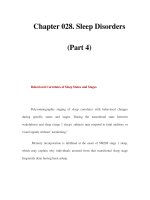
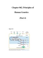
C. Figure I–4 outlines the differential diagnosis for hyponatremia.
It excludes pseudohyponatremia (increased serum lipids) and
hyponatremia with hypertonicity (increased serum glucose).
IV. Database. History and physical exams are of paramount importance
in determining proper course of treatment. Identifying exact causes
of hyponatremia enables clinician to provide safe and appropriate
234 I: ON CALL PROBLEMS
correction of serum sodium concentration. Proper treatment is of
particular importance due to potential neurologic sequelae of abnor-
mal serum sodium. Cerebral edema develops when serum sodium
decreases very rapidly. In hyponatremia associated with severe
intravascular volume depletion, neurologic sequelae may develop as
a function of hypotension, or the development of a cerebral venous
sinus thrombosis.
A. Physical Exam Key Points
1. Vital signs and general appearance. Mental status changes,
weakness, muscular cramps, and hypotension may all be
associated with hyponatremia or decreased intravascular
volume.
2. Fluid status. Assess total body water (including both intracel-
lular and extracellular volume). Assess volume status by check-
ing mucous membranes, presence of tears, capillary refill,
peripheral edema, ascites, jugular venous distention, tachycar-
dia, hypotension, and murmurs.
3. Abdomen. Palpate for masses or organomegaly consistent
with congestive heart failure.
B. Laboratory Data
1. Electrolytes, including BUN, creatinine, and glucose.
2. Serum osmolality as compared with urine osmolality and urine
sodium and creatinine.
Hyponatremia
↑ ECFV↓ ECFV
Urine Na < 20 Urine Na > 20
Cirrhosis
Nephrotic syndrome
Congestive heart
failure
Execssive free water
intake
Acute or
chronic renal
failure
SIADH
Urine Na < 20
Urine Na > 20
Vomiting
Diarrhea
Gastric fistulas
NG drainage
Sweating
Third space
losses
Decreased solute
intake
Diuretics
Salt wasting
Proximal RTA
Adrenal insufficiency
Pseudohypoaldosteronism
Figure I–4. Differential diagnosis of hyponatremia. (↓ = decreased; ↑=increased; ECFV =
extracellular fluid volume; Na = sodium; NG = nasogastric; RTA = renal tubular acidosis;
SIADH = syndrome of inappropriate secretion of antidiuretic hormone.)
50. HYPONATREMIA 235
3. Consider serum pH, determination of anion gap, liver function
tests, thyroid function tests, cortisol levels, and aldosterone
levels.
4. Plasma triglycerides are useful in identifying pseudohypona-
tremia. Serum glucose dilutes serum sodium because it is
hyperosmolar and pulls free water into the intravascular space
(hyponatremia with hypertonicity).
C. Radiographic and Other Studies. Radiographic studies typically
are not helpful unless clinician suspects an underlying malignan-
cy participating in the cause of hyponatremia.
1. Chest x-ray. Helps to rule out heart failure, as well as identify
heart size as a factor in determining volume status.
2. CT scan of head. May help rule out intracranial mass, hemor-
rhage, or sinus thrombosis; however MRI is more sensitive for
most tumor masses.
3. CT scan or ultrasound of abdomen. May be helpful to deter-
mine ascites, portal hypertension, or renal or adrenal masses.
V. Plan
A. Symptomatic Hyponatremia. In patients such as the infant with
seizures described in the opening problem, rapid but modest cor-
rection of serum sodium concentration is of paramount impor-
tance. Seizures that develop as a result of hyponatremia are
difficult to treat unless serum sodium is corrected.
1. Initial goal. Do not attempt to correct to a normal sodium con-
centration (> 135 mEq/L), but rather to raise serum sodium to
a level at which seizures may be controlled (typically > 120
mEq/L). This can be performed by administration of 3% saline.
2. Rule for 3% saline administration. Administration of 1 mL/kg
of 3% saline will raise serum sodium by approximately
1.6 mEq/L.
3. Considerations. Keep in mind that seizures may have devel-
oped due to rapid decrease in serum sodium and cerebral
edema. Once seizure activity is controlled, this therapy should
be held and more definitive treatment initiated. Administration
of 3% saline is not appropriate for asymptomatic hypona-
tremia. Ideal rate of rise of serum sodium should not exceed
1 mEq/h once seizures are controlled. This management
should occur in consultation with a pediatric nephrologist and
intensivist.
B. Asymptomatic Hyponatremia
1. Hypovolemic hyponatremia (decreased ECFV)
a. Estimate total fluid deficit.
b. Use 0.9% normal saline for maintenance fluids plus deficit.
c. Consider ongoing losses when determining fluid rates.
2. Hypervolemic hyponatremia (increased ECFV)
a. Low urine sodium (edematous states)
236 I: ON CALL PROBLEMS
i. Water and sodium restriction (two-thirds maintenance).
ii. Consider loop diuretics.
b. High urine sodium. Water restriction (two-thirds main-
tenance).
VI. Problem Case Diagnosis. The 3-month-old infant had hyponatrem-
ic-induced seizures as a result of gastroenteritis. Urine sodium value
was < 20 mmol/24 h (normal is 40–120 mmol/24 h). CT scan of the
head was normal. Patient showed clinical improvement over the next
2 days once sodium imbalance was gradually corrected.
VI. Teaching Pearl: Question. What neurologic condition is associated
with a rapid increase in serum sodium?
VII. Teaching Pearl: Answer. Central pontine myelinolysis develops in
patients who experience a rapid increase in serum sodium and
hence is a risk factor in treatment of hyponatremia.
REFERENCE
Verbalis JG. Hyponatremia epidemiology, pathophysiology and therapy. Curr Opin
Nephrol Hypertens 1993;2:636.
51. HYPOPHOSPHATEMIA
I. Problem. A 10-year-old boy with cerebral palsy and seizures who
was treated with divalproex sodium is admitted with acute respiratory
illness. His serum phosphate level is 1 mg/dL (normal for this age:
3.7–5.6 mg/dL).
II. Immediate Questions
A. Does patient have any acute symptoms related to hypophos-
phatemia? Patients with moderate to severe hypophosphatemia
(< 1 mg/dL) may have many systemic manifestations and need to
be promptly treated. Symptoms can include cardiomyopathy with
heart failure; muscle weakness that can lead to rhabdomyolysis;
hemolysis; and encephalopathy, seizures, and coma.
B. Are there acute factors that have resulted in severe
hypophosphatemia? Most cases of hypophosphatemia result
from a shift of phosphate from extracellular to intracellular fluid.
Factors causing this shift can be severe and life threatening and
include refeeding syndromes and treatment of diabetic ketoacidosis
(DKA).
C. Pertinent Historical Information. Ask about diet, medications,
underlying conditions, and relevant family history.
III. Differential Diagnosis. Hypophosphatemia usually results from one
of the following processes: shift of phosphate into the intracellular
compartment, renal losses, or GI losses. Changes can be acute,
chronic, or a combination.
51. HYPOPHOSPHATEMIA 237
A. Transcellular Shift From Extracellular to Intracellular
Compartment
1. Nutritional repletion or refeeding syndrome. Can occur with
enteral or parenteral nutrition in patients who are malnourished
or those with anorexia nervosa or AIDS.
2. DKA and insulin therapy. Renal losses are also involved.
3. Respiratory alkalosis. Increased renal losses are also
present.
4. Sepsis. Especially gram-negative and toxic shock syndrome.
5. Leukemia with blast crisis.
B. Increased Urinary Losses
1. Renal tubular defects. Fanconi syndrome (may be primary or
acquired), X-linked hypophosphatemic (XLH) rickets, post–
renal transplantation status.
2. Hyperparathyroidism. Primary (rare in children) or secondary
to vitamin D deficiency or other nonrenal causes.
3. Diuretic phase of acute tubular necrosis (ATN).
4. Postobstructive diuresis.
5. Post–renal transplantation status.
C. Increased GI Losses
1. Use of oral phosphate-binding antacids.
2. Decreased intake. Starvation, anorexia nervosa, protein-
calorie malnutrition. At high risk for refeeding syndrome (see III,
A, 1, earlier). Premature infants require phosphate supple-
mentation.
3. Malabsorption syndromes.
4. Vitamin D deficiency. Low levels of vitamin D due to dietary
deficiency or lack of sunshine, malabsorption, or liver disease.
5. Vitamin D–dependent rickets. Block in 1,25-dihydroxyvita-
min D formation (type 1) or abnormal receptor (type 2).
IV. Database
A. Physical Exam Key Points
1. Vital signs and general appearance
a. Temperature. Severe hyperthermia can cause hypophos-
phatemia through transcellular shift. Fever may be a sign of
sepsis or toxic shock.
b. Respiratory rate. May be a sign of respiratory alkalosis.
c. Body mass. Look for evidence of malnutrition or short
stature, as well as cystinosis or congenital Fanconi
syndrome.
2. Heart. Look for evidence of heart failure as result of severe
depletion.
3. Neuromuscular. Assess for confusion, coma, and muscle
weakness. Muscle tenderness may be a sign of rhabdomy-
olysis.
4. Skeletal. Look for bowing, rachitic rosary, and flared growth
plates at wrists and knees as sign of rickets.
238 I: ON CALL PROBLEMS
5. Skin. Large café au lait spots may point to McCune-Albright
syndrome.
B. Laboratory Data
1. Basic metabolic panel. Low bicarbonate level may point to
acidosis or changes secondary to respiratory alkalosis.
Fanconi syndrome may be associated with low bicarbonate,
low potassium, and possibly elevated creatinine levels. Low
calcium level points to rickets (not XLH) or hungry bone syn-
drome. High calcium level may point to hyperparathyroidism,
but in children most have secondary hyperparathyroidism with
normal or low calcium.
2. ABGs. Evaluate acid-base status; look for respiratory alkalosis
or metabolic acidosis.
3. CBC with differential. Hypophosphatemia may cause hemol-
ysis and thrombocytopeni a. Increased WBC with left shift may
suggest sepsis.
4. Creatinine phosphokinase. Check for rhabdomyolysis if
muscle tenderness is present.
5. Uric acid. Low in patients with volume overload or Fanconi
syndrome.
6. Vitamin D levels. 25-Hydroxyvitamin D is diagnostic of vitamin
D deficiency. No need to routinely check 1,25-dihydroxyvitamin
D levels.
7. Urine studies. Spot urine for phosphorus and creatinine
allows measurement of phosphate excretion to see if etiology
is increased renal loss, as in Fanconi syndrome. Fanconi syn-
drome is characterized by glucosuria, renal tubular acidosis,
aminoaciduria, and excretion of small-molecular-weight pro-
teins such as 
2
-microglobulin.
C. Radiographic and Other Studies. Consider skeletal survey to
look for changes characteristic of rickets, osteomalacia, or hyper-
parathyroidism. Changes may not point to exact etiology; antacid
abuse can lead to osteomalacia.
V. Plan. Mild hypophosphatemia is a common finding in hospitalized
patients, usually due to transcellular shifts of phosphate into intra-
cellular fluid, and requires no specific therapy. Moderate
hypophosphatemia can be treated with oral supplementation, but
severe or symptomatic hypophosphatemia may require careful par-
enteral correction.
A. Moderate Hypophosphatemia (1–2 mg/dL in adolescents;
2–3 mg/dL in infants and young children)
1. Dietary replacement. Milk contains 1 g inorganic phosphate
per liter. Avoid low-phosphate formulas (Similac PM 60/40) or
breast milk as replacement.
2. Enteral supplements. Potassium phosphate can be given as
an oral supplement at a dose of 250–750 mg q6h, depending
on body size. Commonly available supplements are Neutra
51. HYPOPHOSPHATEMIA 239
Phos or K-Phos Neutral, which come as 250-mg tablets, cap-
sules, or packets. Contents can be diluted with 75 mL water or
taken with food. Monitor calcium to avoid hypocalcemia. Watch
for diarrhea. Phosphosoda (Fleet Phosphosoda) can be given
orally or as an enema at a dose of 15–30 mL three to four times
daily.
B. Severe Hypophosphatemia (< 1 mg/dL in adolescents; < 2 mg/dL
in children younger than 12 years)
1. Enteral supplements. As listed under moderate hypophos-
phatemia, earlier; use for asymptomatic hypophosphatemia.
2. Parenteral phosphate. Usually used for symptomatic
hypophosphatemia. Avoid with renal failure. Potassium phos-
phate can be given IV at a dose of 2.5 mg (0.08 mmol)/kg body
weight in
1
/
2
normal saline (NS) over 6 hours or, for sympto-
matic patients, at 5 mg (0.16 mmol)/kg body weight in
1
/
2
NS
over 6 hours. Monitor calcium, phosphate, and potassium
every 6 hours. Monitor BP. Stop parenteral replacement when
serum phosphate is > 2 mg/dL.
C. Treatment of Primary Etiology. After emergency treatment,
recognition and treatment of primary cause is important. May
require vitamin D analogues (see Chapter 46, Hypocalcemia,
p. 221).
VI. Problem Case Diagnosis. The 10-year-old patient has Fanconi syn-
drome, likely due to divalproex sodium administration. In addition to
low serum phosphate level, he had low serum potassium level, meta-
bolic acidosis, glucosuria, and a very large renal leak of phosphorus.
He required large amounts of IV phosphorus and bicarbonate, but
the renal tubular defect improved after stopping divalproex sodium.
VII. Teaching Pearl: Question. Why is the expression of serum phos-
phate as milliequivalents per liter (mEq/L) uniquely confusing as
compared with other ions.
VIII. Teaching Pearl: Answer. Because the average charge of phos-
phate changes at physiologic pH (charge at pH 7.4 is −1.8), the
valency and the value for milliequivalents per liter varies with
changes in serum pH. Expression of phosphate in millimoles per liter
(mmol/L) and milligrams per deciliter (mg/dL) avoids this problem.
REFERENCES
Hruska KA, Lederer ED. Hyperphosphatemia and hypophosphatemia. In Favus M,
ed. Primer on the Metabolic Bone Disorders of Mineral Metabolism, 5th ed.
American Society of Bone and Mineral Research, 2003:286–288.
Rubin MF, Narins RG. Hypophosphatemia: Pathologic and practical aspects of its
therapy. Semin Nephrol 1990;10:536.
Subramanian R, Khardori R. Severe hypophosphatemia: Pathophysiologic implica-
tions, clinical presentations, and treatment. Medicine 2000;79:1.
240 I: ON CALL PROBLEMS
52. HYPOTENSION
I. Problem. A 2-year-old boy who was admitted earlier in the day with
diarrhea and dehydration now has a BP of 64/33.
II. Immediate Questions
A. What are the vital signs? Is patient adequately perfused? Are
airway, breathing, and circulation (ABCs) compromised?
Hypotension represents a medical emergency and requires
immediate assessment and treatment. To determine lowest
acceptable systolic BP for age (represents fifth percentile for age),
use the guidelines in Table I–12. If systolic BP falls below these
ranges, patient is considered to be hypotensive and metabolic
demands of the body for both oxygen and nutrients may not be
met. As treatment is occurring, a simultaneous search for cause
of hypotension should begin.
B. Is patient tachycardic? Sinus tachycardia is the body’s first
modality to maintain adequate cardiac output in the face of hypo-
volemia and suggests intravascular volume depletion.
C. How was BP measured? Be sure that cuff size is appropriate.
Cuff that is too large may give a falsely low BP. Agitation and
movement may alter result and make measurement inaccurate.
D. What has urine output been? Urine output is the best noninva-
sive marker of end-organ perfusion and, in the presence of normal
renal function, provides an accurate reflection of intravascular
volume status. For pediatric patients, urine output should be at
least 1 mL/kg/h.
E. What is patient’s mental status? The body does all it can to
maintain perfusion to heart, brain, and adrenal glands in the face
of hypotension or inadequate cardiac output. If mental status is
not normal, assume that cerebral perfusion has been compro-
mised. This represents an even more urgent medical emergency
and may require immediate attention to airway as fluid resuscita-
tion is occurring.
F. Are invasive monitors in place? If so, measuring central
venous pressure (CVP) or wedge pressure provides an objective
TABLE I–12. LOWEST ACCEPTABLE SYSTOLIC BLOOD
PRESSURE FOR AGE
Age Lowest Acceptable Systolic BP (mm Hg)
Birth–1 mo 60
1 mo–1 y 70
1–10 y 70 + (2 × Age in years)
10 y or older 90