A Lange Medical Book Pediatrics on call - part 8 ppsx
Bạn đang xem bản rút gọn của tài liệu. Xem và tải ngay bản đầy đủ của tài liệu tại đây (783.91 KB, 82 trang )
2. DEFICIT REPLACEMENT 545
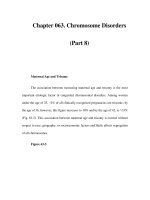
TABLE IV–4. COMPOSITION AND DAILY PRODUCTION OF BODY FLUIDS
Electrolytes (mEq/L)
Average Daily
Fluid Na
+
Cl
−
K
+
HCO
3
−
Production (mL)
Sweat 50 40 5 0 Varies
Saliva 60 15 26 50 1500
Gastric juices 60–100 100 10 0 1500–2500
Duodenum 130 90 5 0–10 300–2000
Bile 145 100 5 15 100–300
Pancreatic juice 140 75 5 115 100–800
Ileum 140 100 2–8 30 100–9000
Diarrhea 120 90 25 45 —
Modified and reproduced with permission from Gomella LG, Haist SA, eds. Clinician’s Pocket
Reference, 10th ed. McGraw-Hill. Copyright 2002.
TABLE IV–5. COMPOSITION OF COMMONLY USED CRYSTALLOID SOLUTIONS
Electrolytes (mEq/L)
Fluid Glucose (g/L) Na
+
Cl
−
K
+
Ca
+
HCO
3
−
kcal/L
D
5
W (5% 50 — — — — — 170
dextrose
in water)
D
10
W (10% 100 — — — — — 340
dextrose
in water)
D
20
W (20% 200 — — — — — 680
dextrose
in water)
D
50
W (50% 500 — — — — — 1700
dextrose
in water)
1
/
2
NS — 77 77 — — — —
(0.45% NaCl)
NS — 154 154 — — — —
(0.9% NS)
3% NS — 513 513 — — — —
D
5
1
/
4
NS 50 38 38 — — — 170
D
5
1
/
2
NS 50 77 77 — — — 170
(0.45% NaCl)
D
5
% NS 50 154 154 — — — 170
(0.9% NaCl)
D
5
LR (5% 50 130 110 4 3 27 180
dextrose
in LR)
LR — 130 110 4 3 27 <10
NS = normal saline; LR = lactated Ringer.
Modified and reproduced with permission from Gomella LG, Haist SA, eds. Clinician’s Pocket
Reference, 10th ed. McGraw-Hill. Copyright 2004.
546 IV: FLUIDS AND ELECTROLYTES
Patients receiving fluid and electrolyte replacement therapy should be
closely monitored. Accurate recording of intake and output, and weight;
monitoring of blood chemistries; and assessment of vital signs and clinical
status are important to prevent over- or underhydration.
REFERENCES
Boineau FG, Lewy JE. Estimation of parenteral fluid requirements. Pediatr Clin North
Am 1990;37:257–264.
Greenbaum LA. Pathophysiology of body fluids and fluid therapy. In: Behrman RE,
Kliegman RM, Jenson HB, eds. Nelson Textbook of Pediatrics, 17th ed. Saunders,
2004:190.
Haist SA, Robbins JB. Internal Medicine On Call, 3rd ed. McGraw-Hill, 2002.
Hill LL. Body composition, normal electrolyte concentrations, and the maintenance of
normal volume, tonicity, and acid-base metabolism. Pediatr Clin North Am
1990;37:241–256.
Jospe N, Forbes G. Fluids and electrolytes—Clinical aspects. Pediatr Rev
1996;17:395–403.
Kallen RJ, Lonergan JM. Fluid resuscitation of acute hypovolemic hypoperfusion
states in pediatrics. Pediatr Clin North Am 1990;37:287–294.
547
V.
Blood Component Therapy
1. BLOOD COMPONENTS AND THEIR USES
IN PEDIATRICS
Many blood products are available in the United States (Table V–1).These
products have never been safer, but they can transmit disease. For this
reason, children should only receive blood products when conservative
measures (eg, crystalloid infusions for acute blood loss) have failed.
Safe Blood Transfusions
In the United States, RBCs, most often received from donors, are carefully
screened to prevent transmission of infectious agents. Platelets are often
derived from apheresis, either stored by the recipient or by a person well
known to him or her. Plasma and other plasma-derived blood factors (clotting
factor concentrates, immune globulins, and protein-containing plasma volume
expanders) are derived from paid donors, with pooled blood fractionated to
remove impurities and infectious agents. Of note, pooled plasma derivatives
are more likely to cause an infection than are whole blood–derived products.
General historical questioning, specific individual questioning, laboratory
screening, and purification techniques maintain blood safety (Table V–2).
Infectious diseases and agents that can be transmitted through blood
products are listed in Table V–3.
Safety can be maintained only with strict adherence to blood product
transfusion pathways. Before injection of any blood product, at least two
people should check the blood bag and patient to be sure that the right
blood product is being administered to the patient.
2. TRANSFUSION REACTIONS
All blood products, especially multidonor plasma and cryoprecipitate, may
result in transfusion reactions. These reactions include urticaria (hives),
fever, nausea, headaches, and pruritus (itching). Rarely, anaphylaxis
occurs. Antihistamines, antipyretics, and epinephrine should be available
at the bedside for any patient receiving a blood product transfusion.
Two significant post-transfusion reactions can occur with blood products,
especially with gamma globulins.
1. Inflammatory reaction. This reaction can occur hours to a day after
transfusion and consists of severe headache and ague (fever and chills),
lethargy, and nausea. Inflammatory reaction is most common in repeated
transfusions and will disappear once transfusion is discontinued.
2. Anaphylactoid reaction. This reaction results from complement
activation and consists of flushing, hypotension, dyspnea, ague,
nausea, and back pain.
Copyright © 2006 by The McGraw-Hill Companies, Inc. Click here for terms of use.
548 V: BLOOD COMPONENT THERAPY
TABLE V–1. BLOOD PRODUCTS AND INDICATIONS FOR TRANSFUSION
Blood Product Type Indications
Red blood cells Whole blood (rarely used) Severe anemia (Hgb
(RBCs) Packed RBCs (whole blood less 70% of usually < 7 g%) or acute,
plasma; most commonly used in US) severe, traumatic blood
or acute, severe, traumatic blood loss) loss)
Leukocyte-poor RBCs (for patients with
history of febrile reactions to blood
products or who will receive many
transfusions)
Washed RBCs (to prevent
host-versus-graft disease in IgA-
deficient recipients and others)
CMV-free RBCs (for potential trans-
plantation patients)
Frozen, stored RBCs (for presurgical
self-transfusion)
Platelets
a
— Potential clotting disorder
due to thrombocytopenia
(platelets < 10,000 or
< 20,000 if surgery
planned) or clinically
significant quantitative
platelet defect
Plasma
b,c
Available products include: Intravascular fluid
Fresh-frozen plasma (FFP); may not depletion, not responsive
supply clotting factors V and VIII to crystalloid, or bleeding
Single-donor plasma; safer than FFP due to depletion of
but otherwise same problems clotting factors
Clotting factor Cryoprecipitate has high levels of VIII, Factor deficiencies or
concentrates von Willebrand factor, and fibrinogen acute liver failure (eg,
Genetically engineered factor VIII con- Wilson disease)
centrates only for factor VIII deficiency
Vitamin K–dependent factor concentrate
has factors II, VII, IX, X, and proteins C
and S; associated with hepatitis and
thrombus formation
Immune globulins Nonspecific gamma globulin and Guillain-Barré, Kawasaki,
intravenous gamma-globulin (IVIG) and other autoimmune
Specific gamma globulins for rabies diseases (eg, ITP)
(RIG), hepatitis (HBIG), varicella Prevention after exposure
(VZIG), and other uses to specific diseases
RhoGAM Prevention of Rh
sensitization and
treatment of ITP
Protein-containing — Intravascular fluid
volume expanders depletion
CMV = cytomegalovirus; Hgb = hemoglobin; ITP = idiopathic thrombocytopenic purpura.
a
In platelet dysfunction, DDAVP (desmopressin acetate) may alleviate clotting disorder without transfusion.
b
Single-donor plasma is safer than multidonor products (cryoprecipitate).
c
If thrombocytopenia is due to autoantibodies or other consumptive problems, transfusions are rarely
effective.
a. If patient develops these symptoms, immediately discontinue
transfusion and administer the following agents.
i. Diphenhydramine (Benadryl), 0.25–1.0 mg/kg per dose PO
or IV q2–6h.
ii. Steroids, 2 mg/kg/dose, to maximum of 60 mg.
iii. Epinephrine, 1:1000 0.01 mL/kg per dose SQ, to maximum of
0.5 mL.
b. Vasopressors. Administer if the preceding agents do not raise
BP to a safe level.
2. TRANSFUSION REACTIONS 549
TABLE V–2. METHODS USED TO MAINTAIN BLOOD SAFETY
Screening Technique Description
General history Interview-style questions:
Has donor ever had blood donation refused?
Current or chronic illnesses?
Presence of fever?
Individual history Interview-style questions:
Any high-risk sexual behaviors in donor or donor’s partner(s)?
Any injected drug use in donor or donor’s partner(s)?
Any overseas travel or history of past infection with HIV, HBV,
HCV, or parasites?
Laboratory screening Detection of HIV-1 and -2, HBV, HCV, HTLV-1 and -2, syphilis
Purification techniques Heat, fractionation, or chemical treatment consistent with
maintaining activity of agent
HBV = hepatitis B virus; HCV = hepatitis C virus; HIV = human immunodeficiency virus; HTLV = human
T-lymphotropic virus.
TABLE V–3. INFECTIOUS AGENTS THAT CAN BE TRANSMITTED THROUGH BLOOD
PRODUCTS
Category Agents and Diseases
Bacteria Staphylococcus (all types), Streptococcus (all types), occasional
gram-negative organisms
Parasites Malaria, Chagas disease
Prions Jakob-Creutzfeldt disease, mad cow disease
Tick-borne agents Babesia, Rickettsia, Borrelia, Ehrlichia
Viruses Tested agents: HIV-1 and -2, HBV, HCV, HTLV-1 and -2
Not currently tested: CMV, parvovirus B19, HAV, HGV, transfusion-
transmitted virus, SEN virus, human herpesvirus-8, West Nile virus
CMV = cytomegalovirus; HAV = hepatitis A virus; HBV = hepatitis B virus; HCV = hepatitis C virus;
HGV = hepatitis G virus; HIV = human immunodeficiency virus; HTLV = human T-lymphotrophic virus.
550 V: BLOOD COMPONENT THERAPY
REFERENCES
Ambruso DR, Hays T, Lane PL, Nuss R. Hematologic disorders. In: Hay WW Jr, Levin
MJ, Sondheimer JM, Deterding RR, eds. Current Pediatric Diagnosis & Treatment,
17th ed. McGraw-Hill, 2005:855–910.
Pickering LK, ed. Red Book 2003 Report of the Committee on Infectious Diseases,
26th ed. American Academy of Pediatrics, 2003.
Strauss RG. Risk of blood component transfusions. In: Behrman RE, Kliegman RM,
Jenson HB, eds. Nelson’s Pediatrics, 17th ed. Saunders, 2003:1646–1650.
Truman JT. Complications of blood transfusions. In: Burg FD, Ingelfinger JR, Poplin
RA, Gershon AA, eds. Gellis & Kagan’s Current Pediatric Therapy, 17th ed.
Saunders, 2002:675–676.
551
VI.
Ventilator Management
1. INDICATIONS FOR VENTILATORY SUPPORT
Respiratory failure can be divided into two categories: hypoxemic (type
I) respiratory failure and hypoventilatory (type II) respiratory failure.
Although hypoxemic respiratory failure is more common, both varieties of res-
piratory failure are seen in pediatric patients. Ventilatory support is
indicated when adequate gas exchange cannot be independently
achieved or maintained.
I. Hypoxemic Respiratory Failure. Inability to oxygenate is an impor-
tant indication for ventilatory support. Oxygenation can be determined
by measurement of pulse oximetry (SpO
2
) or the partial pressure of
oxygen in arterial blood (PaO
2
). By evaluating PaO
2
in the context of
the fraction of inspired oxygen (FiO
2
) employed, objective criteria for
hypoxemic respiratory failure can be established. A PaO
2
/FiO
2
(P/F)
ratio < 200 is consistent with acute respiratory distress syndrome
(ARDS), whereas a ratio between 200 and 300 is consistent with
acute lung injury. Patients with a P/F ratio < 300 or SpO
2
< 90–93% (in
the absence of cyanotic heart disease) require additional support,
especially if they demonstrate signs of inadequate oxygen delivery,
such as tachycardia, metabolic acidosis, or end-organ dysfunction.
Although these patients may be managed initially with high oxygen
delivery systems, their disease may progress to a point at which ven-
tilatory support is required. Because of their physiologic instability and
potential need for advanced therapies, these patients should be closely
monitored in a pediatric intensive care unit.
II. Hypoventilatory Respiratory Failure. Carbon dioxide clearance is
the main function of ventilation. The adequacy of ventilation can be
monitored by either end-tidal carbon dioxide (ETCO
2
) measurement
or measurement of the partial pressure of carbon dioxide (PaCO
2
) in
arterial blood. Although a PaCO
2
above the normal range for age is
consistent with hypoventilatory respiratory failure, it must be consid-
ered in the context of the clinical situation. A patient with status asth-
maticus may have maximized his or her minute ventilation and have
a PaCO
2
rise into the normal range as a result of hypoventilatory res-
piratory failure. Conversely, a patient with bronchopulmonary dyspla-
sia may have developed a metabolic compensation such that the
arterial pH is in the normal range despite chronic ventilatory failure.
Evaluation of physical exam findings, pH, and PaCO
2
are all required
to determine the need for ventilatory assistance. Respiratory acidosis
with rapidly falling pH or pH < 7.25, rapidly rising PaCO
2
, and deterio-
rating mental status secondary to CO
2
“narcosis” are all indications
for ventilatory support. The factors involved in determining the need
for ventilatory support can be evaluated in the following manner.
Copyright © 2006 by The McGraw-Hill Companies, Inc. Click here for terms of use.
552 VI: VENTILATOR MANAGEMENT
A. Respiratory Drive. Does patient have the drive to breathe?
Breathing control issues are not uncommon in pediatric patients.
Premature infants have immature respiratory drive centers that
place them at risk for central apnea. This risk is heightened by
intercurrent electrolyte imbalances, hypothermia, and infections.
The immature respiratory center is also more sensitive to the res-
piratory depressant effects of anesthetics, narcotics, and seda-
tives until 48–52 weeks’ gestational age. Older children are also at
risk for respiratory drive dysregulation secondary to metabolic
derangements, central sleep apnea, intoxication, primary CNS
infection or disease, or traumatic brain injury.
B. Respiratory Muscle Strength. Does patient have the strength to
breathe? Anatomic differences in infants and small children result
in a greater breathing workload. Airway resistance is higher than
in older children and adults due to smaller airway caliber, greater
chest wall compliance, relative weakness of the intercostal mus-
cles, and greater fatigability of the diaphragm. The intercurrent
disease state may accentuate these anatomic and physiologic
conditions, tipping the balance of the strength/workload relationship.
The combination of insufficient respiratory drive and inadequate
muscle strength for workload results in failure of the so-called respira-
tory pump, leading to type II (hypoventilatory) respiratory failure.
C. Extrathoracic Airway. Is obstruction present? The tissues of
the extrathoracic airway may be subject to infection or inflam-
mation, intrinsic masses or compression from extrinsic masses,
or malacia. In addition to fixed anatomic obstruction, there may be
functional obstruction as a result of obstructive sleep apnea or
pharyngeal hypotonia that is exacerbated by sedative and anal-
gesic medications.
D. Intrathoracic Airways and Gas-Exchanging Units. Is there
dysfunction at this level? The smaller size of the intrathoracic air-
ways and alveoli, absence of collateral alveolar ventilation, and
similarity between alveolar closing capacity and functional residual
capacity in infants and small children increases the likelihood of
ventilation-perfusion (V/Q) imbalance secondary to atelectasis.
Primary lung injury (eg, from infection or traumatic injury) or sec-
ondary lung injury due to the release of inflammatory cytokines
may also lead to respiratory failure.
The combination of extrathoracic airway obstruction and dysfunction
of the intrathoracic airways and gas-exchanging units results in the
failure of the lung, leading to type I (hypoxemic) respiratory failure.
E. Other Concerns. Is there a concurrent medical condition for
which intubation and ventilation would be beneficial? The practi-
tioner may want to maintain airway and ventilatory control in a
patient whose condition does not directly affect ventilatory ade-
quacy, or provide specific therapies that are best facilitated while
patient is intubated and ventilated.
2. VENTILATION OPTIONS AND CLASSIFICATION
I. Ventilation Options
A. Negative Pressure Ventilation
1. Description. Negative pressure ventilation is performed by
placing patient into a chamber or body suit device, within which
negative pressure can be generated. Creation of negative pres-
sure around the thorax results in a pressure gradient that favors
gas flow from the atmosphere, through the natural airway, and
into the lungs. Exhalation occurs when negative pressure is dis-
continued and the natural elastic recoil of the pulmonary system
promotes lung emptying. Negative pressures up to −30 cm H
2
O
may be used, cycled at varying rates and inspiratory times as
needed to optimize gas exchange. Supplemental oxygen may
be introduced to patient’s natural airway.
2. Advantages. The advantage to negative pressure ventilation
is that it avoids tracheal intubation and may allow patients to be
free from ventilatory support for intermittent periods, as tolerated.
For patients with “passive” pulmonary circulation, negative
pressure ventilation also augments pulmonary blood flow.
3. Disadvantages. The disadvantages of negative pressure ventilation
include the relative inaccessibility to patient and limitations in
patient positioning while in the negative pressure device, the rela-
tive inefficiency of ventilation compared with positive pressure
techniques, the absence of an artifical airway should the natural
airway become obstructed or secretion clearance become sub-
optimal, and the risk of skin breakdown around seal points.These
problems and the use of other ventilatory modalities have made
negative pressure ventilation relatively uncommon.
B. Positive Pressure Ventilation by Mask
1. Description. Both continuous positive airway pressure and
bilevel positive airway pressure may be delivered by a mask
device. Mask devices may cover either the nose or both nose
and mouth. Nasal masks may be more comfortable and provide
more ready access to the oropharynx for suctioning but may be
less efficient secondary to air leak from the mouth than a full
face mask device. A properly fitting mask with minimal leakage
is essential to success. Mask ventilation is generally well toler-
ated, although reassurance and the occasional judicious use
of sedation may be necessary.
a. Continuous positive airway pressure (CPAP). CPAP may
recruit alveoli, restore functional residual capacity, and
diminish pulmonary edema, improving oxygenation and pul-
monary mechanics. Airway pressures of 3–12 cm H
2
O are
commonly tolerated, but achieving pressures above these
levels may be difficult with a mask system. (See also later
discussion, pp. 558, 563.)
2. VENTILATION OPTIONS AND CLASSIFICATION 553
554 VI: VENTILATOR MANAGEMENT
b. Bilevel positive airway pressure (BiPAP). BiPAP may pro-
vide significant ventilatory support during both acute and
chronic respiratory failure, although not as efficiently as pos-
itive pressure ventilation via a tracheal tube. BiPAP may be
delivered by three different modes: a spontaneous mode, in
which each patient-initiated breath is supported; a timed
mode, in which a predetermined number of breaths is deliv-
ered per minute independent of patient effort; and a timed-
spontaneous mode, which combines the attributes of these
two modes. Settings to be manipulated include inspiratory
positive airway pressure (IPAP), expiratory positive airway
pressure (EPAP), and inspiratory time and mechanical
breath rate (when timed or timed-spontaneous modes are
employed). Supplemental oxygen can be added into the
system as needed. When initiated, low IPAP and EPAP
levels are used to allow patient acclimation to the device,
and subsequently increased as tolerated. An initial
IPAP/EPAP setting of 6/3 cm H
2
O may be used, and this can
be increased to as high as 20–30/10–12 cm H
2
O. Pressures
higher than this may be hard to maintain and suggest that
another ventilatory modality may be needed.
2. Advantages. As with negative pressure ventilation, the advan-
tages of mask ventilation include avoidance of tracheal intuba-
tion and the opportunity to allow patients to be free of
ventilatory support for periods of time as tolerated.
3. Disadvantages. Potential complications of mask ventilation
include gastric distention and aspiration, although this has not
been reported in pediatric case series; the relative contraindi-
cation to oral or nasogastric tube feedings during therapy; and
the risk of skin breakdown due to pressure from the mask. The
lack of airway protection with mask ventilation also limits its use
in patients with diminished or absent airway protective reflexes.
C. Positive Pressure Ventilation via Tracheal Tube. Mechanical
ventilatory support via tracheal tube is the most efficient and most
common ventilation method employed. Tracheal tubes include
translaryngeal tubes placed via nose or mouth and tubes placed
via tracheostomy. A wide variety of modes for providing positive
pressure ventilation through a tracheal tube are available.
II. Ventilator Classification. Ventilators can be classified in various
ways based on their mechanical characteristics and the means by
which they deliver gas to patients. The most common variables used
to classify ventilators are initiation, mode, and cycle control.
A. Initiation. The inspiratory cycle may be initiated by time (ie, a
breath is initiated after a certain time period has elapsed); by pres-
sure (ie, a breath is initiated after a certain amount of negative
pressure is generated by patient’s spontaneous respiratory
effort); or by flow (ie, a breath is initiated after a certain amount of
gas flow is generated by patient’s spontaneous respiratory effort).
These initiating factors may also be used together.
B. Mode. The amount of gas delivered during inspiration is deter-
mined by the mode. The two modes of ventilation are pressure
control, in which gas is delivered to a predetermined amount of
pressure, and volume control, in which gas is delivered to a pre-
determined amount of volume.
C. Cycle Control. Cycle control refers to the parameter that termi-
nates inspiration. The four methods of cycle control are time
cycled, in which inspiration continues for a predetermined amount
of time; volume cycled, in which inspiration continues until a pre-
determined volume of gas is delivered; flow cycled, in which inspi-
ration continues until a certain gas flow is achieved; and pressure
cycled, in which inspiration continues until a predetermined pressure
is achieved.
D. Variations. Ventilator classifications can include such variations
as time-initiated, pressure-control, flow-cycled ventilation; pres-
sure-initiated, volume-control, time-cycled ventilation; and so
forth. In pediatric patients, initiation may be time, pressure, or flow
dependent; pressure and volume control modes are both used;
and time and flow cycle control are most commonly employed.
3. VENTILATOR SETUP
I. The Ventilator. Today’s ventilators provide a variety of ventilatory
modality options to practitioners at the bedside. Despite this, some
basic concepts apply to any mode chosen. In general, a mode of ven-
tilation that will achieve the practitioner’s goals for oxygenation and
ventilation with the least amount of toxicity should be chosen. High
concentrations of supplemental oxygen can directly injure the lung
and the immature retina. A focus on strategies to reduce the FiO
2
to <
50–60% in a timely manner is warranted. Exposure of the lung to
excessive inflating pressures and volumes may directly injure the lung
and initiate an inflammatory response that may result in secondary
lung injury. Recent research suggests that limiting tidal volumes to
5–8 mL/kg may be protective against these effects in patients with
acute respiratory distress syndrome (ARDS).
II. Ventilatory Modes. Keeping in mind that a variety of choices for ven-
tilatory support exist, all involve one of two modes or limits: volume or
pressure. The amount of gas delivered during inspiration will be
determined either by the amount of pressure or by the amount of
volume preset by the practitioner.Volume control modes of ventilation
offer the advantage of consistently applied minute ventilation despite
changes in compliance of the respiratory system. The potential
danger of this approach is that excessive peak inspiratory pressures
may be achieved should compliance decrease, leading to the risk of
3. VENTILATOR SETUP 555
556 VI: VENTILATOR MANAGEMENT
barotrauma. Pressure control modes of ventilation allow for control of
the peak inspiratory pressure, but minute ventilation may vary as
compliance changes occur. With this in mind, volume or pressure
control (preset) ventilation can be performed in a variety of ways,
depending on patient needs and practitioner preferences.
A. Control Mode Ventilation. This mode of ventilation is time initiated;
volume or pressure controlled; and volume, pressure, or time
cycled. It is insensitive to patient effort or response. As a result, no
support to spontaneous respiratory efforts is provided. Control
mode ventilation is appropriate in the operating room or for
patients in the pediatric intensive care unit who are sedated and
neuromuscularly blocked, and is infrequently used.
B. Assist-Control Ventilation. This mode is pressure, time, or flow
initiated; volume or pressure controlled; and volume, pressure,
time, or flow cycled. The ventilator senses a sub-baseline pres-
sure or flow when patient makes an adequate respiratory effort,
initiating a mechanical breath. The sub-baseline pressure or flow
needed to trigger a ventilator breath is determined by the sensi-
tivity that is set on the ventilator. A control rate is initiated when
patient effort is inadequate or absent, providing safety should the
patient become hypopneic or apneic. Thus, assist-control ventila-
tion allows spontaneous breathing with the safety of a backup
mechanical ventilatory rate. All breaths are fully supported by
preset ventilator parameters. As a result, the preset volume or
pressure must be decreased during the process of weaning from
mechanical ventilatory support when this mode is used.
C. Intermittent Mandatory Ventilation. This mode is time initiated;
volume or pressure controlled; and volume, pressure, time or flow
cycled. A continuous gas flow system is also present on most ven-
tilators that provide this mode of ventilation. Positive pressure
breaths are delivered independent of patient effort. The continu-
ous gas flow system allows breathing of a fresh gas source during
spontaneous respiratory effort by the patient. As a result, inter-
mittent mandatory ventilation allows for spontaneous breathing of
fresh gas with the safety of a backup rate, but the lack of coordi-
nation between mechanical breaths and patient efforts may result
in ventilator-patient dyssynchrony and is not generally well toler-
ated. As a result, this method of ventilation is not commonly used.
D. Synchronized Intermittent Mandatory Ventilation. This mode
is pressure, time, or flow initiated; volume or pressure controlled;
and volume, pressure, time, or flow cycled. Mechanical ventilatory
breaths up to the number prescribed by the practitioner are syn-
chronized with patient respiratory effort as sensed by patient’s
development of a negative inspiratory pressure or flow that
exceeds the limit set by the ventilator’s sensitivity setting. As a
result, full ventilatory support can be provided by setting the ventila-
tor to provide enough breaths to deliver adequate minute ventilation.
Furthermore, partial support can be provided by decreasing the
preset mechanical breath rate, allowing patient to contribute to
minute ventilation by spontaneous respirations. Patient-ventilator
synchrony occurs, making this method of ventilation more com-
fortable for patient. A demand flow system provides a fresh gas
source to patient during spontaneous respiration if patient gener-
ates a sufficient negative inspiratory force to open the demand
valve. The volume of gas provided is proportional to patient effort
and is usually small unless it is augmented by the addition of pres-
sure support. This is in contrast to assist-control ventilation, in
which all breaths receive the full amount of preset support. As a
result, overventilation and development of respiratory alkalosis
are less likely with synchronized intermittent mandatory ventila-
tion as compared with assist-control ventilation. Also, with syn-
chronized intermittent mandatory ventilation, a mechanical breath
can be maintained at full tidal volume or inspiratory pressure
during the process of ventilator weaning, decreasing the potential
for development of atelectasis that may occur with assist-control
ventilation (for which the tidal volume or inspiratory pressure must
be decreased during the weaning process). For both synchro-
nized intermittent mandatory ventilation and assist-control venti-
lation, patient’s work of breathing is determined by trigger
sensitivity, response time of the ventilator, and inspiratory flow
rate of the gas provided.
E. Pressure Support Ventilation. This mode of ventilation is pres-
sure or flow initiated; pressure controlled; and flow or time cycled.
As patient makes a spontaneous respiratory effort, a sub-baseline
pressure or flow is detected by the ventilator, activating ventilator
gas flow to achieve a preset inspiratory airway pressure. This
inspiratory pressure is maintained until either the inspiratory flow
generated by patient decreases to a preset level below the maxi-
mum flow rate (commonly 75–90%) or the time cycle is complete.
The amount of volume delivered depends on the preset level of
pressure support, patient effort, and compliance of the pulmonary
system. Pressure support ventilation can provide full ventilatory
support when a high enough preset pressure level is selected,
partial support, or only enough support to overcome the resist-
ance to gas flow imposed by the endotracheal tube (ETT), venti-
lator tubing, and ventilator demand valves. It may also be
combined with other ventilatory support modes (eg, synchronized
intermittent mandatory ventilation or continuous positive airway
pressure [CPAP]). Pressure support ventilation usually involves
minimal work of breathing and is comfortable for patients.
Because there is no control element to this form of ventilatory
support, care must be taken when using it in patients who may
become hypopneic or apneic.
3. VENTILATOR SETUP 557
558 VI: VENTILATOR MANAGEMENT
F. CPAP. In this mode, continuous positive airway pressure is main-
tained in spontaneously breathing patients. No mechanical
breaths are provided. CPAP may be used independently in
patients who only require distending pressure to maintain func-
tional residual capacity or for stenting of airways when malacia is
present. It may also be used during spontaneous breathing trials
as part of the process of weaning a patient from mechanical ven-
tilatory support. CPAP may be combined with pressure support
ventilation during weaning trials.
III. Ventilator Settings
A. Primary Controls. Initial ventilator settings should be determined
based on patient pathophysiology and goals of the bedside clinician.
The primary controls that need to be set initially include the mode
of ventilation (as previously discussed), tidal volume (when a
volume mode is used) or peak inspiratory pressure (when a pres-
sure mode is used), positive end-expiratory pressure, mechanical
breath rate, inspiratory time and resultant inspiratory-to-expiratory
(I:E) ratio, and FiO
2
.
B. Tidal Volume (V
T
). V
T
is chosen based on body weight.
Commonly a V
T
of 7–10 mL/kg is chosen. Recent studies in adult
patients with ARDS suggest that a low V
T
strategy of ventilation,
whereby 4–8 mL/kg V
T
is employed, is associated with less mor-
bidity and mortality in patients with this condition. This strategy is
thought to limit stretch injury to diseased alveoli and reduce sec-
ondary lung injury. An adequate V
T
should result in good chest rise
and air entry bilaterally and generate positive inspiratory pressure
< 30–35 cm H
2
O.
C. Peak Inspiratory Pressure (PIP). PIP is chosen to provide effective
support at the minimal pressure possible. In children with normal
lung compliance (eg, postsurgical procedure), PIP of 20–25 cm
H
2
O is often adequate. In small or premature infants, a lower PIP
can often be used. In patients with disease processes that worsen
compliance or require high minute ventilation, a higher PIP is nec-
essary. PIPs > 30–35 cm H
2
O are avoided, if possible, because
the risk of barotrauma increases above these levels.
D. Positive End-Expiratory Pressure (PEEP). PEEP provides
CPAP throughout the expiratory phase. For patients breathing
through their natural airway, glottic closure at the end of expiration
will generate PEEP of 3–5 cm H
2
O. The passage of a tracheal
tube through the glottis prevents this process from occurring.
Therefore, a “physiologic” amount of PEEP is commonly used for
all intubated patients. PEEP helps to maintain expiratory airway
patency, restoring functional residual capacity and decreasing
closing volume. Atelectasis may be prevented and alveoli that
have already become collapsed may be recruited. As a result,
total respiratory system compliance decreases and the distribution
of pulmonary blood flow to better ventilated lung units is improved.
The major effect seen with use of PEEP is an improvement in oxy-
genation. Excessive amounts of PEEP, however, will worsen oxy-
genation as alveolar capillaries are collapsed. Thus, it may be
necessary to provide a trial using varying levels of PEEP to deter-
mine the “best PEEP” for patient’s pathophysiology. Other poten-
tial adverse effects of PEEP include a decrease in cardiac output
secondary to decreased pulmonary venous return (preload) and
increased pulmonary vascular resistance, a decrease in cerebral
perfusion pressure secondary to decreased cardiac output and
decreased cerebral venous drainage, alveolar overdistention and
resultant air-leak phenomenon, and potential fluid retention and a
decrease in urine output related to a complex interaction of neu-
rohumoral and cardiovascular responses.
E. Mechanical Breath Rate. The mechanical rate of ventilation is
usually selected based on the physiologic or age-appropriate rate
for the patient with normal minute ventilation requirements. If the
underlying disease requires increased minute ventilation, the ven-
tilator rate may be increased as needed. However, consideration
must be given to the effect of rate on inspiratory and expiratory times.
As rates are increased, less cycle time is available for expiration. If
expiratory time is decreased excessively, patient’s expiration may
not be allowed to complete, leading to decreased minute ventila-
tion and air trapping with resultant worsening of the ventilation-
perfusion (V/Q) relationship. Conversely, patients with conditions
that decrease gas flow on expiration may require a decrease in
the mechanical ventilator rate to allow enough expiratory time for
completion of expiration.
F. Inspiratory Time. When the mode of ventilation includes a time
cycle, the inspiratory time is set directly or by setting an I:E ratio.
Inspiratory times and I:E ratio are commonly set to physiologic
and age-appropriate levels in patients with normal or minimally
altered pulmonary physiology. In patients with impaired oxygena-
tion, increasing the inspiratory time will favor additional alveolar
recruitment. In patients with ARDS, a strategy of so-called inverse
I:E ratio ventilation, in which inspiratory time exceeds expiratory
time, may be employed in an attempt to optimize alveolar recruit-
ment. The impact on ventilation due to a decreased expiratory
time and hemodynamic complications related to an increase in
intrathoracic pressure must be considered. Conversely, patients
with airway obstruction (eg, those with status asthmaticus) may
benefit from a decreased inspiratory time to increase the time
available for expiration.
G. FiO
2
. Upon initiation of mechanical ventilatory support, it is
common to begin with an FiO
2
setting of 100% until patient is sta-
bilized. Subsequently, there should be rapid efforts to decrease
FiO
2
based on SpO
2
or ABG measurements. The lowest FiO
2
that
3. VENTILATOR SETUP 559
560 VI: VENTILATOR MANAGEMENT
achieves adequate oxygenation should be employed. Generally,
FiO
2
of 60% is considered nontoxic, although this will be influ-
enced by amount and duration of exposure, atmospheric pres-
sure, underlying disease state, and individual variation.
IV. Further Considerations
A. Sedation and Analgesia. Provide sedation and analgesia ade-
quate to manage the stress and discomfort associated with an in
situ ETT, mechanical ventilation, and underlying disease process.
These pharmacologic adjuncts will also blunt the patient’s ability
to displace the ETT.
B. Safety. Soft physical restraint of the hands may be required in addi-
tion to pharmacologic measures for patient safety. Uncontrolled dis-
lodgement of the ETT can have disastrous consequences.
C. Gastric Care
1. Perform gastric decompression with a nasogastric or orogastric
tube in all intubated patients unless there are contraindications.
2. Consider gastric buffering for prevention of gastric ulceration,
using nasogastric feedings or pharmacologic agents, when the
course of ventilation is prolonged or the underlying disease
state or treatments increase the risk of ulcer disease.
D. Pulmonary Care. Apply pulmonary toilet. The presence of an
ETT and the use of sedative medications will blunt the normal
bronchociliary mechanisms and cough reflexes for secretion
clearance. Additional therapies may be needed to address the
underlying pulmonary pathophysiology.
E. Skin Care. Meticulous skin care to prevent skin breakdown is
essential.
F. Nutrition. Provide adequate nutritional support in a timely
manner to meet the needs of the growing child recovering from an
acute illness. Enteral feedings are always preferred when possible.
G. Electrolyte and Acid-Base Balance. Normalization of elec-
trolyte and acid-base imbalances is needed to optimize patient
strength and respiratory drive prior to attempts to decrease
mechanical ventilatory support.
4. MODIFICATION OF VENTILATOR SETTINGS
After initiation of ventilatory support, ongoing attention is needed to opti-
mize ventilatory strategy as the patient’s pathophysiologic condition
evolves. Multiple sources of data are available for analysis of patient condition;
however, the cornerstone of this process remains the physical exam.
Evaluation of adequacy of chest rise, quality of breath sounds, patient’s
color and perfusion, respiratory rate and work of breathing, and quality of
other end-organ functions will tell volumes about the adequacy of mechan-
ical ventilatory support being provided. Additional information is provided
by continuous pulse oximeter (SpO
2
) and end-tidal carbon dioxide (ETCO
2
)
measurements, intermittent ABG analysis and chest x-ray interpretation,
and review of mechanical ventilator parameters, such as peak inspiratory
pressure (PIP), exhaled tidal volume (V
T
), and spontaneous minute venti-
lation. Above all, remember to treat the patient and not the ventilator.
I. Adjustments to Oxygenation. Generally, in patients without cyan-
otic heart disease, SpO
2
≥ 93% or PaO
2
> 60 torr is acceptable.
A. To Decrease PaO
2
1. FiO
2
can most easily be decreased based on SpO
2
measure-
ment. Once FiO
2
is ≤ 60%, further decreases may be achieved in
5–10% increments. In patients with normal ventilation-perfusion
(V/Q) relationships, PaO
2
will decrease by 7 mm Hg for each
1% decrease in FiO
2
.
2. If positive end-expiratory pressure (PEEP) is above so-called
physiologic levels (3–5 cm H
2
O) and FiO
2
has been reduced to
≤ 40%, then decreases in PEEP in increments of 1–2 cm H
2
O
may be performed until physiologic levels are reached.
B. To Increase PaO
2
1. FiO
2
may be increased in response to a low PaO
2
or SpO
2
;how-
ever, it is preferable to maintain FiO
2
at ≤ 60%. Should this not
resolve the problem, other strategies should be pursued.
2. PEEP can be added in increments of 2–4 cm H
2
O to improve
oxygenation by improved alveolar recruitment, functional resid-
ual capacity, and V/Q matching. The effects of increased PEEP
are not immediate and may not be apparent for 1 hour or more.
If PEEP levels of 12–15 cm H
2
O are not effective, other strate-
gies should be pursued.
3. Increasing the inspiratory time and performing inverse inspiratory-
to-expiratory (I:E) ratio ventilation will have an impact on oxy-
genation by increasing mean airway pressure and the time
over which alveoli are distended. It can be used in conjunction
with other measures.
4. Increasing ventilation will have some impact on oxygenation
(as shown by the alveolar gas equation at the end of this section),
although this is commonly minimal.
II. Adjustments to Ventilation. Maintaining PaCO
2
or ETCO
2
of 35–45
torr with a normal pH is commonly the goal of ventilation. However, if
excessive amounts of ventilation are required to achieve this, so-
called permissive hypercapnia ventilation may be performed, in which
CO
2
levels are allowed to increase as long as pH can be maintained
at ≥ 7.20–7.25 by metabolic compensation or the use of exogenous
buffer. This allows mechanical ventilatory support to be decreased to
potentially less toxic levels.
A. To Decrease PaCO
2
1. Increase mechanical breath rate.
2. Increase V
T
or PIP.
3. Check for system leaks.
4. Optimize patient-ventilator synchrony.
4. MODIFICATION OF VENTILATOR SETTINGS 561
562 VI: VENTILATOR MANAGEMENT
B. To Increase PaCO
2
1. Decrease mechanical breath rate.
2. Decrease V
T
or PIP.
3. If an assist-control mode of ventilation is being used, consider
changing to synchronized intermittent mandatory ventilation.
Recall that with assist-control, all breaths receive full mechan-
ical ventilatory support.
4. Determine whether patient’s spontaneous effort is producing
the hyperventilation, and determine the cause. Possible
causes include hypoxemia, pain, anxiety, fever, metabolic aci-
dosis, and CNS injury. Treat the underlying problem.
5. Rule out mechanical problems that may be increasing the
mechanical ventilatory rate (eg, autocycling related to a low
trigger sensitivity).
III. Weaning
A. Criteria. Decreasing the amount of mechanical ventilatory sup-
port applied to a patient in preparation for discontinuation of ven-
tilation is termed weaning. An aggressive approach to weaning is
commonly warranted, because an unnecessary extension of the
period of time during which mechanical ventilatory support is
applied increases the chances for associated morbidities. Several
criteria should be considered as ventilator weaning is entertained.
1. The original indication for the application of mechanical venti-
latory support should be resolving or no longer existent.
2. There should be no new indication for mechanical ventilatory
support.
3. Other organ systems should be functioning adequately.
Hemodynamics should be acceptable. Neurologic function
should be such that an appropriate drive to breathe and airway
protective reflexes are present when extubation is being con-
sidered. The amount of tracheal secretions and the frequency
of suctioning should be considered.
4. Oxygenation, ventilation, and acid-base balance should be
adequate.
5. Patient should have the strength to breathe and not have
excessive work of breathing. Patient’s physical exam should be
notable for absence of excessive tachypnea, retractions, nasal
flaring, or accessory respiratory muscle use while maintaining
adequate oxygenation and ventilation. Vital capacity (VC) and
maximum negative inspiratory force (NIF) measurements
can be performed at the bedside. VC of > 10–15 mL/kg and
NIF >−20 cm H
2
O generally correlate with adequate strength
for spontaneous breathing.
6. Prior to extubation, patency of the extrathoracic airway should
be considered.The presence of a leak with deflation of the ETT
cuff, or with inspiratory pressures of < 30 cm H
2
O with an
uncuffed ETT, suggests adequate airway patency.
B. Weaning Techniques
1. Synchronized Intermittent Mandatory Ventilation (SIMV)
a. Description. In this mode of ventilation, the number of fully
supported breaths provided by the ventilator is determined
by the set rate, while patient may breathe spontaneously
from a fresh gas source. In addition, pressure support is
often added to decrease the work of spontaneous breathing
secondary to the resistance of the demand valves and circuit.
Weaning is performed by decreasing the set mechanical
breath rate, thus allowing patient’s spontaneous effort to
provide an increasing amount of the minute ventilation. If
patient does well at a minimum set rate, extubation may pro-
ceed, or a brief testing period using continuous positive
airway pressure (CPAP) or a T-piece may be used.
b. Considerations. The advantages to this approach include
the presence of a backup rate and alarms should patient
become apneic, a graded assumption of the work of breath-
ing, and complete inspiration with set breaths to help
decrease the chance of atelectasis development.
2. Pressure Support Ventilation (PSV)
a. Description. This mode of ventilation also allows the appli-
cation of a variable amount of support to patient’s sponta-
neous breathing effort. Weaning is performed by decreasing
the level of pressure support from one that may provide full
or partial ventilation to one that only overcomes the resist-
ance of the ventilator and endotracheal tube (ETT). It may
also be used in combination with SIMV or CPAP.
b. Considerations. The advantage of this approach is that the
level of mechanical ventilatory support can be gradually
decreased as patient improves, allowing patient to set his or
her own rate, inspiratory time, and expiratory time. The dis-
advantage of this approach is the lack of a backup rate
should patient become fatigued or apneic.
3. CPAP
a. Description. CPAP allows for spontaneous breathing with
the application of a continuous distending pressure. It may
also be combined with PSV. As patient improves, he or she
is weaned by switching from an assisted or controlled mode
of ventilation to CPAP for trial periods of spontaneous venti-
lation with no mechanical ventilatory support.
b. Considerations. Prolonged periods of CPAP are not commonly
used because patient may become fatigued secondary to resist-
ance created by the ETT and ventilator circuit. The absence of a
safety backup rate must also be considered.
4. T-Piece
a. Description. Patient may be removed completely from ven-
tilatory support and allowed to breathe spontaneously
4. MODIFICATION OF VENTILATOR SETTINGS 563
564 VI: VENTILATOR MANAGEMENT
through the ETT, which is connected to a constant flow of
fresh gas.The weaning process involves increasing the time
patient uses the T-piece.
b. Considerations. This method is not generally used in pedi-
atric patients because of the high resistance to gas flow
through smaller pediatric ETTs and the associated exces-
sive work of breathing necessary for spontaneously ventila-
tion. Also, no ventilator alarms are available, creating a
safety issue.
5. SPECIAL MODES OF VENTILATION
I. Inverse Ratio Ventilation. In patients with inadequate oxygenation,
increasing the inspiratory time so that it equals or exceeds the expira-
tory time will increase the mean airway pressure without increasing
peak inspiratory pressures (PIPs) and will increase the time over
which noncompliant alveoli may be recruited (see Ventilator Settings,
III, F, p. 559). As a result, oxygenation is usually improved. However,
there have been no studies that demonstrate an improved outcome in
patients with hypoxemic respiratory failure when inverse ratio ventila-
tion is used versus other modalities. Ventilation may be impaired as
the time for expiration is decreased. The long inspiratory times are
uncomfortable for patients, such that deep sedation or anesthesia and
neuromuscular blockade are needed.
II. Pressure-Regulated Volume Control (PRVC) Ventilation. PRVC is
a hybrid mode of ventilation, combining aspects of volume and pres-
sure control ventilation. Originally, PRVC provided only controlled
ventilation. Newer ventilator products now include PRVC with pres-
sure support to assist spontaneous respiratory effort. With PRVC, a
tidal volume (V
T
) and minute ventilation goal is set by the practitioner.
Decelerating inspiratory flow pattens are adjusted by the ventilator to
deliver the set V
T
at the lowest PIP possible. With changes in pul-
monary compliance, however, PIPs are allowed to change by only
3 cm H
2
O per breath. As a result, the prescribed V
T
may not be deliv-
ered during these breaths. In patients with poor pulmonary compli-
ance, PRVC may be useful for providing a goal minute ventilation
with minimalization of PIP and resultant barotrauma.
III. Airway Pressure Release Ventilation (APRV). With this mode of
ventilation, there is an intermittent decrease, or release, of continuous
positive airway pressure from a preset high level to a preset low level.
Patient may breathe spontaneously from a fresh gas source at either
pressure level. Thus, deep sedation can be minimized and neuromus-
cular blockade avoided. In patients with acute lung injury, atelectatic
alveoli can be recruited and stabilized without excessive PIPs while
allowing spontaneous ventilation augmented by transient releases of
airway pressure. Pediatric experience with APRV is minimal.
IV. High-Frequency Oscillatory Ventilation (HFOV). Of the several
methods of high-frequency ventilation, HFOV is the most commonly
used in pediatrics. Tidal volumes at or below dead space volume are
introduced into the airway at a rate of 180–900 times per minute.
Inspiration and expiration are both active. The result is the generation
of a high mean airway pressure with minimal variation of airway pres-
sure amplitudes around the mean as oscillation occurs. This strategy
helps to recruit atelectatic alveoli without overstretching the gas-
exchanging units, minimizing volutrauma. HFOV has been found to be
effective in the management of children with acute respiratory distress
syndrome and pulmonary air leak syndromes. Disadvantages of HFOV
include the need to minimize tracheal suctioning, because with every
circuit disconnection, alveolar recruitment is lost and must be reac-
quired over 1–2 hours; potential hemodynamic decompensation due to
decreased preload from high intrathoracic pressure; and the common
need for deep sedation or anesthesia and neuromuscular blockade.
6. EQUATIONS
I. Metabolic Acidosis
Expected PaCO
2
= 1.5 × [HCO
3
] + 8 (± 2)
II. Metabolic Alkalosis
Expected PaCO
2
= 0.7 × [HCO
3
] + 21 (± 2)
III. Respiratory Acidosis or Alkalosis
A. Bicarbonate. For every 10 torr change in PaCO
2
, HCO
3
changes
by 2 if acute, 4 (± 1) if chronic.
B. pH
1. Acute change. For every 10 torr change in PaCO
2
, pH changes
by 0.08.
2. Chronic change. For every 10 torr change in PaCO
2
, pH
changes by 0.03.
IV. Alveolar Gas Equation (Simplified)
PA O
2
= [FiO
2
× (P
B
− PH
2
O)] − PaCO
2
/RQ
In which PAO
2
= alveolar oxygen tension; P
B
= barometric pressure (760
torr at 1 atm); PH
2
O = water pressure (47 torr); and RQ = respiratory quotient
(0.8 with mixed fuel).
V. Arterial Oxygen Content (CaO
2
)
CaO
2
= (Hgb × 1.36 × SaO
2
) + (PaO
2
× 0.003)
6. EQUATIONS 565
566 VI: VENTILATOR MANAGEMENT
VI. Oxygen Delivery (DO
2
)
DO
2
= CaO
2
× CO × IO
CO (cardiac output) = Heart rate × Stroke volume
VII. P/F Ratio
P/F ratio = PaO
2
/FiO
2
PaO
2
is in mm Hg, and FiO
2
= 50% = 0.5).
VIII. Oxygenation Index (OI)
OI = [(MAP × FiO
2
)/PaO
2
] × 100
In which MAP = mean arterial pressure.
REFERENCES
Arnold JH, Hanson JH, Toro-Figuero LO, et al. Prospective, randomized comparison
of high-frequency oscillatory ventilation and conventional mechanical ventilation in
pediatric respiratory failure. Crit Care Med 1994;22:1530–1539.
Dreyfuss D, Saumon G. Ventilator-induced lung injury. Am J Crit Care Med
1998;157:294–323.
Padman R, Lawless ST, Kettrick RG. Noninvasive ventilation via bilevel positive
airway pressure support in pediatric practice. Crit Care Med 1998;26:169–173.
Rodgers MC, ed. Textbook of Pediatric Intensive Care, 3rd ed. Williams & Wilkins,
1996.
567
VII.
PreoperativeManagement
1. PREOPERATIVE ASSESSMENT
All children undergoing surgery should have a preoperative assessment
and preparation prior to the surgical procedure. Goals of the evaluation
are twofold: (1) to identify and optimize management of the surgical con-
dition as well as any coexisting diseases, and (2) to introduce patient,
family, and other caretakers to the perioperative process. The anesthesi-
ologist and surgeon usually decide the anesthetic plan. The pediatric con-
sultant can assist with their ascertainment of complete and correct patient
information, including patient’s baseline physiologic and mental status,
current medication, and pertinent psychosocial issues. Table VII–1 outlines
common issues that may lead to perioperative pediatric consultation.
I. Key Considerations in Perioperative Consultation
A. Patient Condition That Requires Surgical Treatment. As a
medical consultant, the pediatrician usually is not being asked to
provide an opinion as to the need for surgery or to select among
various surgical options; however, a good understanding of the
planned intervention allows him or her to better contribute to
patient care. Surgical interventions that produce large degrees of
tissue trauma and blood loss are generally more likely to require
an understanding of baseline metabolic and hematologic labora-
tory values than are less-extensive procedures. Long procedures,
even when they cause relatively minor tissue trauma, may
demand intraoperative dosing of chronic medications or other
considerations.
B. Presence of Coexisting Conditions. This consideration is partic-
ularly important for children with complicated medical conditions.
Clarify and clearly document the specific diagnosis, degree of
physiologic compromise, and routine management of these condi-
tions, and address any needed modifications to the surgical and
anesthetic plan. Use the American Society of Anesthesiologists
(ASA) classification system to communicate effectively (see later
discussion and Table VII–3).
C. Patient Medication Use. NPO status pre- and postoperatively,
length of time intraoperative, degree of fluid or blood loss and
replacement, and extent of tissue trauma may modify drug distri-
bution and metabolism. Clarify drug dosage and intervals, appro-
priate drug levels, alternative medications for periods when enteral
intake is prohibited, and need for intraoperative administration of
maintenance medications. In general, preoperative “NPO orders”
do not preclude oral medications. Advise patients to take their rou-
tine oral medications on the morning of surgery with a few sips of
Copyright © 2006 by The McGraw-Hill Companies, Inc. Click here for terms of use.
TABLE VII–1. COMMON ISSUES THAT MAY LEAD TO PERIOPERA
TIVE PEDIATRIC CONSULTATION
Pediatric Consultant’s Potential
Implications for Surgery Contribution to Perioperative
System and Condition
and Anesthesia
Preparation and Management
Comments
Acute upper respiratory
infection
Apnea of prematurity
Bronchopulmonary dysplasia
Patients have an increased
incidence of laryngospasm,
bronchospasm, secretion
occlusion of tracheal tube
Postanesthetic apnea may
occur in young infants, par-
ticularly those with history
of prematurity
Postoperative respiratory fail-
ure due to interaction of
patient’s respiratory disease
with:
Help ascertain if patient’s signs
and symptoms represent acute
or chronic condition
Recommend perioperative med-
ications and dosages if needed
(antibiotic,
β-agonists,
steroid)
Help make best estimate of
infant’s postconceptual age
Provide accurate diagnosis and
characterization of severity of
pulmonary condition
Elective surgery and anesthesia during
acute illness is not advisable, but sus-
pected upper respiratory infection is
so common during young childhood
that an overly conservative posture
regarding this issue will result in fre-
quent and unnecessary cancellation of
needed surgery
Parents’ stated perception that their
“child is ill” was best predictor of peri-
operative laryngospasm in several
recent studies
Etiology of postanesthetic apnea is
unclear, but maturity of respiratory
control center and history of prema-
ture birth seem to be independent risk
factors; we have adopted practice of
requiring automatic hospital admis-
sion for postoperative overnight moni-
toring for infants younger than 52 wk
postconception
Respiratory System
568