Practical Pediatric Gastrointestinal Endoscopy - part 9 docx
Bạn đang xem bản rút gọn của tài liệu. Xem và tải ngay bản đầy đủ của tài liệu tại đây (322.82 KB, 22 trang )
168 CHAPTER 7
FURTHER READING
Ament ME, Gershman G. Pediatric colonoscopy. In:Waye JD, Rex DK,
WilliamsCB (eds), Colonoscopy. Principles and Practice. Oxford:
Blackwell Publishing; 2003:624–9.
Arain Z, Rossi TM. Gastrointestinal bleeding in children: an overview of
conditions requiring non-operative management.Semin Pediatr Surg
1999;8:172–80.
Balsells F, Wyllie R,Kay M, Steffen R. Use of conscious sedation for low
and upper gastrointestinal endoscopic examinations in children, ado-
lescents
, and young adults: a twelve-year review. Gastrointest Endosc
1997;45:375–80.
Berkelhammer C, Caed D,Mesleh G, et al. Ileocecal intussusception of
small-bowel lymphoma: diagnosis by colonoscopy.JClin Gastroen-
terol 1997;25:358–61.
Wengrower D, Goldin E,Libson E,Okon E. Burkitt’slymphomainan
old patient with diarrhea: ileoscopic diagnosis. Am J Gastroenterol
1988;83:696–8.
Cotton PB,WilliamsC. Practical Gastrointestinal Endoscopy.The Fun-
damentals
,5th edn. Oxford: Blackwell Publishing; 2003.
Cynamon H, Milor D, Andres J. Diagnosis and management of colonic
polyps in children.JPediatr 1989;114:593–6.
Dahshan A,Lin C, Peters J, et al. A randomized, prospective study to
evaluate the efficacy and acceptance of three bowel preparations for
colonoscopy in children. Am J Gastroenterol 1999;94:3497–501.
El-Baba M, Tolia V, Lin C, Dajani A. Absence of bacteremia after gastroin-
testinal procedures in children. Gastrointest Endosc
1996;44:37–81.
Elitsur Y, Blenkenship P,Lawrence Z. Propofol sedation for endoscopic
procedures in children. Endoscopy 2000;32:788–91.
Farley DR, Bannon MP,Scott PZ, et al.Management of colonoscopic
perforations.Mayo Clin Proc 1997;72:729–33.
Fox VL. Colonoscopy. In:Walker WA, Durie PR,Hamilton JR,Walker-
Smith JA,WatkinsJB (eds), Pediatric GastrointestinalDisease,2nd edn.
St
.Louis, MO: Mosby; 1996:1533–41.
Fox VL. Pediatric endoscopy. Gastrointest Endosc Clin NAm2000;10:
175–94.
Garbay JR,Suc B, Rotman N,Fourtanier G, Escat J. Multicenter study of
surgical complications of colonoscopy. Br JSurg 1996;83:42–4.
Gedebou TM, Wong RA, Rappaport WD, et al. Clinical presentation and
management of iatrogenic colon perforations. Am JSurg 1996;172:
454–8.
Goldin E,Libson E. Intussusception in intestinal lym
phoma: the role of
colonoscopy. Postgrad Med J 1986;62:1139–40.
Goldman H, ProujanskyR. Allergic proctitis and gastroenteritis in chil-
dren. Am JSurg Path 1986;10:75–86.
Gremse DA,Sacks AI, Raines S. Comparison of oral phosphate
to polyethylene glycol-based solution for bowel preparation for
colonoscopy in children.JPediatr Gastroenterol Nutr 1996;23:586–90.
Gupta SK, Fitzgerald JF, Croffie JM, et al. Experience with juvenile polyps
in
North American children: the need for pancolonoscopy. Am J Gas-
troenterol 2001;96:1695–7.
Haens GD, Rutgeerts P. Endoscopy of inflammatory bowel diseases. In:
Waye JD, Rex DK, WilliamsCB (eds), Colonoscopy. Principles and
Practice. Oxford: Blackwell Publishing; 2003:573–81.
PEDIATRIC COLONOSCOPY 169
Hassall E, Barclay GN, Ament ME. Colonoscopy in childhood. Pediatrics
1984;73:594–9.
Haubrich W. Anatomyof the colon. In:Haubrich W, Schaffner F(eds),
Gastroenterology,Vol 2, 5th edn. Philadelphia, PA:WB Saunders;
1995:1573–91.
Hertzog JH, Campbell JK, Dalton HJ, Hauser GJ. Propofol anesthesia for
invasive procedures in ambulatory and hospitalized children: experi-
ence in the pediatric intensive care unit. Pediatrics 1999;103:E3
0.
Hoppin A.Other neoplasms. In:Walker WA, Durie PB,Hamilton JR,
Walker-Smith JA,Watkins JB (eds), Pediatric Gastrointestinal Disease :
Pathophysiology, Diagnosis, and Management, 3rd edn.Hamilton,
ON: BC Decker; 2000:810–20.
Hyar W, Neale K, Fell J, et al. At what age should routine screening
start in children at risk offamilial adenomatous polyposis? J Pediatr
Gastroenterol Nutr 200
3;31(suppl 2):135.
Jerkis S, Rosewich H, ScharfJG, et al. Colorectal cancer in two pre-
teenage siblings with familial adenomatous polyposis. Eur J Pediatr
2005;16:306–10.
Kawamitsu T, Nagashima K, Tsuchiya H, et al. Pediatric total colono-
scopy.JPediatr Surg 1989;24:371–4.
Ker TS, Wasseberg N, Bear RWJr. Colonoscopic perforation and bleed-
ing of the colon can be treated safely without surgery. Am Surg
2004;70:922–4.
Perisic
V. Colorectal polyps: an important cause of rectal bleeding. Arch
Dis Child 1987;62:188–9.
Pinfield A,Stringer MD. Randomised trial of two pharmacological meth-
ods of bowel preparation for day case colonoscopy. Arch Dis Child
1999;80:181–3.
Radhakrishnan CN, Bruce J. Colorectal cancer in children without any
predisposing factors. A report of eight cases and review of the litera-
ture. Eur J Pediatr Surg 2003;13:66–8.
Reijchrt S, Bure
ˇ
s J,
ˇ
Sirok
´
y M, et al
. A prospective, observational study
of colonic mucosal abnormalities associated with orally administered
sodium phosphate for colon cleansing before colonoscopy. Gastroin-
test Endosc 2004;59:651–4.
Rossi T. Endoscopic examination of the colon in infancy and childhood.
Pediatr Clin North Am 1988;35:331–55.
Rothbaum RJ. Complications of pediatric colonoscopy. Gastrointest En-
dosc Clin NAm1996;6:445–59.
Shaheen NJ, Robertson DJ, Crosby MA, et al.Hyocyamine as a phar-
macological ad
junct in colonoscopy: a randomized, double blinded,
placebo-controlled trial. Am J Gastroenterol 1999;94:2905–8.
Snyder J, Bratton B. Antimicrobial prophylaxis for gastrointestinal pro-
cedures: current practice in North American academic pediatric pro-
grams.JPediatr Gastroenterol Nutr 2002;35:564–9.
Snyder WH. The embryology of alimentary tract with special emphasis
on the colon and rectum. In: Robert Turell (ed), Diseases of Colon and
Anorectum,Vol 1, 2nd edn
. Philadelphia, PA:WBSaunders;1969:3–19.
Sondheimer JM, Sokol RJ, Taylor SF, et al.Safety, efficacy, and toler-
ance of intestinal lavage in pediatric patients undergoing diagnostic
colonoscopy.JPediatr 1991;119:148–52.
Spach DH, Silverman FE,Stamm WE.Transmission of infection
by gastrointestinal endoscopy and bronchoscopy. Ann Intern Med
1993;118:117–28.
170 CHAPTER 7
Tolia V, Chang C. Adenomatous polyp in a four-year-old child.JPediatr
Gastroenterol Nutr 1990;10:262–4.
Valentin J, ed. Alimentary system. In: Annals of the ICRP: Basic Anatom-
ical and Physiological Data for Use in Radiological Protection, Refer-
ence Values. Oxford: Pergamon; 2003:109–17.
Vardley J, Lazenby A,Kornacki S. Collagenous colitis in children. Gas-
troenterology 1993;105:647–8.
Vastyan AM, Wal
ker J, Pinter AB, et al. Colorectal carcinoma in children
and adolescents – a report of seven cases. Eur JSurg 2001;11:338–41.
Waye JD, Bashkoff E.Total colonoscopy: is it always possible. Gastroin-
test Endosc 1991:37:152–4.
Waye JD, Yessayan SA,Lewis BS, Fabry TL. The technique of abdominal
pressure in total colonoscopy. Gastrointest Endosc 1991;37:147–51.
Weaver LT. Anatomy and embryology. In:Walker W
A, Durie PB,
Hamilton JR,Walker-Smith JA,Watkins JB (eds), Pediatric Gas-
trointestinal Disease: Pathophysiology, Diagnosis, and Management,
1st edn.St.Louis, MO: Mosby; 1992:195–216.
WilliamsC, Nicholls S. Endoscopic features of chronic inflammatory
bowel disease in childhood. Baillieres Clin Gastroenterol 1994;8:
121–31.
Winter H. Intestinal polyps. In:Walker WA, Durie PB
,Hamilton JR,
Walker-Smith JA,Watkins JB (eds), Pediatric Gastrointestinal Disease :
Pathophysiology, Diagnosis, and Management, 3rd edn.Hamilton,
ON: BC Decker; 2000:796–809.
171
8
Polypectomy
BASIC PRINCIPLES OF ELECTROSURGERY
The cornerstone of electric cutting and coagulation of a living
tissue is heating of the restricted area by radio frequency (RF)
alternating current without stimulation of nerves and muscles.
When current alternates up to a million times per second, it does
not stimulate muscle and nerve membranes long enough to in-
duce depolarization before the next alternation occurs. Cutting
is produced by rapid and strong heating, which creates evapo-
ration of intracellular and extracellular fluids.
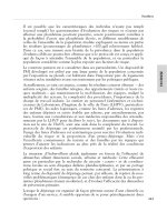
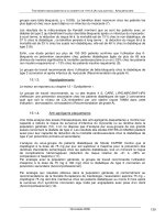
Coagulation is initiated when the speed and degree of tissue
heating is slower and less intense, leading to cellular desiccation.
Specificeffects of different types of RF currents and heat-related
tissue destruction are illustrated in Figs. 8.1 and 8.2.
Several factors regulate the degree of tissue heating:
r
Voltage (V) is the force required to push current through the
tissue.The higher the voltage, the deeper the thermal tissue
destruction.
r
Tissue resistance (R) or impedance (for alternating current) is
the force generated by the tissue to resist electric flow. Itis
directly proportional to the amount of tissue electrolytes.
Resistance increases dramatically during tissue heating and
desiccation. Normal tissue resistance is not uniform; it is the
lowest along the blood vessels and thehighestatthelevel of the
skin.
r
Time (T) is an essential factor of energy (E) regulation, which
can beexpressed as
E(in joules) = P(power in watts) × T
Tissue heating increases with time, although the process is
quite complex:
r
Heating produces water losses and increases resistance
r
Increasing resistance shifts the distribution of current from the
lowest resistance pathway
r
Fluctuation of resistance affects the power output produced by
the generator
r
Someof the released heat is removed from high-temperature
areas by blood flow.The cooling effect of blood flow explains
why the same energy applied to the tissue generates less de-
struction, if delivered slowly.
Practical Pediatric Gastrointestinal Endoscopy
George Gershman, Marvin Ament
Copyright © 2007 by Blackwell Publishing Ltd
172 CHAPTER 8
* Low-voltage current penetrates less through desiccation tissue and has limited
ability to induce deep tissue heating.
** Spikes of high-voltage coagulating current allow a deeper spread through
desiccated tissue and induce more tissue destruction.
Alternating RF Current
Uninterrupted high-
power, low-voltage
current
Interrupted high-voltage
spikes of RF current
lasting 20% of the cycle
Combination of both
currents
Sparks between tissue
and active electrode
Deep penetration of
current across the tissue,
causing desiccation
Relatively greater “cut”
than “coagulating”
tissue effects
Quick tissue heating
up to 500ºC and above
produces vaporization
Coagulating current ** Blended current
Cutting current *
Fig. 8.1 Different types of alternating RF currents and specific tissue
response.
Alternative RF current Tissue resistance
Heat
Above 41.5ºC
• devitalization –
irreversible death
of the tissue
Above 60ºC
Coagulation and
moderate
desiccation
• contraction of
collagen
• hemostasis of
small vessels
• formation of
adhesive
derivatives of
glucose
Above 200ºC
Cabonization
• tissue may
become an
electric
insulator
100ºC Fast
desiccation
• hemostasis of
bigger vessels
secondary to
glue effect of
desiccated
glucose
• tissue sticking
to the active
electrode
Above 500ºC
• tissue
vaporization
cutting
• smoke
production
Fig. 8.2 Temperature-related tissue destruction always induced byRF current.
POLYPECTOMY 173
r
Current density is a measure of RF current (I) that flows
through a specific cross-sectional area (a ):
I
a
=
I
πr
2
The amount of heat generated in the tissue is directly pro-
portional to power density (P), expressed as a square value of
current density multiplied by resistance:
P =
I
a
2
=
I
2
πr
2
×
This important equation implies that power density is in inverted
relationship with the square of the cross-sectional area (π r
2
).
It means that even small tightening of the wire loop produces
a profound effect on tissue heating.This can be illustrated by
polypectomyof a 1-cm polyp.
If a snare decreases the diameter of a polyp in half, the cross-
sectional area at the level of the loop will be only 0.2 cm
2
. Itis
4 times less than the cross-sectional area at the basis of a polyp
and about 500 times less than that of skin under a 10 × 10 cm
plate of the “return’’ electrode.
If 0.2 A electric current is applied through the snare, it pro-
duces a current density of 1, 0.25, and 0.002 A/cm
2
at the level
of the loop, polyp basis, and skin level, respectively.The fall of
power density, i.e., power actually delivered to the tissue and
generated heat, is even more dramatic:from 1A/cm
2
× R at the
level of the loop to 0.06 A/cm
2
× R and 0.000004 A/cm
2
× R at
the basis of thepolypand skin under the return electrode, respec-
tively. Narrowing of a cross-sectional area by aclosing snare pro-
duces the most significant effect on heat production compared
with increasing power setting and timeof electric current ap-
plication. It also allows one to perform a polypectomy at a low
power, using a coagulating mode safely.
The law of currentdensityis vital for polypectomy. Narrowing
of a cross-sectional area is the most important safety technique,
which produces a coagulation of core vessels of the polyps be-
fore cutting, restricts the area of maximal tissue heating around
the loop, and limits tissue destruction of the deep b
owel wall
layers.
SNARE LOOPS
Commercially available snares vary bysize, configuration of
the loop, design and mechanical characteristics of the handles
and, wire thickness. Reusable snares often loose their mechan-
ical properties and can peel and break at the tip. Disposable
snares are more durable and predictable.The thickness of the
wire loop and handle “behavior’’ can significantly affect the
174 CHAPTER 8
Fig. 8.3 Snare preparation before polypectomy: marking of so-called
closing point on the handle of the snare.
results of polypectomy.Snares with thick wire loops have two
important advantages:
r
Decreased risk of snapping a polyp without adequate coagu-
lation
r
Large surface contact with tissue and better coagulation.
A standard snare with an opening diameter of 2.5 cm can be
used for different size polyps. A special small or “mini’’ snare
(1-cm loop) has been designed for polyps less than 1 cm. Itis
important for endoscopists to find an “optimal’’ snare for routine
practice in order to avoid unexpected “surprises’’ during cutting
or coagulation.
A chosen snare should be fully open and then closed to the
point when just the tip of a wire loop is outside of outer sheath.
Marking of the so-called closing point on the handle of the snare
(Fig.8.3)
serves two important safety features:
r
Protects from premature cutting of asmall sessile or peduncu-
lated polyp without an adequate coagulation
r
Alerts the endoscopist to partial polyp ’s head entrapment or
underestimation of the stalk size.
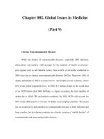
It is very important to check how far the tip of a wire loop
is retracted into the outer plastic sheath when a snare is fully
closed.The distance of15mm reassures an adequate squeezing
pressure (Fig. 8.4). If the stalk of a large polyp is not squeezed
adequately, it compromises the coagulation of core vessels by
two reasons:
r
Blood vessels remain open and blood flow continues produc-
ing a cooling effect but, more importantly,
POLYPECTOMY 175
15mm
Fig. 8.4 Squeezing pressure. A fifteen mm retraction of the wire into
the plastic sheath provide an optimal narrowing of the polyp base or
the stalk for adequate constriction of the blood vessels and generation
of an appropriate power density.
r
a cross-sectional area is not narrow enough to concentrate the
current flow to an appropriate power density to coagulate the
core vessels.
Closure of a snare loop with excessive pressure caninducepre-
mature cutting before coagulation. Both conditions could lead to
significant bleeding.
POLYPECTOMY ROUTINE
Polypectomyisthemost common therapeutic procedure in pe-
diatric gastrointestinal endoscopy. It can besimple or more
complex depending on the size or location o f the polyp and per-
sonal experience. No matter how easy the polyp appears to the
endoscopist, it is always wise to follow a simple rule: safety be-
fore action.
SAFETY ROUTINE
It is always useful to routinely inspect the snare and genera-
tor as well as to prepare hemostatic equipment such as detach-
able loops, metal clips, and needle for epinephrine injection.
The polypectomy snare should be checked for smooth opening,
thickness of the wire (a thin snare predisposes to a premature
cut of asmall polyp before appropriate coagulation), adequate
squeezing pressure, and closing point. Itisextremely important
to test a generator to find a minimal power setting, which is nec-
essary to induce whitening and swelling of the tissue inside a
wire loop. It should be done at least once byadjusting the power
output according to the effect of
short (2–3s) burst of coagulat-
ing current until a visible effect is achieved.Thegenerator setting
should be inspected routinely before the procedure to avoid an
accidentally high power setting. A foot pedal should be conve-
niently positioned in front of the endoscopist. A teaching session
with an assistantor a technician isimportant for safe andoptimal
manipulations with a snare during opening or closure.
176 CHAPTER 8
SAFETY CONDITIONS AND TECHNIQUES
A good bowel preparation is essential not only for optimal view
and positioning of the loop around a polyp stalk or base, but also
to avoid an accidental burning or coagulation of normal mucosa.
A large amount of liquid or solid stool increases the chance of
missing a small and even a good size polyp. Anobscure view
often leads to excessive use of air and bowel stretching, which
makes the bowel wall thinner.
Sudden patient irritability, unexpected awaking, or move-
ments complicate polypectomy especially during a snare closure
and should be prevented by adequate sedation.
The technique of polypectom
y consists of three important el-
ements:
1 Navigation of the scope to an optimal position, angle, and
distance to a polyp
2 Placement of a wire loop around a polyp
3 Cutting.
A 6o’clock position is an ideal one for polypectomy. A loca-
tion of a polyp between 4 and 5 o’clock and 7 and 8 o’clock is
suboptimal. Polypectomy is very difficult and somewhat unsafe
if a polyp is located on the upper aspect of alumen between 9
and3o’clock.
An ideal 6 o’clock position could be created by clock- or coun-
terclockwise rotation of the shaft and downward deflection of
the tip. Careful assessment of stalk size and location of a polyp
is obligatory before polypectomy. It can be done by rotation,
advancement of a scope beyond a polyp, and pulling the shaft
backward.Once an optimal position and clear view of a polyp
is achieved, the scope is moved toward the polyp base. An ideal
distance form the tip of the scope to a polyp is 1–2 cm unless a
polyp is hiding beyond a fold. In this case the tip of the closed
snare should be positioned just above the fold and pressed down
to reveal the polyp.The sameeffect can be achieved by manipu-
lations with the use of a closed snare.
All manipulations with a snare should
be slowly done. Itis
opened just enough to embrace a polyp.Full opening of a snare
makes the wire less controllable.
Fig. 8.5 The snare is placed
around the polyp.
Snaring a sessile polyp at 6 o’clock position is easy if the wire
loop is horizontal to the polyp.Simple downward tip deflection
is needed to move a loop and encircle a polyp. If an opened
wire loop creates an angle to the base of a polyp, the shaftof the
scope should be rotated toward the polyp until it is caught.The
technique is modified if a sessile polyp is located between 4 and
5 o’clock or 7 and 8 o’clock and attempts to establish an ideal
6o’clock position have failed.The shaft is slightly rotated away
from a polyp.The snare is opened more than usual to makeit
less rigid and slide toward the polyp (Fig. 8.5). Once the polyp is
POLYPECTOMY 177
inside the loop, the scope is rotated slowly toward the polyp to
align the plane of a snare with the axis of a bowel lumen.Then
the snare is closed slowly and moved forward until it reaches the
base of the polyp. At this moment the snareshould becompletely
closed (Fig.8.6).
Fig. 8.6 The snare is closed tight
but not enough to amputate the
polyp.
Occasionally, a backward snaring is more effective, especially
if the polyp is more than 1.5 cm in length. An open loopispointed
down to the area where a polyp head touches the bowel wall.
When the snare is advanced, tissue resistance creates a bowing
effect and induces a loop opening. As a result, the loop slides
between the mucosa and the polyp head. An additional clock-
wise rotation of the tip using both knobs swings a wire loop
under the polyp head. If the position of the snare is satisfactory,
the snare is slowly closed tight enough for polypectomy.
If a polyp is facing away from the tip, the snare is advanced
and opened slowly until the tip of the wire is beyond the polyp’s
head.The tip of the scope is de
flected down slightly to move the
wire loop below the polyp. After that the snare is pulled back
until the head of the polyp is inside the loop and the wire is just
under the polyp head.The snare is closed slowly and advanced
toward to the polyp to prevent sliding of the wire along the
stalk.
Advancement of the snare toward the polyp during wire loop
closure is a key element to polyp snaring. It secures a polyp
within the loop and allows precise navigation of the snare.The
capturing of asmall polyp with a standard snare may be chal-
lenging. A slight decompression of the bowel may elevate a
polyp above a wire loop and facilitate a capture.
The technique of polypectomyisdifferent when applied to
small polyps less than 5 mm, broad-based polyps more than
15 mm, or pedunculated polyps m
ore than 20 mm. Diminutive
or small sessile polyps less than 5 mm can beremoved safely by
cold biopsy forceps.Two helpful hints are as follows:
1 If a polyp is located on the edge of a fold, position the tip of
the colonoscope within a distance of2cm from the polyp, open
the forceps and place the open cusps perpendicular to the fold
just above the polyp, and close it. Avoid pushing the forceps
up against the mucosa as it will stretch the tissue and result in
suboptimal sampling.
2 If asmall polyp is between the folds, try to position the snare
with cusps opened horizontally and just enough to outline the
polyp. Advance the forceps forward slightly to cover the polyp
and close the forceps slowly. An alternative technique consists
of
r
opening the forceps with cusps vertical to the folds,
r
positioning the lower cusp just below the polyp to avoid
grasping normal mucosa, and
r
closing a forceps.
178 CHAPTER 8
A large sessile polyp is rare in children except in patients with
Peutz-Jegher’s syndrome. Polyps more than 2.5–3cm are usually
located in the small intestine, primarily in the jejunum. If the size
of a polyp is between 10 and 15 mm, a single-cut polypectomy
may besafeif advancement of a snare with captured polyp does
not produce synchronous movements of the underlying wall.
This indicates that submucosa and muscularis propria are not
trapped within the wire loop.
Piece-meal technique: Piece-meal technique is used for piece-by-
piece removal of a large broad-
based polyp, more than 15 mm. A
submucosal injection of saline, hypertonic saline, or epinephrine
(1:10,000) solution before polypectomy decreases the risk of the
transmural burns.
Injection at site proximal on the polyp is performed first if
possible,followed byinjections at the distal edge and both sides
of a polyp. Injection of 3–10 cc of a chosen solutionat three to four
sites is usually adequate to create a liquid “cushion’’ under the
polyp.The needle should be oriented tangentially to minimize
the risk of transmural injection.
Once again, a broad-based polyp more than 15 mm should be
removed in pieces to minimize the risk of perforation.The risk
of bleeding is not high since blood vessels in such polyps are
much smaller than in large pedunculated polyps.
The piece-meal technique consists of placement of a wire loop
diagonally across a polyp and removing the polyp in few pieces.
The remaining central area is cut at the end. Excessive closing
pressure should be avoided because it may compromise initia-
tion of cutting due to lack of electric arc from
the active electrode
to the tissue. In addition, decreased wire–tissue contact area in-
creases current density, which may induce excessive desiccation
and cease current flow.
Polypectomyof pedunculated polyps more than 2 cmmay
be challenging. Attention should be paid to proper positioning
of the wire loop at the narrowest portion of a stalk right below
a polyp head.Thickblood vessels in the middle of a stalk re-
quire slow desiccation for complete coagulation and hemostasis
before the final cut. Endo-loop
R
and clipping devices should
be available for immediate action. Itisquite difficult to avoid
direct contact of a large pedunculated polyp with normal mu-
cosa during polypectomy.However, attempts should be made
to keep a snared polyp close to the center of the bowel lu-
men to minimize thermal destruction of adjacent tissue. Care-
ful inspection of a long stalk should precede any manipulations
with a snare.The location of the polyp base and position of the
long stalk are crucial for optimal approach to the polyp.The
snare is advanced forward to the lowest point of the polyp head
and opened slowly until the loop is big enough to embrace the
polyp.
POLYPECTOMY 179
Further manipulation with the snare should be coordinated
with either right or le ft torque of the shaft toward the 6 o’clock
direction. Backward snaring may be useful.The reduction of
a polyp size by piece-meal technique with prior injection o f
epinephrine solution (1:10,000) into a stalkbelow the polypec-
tomy site is the last option to complete the procedure.
After successful capture and adequate tightening of the wire
loop, a polyp less than 10 mm is removed by using a low-power
coagulating current (15–18 W) continuously for 2–3 seconds and
by slow closureof a snare after whitening and tissue swelling has
occurred. Amodified technique is applied to sessile polyps less
than 15
mm or large pedunculated polyps with a small pseudo
stalk. Injection of saline or epinephrine (1:10,000) solution un-
derneath the polyp head protects deep tissue from desiccation
and decreases mobility of the polyp, which simplifies a place-
ment of the wire loop without trapping a part of the polyp head.
A slightly longer duration of coagulation (2–3 cycles) may be
necessary for adequate coagulation of blood vessels.
Ablended current up to 20–25 W may be reasonable for
polypectomyof a broad-based polyp, using a piece-meal tech-
nique.
Different electrosurgical generators have different setting sys-
tems: a dial type syste
m with a scale from 0 to 10. Usually, a
setting point between 2.5 and 3 are equivalent to a low power
of15–20 W; anumeric-type system, when displayed, numbers
represent current power in watts.
An endoscopist should become familiar with the particular
electrosurgical generator available for his or her practice to avoid
an application of excessively high power above 30W,which
could lead to a transmural tissue necrosis.
A polypectomy can be performed during colonic intubation
or withdrawal phase of colonoscopy.The decision is made based
on the sizeof the polyp. It is wise to re moveasmall sessile or
pedunculated polyp as soon as it was discovered to eliminate the
chance of
missing this polyp later on. Removal of a large polyp is
more convenient after the entire colon has been inspected except
in the case when the position of a polyp is ideal for polypectomy.
Careful examination of the colon (especially behind the folds)
can be accomplished by circumferential rotation of the tip and
the shaft, aspiration of excessive fluid, and repeat insertion of
the scope for a few segments if the bowel quickly slipped away
from the tip.
After polypectomy, polyps less than 10 mm can be easily
sucked into a
biopsy channel and eventually into a filtered polyp
suction trap.Water irrigation and proper orientation of a suction
nostril at the tip of a scope facilitate the recovery process.
During polypectomy, attention should be paid to remove
polyps and to observe the direction where it falls.The first place
180 CHAPTER 8
to look for a hiddenpolyp is in a pooloffluid. If a polypis not dis-
covered,flush some water and watch where it flows: backflow
indicates that the polyp is distal to the tip of the scope.
Nylon polyp retrieval nets or metal baskets can be used for
removal of multiple polyps. Grasping of a large polyp by the
snare is the most reliable way to bring it to the rectum.Manual
assistance in the recovery of a specimen may be necessary to
squeeze a large polyp more than 3 cm through the anus.
COMPLICATIONS
Three types of complications can occur after polypectomy.The
most common one is bleeding. In contrast to adults, a delayed
bleeding within 2 weeksafter the procedure is quite rare. Imme-
diate onset of bleeding is more common, although the incidence
of this complication is less than 1% in infants and children.This
may reflectasmaller size, the number of polyps, and the absence
of comorbid conditions such as hypertension and atherosclero-
sis. A slow oozing from the polpypectomy site is easy to control
byinjection of epinephrine solution
(1:10,000) or by bipolar or
argon plasma coagulation (Fig. 8.7).
Fig. 8.7 APC is useful tool of
hemostasis. Bleeding after
polypectomy was successfully
controlled by argon plasma
probe.
The risk of arterial bleeding always exists right after polypec-
tomyof a large pedunculated polyp due to incomplete
coagulation of thick vessels. Endoscopic hemostasis should be
prompt before a large amount of blood and clots make the bleed-
ing vessel invisible. A temporary hemostasis can be achieved
almost immediately by resnare and tightening of the stalk. Af-
ter a few minutes, the wire loop should be replaced by the
Endo-loop
R
for permanent hemostasis. In addition, injection of
epinephrine below the Endo-loop
R
can augment a hemostatic
effect.
FURTHER READING
Cappell MS, Abdullah M. Management of gastrointestinal bleeding in-
duced by gastrointestinal endoscopy. Gastrointest Endosc Clin NAm
2000;29:125–67.
Jalihal A,Misra SP, Arvind AS, Kamath PS. Colonoscopic polypectomy
in children.JPediatr Surg 1992;27:1220–2.
Tappero G, Gaia E , DeFiuli P, et al. Cold snare excision of small colorectal
polyps. Gastrointest Endosc 1992;38:310–13.
Waye JD. Endoscopic mucosal resection of colon polyps. Gastrointest
Endosc Clin NAm2001;11:537–48.
Waye J
D. New methods of polypectomy. Gastrointest Endosc Clin NAm
1997;7:413–22.
181
9
Chromoendoscopy
Chromoendoscopy is the topical application of dyes to the gut
mucosa, carried out in order to allow or improve the endo-
scopic localization and characterization of a specific tissue or
lesion. Generally, the identification of a lesion can be accom-
plished either by a positive or negative staining; i.e., the dye
either stains the lesion or the normal mucosa surrounding it.
Chromoendoscopy can be used in combination with optical en-
hancement (magnification endoscopy) to further increase the
yield of biopsy particularly in case of suspect dysplasia or can-
cer. Although it was developed and first used some30 years ago,
chromoendoscopy is seldom used in everyday clinical practice
foranumber of reasons. Apart from
highlighting mucosal le-
sions that have to be biopsied or removed, the superiority of
chromoendoscopy on standard endoscopy and histologyhas not
been demonstrated yet.The recognition and interpretation of le-
sions imply a degree of subjectivity and the procedure requires
someextra time.Fortunately, dysplasia and cancer are an uncom-
mon occurrence in the gastrointestinal (GI) tract of infants and
children, and thus their recognition isnot such a relevant issue as
in adult gastroenterology.On the other hand, chromoendoscopy
techniques are simple,quick, inexpensive, and generally safe
and the equipment needed is widely available.Furthermore, in
large pediatric GI ref
erral centers, conditions where endoscopic
surveillance for the detection of dysplasia are indicated – such
as Barrett’s esophagus, early onset inflammatory bowel disease
(IBD), or familial polyposis syndromes – may well be seen.Fi-
nally, the recent development of therapeutic endoscopic tech-
nologies such as mucosal resection and photodynamic therapy,
which re quire a precise tissue locali zation and characterization,
have produced a renewed interest in chromoendoscopy world-
wide.
INDICATIONS
Esophageal disorders
One potential indication of chromoendoscopy in the pediatric
esophagus is intestinal metaplasia, i.e., Barrett’s esophagus. If
this condition is suspected, the main aim of chromoendoscopy is
to help increase the diagnostic yield of endoscopic biopsies. Pos-
itive staining with methylene blue could also be used to identify
Practical Pediatric Gastrointestinal Endoscopy
George Gershman, Marvin Ament
Copyright © 2007 by Blackwell Publishing Ltd
182 CHAPTER 9
endoscopically invisible intestinal metaplasia of the cardia re-
gion, which may exist in patients with gastroesophageal reflux
disease (GERD). However, it is questionable if methylene blue
staining should be applied to all patients with long-standing
GERD who undergo upper endoscopy, because intestinal meta-
plasia can also be found in asymptomatic individuals and the
advantage of methylene blue stainingoverrandombiopsy is con-
troversial. In adult patients with short-segment Barrett’s esoph-
agus, the sensitivity of methylene blue staining for the detection
of intestinal metaplasia varies from 60 to 98% but is generally
higher than that of randombiopsies. Ab
normal methylene blue
staining can also be helpful in delineating dysplastic or malig-
nant areas for endoscopic treatment such as mucosal resection or
photodynamic therapy. If mucosectomy is planned, a minimum
amount of methylene blue injected with saline into the underly-
ing submucosa will stain it blue, thereby facilitating an accurate
removal of the mucosal lesion. In patients who have undergone
mucosal ablation, chromoendoscopy could also help distinguish
the regenerating squamous epithelium from residual Barrett’s
mucosa.Lugol’s solution has also been used in follow-up en-
doscopic examination of young patients who have been treated
for Barrett’s esophagus or dysplasia, in order to promptly detect
remnants of unstained Barrett’s epithelium.
Studies in adults have shown that chromoendoscopy with
Lugol’s solution is superior to conventional endoscopy for the
detection of severe dysplasia and early squamous cell carci-
nomaof the esophagus. In a Chinese population with high
esophageal cancer rate, chromoendoscopy with Lugol’s solu-
tion showed a sensitivity of 62–96% and a specificity of 63%.
However, esophageal dysplasia and cancer are extremely un-
common in pediatric patients, and it should be kept in mind that
Lugol’
s solution can also stain an inflamed esophageal mucosa,
namely, reflux esophagitis.Other staining techniques such as in-
digo carmine and acetic acid have been proposed in association
with magnification endoscopy to detect Barrett’s esophagus and
dysplasia.Stainingwith toluidine blue has been reported to have
a very high (98%) sensitivity for Barrett’s esophagus, but cannot
distinguish between gastric and intestinal metaplasia.
Although studies in adults have shown promising results, so
far there are insufficient data supporting a routine use of chro-
moendoscopy for detecting Barrett’s esophagus and dysplasia in
children.
Helicobacter pylori infection and
related disorders
Todate, there are no clear-cut indications for the use ofchromoen-
doscopy to detect specific gastric disorders in clinical practice.
CHROMOENDOSCOPY 183
At least two reactive dyes, however, deserve attention and may
prove useful in the near future. Congo red stains acid-secreting
mucosa and has been used in adult patients to detect gastric
atrophy, which appears as an area of negative staining on the
darkblue/blackbackground of the normal mucosa of the gas-
tric fundus and body. Phenol red turns from yellow to red in the
presence of alkaline pH, such as that related to the hydrolysis of
urea by urease-producing H.pylori, andhas been used to map the
extent of H.pylori colonization in the stomach. Both these stain-
ing techniques could therefore find an application in pediatric
patients with long-standing or refractory H.pylori
infection.
Celiac disease
Gluten-sensitive enteropathy (celiac disease) usually results in
endoscopically visible changes of the duodenal mucosa, in-
cluding a “mosaic’’ pattern, loss or indentation (scalloping) of
Kerckring’s folds, and a visible vascular pattern. Chromoen-
doscopy with methylene blue emphasizes the mosaic pattern,
though it does not seem to increase the diagnostic yield of
endoscopy, at least when performed byexperienced gastroen-
terologists. In one study, indigo carmine scattering combined
with magnification endoscopy proved superior to standard en-
doscopy for the detection of small bowel enteropathy, mainly
because it was able to distinguish
between total and partial vil-
lous atrophy.However, since the diagnosis of celiac disease is
established by histology and not by endoscopy, duodenal biop-
sies should betaken whenever celiac disease is suspected, irre-
spective of the endoscopic appearance of the duodenal mucosa.
Therefore, the major contribution of chromoendoscopy in celiac
disease is to allow for better targeting – and consequently some
sparing – of duodenal biopsies.
Polyposis syndromes
Chromoendoscopy may be very useful to detect smaller lesions
in the duodenum of patients with familial adenomatous poly-
posis (FAP). Small flat duodenal adenomas may in fact go unno-
ticed during standard endoscopy and even capsule endoscopy,
but can be identified as negative-staining lesions when an ab-
sorptive dye such as methylene blue is sprayed onto the mu-
cosa. In colonic polyposis, the main aim of chromoendoscopy is
the same as in the duodenum, i.e., to increase the detection rate
by facilitating the identification of small flat polyps, especially
adenomas.T
he preferred dye for the detection of colonic polyps
is indigo carmine, a contrast stain that pools in areas of mucosal
irregularity and often gives a three-dimensional effect, which is
particularly useful for the detection of small protruding lesions.
184 CHAPTER 9
Needless to say, magnification endoscopy and high-resolution
endoscopy can add to the accuracy of the technique. In adult
studies, left-sided or total colonic indigo carmine staining signif-
icantly increased the detectionrateofsmall flat or depressed ade-
nomas. Chromoendoscopy can also help distinguish between
hyperplastic and adenomatous polyps, as they produce differ-
ent staining patterns. In a recent multicenter study, more than
90% of colonic polyps were correctly classified according to the
staining pattern, and for adeno matous polyposis the sensitivity
and specificity were 82% and the negative predictive value was
88%.
Inflammatory bowel disease
In IBD, the greatest potential for chromoendoscopy is the ability
to early detect dysplasia or cancer in patients with long-standing
ulcerative colitis. Colonic dysplasia and colitis-related colon can-
cer may occasionally be a problem also in pediatric patients, as
in case of ulcerative colitis presenting before 10yearsofage, espe-
cially if associated with sclerosing cholangitis. In a randomized
controlled trial on 174 patients with long-standing ulcerative col-
itis, total colonic methylene blue staining was clearly superior to
conventional surveillance endoscopy with biopsy for the detec-
tion of early neoplasia (32 vs 10 overall intraepithelial lesions;
24 vs 8 low-grade; and 24 vs 10 in flat mucosa).
Other indications
In the duodenal bulb, methylene blue spray can help identify
areas of gastric metaplasia, which is a marker of inflammation
such as that related to H.pylori infection.Methylene blue was
also used to identify the minor papilla in patients with pancreas
divisum.
APPLICATION TECHNIQUE
Fig. 9.1 The tip of a pediatric
ERCP catheter pushed through
the biopsy channel is seen in the
distal duodenum, prior to dye
spraying.
Equipment
Special reusable spray catheters such as those used for en-
doscopic retrograde cholangiopancreatography (ERCP)(e.g.,
Olympus PW-5L1) are preferable.The biopsy channels of all
modern pediatric videoendoscopes allow the passage of such
catheters (Fig. 9.1). It is also convenient to use a new biopsy chan-
nel cap in order to minimize the leakage of dye. Endoscopists and
support staff with less experience in chromoendoscopy should
be particularly careful, as most dyes can produce a fairly per-
sistent staining of skin and clothing. Depending on the specific
indication and need, different type of stains can be used, i.e.,
CHROMOENDOSCOPY 185
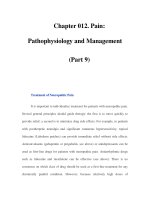
Staining Main clinical
Dye (%) mechanism Color application(s)
Methylene blue
(0.5%)
Absorption into intestinal
epithelial cells
Blue Intestinal metaplasia in
esophagus (Barrett’s)
Intestinal metaplasia in
stomach
Gastric metaplasia in
duodenum (negative
staining)
Celiac disease
Lugol’s solution
(1–5%)
Binding to glycogen-
containing cells
Dark green/
brown or black
Squamous esophageal cancer
(negative staining)
Residual postablation Barrett’s
(negative staining)
Esophagitis (negative staining)
Toluidine blue
(1%)
Binding to nuclear DNA
of malignant cells
Blue Squamous esophageal cancer
Indigo carmine
(0.1–0.5%)
Pools in mucosal
crevices and pits
Indigo
(blue/violet)
Small, flat, or superficial
polyps
Barrett’s esophagus
Dysplasia or cancer in
ulcerative colitis
Congo red
(0.3–0.5%)
Stains acid-producing
mucosa (pH <3)
Turns red to dark
blue/black
Mapping of acid-secreting
mucosa
Gastric cancer, gastric atrophy,
and intestinal metaplasia
(negative staining)
Phenol red
(0.1%)
Stains alkalinized
mucosa
Turns yellow to
red
Mapping of H. pylori-infected
mucosa
Gastric metaplasia (negative
staining)
India ink (1%) Staining of mucosa at
site of injection
Black
(permanent)
Site of endoscopically
removed polyp
Lesion to be removed
surgically
Table 9.1 Types of staining.
stains that are absorbed by the mucosa (vital stains), stains that
produce contrast (reactive stains), and stains for tattooing of the
mucosa (Table 9.1).
Methylene blue
Methylene blue is actively absorbed by the intestinal epithe-
lium and does not stain nonabsorptive tissues such as the nor-
mal esophageal or gastric mucosa.Optimal staining requires
186 CHAPTER 9
washing of the mucosa with a mucolytic agent such as N-
acetylcysteine prior to spraying a 0.25–0.5% solution of the dye
and subsequent washing with water.The absorptive intestinal
epithelium – including metaplastic epithelium as in Barrett’s
esophagus – is stained blue, whereas the nonabsorptive epithe-
lium – such as ectopic gastric metaplasia – is delineated as an
area of negative staining against a blue-stained background.The
presence of dysplasia or early malignancy within Barrett’s ep-
ithelium result in inhomogeneous staining as a consequence of
the differential absorption of methylene blue from cells that are
depleted of gob
let cells and have less cytoplasm.Methylene blue
is generally considered to besafe.However, it has been reported
that, once photosensitized by white light, methylene blue may
induce oxidative damage of the DNA and although it does not
usually stain the dysplastic intestinalepithelium,thereisconcern
that it may increase the risk of carcinogenesis in patients with
Barrett’s esophagus.The parents of patients in whommethylene
blue staining is being used should be warned that their child’s
urine and stool might temporarily acquire a green-bluish color.
Lugol’s solution
Lugol’s solution contains iodine, which has a special affinity for
the glycogen contained in squamous epithelia.For this reason
it is most comm
only used in the esophagus, where the normal
squamous epithelium is stained green/brown to darkbrown
or black.Malignancy, dysplasia, metaplasia, or even simple in-
flammation is associated with glycogen depletion and the af-
fected mucosa will thus appear as an unstained area on a dark
stained background.Severeallergicreactionstoiodinehavebeen
reported, so allergy to iodine should be carefully excluded in
patients who are undergoing chromoendoscopy with Lugol’s
solution.
Toluidine blue
Toluidine blue is a basic dye that binds to the nuclear DNA of
epithelial cells, and therefore can be used to identify tissues with
an increased DNA synthesis such as malignancy.Toluidine blue
staining has
been mainly used in the endoscopic screening for
malignant gastric ulcers and early squamous esophageal cancers
in at-risk populations, e.g., heavy alcohol drinkers and smokers.
Indigo carmine
Indigo carmine is the most widely used contrast stain and is es-
pecially useful to identify and define the margins of neoplastic
CHROMOENDOSCOPY 187
lesions. Indigo carmine, in fact, typically pools in areas of mu-
cosal irregularity, which are stained indigo (blue/violet) color.
After washing, pits, grooves, and edges of the lesion are high-
lighted and this may produce a three-dimensional effect, which
is particularly useful for the detectionof small superficial lesions.
Indigo carmine at a concentration of 0.1–0.5% is usually sprayed
onto the gut mucosa, but may also be given orally in a capsule.
Although mostly utilized to identifysmall superficial polyps,
indigo carmine has been applied in several other conditions
such as Barrett’s esophagus
, gastric cancer, sprue, and ulcerative
colitis.
Congo red
Congo red reacts to an acidic pH by changing from red to dark
blue/black. Its major application is the identification and map-
ping of nonsecretory gastric mucosa such as that of gastric atro-
phy, intestinal metaplasia, and gastric cancer, which will appear
red in contrast to blue/black secretory areas. A stimulation of
acid production with pentagastrin is therefore necessary before
staining.
Phenol red
Phenol red is also a reactive dye, but unlike Congo red it reacts to
an alkaline pH by changing from yellow to red. Patients should
undergo pretreatment with a proton pump inhibitor and an anti-
cholinergic
, plus the local application of a mucolytic.Once 0.1%
phenol red and 5% urea have been sprayed onto the gastric mu-
cosa of H.pylori-infected individuals, the alkalinized mucosa is
stained red, whereas areas of intestinal metaplasia in the stomach
will stain negative.
Acetic acid
Acetic acid is a newcomer to GI chromoendoscopy. Preliminary
studies suggest that acetic acid stain may help identify Barrett’s
esophagus as well as duodenal atrophy in celiac disease by de-
lineating the features of the metaplastic or atrophic intestinal
epithelium.
India ink
When injected into the mucosa,1% india ink produces a perma-
nent black staining. I
ndia ink can beinjected superficially into
the mucosa to mark the site where a worrisome polyp has been
188 CHAPTER 9
endoscopically removed, or it can beinjected deeper to mark a
lesion that has to beremoved surgically.
Patient’s sedation
Because the main aim of chromoendoscopy is to allow for the
visualization of small and fine features of the gut mucosa, the
whole procedure can be rendered completely useless if the pa-
tient is restless or agitated.Therefore, unless the patient is fully
cooperative – which is the exception rather than the rule in pe-
diatric endoscopy – an adequate sedation is mandatory to main-
tain the patient still throughout the procedure. Conscious se-
dation with midazolam 0.05–0.20 mg/kg intravenous (IV) may
not besufficient in infants or very anxious children, where deep
sedation with propofol or a brief general anesthesia may
be
necessary.
Preparation of the mucosa
There is no doubt that chromoendoscopy gives better results
when the gut mucosa to beexamined is cleared frommucus (and
blood, bile, or food debris; if present). So, whenever possible, the
mucosa should be washed prior to staining. Abetter washing
is obtained ifforceful pressure is applied with a syringe either
through the spray catheter or directly into the biopsy channel. If
absorptive dyes such as methylene blue or Lugol’s solution are
to be used, the mucosa should be washed with a few milliliters of
10% N-acetylcysteine to adequately remove mucus
.Once the tis-
sue has been stained, a wash with water or saline can remove the
excess, nonabsorbed dye. If the vision is disturbed by bubbles or
foam, asmall volumeof antifoam preparation (e.g., simethicone,
10–20 drops) can be added to the wash. A spasmolytic drug such
as hyoscine N-butylbromide can beadministered IV to reduce
peristalsis or smooth muscle spasm and maximize visualization
o
f the mucosal area of interest. As mentioned above, when a pH-
sensitive dye is used, acid secretion should be either stimulated
or suppressed, depending on the dye being used.
Staining technique
The technique for staining is fairly simple.Oncethegutareaof in-
terest has been reached and adequately washed (see above), the
endoscope and the tip of the catheter should be directed toward
the mucosa with a combination of clockwise and counterclock-
wise rotation movements, and the dye should be sprayed onto
the mucosa while the tip of the endoscope is gently and slowly
withdrawn.The only exception is india ink staining, which is
in fact a permanent tattoo of the mucosa and as such requires
CHROMOENDOSCOPY 189
1 Strict patient selection: patients with histologically proven ulcerative colitis and at least 8 years’ duration in
clinical remission; avoid patients with active disease
2 Unmask the mucosal surface: excellent bowel preparation; remove mucus and remaining fluid in the colon
when necessary
3 Reduce peristaltic waves: when drawing back the endoscope, a spasmolytic agent should be used if
necessary
4 F ull-length staining of the colon: in ulcerative colitis, perform panchromoendoscopy rather than local
staining
5 Augmented detection with dyes: vital staining with 0.4% indigo carmine or 0.1% methylene blue should be
used to unmask flat lesions more frequently than with conventional colonoscopy
6 Crypt architecture analysis: using magnification endoscopy all lesions should be analyzed according to the
pit pattern classification; whereas pit pattern types I–II suggest the presence of nonmalignant lesions, staining
patterns III–IV suggest the presence of intraepithelial neoplasias and carcinomas
7 Endoscopic targeted biopsies: perform targeted biopsies of all mucosal alterations, particularly of
circumscribed lesions with staining patterns indicative of intraepithelial neoplasias and carcinomas, i.e., pit
patterns III–IV
From: Kiesslich and Neurath 2004.
Table 9.2 “SURFACE’’ guidelines for chromoendoscopy in ulcerative
colitis.
injection into the mucosa or submucosa.Once satisfactory im-
ages are obtained, it is always advisable to take photographs of
the stained mucosa, in order to compare staining features with
the histological abnormalities, to assess interobserver variability
and also to monitor the improvement of the staining technique
overtime. Recently, guidelines have been proposed for optimal
chromoendoscopy in ulcerative colitis (Table 9.2), but most of
these guidelines do apply to chromoendoscopy in general.
RECOGNITION OF THE LESIONS
Barrett’s esophagus and related disorders
Methylene blue is absorbed by the intestinal epithelium, so it has
been used for the endoscopic detection of the intestinal metapla-
sia typical of Barrett’s esophagus, especially when the diagnosis
is uncertain as it may be in short-segment Barrett’s.The staining
is usually homogeneous, but in short-segment Barrett’sitmay
besomewhat patchy due to the presence of nonintestinal colum-
nar cells.More importantly, in Barrett’s esophagus the pattern of
methylene blue staining is irregular and heterogeneous if dys-
plasia or cancer is present (Fig. 9.2). Heterogeneously stained
or light blue/unstained areas should be biopsied with particu-
lar care in search of high-grade dysplasia and early adenocarci-
nom
a. IfLugol’s solution is used, Barrett’s epithelium, dyspla-
sia, or carcinoma will appear as areas of negative staining on the
dark green/brown stained background of the normal squamous
epithelium.