Paediatrics & Child Health - part 9 pot
Bạn đang xem bản rút gọn của tài liệu. Xem và tải ngay bản đầy đủ của tài liệu tại đây (129.58 KB, 23 trang )
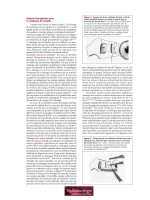
Testicular torsion
D: The testicle twists on the spermatic cord, occluding its blood supply from the
testicular artery.
A: Intravaginal torsion: the typical testicle is covered by the tunica vaginalis,
which attaches to the posterolateral surface of the testicle and allows for little
mobility. Torsion may be idiopathic or due to the congenital bell clapper
deformity (12% of cases). In this condition patients have an inappropriately
high attachment of the tunica vaginalis, so the testis can rotate freely on the
spermatic cord within the tunica vaginalis.
Extravaginal torsion (5%): develops antenatally in the spermatic cord,
proximal to the attachments of the tunica vaginalis.
A//R: DD:
Torsion of testicular appendage (hydatid of Morgagni):
(1) Occurs a little earlier (7–12 years).
(2) Causes less pain (patient can walk without pain).
Epididymitis, orchitis, epididymo-orchitis:
(1) Occurs in older patients and onset of pain is gradual and usually associated
with dysuria.
(2) Commonly 28 to reflux from UTI or STD (gonococcus, chlamydia).
(3) May be 28 to an underlying congenital, acquired, structural, or urologic
abnormality.
Hydrocele: painless swelling that transilluminates.
Testicular tumour: insidious onset of scrotal enlargement, usually painless.
Idiopathic scrotal oedema: scrotal skin is thickened, oedematous, and often
inflamed. The testis is not tender and is of normal size and position.
Acute appendicitis: torsion may mimic an acute abdomen.
E: 1/4000 boys. L > R. Bilateral in 2%.
Peak age intravaginal torsion: 14 years (range 11–30 years).
H&
E:
Extravaginal torsion: manifests as a firm, hard, scrotal mass. The scrotal skin
characteristically fixes to the necrotic testis.
Intravaginal torsion:
(1) Sudden onset of severe unilateral scrotal pain followed by scrotal swelling
and erythema.
(2) Hard and tender testicle tends to lie high and horizontally in comparison to
the other testis.
(3) May have a history of prior episodes of intermittent testicular pain that has
resolved spontaneously (intermittent torsion and detorsion).
(4) May be associated with nausea and vomiting, abdominal pain, fever, urin-
ary frequency.
P: Twisting of the testicle on the spermatic cord ! venous occlusion and
engorgement, with subsequent arterial ischaemia causing infarction of the
testicle.
I: Diagnosis is clinical.
Doppler USS: may demonstrate reduced/absent blood flow and differentiate
from epididymitis but must not delay surgery.
M: Surgical emergency: torsion must be relieved in < 6h for the testis to
remain viable.
Orchidopexy: if the testis is viable, it is untwisted and fixed to the scrotum.
The contralateral testis should be fixed at the same time.
Orchidectomy: if the testis is not viable. Testicular prostheses are available.
C: Delayed diagnosis or treatment may result in a non-viable testis.
P: Good if emergency surgery is performed without delay.
166
CONDITIONS
Brough / Rapid Paediatrics and Child Health Final Proof 9.7.2004 2:45pm page 166
Tetralogy of Fallot
D: Cyanotic CHD, which consis ts of 4 structural defects to the heart:
(1) Large VSD.
(2) Infundibular and valvular pulmonary stenosis.
(3) RVH.
(4) Overriding of the aorta with respect to the ventricular septum (aorta sits on
top of VSD).
A: Complex anatomical abnormalities arising from the abnormal development of
the right ventricular infundibulum.
A/R: Foetal hydantoin, foetal carbamazepine, FAS, Down syndrome.
E: 5/10 000 live births.
H&
E:
Neonatal: if there is pulmonary atresia, child becomes cyanosed when the
ductus arteriosus closes.
Infants:
. Hypoxic ‘spells’ give rise to pallor or cyanosis with respiratory distress.
. Harsh ejection systolic murmur at left sternal edge/pulmonary area, which
radiates to the back.
. Loud single 2nd heart sound due to loss of the pulmonary valve.
. Parasternal thrust in RVH.
Older children:
. Often adopt a squatting position with ‘spells’.
This improves symptoms by two means:
(1) Squatting " systemic vascular resistance and thereby decreases the right !
left shunt through the large VSD.
(2) Squatting " systemic venous return which improves blood flow to the pul-
monary system and " blood oxygenation.
. May exhibit signs of CHF.
P: Severity of disease is determined by degree of pulmonary outflow
tract obstruction. This results in:
(1) Reduced blood flow into lungs.
(2) Elevation of right ventricular pressure ! RVH.
(3) Resistance to ejection into the pulmonary circulation produces right ! left
shunting through the large VSD and deoxygenated blood going back into
the systemic circulation.
Hypoxic spells: due to "right ! left shunting, which results in a reduction in
pulmonary flow. It is thought to be due to infundibular spasm.
I: CXR: normal/small-shaped heart with uptilted apex 28 to RVH and concave
pulmonary segment, which in severe cases appears ‘boot-shaped’. Dark lungs
due to reduced lung vascularity reflects #pulmonary flow.
ECG: right axis deviation and evidence of RVH.
Bloods: "Hb (polycythaemia 28 to hypoxia).
Echo: confirms diagnosis.
M: Treatment of cyanotic spells: soothe the distressed infant to try and induce
sleep. If prolonged (> 15min), they require treatment with pain relief, sed-
ation (e.g. morphine) and IV propanolol.
Corrective surgical intervention: carried out in early infancy to widen the
pulmonary valve and close the VSD.
C: (1) Hypoxic attacks can result in myocardial infarction, cerebrovascular acci-
dents, and death.
(2) 28 polycythaemia may ! cerebral thromboem bolic events.
(3) Infective endocarditis.
(4) Cerebral abscess.
(5) Delayed growth and development.
167
CONDITIONS
Brough / Rapid Paediatrics and Child Health Final Proof 9.7.2004 2:45pm page 167
Tetralogy of Fallot continued
P: Pre-surgery 30% mortality in the 1st year of life and 75% by 10 years. With
surgery now 90% survive to adult life and 90% of these have a normal life-
style.
168
CONDITIONS
Brough / Rapid Paediatrics and Child Health Final Proof 9.7.2004 2:45pm page 168
Thalassaemia
D: Heterogeneous group of autosomal recessive defects of globin synthesis.
A: b-thalassaemia major: homozygous point mutations/deletions in the
b-globin genes on chromosome 11 ! b
0
(no b-chains)/b
þ
(small amounts).
b-thalassaemia trait: asymptomatic heterozygous carriers, mild microcytic
hypochromic anaemia; may be confused with iron deficiency anaemia.
b-thalassaemia intermedia: several different gene defects cause mild
b-globin synthesis abnormalities with variable clinical features, symptomatic
anaemia, hepatosplenomegaly, and extramedullary haemopoiesis.
a-thalassaemia: #a-globin chain synthesis. There are 4 a-globin genes on
chromosome 16. Severity of disease depends on number of genes deleted.
A/R: Due to geographical distribution individuals affected may also inherit sickle
cell gene.
E: Common in Mediterranean, Middle-Eastern, and SE Asian populations. There
has been a marked reduction due to antenatal diagnosis and termination of
pregnancy.
H: Anaemia and jaundice at 3–6 months (when g-chain synthesis switches to
b-chain synthesis), failure to thrive, recurrent infections.
E: Pallor, jaundice, frontal bossing, and maxillary overgrowth due to marrow
hyperplasia and hepatosplenomegaly due to haemolysis, extramedullary hae-
mopoiesis and iron overload.
P: Reduced synthesis of b -globin chain ! excess of other chains ! precipitation of
erythroblasts and erythrocytes in the bone marrow ! ineffective erythropoiesis,
haemolysis, anaemia, and extramedullary haemopoiesis.
I: Bloods: #Hb, #MCV, #MCH. Film: target cells, nucleated RBCs, "reticulocytes.
Skull X-ray: hair-on-end appearance due to expansion of bone marrow into
the cortex.
Hb electrophoresis: absent/#HbA and "HbF a
2
g
2
ðÞ, "HbA
2
a
2
d
2
ðÞas g and d
chain production continues.
Bone marrow: hypercellular with erythroid hyperplasia.
DNA analysis: for specific mutations (antenatal and postnatal CVS)
M: Medical: blood transfusions (maintain Hb > 10 g/dl), iron chelation with des-
ferrioxamine, vitamin C ("iron excretion), hepatitis B immunisation.
Surgical: splenectomy after 6 years to #blood requirements:
. Before splenectomy: pneumococcus/meningococcus/Haemophilus in-
fluenzae vaccinations.
. After splenectomy: daily penicillin, low-dose aspirin for post-splenectomy
thrombocytosis to #risk of thromboembolism.
BMT: from HLA-matching sibling, 90% success rate.
C: Iron overload: ‘slate-grey’ skin pigmentation ("melanin and haemosiderin),
cirrhosis, hepatoma, short stature, delayed puberty, DM, hypothyroidism,
hypoparathyroidism, cardiomy opathy.
Antibody formation: RBC antibodies, HLA antibodies.
Infections: meningococcal and pneumococcal after splenectomy, Yersinia
enterocolitica in those taking desferrioxamine.
Osteoporosis: 28 to marrow expansion and endocrinological complications.
Hypersplenism: leads to #Hb, #Plt, #neutrophils.
P: Good with regular transfusion and iron chelation. Mortality is mainly due to
infections and heart failure in untreated iron overload. Without transfusions
b-thalassaemia major is fatal.
169
CONDITIONS
Brough / Rapid Paediatrics and Child Health Final Proof 9.7.2004 2:45pm page 169
Tics
D: Tics: stereotyped movements of muscle groups that have no useful function.
Tourette syndrome: chronic idiopathic syndrome with both motor and vocal
tics beginning before adulthood.
A: Genetic: suggested by significantly higher concordance in monozygotic twins
compared to dizygotic twins, and significantly higher incidence in 1st degree
relatives of sufferers.
Acquired: there is a possible subgroup who have antibodies to b-haemolytic
streptococci that crossreact with neurons.
A/R: ADHD in > 30%, OCD in > 20%.
E: Tic disorders: 3–15% of children according to different studies, declining to
2–3% by adolescence.
Tourette syndrome: 0.5–1%. M : F¼ 2:1.
H&
E:
Simple tics: brief movements involving few muscle groups, e.g. eye blinking,
shoulder shrugging, clearing the throat, and humming.
Complex tics: coordinated patterns of succes sive movements involving
several muscle groups, e.g. jumping, touching the nose, echolalia (repeating
another’s speech), and coprolalia (outbursts of obscenities).
Tics are worsened by stress and reduced by absorbing activities, markedly
reduced during sleep and suppressible for brief periods of time.
Tourette syndrome: multiple motor and vocal tics occur (not necessarily con-
currently). Tics occur many times a day, nearly every day for more than 1 year and
frequently vary innature, severity, andlocation. Rage attacksconsist of explosive,
unpredictable outbursts outof proportion to stimulithreatening destruction and
self-injury, followed by immediate remorse.
P: Unknown. Theories include a reduction in the basal ganglia’s inhibition of
undesired motor programmes.
I: Usually none required.
In specific cases investigations may be appropriate to exclude organic cause:
(1) Anti-streptolysin titre, especially if there was sudden onset of tics post
impetigo, pharyngitis, or otitis media.
(2) TFTs to exclude hyperthyroidism.
(3) Serum caeruloplasmin to exclude Wilson disease.
(4) EEG to assess for absence seizures.
To assess for comorbid psychiatric disease: psychometric testing and MSE
for ADHD and OCD.
M: Multidisciplinary team approach:
Supportive: parental education and notify school of diagnosis.
Behavioural therapy: habit reversal.
Medical treatment: neuroleptic drugs (lower dose than for psychosis), dopa-
mine agonists.
Treatment of comorbid psychiatric disease:
(1) OCD: SSRIs.
(2) ADHD: tricyclic antidepressants (psychostimulants such as methylphenidate
used in treating ADHD can cause tics).
C: Stigma associated with outbursts may ! social withdrawal. Interruption in
thought and conversation affects education. Self-injurious behaviour may
arise from depression.
P: Tics can progressively worsen in childhood but abate or diminish markedly by
the age of 18 in 90% of cases. There is significant morbidity associated with
comorbid psychiatric disease.
170
CONDITIONS
Brough / Rapid Paediatrics and Child Health Final Proof 9.7.2004 2:45pm page 170
Toxoplasmosis (congenital)
D: Symptomatic congenital infection with Toxoplasma gondii.
A: Transplacental transmission:
. Occurs in 40% of mothers who have active 18 infection. They may contract
this from eating undercooked poultry, handling cat litter, or from blood
transfusions.
. Latent toxoplasmosis may reactivate in women with HIV and result in con-
genital transmission.
. Risk of transmission is greatest in the 3rd trimester.
. The severity of infection in the foetus depends on the gestational age at
the time of transmission.
Postnatal transmission: infants usually contract a much milder form of the
disease.
A/R: Undercooked meat, handling cat litter, immunocompromise.
E: 1/10 000 live births.
H&
E:
Mother: 18 infection in an otherwise healthy mother can be asymptomatic or
mild; with malaise, painless lymphadenopathy (usually cervical), myalgia, and
a low-grade fever.
1st trimester infection s: may result in foetal death in utero or in the neo-
nate with severe CNS involvement, such as cerebral calcifications and hydro-
cephalus.
2nd–3rd trimester infections: infants are usually asymptomatic at birth and
subsequently may develop the following sequelae:
. Eyes: chorioretinitis with diplopia, scotoma, and/or photophobia.
. Neurological: nystagmus, hypertonicity, and/or seizures.
. Others: jaundice, hepatomegaly, splenomegaly, rash, lymphadenopathy,
bulging fontanelle, micro/macrocephaly.
P: T. gondii is an intracellular protozoan. Lymph nodes typically show reactive
follicular hyperplasia as well as irregular groups of histiocytes around the
edges of the germinal centres.
I: Antenatal diagnosis:
20–24 week USS: in severe infection the foetus may exhibit hydrocephalus.
Cordocentisis: to confirm foetal infection during early pregnancy.
Neonatal diagnosis:
Serology: persistent or rising IgG titres indicate active infection rather than
maternal antibodies.
Sabin–Feldman dye test: measures IgG antibodies.
CT//MRI brain: in cerebral toxoplasmosis there are multiple ring-enhancing
lesions.
M: Detection during pregnancy: counselling, consider termination of preg-
nancy or introduction of spiramycin.
Prevention: avoid eating undercooked poultry, unpasteur ised milk, and un-
cooked eggs, wear gloves when handling cat litter during pregnancy.
Infant: 1-year course of pyrimethamine with folic acid supplements,
clindamycin.
C: Ophthalmic: the asymptomatic infant is at risk of chorioretinitis during
adulthood, and subsequent blindness.
Neurological: hydrocephalus, cerebral calcifications, seizures.
P: Neonates who are symptomatic at birth often die in the 1st month of life.
Infants with severe infection may have persistent hearing and visual impair-
ment and learning disability. Immunocompromised children have a higher
morbidity and mortality.
171
CONDITIONS
Brough / Rapid Paediatrics and Child Health Final Proof 9.7.2004 2:45pm page 171
Transient synovitis (TS)/Irritable hip
D: Arthralgia and arthritis 28 to a transient inflammation of the synoviu m of the
hip.
A: Ligamentous or minor capsular injury.
A/R: Associated: recent URTI (in 50% of children with TS).
Related: juvenile arthritis, arthritis associated with inflammatory bowel dis-
ease, psoriasis and ankylosing spondylitis, reactive arthritis from Campylo-
bacter, Salmonella, and Shigella.
E: Commonest cause of acute hip pain in children aged 2–12 years.
Sex: M:F¼ 2:1.
H: Elicit history of trauma, previous URTI, previous episodes of TS.
General: well-looking child; toxic-looking child is more likely to have septic
arthritis.
Pain: onset over hours to days. Unilateral hip or groin pain is common, may
have radiation to the knee or medial thigh on movement. Usually no pain at
rest.
Limp: child may present with a painless limp or antalgic gait and complain of
pain while walking.
Fever: unusual; may have mild fever (< 388C), if any higher, then septic arthritis
must be considered.
E: Look: leg may be held in flexion and internal rotation, no leg length inequal-
ity (differentiate from other causes of a limping child).
Feel: hip may be tender to palpation.
Move: mild restriction in range of movement, particularly internal rotation
and abduction.
Leg roll: patient is supine whilst involved leg is rolled from side to side –
involuntary muscle guarding occurs on one side when compared to the other.
P: Non-specific inflammation, hypertrophy of synovial membrane, "proteoglycans
in the synovial fluid.
I: Bloods: CRP/ESR normal or slightly ", WCC normal or slightly " .
Blood culture: negative. Urine culture: negative.
AP and lateral X-ray of the pelvis: medial joint space may be slightly wider
in the affected hip. Half to two-thirds of patients with TS may have an accentu-
ated pericapsular shadow. Look for signs of SUFE or LCPD (see chapters).
USS: small joint effusion with bulging of anterior joint capsule.
Joint aspiration: under USS guidance only if septic arthritis is suspected.
M: Once septic arthritis is excluded, management is supportive; avoid weight
bearing on the affected limb, analgesia with NSAIDs.
In severe cases refer to paediatric orthopaedic surgeon for skin traction with the
hip in 458 flexion.
C: May be the initial presentation of LCPD (develops in 1.5% of children with TS)
or SUFE.
P: Improves within days. Most have complete resolution in 2 weeks.
Recurs in up to 15%, mostly within 6 months.
172
CONDITIONS
Brough / Rapid Paediatrics and Child Health Final Proof 9.7.2004 2:45pm page 172
Transient tachypnoea of the newborn (TTN)
D: Acute, self-limiting tachypnoea in the absence of other cause such as meta-
bolic acidosis, RDS, or infection.
A: Infants almost invariably recover fully; therefore it is difficult to define TTN
pathologically.
Hypothesis: thought to be due to delayed resorption of foetal lung fluid.
The delayed resorption causes #pulmonary compliance and #tidal volume with
"dead space.
A/R: TTN is more common after elective Caesarean section and precipitous deliver-
ies. This is thought to be due to #time in labour. Lung liquid is predominately
reabsorbed actively by pneumocytes (type I) as a result of the changes in
hormones, and prostaglandins during labour. If there is no time for this to
occur, the fluid is not reabsorbed.
E: Commonest cause of respiratory distress in full-term infants.
1–2% of newborn infants have respiratory distress; of these 33–50% have TTN.
Other causes: RDS, metabolic acidosis, congenital cardiac disorders.
H: Onset: usually occurs in the first 1–3 h following an uneventful normal pre-
term, term vaginal, or elective Caesarean section delivery.
Duration: most cases resolve within 24–36 h.
E: Early onset of tachypnoea in the neonate; may also display signs of respiratory
distress such as recession; intercostal/subcostal/sternal expiratory grunting,
nasal flaring and in severe cases cyanosis.
P: Prominent perihilar streaking seen on CXR is usually the result of engorge-
ment of the periarterial lymphatics that participate in the clearance of the
alveolar fluid.
I: CXR: prominent perihilar streaking, patchy infiltrates, fluid in the horizontal
fissure, flat diaphragms, and occasional pleural fluid.
ABG: degree of #pO
2
depends on the amount of fluid on the lungs.
Blood culture: to exclude infectious cause of respiratory distress.
M: It is important to exclude other causes of neonatal respiratory distress such as
pneumonia (e.g. group B haemolytic streptococcus), meconium aspiration,
pulmonary haemorrhage, or cerebral hyperventilation that follows birth as-
phyxia.
Management: continual monitoring and supportive care.
(1) Ventilatory support as required including supplemental oxygen and occa-
sionally CPAP.
(2) Maintenance hydration and dextrose IV.
(3) Feeds should be withheld until respiratory rate < 60/min to reduce likeli-
hood of aspiration.
(4) Empirical antibiotics. Discontinue once infectious cause of RDS has been
excluded (negative cultures).
C: Usually no complications if managed with good supportive measures.
P: Excellent.
173
CONDITIONS
Brough / Rapid Paediatrics and Child Health Final Proof 9.7.2004 2:45pm page 173
Transposition of the great arteries (TGA)
D: Cyanotic CHD with transposition of the aorta and the PA.
A: Embryology likely to involve abnorma l persistence of the subaortic conus with
resorption or underdevelopment of the subpulmonary conus (infundibulum).
This abnormality aligns the aorta anterior and superior to the right ventricle
during development.
A/R: Maternal rubella, poor antenatal nutrition, FAS, maternal age > 40 years and
maternal DM.
E: 2/10 000 live births. M : F ¼3:2.
H&
E:
Depends on the extent of intercirculatory mixing and the presence of associ-
ated anatomic lesions.
(1) TGA with intact ventricular septum: prominent and progressive cyan-
osis within < 24 h.
(2) TGA with large VSD: infants may be asymptomatic initially or may exhibit
mild cyanosis when crying. Parasternal heave 28 to RVH. In the first 3–6
weeks infant may exhibit signs of CHF as pulmonary blood flow increases.
(3) TGA with VSD and left ventricular (pulmonary) outflow tract
obstruction: patients present in a similar fashion to tetralogy of Fallot.
(4) TGA with VSD and pulmonary vascular obstructive disease: patients
present with progressive cyanosis, despite early balloon atrial septostomy.
P: Circulations are in parallel instead of in series. Results in systemic (deoxygen-
ated) blood recirculating through the body, and pulmonary (oxygenated)
blood recirculating through the lungs. Survival is reliant on transfer of blood
from each circuit into the other via a patent foramen ovale, PDA, or ASD/VSD.
I: CXR: narrow mediastinum due to AP relationship of the great vessels,
"pulmonary vascular markings due to "pulmonary flow. In severe cases ‘egg-
shaped’ heart due to the hypertrophied right ventricle.
Echo: diagnostic.
M: Medical: pre-op correction of electrolyte abnormalities and a prostaglandin
infusion to maintain patency of the ductus arteriosus.
Radiological intervention: balloon atrial septostomy is performed by pass-
ing a catheter with an expandable balloon at its tip into the left atrium via
the right atrium and foramen ovale. The balloon is inflated within the left
atrium and then pulled through the atrial septum. This tears the atrial
septum, so allowing mixing of the systemic and pulmonary venous blood
within the atrium.
Surgical: subsequent ‘arterial switch procedure’ is performed in the first few
weeks of life. The PA and aorta are transected above the arterial valves and
switched over. The coronary arteries are also transferred across to the new
aorta.
C: CHF, cardiac arrhythmias, progressive pulmonary hypertension, polycythaemia
28 to prolonged hypoxia.
P: The mortality rate in untreated patients is $ 30% in the 1st week, 50% in the
1st month, and 90% by the end of the 1st year. The overall survival rate
following arterial switch operation is 90%.
174
CONDITIONS
Brough / Rapid Paediatrics and Child Health Final Proof 9.7.2004 2:45pm page 174
Turner syndrome
D: Genetic defect of the sex chromosomes in females resulting in the majority of
individuals having the karyotyp e 45XO.
A: 50% of affected individuals have only one X chromosome (45XO). Other
defects include a deletion of the short arm of one X chromosome resulting in
an isochromosome with 2 long arms and no short arms. Others include the
mosaic 45X/46XX and rarely 45X/46XY. Inheritance is sporadic.
A/R: Not associated with "maternal age.
E: 1/2500 live births.
H: Antenatal: > 95%, which results in early miscarriage.
Childhood: secretory otitis media in 50%, which results in conductive hear-
ing loss.
Adolescence: ovarian dysgenesis, which results in infertility in later life.
E: Congenital malformations: congenital heart defects (20%), especially COA,
horseshoe kidney (40%), and ovarian dysgenesis (95%).
Physical signs: neonatal lymphoedema of the hand and feet, neck webbing,
wide carrying angle (cubitus valgus), widely spaced nipples, pigment ed naevi,
and short 4th metacarpal.
Growth and development:
(1) Normal intellectual development (low average).
(2) Short stature (however, growth is normal for 4 years until the ovaries
involute), Turner growth charts are available.
P: Webbed neck is caused by the cystic hygroma of the neck in early foetal life.
I: Antenatal: is occasionally detected with antenatal ultrasound investigation
due to presence of a cystic hygroma or foetal oedema of the neck.
Amniocentesis and chromosomal analysis can be performed to confirm/ex-
clude diagnosis.
Chromosomal studies: patients have a characteristic appearance, but diag-
nosis is from karyotype analysis as patients with Noonan syndrome are pheno-
typically very similar to patients with Turner syndrome.
M: Management is of the individual symptoms:
Surgical treatment:
(1) Congenital heart defects.
(2) Grommets may be inserted if secretory otitis media causes significant hear-
ing loss.
(3) Plastic surgery for the neck webbing.
(4) Removal of gonads if present as 50% become neoplastic.
Hormonal treatment:
(1) Treatment with GH from mid-childhood to " final height .
(2) The gradual introduction of oestrogen replacement in early adolescence
promotes the development of 28 sexual characteristics.
C: Most patients with Turner syndrome remain infertile despite oestrogen re-
placement therapy. Gonads may be present if there is a Y chromosome
present (mosaic).
P: Good with treatment options available; patients can expect to have a normal
lifespan.
175
CONDITIONS
Brough / Rapid Paediatrics and Child Health Final Proof 9.7.2004 2:45pm page 175
Upper respiratory tract infection (URTI)
D: A number of different conditions such as the common cold (coryza), sore
throat (pharyngitis and tonsillitis), and middle ear infection (otitis media).
A: Viruses cause >90% of URTIs.
Coryza: rhinovirus, coronavirus, RSV.
Pharyngitis: adenovirus, enterovirus, rhinovirus, group A b-haemolytic
streptococcus in older children.
Tonsillitis: EBV causing infectious mononucleosis, group A b-haemolytic
streptococcus.
Otitis media: influenza, parainfluenza, enteroviruses and adenovirus, strepto-
coccus pneumoniae, non-typeable Haemophilus influenzae (i.e. not Hib), Mor-
axella catarrhalis.
Underimmunised child: Corynebacterium diphtheriae is a severe, life-
threatening cause of pharyngitis and tonsillitis.
A/R: M > F, URTIs are universally prevalent and are not associated with factors asso-
ciated with low socio-economic class (e.g. household smoking) as are LRTIs.
E: Very common. 2 peaks: starting nursery (2–3 years) and starting primary school
(4–5 years).
H: General: lethargy, poor feeding.
Coryza: sneezing, sore throat, fever is variable.
Pharyngitis//tonsillitis: fever, sore throat, cough, abdominal pain; mesenteric
adenitis is often preceded by a URTI with subsequent enlargement of the
mesenteric lymph nodes.
Infectious mononucleosis: prolonged lethargy, malaise, sore throat.
Otitis media: ear pain; infant may scream and pull at ear, conductive hearing
loss in chronic secretory otitis media.
E: General: toxicity, pyrexia, tachycardia, cervical lymphadenopathy.
Coryza: nasal discharge (rhinitis).
Pharyngitis: the pharynx, soft palate, and tonsillar fauces are inflamed and
swollen.
Tonsillitis: red, swollen tonsils with or without white exudates. Follicular
tonsillitis with white exudates may be due to adenovirus, EBV, or group A
b-haemolytic streptococcus.
Otitis media: tympanic membranes bright red and bulging on otoscopy with
loss of normal light reflex.
P: Macro: reactive inflammation of the URT to infectious agent with production
of serous fluid (rhinitis) and swelling of mucosal lining.
I: Throat swab: may grow Group A -haemolytic streptococcus. Used in compli-
cated tonsillitis/pharyngitis to rule out diphtheria.
Bloods: ASOT, monospot test (EBV).
M: Treat pyrexia: regular paracetamol þ=À ibuprofen may be required to bring
down temperature. Especially important if the child is prone to febrile convul-
sions. Do not use aspirin as may precipitate Reye’s syndrome (severe liver
disease).
Active treatment: oral antibiotics such as penicillin or erythromycin (if peni-
cillin-allergic) for 10 days to prevent rheumatic fever are indicated if group
A b-haemolytic streptococcus grows on throat swab.
Surgical intervention: tonsillectomy is rarely indicated, only when recurrent
tonsillitis is causing significant loss of schooling or upper airways obstruction
and sleep apnoea.
C: (1) Recurrent acute tonsillitis/tonsillar hypertrophy.
(2) Peritonsillar abscess: Quinsy.
(3) Post-streptococcal immunological response, e.g. acute GN.
P: Excellent; duration of illness 1–2 weeks.
‘Treat a cold it lasts a week, don’t treat and it lasts 7 days.’
176
CONDITIONS
Brough / Rapid Paediatrics and Child Health Final Proof 9.7.2004 2:45pm page 176
Urinary tract anomalies
D: Congenital structural abnorma lities of the kidneys.
A: Majority are congenital or inherited defects:
(1) Infantile polycystic kidney disease: autosomal recessive disorder,
which causes multiple cysts in the renal cortex and medulla. The majority
also have liver cysts.
(2) Medullary spong e kidney: inherited condition causing cystic dilation of
the collecting ducts, which allows formation of calculi in these ducts.
(3) Nephronophthisis: autosomal dominant or recessive defect causing mul-
tiple cyst formation at the corticomedullary junction with progressive glom-
erular sclerosis.
(4) Unilateral renal agenesis: congenital.
(5) Ectopic//horseshoe kidney: congenital.
A/R: Associated with other congenital malformations (cardiac, pulmonary, cleft
palate, CNS).
E: Infantile polycystic kidney disease: 1–4/10 000 live births. M ¼F.
Medullary sponge kidney: 1–4/20 000 live births. The rest are very rare.
H&
E:
Antenatal presentation: renal agenesis, horseshoe kidney, and infantile
polycystic disease can present antenatally with oligohydramnios due to #foetal
urine output. In severe cases this results in pulmonary hypoplasia and most of
these infants die from pulmonary complications shortly after birth.
Infantile polycystic kidney disease : bilateral flank masses are usually
detected at birth. If the diagnosis is missed in the neonatal period, children
may present with haematuria, hypertension, hepatosplenomegaly, and failure
to thrive. Older children and adolescents may present with complications from
liver fibrosis such as a GI bleed from oesophageal varices.
Medullary sponge kidney: predisposed to UTIs and renal calculi.
Nephronophthisis: usually presents at age 4–6 with polyuria, 28 enuresis and
polydipsia.
Ectopic renal masses: may be palpable in the pelvis.
Horseshoe kidney: may be palpable as a midline mass with transmitted aortic
pulsation.
P: Abnormal renal position and tissue are usually visually apparent. Kidney(s)
may be enlarged, shrunken, absent, or displaced. Presence of cysts may be
visible through the renal capsule. Intrarenal calculi are common in medullary
sponge kidney.
I: USS: effective and non-invasive.
IVU: allows visualisation of ectopic kidneys and medullary ‘blush’ in sponge
kidneys.
Nuclear renal imaging: DTPA, DMSA , and MAG3 are common radio-labelled
agents used for assessment of kidney function and perfusion.
M: Advice: patient education and genetic counselling.
Of hypertension: ACE inhibitors, Ca
2þ
channel blockers, b-bloc kers, and
diuretics.
Of renal osteodystrophy: calcium supplements, phosphate binders, and
1,25-dihydroxyvitamin D
3
to suppress PTH.
Of renal insufficiency//failure: peritoneal dialysis is the treatment of choice
in infants.
Renal transplantation is the definitive treatment.
C: Hypertension, renal osteodystrophy, UTIs, and calculi.
P: Most conditions ! end-stage renal failure and its associated complications.
Age of onset of symptoms is associated with severity of disease.
177
CONDITIONS
Brough / Rapid Paediatrics and Child Health Final Proof 9.7.2004 2:45pm page 177
Urinary tract infection (UTI)
D: Symptomatic bacteriuria:
(1) Bacteria in the urine, which is not a contaminant of urethral flora; signified
by concomitant pyuria; must have both to diagnose UTI.
(2) Features of GU inflammation at particular sites: kidney (pyelonephritis),
bladder (cystitis), urethra (urethritis).
Asymptomatic bacteriuria: occasionally (2% of females) bacteriuria may be
discovered during investigation of another problem in an asymptomatic child.
Usually does not require treatment in the older child when one would expect
symptoms of dysuria/frequency.
A: Neonates: 70% ascending infection, 30% are of haematogenous origin.
Infants, children, adolescents: almost always ascending infection.
Organisms:
(1) Gram-negative bacteria such as Escherichia coli (90%), Streptococcus
faecalis, and Klebsiella species from the child’s faecal flora.
(2) Proteus (commoner in boys because it is present under the prepuce).
(3) Staphylococcus saprophyticus (common in adolescent girls).
(4) Pseudomonas (usually in children with congenital urinary tract anomalies
(see chapter) or acquired renal problems, e.g. stones).
(5) Adenovirus 11 and 12 in haemorrhagic cystitis.
A/R: Structural anomalies: congenital GU malformations and urinary obstruc-
tion.
VUR: developmental anomaly of the vesicoureteric junctions with lateral dis-
placement of the ureters and a shorter intramural course. 25–50% of 1st
degree relatives have VUR.
E: Neonates to infants: highest occurrence of symptomatic UTI is at <1 year;
usually due to acute pyelonephritis. M > F.
Children: incidence of UTI increases again after 2 years, usually presenting as
cystitis. F > M.
Girls: 3–8%; have shorter urethra and closer proximity of perianal colonic
organisms.
Boys: 0.5–1%.
H: Neonates to infants: history of unexplained fever, irritability, febrile convul-
sions (> 6 months), vomiting/diarrhoea, poor feeding/failure to thrive, pro-
longed neonatal jaundice.
Children: fever with or without rigors, dysuria, frequency/retention, leth-
argy/anorexia, diarrhoea/vomiting, abdominal–loin pain, 28 enuresis.
E: General: fever, dehydration, weight loss.
Specific: loin tenderness, palpable bladder, rarely signs of CRF (see chapter).
P: Acute pyelonephritis: renal parenchymal infection with neutrophil infiltra-
tion; may be due to ascending ureteric infection or haematogenous spread
(bacteraemia).
Chronic pyelonephritis: reflux nephropathy shows cortical scarring and
clubbing of calyces.
I: Urine collection:
Infants: a clean-catch sample into a sterile pot or via suprapubic aspiration is
of great importance. Adhesive plastic bags applied to the perineum may be
contaminated with faecal and/
or genital flora.
Children: MSU.
Urine dipstick: used as a screen for UTIs. Nitrites þ=À leucocytes, blood or
protein in 2 samples need to be present for diagnosis.
Urine microscopy, culture and sensitivity: >10
5
organisms/L of a pure
growth signifies a UTI. Growth of mixed organisms in the absence of WBC
signifies a contaminant. Epethelial cells also signify contamination.
Bloods: "WCC, U&E, "CRP, blood cultures (toxic child).
178
CONDITIONS
Brough / Rapid Paediatrics and Child Health Final Proof 9.7.2004 2:45pm page 178
Urinary tract infection (UTI) continued
Radiological follow-up depends on child’s age: all recei ve USS and DMSA.
. < 1 year: undergo MCUG independen t of USS and DMSA findings.
. Age 1–5 years: undergo MCUG only if USS þ DMSA is abnormal, or MAG3
indirect cystogram if can void on demand.
. Age > 5 years: MAG3 indirect cystog ram if USS þ DMSA is abnormal.
USS: quick, non-invasive, and involves no radiation. It identifies structural
anomalies, scars, or hydronephrosis but is not good at detecting VUR.
Radioisotope scanning (DMSA): IV injection of radio-labelled DMSA is taken
up by the cortical tubular cells and remains bound to them, allowing visualisa-
tion of the renal parenchyma, independent of activity in the pelvicalyceal
system. The scan distinguishes areas of acute inflammation from normal renal
parenchyma and gives valuable information as to whether a particular UTI has
involved the kidneys or not.
MCUG: the child is catheterised and the bladder filled with contrast material.
X-rays are taken as the child voids. It involves significant radiation of the genital
area but is very good at detecting VUR. It should only be performed once the
urine is sterile so as not to precipitate septicaemia. It is an unpleasant, traumatic
investigation and so is replaced by indirect cystography once the child can void
on demand.
MAG3 indirect cystogram: IV injection of MAG3, which is excreted by the
kidneys. Once the whole tracer is visualised in the bladder on screening, the
child is asked to void, and reflux into the ureters can readily be detected.
M: Prompt treatment is important as risk of irreversible renal damage is high
especially in infants.
Treatment: oral trimethoprim. In infants and severely ill children IV cefotax-
ime or gentamicin may be required. Ensure good fluid intake.
Prevention: regular and complete voiding, good hygiene, avoidance of con-
stipation, and long-term low-dose antibiotic prophylaxis (trimethoprim) for
children with recurrent UTIs, reflux, scarring, and whilst awaiting investigations.
C: Chronic pyelonephritis, CRF, and hypertension (3%). VUR accounts for CRF in
20% of children and 5–10% in adults.
P: Infants who present with their 1st symptomatic UTI at age <1 year are signifi-
cantly more at risk of having a recurrence; 30% risk. Preschool presentation of
a UTI has a recurrence rate of 12%.
179
CONDITIONS
Brough / Rapid Paediatrics and Child Health Final Proof 9.7.2004 2:45pm page 179
Varicella (chickenpox)
D: Contagious infectious disease caused by the DNA herpes virus varicella zoster.
A: Antenatal: varicella embryopathy (VE) is caused by transplacental transmis-
sion during maternal infection in 2.2% of foetu ses if < 20 weeks gestation.
Perinatal: varicella of the newborn (VON); severity depends on the time of
maternal infection:
. 21–5 days before delivery: VON appears in first 4 days and there is a good
prognosis.
. 5 days before delivery or 2 days after delivery: VON presents day 6–26; may
be mild or severe (30% mortality).
Postnatal: transmission via the respiratory route; preterm infants are at higher
risk due to lack of placental varicella IgG transfer in the 3rd trimester.
Childhood: virus enters the respiratory tract and undergoes replication in
the regional lymph nodes. At 4–6 days a 18 viraemia spreads the virus to the
reticuloendothelial cells primarily in the spleen and liver. At 11–24 days there is
a28 viraemia to the viscera and skin, which elicits typical skin lesions.
A: Maternal, family contact, and school contact with infected individuals.
E: 15% of pregnant women are susceptible to varicella infection. Incidence of vari-
cella during pregnancy is 1–5/10 000. Household transmission rates are 80–90%.
H&
E:
VE manifestations:
(1) CNS: microcephaly, paralysis, psychomotor retardation, seizures.
(2) Ocular: cataracts, chorioretinitis, microphthalmia, nystagmus.
(3) Musculoskeletal: cicatricial dermatomal skin lesions and scarring, unilat-
eral atrophy of a limb with scarring and paresis, rudimentary digits.
VON manifestations:
(1) Prodrome: poor feeding, mild pyrexia, and malaise.
(2) Rash: morbilliform rash in the prodrome develops into a generalised prur-
itic vesicular rash.
Childhood manifestations:
(1) Prodrome: mild pyrexia precedes skin manifestation by 1–2 days.
(2) Rash: appears in crops at different stages (papule, vesicle, pustule, and
crust). Varicella’s hallmark is the simultaneous presence of different stages
and intense pruritus.
(3) Systemic: abdominal pain, headache, malaise, anorexia, cough, coryza,
sore throat.
P: See A.
I: Varicella infection is usually a clinical diagnosis.
Specific tests: serology (varicella-specific IgM) in foetal blood, detection of
varicella antigens by ELISA, virology from vesicular fluid.
M: Conservative: cool compresses and regular bathing to manage pruritis; dis-
courage scratching to prevent scarring (mittens may be necessary).
Medical: acyclovir is indicated for moderate to severe disease.
VZ Ig: indicated in:
(1) Infants born to mothers with infection 5 days before delivery or 2 days after.
(2) At-risk infants (< 28 weeks or <1000 g).
(3) Exposed seronegative pregnant women.
Prevention: Routine VZ immunisation is available in some countries (USA,
Uruguay).
C: 1/50 cases are associated with complications, including varicella pneumonia
and encephalitis.
P: In otherwise healthy children aged 1–14 years, mortality rate is 2/100 000
cases. Patients with previous VE have a higher incidence of VZ in the first 10
years of life.
180
CONDITIONS
Brough / Rapid Paediatrics and Child Health Final Proof 9.7.2004 2:45pm page 180
Ventricular septal defect (VSD)
D: Acyanotic congenital heart condition.
A: Defect resulting in one or more holes in the ventricular septum. 2 main types:
Perimembranous: adjacent to the tricuspid valve in the region of the mem-
branous septum.
Muscular: defect surrounded by muscle on either side.
A/R: VSD is the most common congenital heart lesion in most chromosomal anom-
alies and syndromes; especially trisomy 21 (Down syndrome), 18, and 13. More
common in preterm infants.
E: 4/1000 live full-term births, 5–7/1000 live preterm births; comprises 30% of all
cardiac defects. It is the commonest of all cardiac defects.
H: Can be asymptomatic or symptomatic depending on the size of the defect.
Symptoms for large defects include recurrent respiratory infections, symptoms
of CHF; dyspnoea, palpitations (older children), and failure to thrive.
E: (1) Small defect: blood flowing through VSD results in a with loud harsh
blowing (high-pitched) pansystolic murmur, and may be associated with a
parasternal thrill.
(2) Large defect: parasternal heave 28 to RVH, systolic murmur is consider-
ably softer. Additional diastolic murmur may be heard due to "flow
through the mitral valve.
P: VSD occurs when any portion of the ventricular septum does not close after
the 7th week of gestation. These defects are single or multiple. VSD can occur
in any portion of the intraventricular septum including the membranous, mus-
cular, inlet or outlet septum, or a combination of locations.
I: CXR: cardiomegaly with prominence of ventricles and PA with "pulmonary
vasculature.
ECG: LVH and RVH.
Doppler þ echo: for diagnostic assessment of size of defect and of CHF.
Shows volume overload of left and right ventricles.
M: Supportive management:
(1) Antibiotic prophylaxis for dental surgery and other minor procedures is
important in all septal defects.
(2) Medical treatment of associated CHF.
Surgical management:
In infancy: if child exhibits failure to thrive, CHF, or if beginnings of pulmon-
ary hypertension.
Later repair: may be indicated if the defect fails to close and continues to
cause a significant shunt. Older children with an unoperated large VSD usually
require cardiac catheterisation prior to surgical closure to assess pulmonary
vascular resistance; if it is too high, then it may be regarded as inoperable.
C: Infective endocarditis, CHF, pulmonary hypertension leading to Eisenmenger
syndrome (cardiac failure with significant right À! left shunt producing cyan-
osis due to pulmonary hypertension).
P: Small defects close spontaneously and have an excellent prognosis. All large
defects remain open. In practice, only $ 25% of children with a VSD will
require surgery for it.
k11
181
CONDITIONS
Brough / Rapid Paediatrics and Child Health Final Proof 9.7.2004 2:45pm page 181
Visual impairment
Visual impairment
E: Severe visual impairment affects 1/ 1000 births.
Developed countries: 50% genetic.
Developing countries: mainly acquired causes.
H: Lack of eye contact with parents, no responsive smiling by 6 weeks, impaired
social bonding, visual inattention, random eye movements, squint, abnormal
perceptual development, delays in mobility, other developmental delays.
E: May be normal if impairment is of cortical origin. However, children may lack
fixation and visual tracking behaviour, or have persistent nystagmus, which is
abnormal at any age. One must always assess visual acuity.
M: Maximise development of compensatory responses and available visual ability
(e.g. correct any refractive errors). Advise parents on providing non-visual
stimulation, ensuring safe environment, special schooling for severely visually
impaired with teaching through Braille.
Strabismus (squint)
D: Abnormal alignment of both eyes. As a result, the eyes look in different direc-
tions and do not focus simultaneously on a single point.
Most commonly horizontal (convergent or divergent), but may be vertical
(hypertropia – upward-looking, or hypotropia – downward-looking).
A: Failure to develop binocular vision.
Non-paralytic: more common and due to refractive error in one/both eyes .
Paralytic: rare and due to paralysis of motor nerves. When onset is rapid may
be due to underlying space-occupying lesion such as a brain tumour.
A/R: Family history.
E: 4/100 children.
H: Neonates often give the appearance of having a squint because of overcon -
vergence, but almost all correct in infancy. Strabismus is a persistent squint
after 2–3 months of age (may be intermittent).
E: Corneal light reflection test: reflection of the light simultaneously off both
corneas does not appear in the same place.
Cover test: squinting eye moves to take up fixation when normal eye
covered.
Fundoscopy and neurological examination.
I: CT//MRI brain: if rapid onset, paralytic squint.
M: Refer all children with squints after 2–3 months to orthoptist and ophthal-
mologist.
Principles of treatment: develop best possible vision for each eye;
. Correct any underlying defect, e.g. cataract
. Correct refractive error with glasses
. Treat amblyopia with patch occlusion therapy if it occurs
Non-paralytic strabismus can be controlled by glasses that correct for over-
convergence (long-sightedness). Congenital paralytic squints require surgery
as soon as possible to enable the development of good visual function.
C: Amblyopia: in children, when both eyes fail to focus on the same image, the
brain may learn to ignore the input from one eye. If this is allowed to con-
tinue, the eye that the brain ignores will become underdeveloped.
P: With early diagnosis and treatment the defect can usually be corrected.
182
CONDITIONS
Brough / Rapid Paediatrics and Child Health Final Proof 9.7.2004 2:45pm page 182
Visual impairment continued
Congenital cataracts
D: Opacification of lens present at birth.
A: Familial: usually autosomal dominant.
Congenital infection: rubella, CMV, toxoplasmosis, herpes simplex.
Drugs: corticosteroids.
Metabolic: hypocalcaemia, galactosaemia, DM.
Chromosomal: Down syndrome, Turner syndrome, trisomy 13, trisomy 18.
Idiopathic: one-third are sporadic (not associated with other disease).
E: 1/250 live births.
H: Congenital cataracts are present at birth but may not be identified until later
in life. Some cataracts are static, but some are progressive. This explains why
not all congenital cataracts are identified at birth.
E: Loss of red reflex, white reflex in the pupil (cataract, retinoblastoma, or ROP),
photophobia.
P: Insults to developing lens fibres result in opacity.
I: Slit lamp examination. Investigate to exclude associated conditions if
suspected.
M: Surgical removal of cataract, ideally before 2 months.
C: Amblyopia (if surgery delayed), strabismus, glaucoma (postsurgery).
P: Irreversibly impaired vision if untreated.
Amblyopia
D: Reduced vision in one/both eyes, without detectable cause.
A: Interference in visual input to eye during critical period (birth to 6 years) !
atrophy of retinocortical pathways, resulting in loss of visual acuity. If only
one eye sees clearly, it inhibits the eye with a blur; therefore amblyopia is a
neurologically active process.
A/R: Strabismus: fixation with one eye occurs.
Anisometropia: difference in refractive power of eyes, only one eye sees
clearly.
Visual deprivation: any disease blocking images reaching retina, e.g. con-
genital cataracts, ptosis.
E: Affects 2–3% of children. Most cases one eye, rarely both eyes.
H: Blurred vision and delay in fine motor development.
E: Visual acuity difference between eyes. Examination may be normal.
I: CT//MRI: to rule out other organic causes of #vision if examination normal.
M: Correction of any refractive error with glasses.
Patching of the ‘good’ eye for specific periods of the day to force brain to use
the ‘lazy’ eye – until no more improvement occurs.
Treatment of cause if detectable, e.g. cataract.
C: Permanent loss of vision in affected eye, loss of stereopsis (two-eyed depth
perception).
P: Improvement with treatment if started before 7 years.
183
CONDITIONS
Brough / Rapid Paediatrics and Child Health Final Proof 9.7.2004 2:45pm page 183
Whooping cough (pertussis)
D: A respiratory tract infection characterised by paroxysms of coughing followed
by ‘whoop’ (sudden massive inspiratory effort with a narrowed glottis).
A: Caused by the bacterium Bordetella pertussis; has an incubation period of
7–10 (up to 21) days, and is communicable for 3 weeks from the start of
coughing via droplet spread.
A/R: Preterm infants, patients with underlying cardiac, pulmonary, neuromuscular,
or neurological disease are at high risk for complications of pertussis (e.g.
pneumonia, seizures, encephalopathy, and death).
E: UK incidence: 1000/year. Immunisation has decreased the risk of developing
whooping coug h by 80–90%. Previously, epidemics occurred in the UK every 4
years.
Peak age: 3 years. In infants < 6 months it has a much higher mortality.
H&
E:
Pertussis has 3 stages:
Catarrhal stage: duration 1–2 weeks; indistinguishable from common URTIs
with nasal congestion, rhinorrhoea, sneezing, low-grade fever, and the occa-
sional cough. At this stage pertussis is most infectious.
Paroxysmal stage: duration 1–6 weeks; consists of paroxysms of coughing,
followed in the older child by a ‘whoop’, with associated vomiting, dyspnoea,
and possibly seizures. Infants < 6 months do not have the characteristic whoop
but may have apnoeic episodes.
Convalescent stage: duration weeks to months; chronic cough that becomes
less paroxysmal.
Older children and adolescents: may not exhibit distinct stages. Symptoms
in these patients include uninterrupted coughing, feelings of suffocation or
strangulation, and headaches.
P: See A.
I: Bloods: "WCC, absolute lymphocytosis is common, "CRP, U&E.
Immunocytochemistry: direct fluorescent antibody testing of nasopharyn-
geal aspirates is specific but insensitive.
M: Immunisation.
Prophylaxis: erythromycin may be used in the catarrhal phase to decrease
contagiousness of an individual or prophylactically to siblings/close contacts
of a case of pertussis, especially if <1 year of age and not fully immunised.
Respiratory isolation: 5 days after starting antibiotics or until 3 weeks after
the onset of the coughing spasms if the person is not receiving antibiotic
treatment.
Criteria for admission:
(1) Age <6 months due to "mortality in this age group.
(2) Vomiting with dehydration or weight loss.
(3) Respiratory distress þ=À cyanosis.
(4) Apnoea associated with paroxysms.
Notification: pertussis is a notifiable disease (CCDC).
C: Paroxysmal cough: may cause petechiae and conjunctival haemorrhages.
Lack of intake may cause dehydration and weight loss.
Seizures (3%): if encephalopathy follows; one-third die, one-third remain
neurologically impaired, one-third recover fully.
288 infections: otitis media, bronchiectasis, and pneumonia (main cause of
pertussis-related deaths).
P: Usually lasts 6–8 weeks; however, a prolonged illness may occur (‘100-day
cough’). There is significant morbidity and mortality in infants <6 months in
whom apnoea associated with paroxysms may cause sudden death.
184
CONDITIONS
Brough / Rapid Paediatrics and Child Health Final Proof 9.7.2004 2:45pm page 184
APPENDICES
Brough / Rapid Paediatrics and Child Health Final Proof 9.7.2004 12:39pm page 185
Brough / Rapid Paediatrics and Child Health Final Proof 9.7.2004 12:39pm page 186
Taking a history in paediatrics
What is the difference between adult and paediatric
consultations?
(1) History is often given mainly by third party; and may be modified by parent
perception or interpretation.
(2) Extra components to history: pregnancy and birth, feeding history in in-
fancy, immunisations, growth, developmental milestones, behaviour, and
schooling.
(3) History and examination are modified according to the child’s age and
development.
Important points to note before taking a history:
(1) Check if you know the child’s name, age, and gend er.
(2) Introduce yourself; explain who you are and your role in the child’s care.
(3) Remember to address questions to the child if appropriate.
(4) Establishing a good rapport with the child and family is essential.
(5) Make toys available. Observe how the child interacts with parents and
siblings.
Presenting complaint:
(1) It is important to find out what prompted referral to a doctor.
(2) What do the parents think or fear may be wrong?
(3) Use open questions such as ‘What is worrying you about Tom?’
(4) Let the parent tell the story of the presenting problem without interrup-
tions.
(5) Was the child completely well beforehand? Have there been any foreign
travel/sick contacts?
(6) Avoid using medical jargon.
(7) Use the child’s and parents’ own words, and make sure the words they use,
e.g. wheeze, mean the same thing to you.
General enquiry:
(1) Is the child active and lively as usual? Any recent change in behaviour or
personality?
(2) Does the child have the same appetite as usual: eating and drinking usual
amounts?
(3) Is the child growing/gaining weight at a normal rate?
(4) Any fevers, rashes, lumps, pruritis?
Systems review if indicated:
(1) Cardiovascular: cyanosis, exercise tolerance.
(2) Respiratory: grunting, wheeze, cough (nocturnal/chronic), sputum pro-
duction.
(3) GI: jaundice (duration/onset), diarrhoea, vomiting, stool frequency, abdom-
inal pain.
(4) GU: how often is the nappy wet, haematuria, dysuria (older child), sexual
development.
(5) Neuromuscular: feeding ability, abnormal movements, seizures, head-
aches, hearing/vision ability.
(6) ENT: noisy breathing, ear discharge, sore throat, teething.
Past medical history:
(1) In utero: how was the pregnancy? Use of medication, alcohol intake, and
smoking in pregnancy. Rh disease, maternal rubella, other viral infections in
utero.
187
APPENDICES
Brough / Rapid Paediatrics and Child Health Final Proof 9.7.2004 12:39pm page 187
Taking a history in paediatrics continued
(2) At birth: gestation at birth, type of delivery, use of forceps/Caesarean, birth
weight, condition of infant at birth (APGAR scores), need for medical inter-
vention, admission to SCBU, ventilation.
(3) As a neonate: jaundice, fits, fevers, feeding problems, weight gain.
(4) Childhood: operations, illnesses, hospital admission, accidents, injuries.
Immunisations:
Take time to go through what has been administered and compare this to the
recommended schedule.
Growth and development:
(1) Plot growth measurements on appropriate length/height, weight, and head
circumference charts.
(2) Find out usual daily pattern of food intake (breastfeeding/types of formula
feed, intake pattern later).
(3) Infants and toddlers: use ‘screening’ questions that determine develop-
mental progress at hallmark ages for each of the 4 major areas of develop-
ment: gross motor control; fine motor and vision; speech, language, and
hearing; social behaviour and play.
(4) Older children: ask about progress in nursery/school, and parent assess-
ment in comparison with siblings/peers.
Drug history:
(1) Past and present medications plus OTC medication and alternative therap-
ies.
(2) Drug intolerances, adverse reactions, and true allergies.
Family history:
(1) Draw a family tree over 3 generations, age of parent and siblings, medical
problems in the family.
(2) Consanguinity?
(3) Positive family history for atopy, DM, seizures, jaundice, renal disease, TB.
Social history:
(1) Who lives in the household and who provides most of the child’s care?
(2) Does the child live in more than one household? Marital separation/
stresses?
(3) Parental occupation? Economic status? Do they receive financial allow-
ances? Housing?
(4) Factors that might adversely affect child’s health, e.g. household members
smoking? (very important).
(5) Check whether child is on the Child Protection Register.
Closing questions:
(1) Is there anything else that is worrying you?
(2) Is there anything else I should know or anything I have forgotten to ask
you?
188
APPENDICES
Brough / Rapid Paediatrics and Child Health Final Proof 9.7.2004 12:39pm page 188