The Fetal Matrix: Evolution, Development and Disease - part 3 pps
Bạn đang xem bản rút gọn của tài liệu. Xem và tải ngay bản đầy đủ của tài liệu tại đây (274.61 KB, 27 trang )
39 Maternal–fetal ‘conflicts’
most genes on the X chromosome have evolved to be optimal in a monoallelic state
rather thanaffected by the potential forheterosis (orheterozygosity). In mice, oneX
chromosome is silenced 3.5 to 4.5 days after fertilisation, the choice of chromosome
usually being random. In humans, X inactivation may start by the eight-cell stage.
Similar considerations of gene silencing on one chromosome apply to individual
genes on other chromosomes. As each allele comes from a different parent, one
can envisage situations where evolution found it advantageous to determine which
allele is silenced so that only a maternal or paternal effect can be exerted. This
parent-specific silencing of an allele is called imprinting.
12
The most common mechanisms for gene silencing involve chemical modifica-
tion by methylation of cytosine nucleotides, within special DNA sequence regions
(called CpG islands).This alters gene structure and functiondrastically and canstop
the gene from being transcribed into RNA and thus into protein. DNA methylation
is used to determine which of the alleles (maternally- or paternally-derived) of a
particular gene will take precedence under given conditions. About 50 to 100 genes
show maternal/paternal imprinting and many of themare involved in theregulation
of fetal growth and development. The best-described example of genomic imprint-
ing involves a hormone termed ‘insulin-like growth factor-2’ (usually abbreviated
to IGF-2).
The hormone IGF-2 is made in a range of embryonic tissues and is a very impor-
tant promotor of embryonic growth. It acts on receptors (called IGF type 1 recep-
tors) in the targettissues to promote cell division, differentiation, and hence growth.
It turns out that IGF-2 expression is imprinted such that only the paternal allele is
expressed. Thus the production of IGF-2 and hence, potentially, embryonic growth
are driven more by the paternal than the maternal genome. But we have just sug-
gested that the mother must hold her own in the ‘conflict’ otherwise the fetus would
outgrow her pelvic canal. There is another type of receptor to which the IGF-2 can
bind (termed the IGF type-2 receptor), which does not activate cell growth but
instead effectively destroys IGF-2 so it cannot act. And the crucial point is that
the gene for the type 2 receptor is also imprinted, but this time it is the maternal
allele that is active and the paternal allele that is silenced. Thus the mother has a
simple and effective way of regulating the amount of active IGF-2 available to the
conceptus, and hence of limiting fetal growth. Here is an example of processes we
12
It is unfortunate that some words are used in biology with several different meanings. Cloning is one such
word that has very different meanings to reproductive biologists, cell biologists and to botanists. Similarly,
imprinting is used to describe two very different processes by two groups of biologists. Molecular biologists
use it to describe the genomic imprinting and gene silencing that we are describing here. However, animal
ethologists have a quite separate use of the word to describe a biological mechanism – the imprinting
of emotional attachment. The most famous example of the latter is the goose of Konrad Lorenz which,
because some birds will identify the first animal they sight as their mother, imprinted Lorenz rather than
agoose as its mother!
40 Mother and fetus
are only just beginning to understand by which balanced competition between the
maternal and paternal genomes determines fetal growth.
Another way to promote fetal growth is via placental growth. It would appear, at
least in the mouse, that similar mechanisms of genomic imprinting involving the
IGF system may also influence placental growth.
The idea of such a competition between our parents (or at least their genomic
DNA!) occurring during our fetal existence has been explored by David Haig, a
theoretical biologist from Harvard. His thesis is that the competition is based on
differing evolutionary pressures acting on the two sexes, with respect to the way
in which they respond to the parental drive to pass on their genomic DNA. The
pressure on the male is to pass his DNA to an offspring who will grow to be as big
and strong as possible – hence the paternally imprinted drive for enhancing fetal
growth, for example via the IGF-2 gene. Because males can potentially father large
numbers of offspring, the goal of ensuring passage of DNA to future generations
is adequately met by the father passing his DNA to one offspring, provided that
this offspring grows to reproductive age and is fit to compete with rivals. The
evolutionary pressure on the mother is somewhat different. For her, reproduction
itself is a risky business and she must ensure that she survives pregnancy to produce
more offspring if possible – hence her need to regulate fetal growth according to
her body physique. This is a rather simplistic view of the evolutionary forces that
may have acted on the primitive mammal in the mists of prehistory, but, as we shall
see, the operation of such processes even during contemporary human pregnancy
may have important consequences for the health of the offspring in later life.
Maternal constraint in late gestation
We have described the need for the mother to be able to limit fetal growth so
that the fetal head can pass through the birth canal. The concept of maternal–
paternal conflict has been introduced. Genomic imprinting is one of the early
gestation mechanisms by which the mother can limit fetal growth to match her
pelvic size. This however is not the only mechanism and it is critical that there are
others operating in late gestation, when the fetus is rapidly growing and putting
on weight. This is obviously most important in monotocous species (i.e. those that
usually have only one fetus at a time) such as the human, sheep, elephant and
horse. They remain poorly understood processes but the sum total of these various
mechanisms is termed maternal constraint.
Maternal constraint can be defined as the set of mechanisms monotocous species
use to limit the growth of the fetus so that pelvic delivery is still possible. As farmers
of sheep and cattle know, there is a very narrow margin of safety in this matter,
and obstructed labour (dystocia) is quite common if the fetus grows too large.
41 Maternal constraint in late gestation
While dystocia has been selected against in wild populations, in farmed animals
(where selection has been done by the breeders to magnify certain characteristics)
the risk of dystocia becomes greater. In humans obstructive labour with a large fetus
is particularly risky for both mother and fetus, and maternal constraint has been
critical for the survival of the hominid species. The degree of human development
at birth is a compromise brought about by two differing evolutionary realities –
the adoption of an upright posture and the development of a large cerebral cortex
to deal with higher processes and language. The latter determines that the mature
human must have a large head size. The former determines the pelvic canal must be
narrow so as to allow the abdominal contents to stay intra-abdominal! The result
is that the human is born in a relatively immature neurological state, and fetal
size must be very carefully calibrated by maternal constraint so as to allow vaginal
delivery. We propose that maternal constraint has another very important role that
will be discussed in chapters 7 and 8.
The genetics of birth size provide further evidence to support therole of maternal
factors. The correlation of birth size
13
between twins suggests that about 35 per cent
of the variation in the weight of one can be explained (or perhaps better, predicted)
by knowing the weight of the other. This might then be seen as the genetic com-
ponent of variation in growth. A very similar size of effect is seen in siblings with
the same mother and father. In half-siblings with the same mother the correlation
is still high, but in half-siblings with different mothers but the same father there
is no significant correlation. Even when we go to cousins, the same thing applies –
maternally-related cousins have more correlation in birth weights than paternally-
related cousins. Clearly the paternal genome has less direct influence on birth
size and there are important maternal factors, some of which are direct genetic
mechanisms but most of which reflect the maternal environment.
Smaller mothers have smaller pelvic canals and thus it is essential that there is
some link between maternal size and the capacity of the fetus to grow. It is easy to
imagine that if fetal growth was determined solely by paternal and maternal genes
that it would be a fatal combination for a slight 148-cm women to bear a child to an
ectomorphic (big-framed) 185-cm tall man – yet such matings between tall men
and short women are not uncommon. Moreover, postnatal growth is not subject
to constraint and is much more genetically determined than is prenatal growth.
This results in height in adulthood being largely genetically determined (in the
absence of disease in childhood) and adult height of an individual is correlated well
with the average height of his or her parents. So we can see how tall men can have
13
Such correlation is usually measured by a correlationcoefficient. This is a statistical tool for measuring
the strength of a relationship between two variables. In statistical terms the square of the correlation
coefficient, expressed as a percentage, gives the fraction of variance in one variable that is explained by the
other.
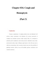
42 Mother and fetus
Correlation between birth
weights of relatives
a
N.E. Morton (1955),
Ann. Hum. Gen.
20,125 34;
b
E.B. Robson (1955),
Ann. Hum. Gen.
19, 262 8
Description of sample
Correlation of
birth weights
r
(
n
)
Maternal half-sibs (adjacent birth rank)
0.581 (30)
a
Full sibs (adjacent in birth rank, non-consanguinous parents)
0.523 (367)
a
Full sibs (adjacent in birth rank, parents first cousins)
0.481 (442)
a
Full sibs (one sib intervening)
0.425 (654)
a
Full sibs (two sibs intervening)
0.363 (153)
a
Paternal half-sibs
0.102 (168)
a
First cousins, maternal sisters
0.135 (554)
b
First cousins, maternal brothers
0.015 (288)
b
Fig. 2.3 The correlations between birth weights among relatives show maternal vs. paternal influ-
ences on this complex phenotype (birth weight) (r is the correlation coefficient, n is the
number of subjects). Correlations are stronger between siblings sharing the same mother
but different fathers than vice versa. The correlation in birth weight between maternal sib-
lings is also diminished as they are separated by one or more siblings in birth rank, suggesting
that maternal constraint processes change in magnitude with parity.
relatively tall adult sons even if their partners are short: maternal constraint limits
fetal growth to match maternal size, but postnatal growth can still be determined
by the paternal genotype.
The most famousdemonstration of maternal constraint comes from studies done
in the 1930s with horses. Breeders had known that depending on the choice of the
stallion and mare, certain characteristics would appear in the foal. So what would
happen if a large breed of horse, say a Shire horse of the type used traditionally to
pull the plough, was crossed with a diminutive breed such as the Shetland pony?
The cross could be done in two ways–aShetland mare with a Shire stallion or vice
versa. This experiment demonstrated dramatically that the size of the foal was very
different in each cross even though both crosses had similar genotypes (50 per cent
Shire, 50per cent Shetland).If a Shire stallion was crossed with aShetland mare (the
experiment was first performed using the then relatively new technique of artificial
insemination, which solved an obvious practical problem!), then the resulting foal
was closer in size at birth to a pure-bred Shetland foal than to a Shire foal. If on the
other handa Shire mare was crossed with aShetland stallion, then the foalwas much
43 Maternal constraint in late gestation
Newborn foals
Parents
Fig. 2.4 Outcome of crosses between the large Shire horse and the tiny Shetland pony, reported
by Walton and Hammond in 1938. A Shire mare crossed with a Shetland stallion produces
a foal of similar size to a pure-bred Shire foal (left). A Shetland mare crossed with a Shire
stallion, on the other hand, produces a much smaller foal similar in size to a pure-bred
Shetland foal (right). Maternal size determines the degree of prenatal constraint of fetal
growth.
larger and was closer to a Shire foal. These studies strongly suggested the presence
of maternal constraint: a Shetland mare in some way limited the growth of a fetus
with a genotype that would be too large to deliver through her pelvis. The reciprocal
cross also showed that the same genetic composition could lead to a larger fetus if it
grew in a larger uterus – this was the first suggestion that mammalian fetal growth
is normally constrained below its maximal rate by the uterine environment.
We do not fully understand the mechanisms of such maternal constraint. The
genomic imprinting of IGF-2 secretion and IGF-2 clearance receptors is one partial
explanation. But late in gestation IGF-2 seems less important as a fetal growth
regulator – although it may be more important for matching placental transport to
fetal demands. Instead, the closely related hormone, IGF-1, becomes the primary
regulator of fetal (as opposed to embryonic) growth. IGF-1 is not imprinted but
there is preliminary evidence that it too is extracted from the fetal circulation by
the placenta, particularly if levels get too high. Thus it may be that the placenta acts
as a ‘governor’, placing a maximum limit on fetal IGF-1 levels. The most favoured
explanation however is that uterine size is correlated with pelvic size and maternal
stature. The smaller pelvis and uterus thus have a smaller vasculature, which limits
the nutrient supply to the placental bed and hence delivery of nutrients to the fetus,
and this constrains fetal growth.
44 Mother and fetus
The phenomenon of maternal constraint has been shown elegantly in recent
years using embryo-transfer techniques in several species – while such studies
have eliminated genetic confounders in their interpretation they do not change
the conclusions reached. Data is now also available from studying the offspring
of human-assisted reproduction. One technique is that of oocyte donation where
the egg from one woman is harvested, fertilised with sperm in a test-tube and
placed as a fertilised embryo into the uterus of a recipient mother. This would
most likely happen where the recipient mother has an inability to make eggs. The
birth size of the fetus born in such scenarios correlates with the size of the recipi-
ent mother, again showing that the size of the uterus in which the embryo/fetus
growsdetermines birth size more than its genetic origin. Such embryo-transfer
experiments also argue against the sole operation of another genomic mech-
anism that has been suggested for maternal constraint – that is the role of
mitochondrial DNA.
14
In humans, maternal constraint operates in all pregnancies. However, some situ-
ations are associated with greater degrees of constraint than others. The most
obvious is small maternal size. In populations such as those of India where the
combination of genotype plus many generations of poor nutrition, infection and
disease have led to small skeletal size in mothers, most babies are subject to serious
maternal constraint, and the mean birth size is 25 per cent less than in developed
countries. Other major causes of maternal constraint are the first pregnancy and
maternal age. Multiplepregnancy isa special form of enhanced maternal constraint,
where the limited capacity to deliver nutrients is exaggerated by the greater demand
of twins or triplets.
It is well described that the first baby to a mother is on average smaller at birth
than hersubsequent babies by about 200grams. It isalso true both in humans and in
animals that adolescent mothers give birth to smaller fetuses than do mothers who
are fully mature. Both of these related situations appear to be examples of increased
maternal constraint. The mechanisms are not fully understood, although it may
be an erroneous assumption that the mechanisms of primiparous
15
and adoles-
cent maternal constraint are the same as those underpinning maternal constraint
associated with limiting the effect of the paternal genome. It is suggested from
work in sheep that in adolescent pregnancy the mother competes with the placenta
14
Mitochondria located in special organelles within a cell’s cytoplasm are important in cellular energy
homeostasis. They carry some DNA of their own, coding for a small number of genes involved in cellular
energy homeostasis. When cells divide, the mitochondria split to end up in the daughter cells. Because
sperm have no cytoplasm and eggs do, theonly mitochondrial DNA in fertilised eggs is of maternal origin.
As the fertilised eggs are the progenitors of all cells in the organism, all the mitochondria and thus all
mitochondrial DNA is of maternal origin. This is an interesting mechanism because it allows passage of
genomic information only down the maternal inheritance line.
15
Primiparous refers to the first pregnancy, multiparous to subsequent pregnancies.
45 Maternal constraint in late gestation
and fetus for substrates to complete her somatic (i.e. musculo-skeletal) growth.
As the placenta produces hormones such as placental growth hormone, which are
intended to alter maternal metabolism to favour nutrient supply to the fetus, it may
be in these situations that these hormones exert an inappropriate anabolic drive in
the mother.
An explanation for the reduced birth size in the first born is the impact of
pregnancy on the uterine vascular bed. The blood vessels in the non-pregnant
uterus are small and very tortuous, and blood flow is low. In pregnancy, under
the impact of oestrogensandprostanoidsmade bythe placenta, these vessels become
much more relaxed and dilated to permit more flow. Just as elastic bands are more
stretchableafter they have been stretched once, the firstpregnancy makesthe uterine
vesselsmore pliant in thesecond andsubsequentpregnancies. Greater uterine blood
flow and better placental bed formation are reflected in better nutrient supply to
the fetus.
This phenomenon of primiparous maternal constraint is now of great impor-
tance. For example in the Western world, where the number of children born has
fallen, over 50 per cent of babies are now from primiparous pregnancies, whereas
100 years ago the proportion would have been under 20 per cent. The impact of
changed family practices in China is even more dramatic and may be a time bomb
in relation to the changing pattern of disease – we will discuss this in chapter 8.
Thus theproportion ofbabies born where maternal constraint isa major feature has
risen, despite the increase in maternal size during this period. The high proportion
of teenage pregnancies is an additional contributor in many populations.
Maternal constraint is also seen in polytocous species, and is manifest in the
inverse relationship between fetal size and fetal number. For example, in the pig
the average size of a piglet in a litter of four is greater than in a litter of twelve. This
would suggest that there is a limitation in the supply of nutrients to the multiple
fetuses and this is part of the explanation of maternal constraint.
We see this echoed in humans with multiple pregnancy. Even allowing for
prematurity, the average size of triplets is less than that of twins, which in turn
is less than that of singletons. The rapid increase in multiple pregnancy in devel-
oped countries owing to increasing maternal age and, particularly, the increased
use of assisted reproductive techniques, needs to be considered.
Maternal constraint is thus a general phenomenon, and in chapter 7 we suggest
that its importance in evolutionary terms may be broader than just ensuring a
match between maternal size and fetal growth. However it is particularly in the
human that it may be of greatest importance as it allowed our ancestors to adopt
the upright position by balancing the needs of protecting fetal development against
the problem of too wide a pelvic canal, risking our abdominal contents prolapsing!
We discuss this in chapter 8.
46 Mother and fetus
Brain growth
Brain growth has quite a distinct pattern of growth from that of other organs.
The number of neurons is almost entirely determined in fetal life and is largely
completed in mid-gestation. Essentially no neuronal stem cell proliferation occurs
afterbirth, exceptfor a small amountinthearea of thebrain associated with memory
(the hippocampus). Neurons are the cells that carry out brain function but they are
supported and nourished by glial cells. These also largely develop in fetal life but the
peak of glial cell formation is somewhat later than for neurons, occurring in the last
weeks of pregnancy. Brain development is very complex – involving the processes
of stem cell proliferation, migration, axon and dendrite formation, differentiation
into neurons and glia, then differentiation into the myriad forms of neuron with
different neurotransmitters and the formation of billions of connections. There
is also a carefully coordinated pattern of cell death as neurons that do not make
connections are weeded out. Indeed neurons die at a great rate from fetal life and
throughout the rest of life.
This complexity means that the fetal brain is very sensitive to environmental
stimuli that might irreversibly damage it. Fortuitously much of brain function is
relatively plastic because of the redundant excess of neurons and connections, but
nevertheless we now recognise that many neurological and psychiatric diseases may
have their origin, in part, in fetal life. For example, autism is likely to be associated
with problems in forming connectivity properly in the fetal brain. Some forms of
schizophrenia are associatedwith a small head circumferenceatbirth suggesting that
aprenatal factor plays a role in some cases, although whether this is environmental
or genetic or both is not entirely clear. The fetal alcohol syndrome is an example of
atoxin interfering with the correct migration of brain cells within the developing
brain. Also oxygen lack or infection can cause irreversible damage to the brain and
lead to conditions such as cerebral palsy.
During fetal life many specific adaptations ensure protection of the blood supply
and oxygen delivery to the developing brain.
16
Considered as a proportion of body
size and energy consumption, the fetal brain is relatively larger than the adult brain,
even though much increase in brain size occurs after birth (largely as myelin – a
16
We now know that there are also mechanisms which ‘spare’ the developing heart and the liver. Adequate
cardiacfunctionis obviously essentialforhealth, butit isonlyrecentlythatliversparing hasbeen recognised.
The liver is a major source of growth factors and nutrients anditplays a role in determining the blood-lipid
profile. In fetal life the liver may also metabolise hormones such as cortisol that have crossed the placenta.
Blood returning from the placenta in the umbilical vein can be diverted to the liver, to maintain such
functions, or bypass it, which may assist the developing heart and brain. It is likely that such ‘liver sparing’
occurs when the nutritional challenge is mild but that this is overridden by ‘brain and heart sparing’ if the
challenge becomes greater. But even the liver-sparing response can produce detrimental consequences for
later health as will be discussed in chapter 6.
47 The physical phenotype at birth
fatty acid substance that helps electrical conduction along the axons of neurons –
forms in the brain). Some of these adaptations include the special structural shunts
in the liver and heart and great vessels that preferentially send the blood leaving
the placenta in the umbilical vein (which is oxygen rich)
17
directly to the brain.
For these reasons, when the fetus is born small the reduction in body size is often
disproportionate and there is a relative preservation of head growth – this is called
asymmetrical fetal growth retardation.
The physical phenotype at birth
It is the phenotype that interests every parent at birth. In most cases the mother (but
not necessarily the father!) knows the fullgenotype already (i.e. whois the biological
mother, who is the biological father). The first questions at birth are the same for
every parent – does the baby have ten fingers and toes, is it a boy or girl and how
heavy is it? The physical phenotype at birth (e.g. height, weight, head circumference
etc.) is a consequence of several factors – the genotype, the maternal environment
and the fetal responses to it, and gestational length. Obviously, premature babies
are smaller than term babies but, in turn, prematurity is more common in growth-
retarded babies.
Fetal and birth size are determined in thenormal fetus by theinteraction between
the genomic drive to grow and develop and the supply of nutrients from mother via
the placenta. Normal fetal growth reflects the interaction. In turn this interaction
is compounded by many transient variations in the fetal environment. In the sheep
fetus we know that just two days of undernutrition of the ewe will send a transient
nutritional signal to the fetus to reduce its IGF-1 levels and slow down growth.
On re-feeding with glucose, growth starts again. If there is a transient dip in fetal
oxygenation this too will transiently reduce IGF-1 levels and have a temporary
effect on growth rate. Maternal exercise can affect blood supply to the uterus and so
overzealous exercise in late gestation can affect fetal growth. Even how the mother
lies in bed in late pregnancy can be important because if she lies supine the weight
of her uterus may compress her abdominal vessels and reduce uterine blood flow.
What the mother eats can affect placental nutrient transfer. Maternal health status
(e.g. infection), her macro-environment (e.g. altitude) and her behaviour (e.g.
smoking or drug taking) all impact on the fetal environment and thus on the fetal
pattern of growth. Thus in every pregnancy there are many environmental factors
that can lead to transient changes in fetal growth and may affect birth size.
17
The umbilical circulation is the one circulation other than the postnatal pulmonary (lung) circulation in
which oxygen content is higher in a vein than in an artery.
48 Mother and fetus
Abnormally impaired fetal growth generally occurs for one of several reasons.
18
It may occur because of gross genetic abnormalities – for example, a mutation in the
IGF-1 gene or its receptor will cause severe fetal growth retardation because IGF-1
is the most important fetal growth factor in the second half of pregnancy. Insulin is
the other major fetal growth factor (acting in part by regulating IGF-1) and genetic
defects in the insulin receptors or its action can also cause severe fetal growth
retardation. Toxins from smoking or toxic infections such as rubella that interfere
with the fetus or placenta are major causes of intrauterine growth retardation.
In tropical countries malarial infestation of the placenta is of particular concern
because it interferes with normal placental function, and the parasite load utilises
energy that should otherwise be available for delivery to the fetus. However, most
cases of impaired fetal growth are caused by placental interruption of the supply
line – poor placental function associated with pre-eclampsia is a very common
cause of intrauterine growth failure. Interruption to the supply line can be at many
levels, from the maternal blood supply to the uterus (e.g. maternal heart disease) to
anatomical problems with the umbilical cord delivering nutrients from theplacenta
to the fetus.
Finally some small babies are just small because they have small mothers, not
because of any specific disease state.
Fetal growth must be seen as an integrated readout of the many gene–
environmental interactions that happen during fetal life. Depending on when in
pregnancy the fetus changes its growth rate in response to an external cue, the
birth-size phenotype might be affected differently – it is generally stated that late in
pregnancy, when the fetus has considerable soft tissues (fat, viscera, muscle), fetal
undernutrition leads to a thin baby, whereas earlier in gestation undernutrition
of the fetus will affect linear growth as well: thus the baby will be shorter and
lighter. Head growth is relatively protected because the fetal adaptations of chang-
ing regional blood flows attempt to preserve blood supply to the fetal head at the
expense of the trunk in adverse situations. In reality this is a gross simplification
19
but it is easy to see how insults or circumstances at different times can interact
to give avast plethora of different birth-size phenotypes – fat or thin, long or
short, large or small head, large or small abdomen (reflecting liver growth) etc. If
only we could read accurately what the fetus was telling us from the detail of its
18
Birth size must be interpreted relative to gestational age. There are normal standards for various measures
of birth size – weight, length and head circumference being the most common. Intrauterine growth
retardation (IUGR) is a term used to describe birth size outside the normal range – the alternative term is
small for gestational age (SGA). We prefer the latter as IUGR implies that the mechanisms ofthe reduction
in birth size are known andthatitis pathological, whereas not allsmallfetuses are necessarily pathologically
growth impaired.
19
Forexample recent studies have shown that altered maternal food intake at conception can alter the
development of a variety of hormonal systems in the fetus but these only become manifest in late gestation
as reduced fetal growth.
49 Developmental plasticity
birth phenotype, we might know much more about how it had integrated its envi-
ronmental experiences from conception to birth with the genomic information it
inherited at conception.
Birth size can also be increased under some conditions. One way is genetic. For
exampleiftherehasbeenafailureofsuppressionofoneofthetwocopies of the IGF-2
gene by imprinting the maternal copy, then the fetus expresses twice the normal
amount of IGF-2: – this is the cause of the rare Beckwith–Weidemann syndrome,
which leads to a very big baby at birth witha number ofhormonal abnormalities and
an increased risk of some forms of childhood cancer. But the commonest cause of
big babies is excessive glucose supply to the fetus. This happens either if the mother
is diabetic or prediabetic. Because glucose supply to the fetus is so important during
pregnancy, the placenta makes hormones that induce some insulin resistance in the
mother. She thus primarily uses fat for her own energy needs and gives the glucose
to the fetus andplacenta. But if the mother has a latent diabetictendency, this will be
exposed by these placental hormonal changes and insulin resistance induced, and
thus glucose delivery to the fetus becomes excessive. But bone and muscle growth
primarily require amino acids, and the supply of these remains somewhat limiting.
High levels of glucose in the fetus make its pancreas secrete more insulin and this,
together with high energy availability, promotes storage of the excess energy as fat.
Thereforethe fetusof adiabetic mother hasonly a smallincrease in muscle and bone
size (i.e. length) but is rather obese and has a high birth weight. The implications
of this are discussed further in chapter 8.
Developmental plasticity
It will be clear to the reader that the development of the mature organism from
a single egg can take multiple paths and lead to a range of phenotypes. These
different pathways result from the interaction of the environment with the genome
both before and after birth. The mechanisms involved can encompass any of the
developmental processes we have already referred to – changes in cell number, in
proliferation rate, in differentiation, in gene expression, to name but a few. The
capacity of cells, organs and systems to change their function or state is referred
to as plasticity. Essentially development is all about plasticity as the embryo starts
as a single cell and ends up as a mature adult with fully differentiated organs and
cell types. This global set of plastic responses is termed developmental plasticity
but plasticity is not limited solely to development. Some tissues remain plastic
throughout life – for example, although muscle cells cannot change in number in
adulthood they can change their characteristics and size when subject to repeated
exercise, such as in body-builders. Equally such hypertrophied muscles return to
areduced state when not exercised. This is reversible plasticity. A skin wound in
50 Mother and fetus
adults or older children leaves a scar: the skin proteins and cells that form the scar
derive from the same cells that form normal skin and its underlying proteins, so to
this degree they are plastic; but the scar, once formed, does not disappear – this is
an example of irreversible plasticity.
Developmental plasticity is generally irreversible. At the most obvious level,
once a stem cell has been committed to a lineage – say of ectodermal cells (e.g. liver
cells) – that is an irreversible commitment of that cell, and its progeny must be
liver cells. The basic programme for development from a single cell to a complete
organism is obviously entirely located within the genome of the fertilised zygote,
so developmental plasticity itself is inherently genomically determined. However
it is the capacity of the direction, timing or magnitude of each developmental
plastic event to be influenced by the environment that leads to the multitude of
phenotypes possible fromasingle genotype and that is thefocusofthisbook.During
early development many environmental influences can act on the programme of
embryonicandfetal development to amend or influence plastic change. For example
exposure to the male sex steroids at a point in development when the genitalia
are forming can lead to permanent changes in their appearance. While we have
focused here on structural plasticity, plasticity can be functional, e.g. in the set-
point of a physiological process. For example there may be a permanent change in
the expression of a gene controlling the number of receptors for a hormone and
this will be reflected in altered hormone sensitivity.
Permanent plastic responses of this type play an important role in the develop-
mental origins of adult disease, which we will discuss in chapter 6.
Critical windows of development
Birthdefects and malformations can arise in one of two ways – either there is a
genetic defect so that the orchestrated programme of development is defective,
or there is an environmental factor (called a teratogen) that somehow affects the
genetic programme. Studies ofthe latter give us insight into an importantconcept in
development – that is of critical windows.Thalidomide is an example of a teratogen,
and its effect in producing limb and hand abnormalities was critically linked to
when in pregnancy the mother ingested it: the earlier in pregnancy the more gross
the limb malformation. While most teratogens are drugs or chemicals, infectious
agents such as rubella can also be teratogenic leading to malformations of the fetal
heart. Although the teratogenic effects of many drugs are clearly understood, there
remains much uncertainty about xenobiotics such as pesticide residues etc.
Te ratogens act by interfering with the developmental programme at a critical
point in time to cause an overt anatomical malformation. This might arise as an
acute insult,e.g. from exposure to a drug or chemical for only a short period oftime.
51 Critical windows of development
POSITION
OF MEATUS
AGENT
WEEK OF GESTATION
6
10 14 18 22
2
MEDROXYPROGESTERONE
MEDROXYPROGESTERONE
NOR-ETHISTERONE
UNKNOWN
HYDROXYPROGESTERONE
CAPROATE
NOR-ETHISTERONE
MEDROXYPROGESTERONE
NOR-ETHISTERONE
METHYLESTRADIOL
NOR-ETHISTERONE
ETHINYLESTRADIOL
NOR-ETHISTERONE
Fig. 2.5 Cases of hypospadias in male offspring related to time of beginning of maternal treat-
ment with various estrogens and progestins. Note that the closer the time of treatment to
beginning of penis formation the more severe the hypospadias. Redrawn from Schwarz and
Yaffe (1980).
An example of this can be seen in the male fetuses exposed to certain hormonal
medications containing forms of progestogenic steroids. Between the seventh and
twelfth week of pregnancy the male fetus develops his penis under the influence of
a derivative of the male hormone testosterone made by the fetal testis. The urethra
is the urinary channel that in the mature penis exits at its tip. As the penis develops,
the urethra first opens at the base of the penis and the penis gradually wraps around
the urethral tube – so that as the penis matures the urethral opening progresses
along itsunderside until it reaches the tipat about thetwelfth week after conception.
The progestins act as anti-testosterones and can cross the placenta. If the mother
is exposed to such progestins between the seventh and twelfth week
20
then the
maturation of penile growth is interfered with and the urethra opens not on the tip
but underneath the penis – this is called hypospadias. The degree of hypospadias
can be related to when exposure to steroid occurred – the closer to 7 weeks the
closer the urethral opening is to the base of the penis, the closer to 12 weeks the
closer to the tip of the penis. But after 12 weeks, when the penis is fully formed,
progestins and oestrogens have no effect on penile development. Thus the critical
window for affecting penile development is 7 to 12 weeks.
Another example of a critical window relates to the sexual behaviour of rats. As
in most species, rats have quite distinct patterns of sexual behaviour, with males
taking mounting positions and females submissive positions. This behaviour is
controlled by a part of the brain known as the hypothalamus. The hypothalamus
is crucial for the regulation of reproduction and other hormonal mechanisms such
20
They used to be prescribed as drugs to reduce the risk of miscarriage.
52 Mother and fetus
as the response to stress. In the rat, which has a very immature brain at birth,
the development of the hypothalamus largely occurs after birth. If a female rat
pup is exposed to the male hormone testosterone between three and ten days after
birth, the hypothalamus will develop like a male hypothalamus, and at adulthood
the female will show male-like mounting behaviour. Conversely, a male rat pup
castrated before this critical period will not develop male behaviour. But if a female
is exposed to testosterone before 3 days or after 12 days it will have no effect on
her adult sexual behaviour. Obviously there is a critical window in hypothalamic
development when exposure to themale hormone will irreversibly produce changes
in wiring leading to adult male-like behaviour.
It is much more debatable whether such a window relating to sexual behaviour
occurs in humans – if it did it must occur some time in fetal life as the human
brain is much more mature at birth than the rat. There have been claims that girls
born with adrenogenital syndrome (a genital defect in the way the adrenal gland
makes steroid hormones sothat itmakes more androgens than cortisol) have altered
psychosexual development and that this in turn reflects the elevated androgen levels
they were exposed to in utero. East German scientists, prior to the destruction of
the Berlin wall, presented data to suggest that male fetuses exposed to stress in utero
had lower testosterone levels and they claimed this was the biological origin of male
homosexuality. This claim may have better suited theideals ofMarxism, butneither
observation nor claim has achieved standing in the scientific community.
Two important questions arise from the above discussion. First, are there insults
that cause less obvious but nevertheless permanent changes in function if the fetus
is exposed to them in utero? Second, are there environmental factors that affect
the developmental programme in the later part of pregnancy, after organ for-
mation is largely complete, but while functional development is still occurring?
We will primarily address both questions in later chapters but the short answer
to both is ‘yes’. There are many functional abnormalities that have their origin in
environmental factors acting in early development. Furthermore, while gross organ
development is achieved in humans in the first half of pregnancy, the finer aspects
and functional maturation extend throughout the fetal phase into childhood, and
permanent changes in function can beachievedbyenvironmental factors operating
throughout this period.
Prematurity: an alternative strategy
Prematurity has a variety of causes. Some cases are due to infection ascending the
cervix and entering the amniotic fluid and membranes. This leads to an inflamma-
tory response causing induction of prostaglandins in the uterine muscle, leading to
contractions. Perhaps 40 per cent of cases of premature labour have such a cause.
53 Maternal nutrition
The origin of the ascending infection is unknown – one study has suggested that
intercourse late in pregnancy might be a factor but there is disagreement about
this. Most premature labour has an unknown cause although recent studies in
sheep raise the possibility that at least in that species it has its origin very early in
pregnancy.
Babies who are born premature often seem more mature in some functional
aspects than we would expect. This is even more so the case in growth-retarded
fetuses born prematurely. This is because the fetal period prior to premature labour
may have been associated with higher cortisol levels in the fetal blood than a normal
pregnancy at the same postconceptionalage.Arise in cortisol (the primary hormone
made by the cortex of the adrenal gland) occurs in the blood of the fetus prior to
delivery in all mammals. After birth, cortisol is best known as a stress hormone and
it plays a major role in regulating blood pressure, fluid balance and blood glucose
levels and assisting the body in responding to stress. It also reduces the immune
response, hence its powerful medical use as an anti-inflammatory drug.
The rise in fetal cortisol is responsible for the maturation of many organs before
birth. Elevated cortisol levels are therefore likely to be the cause of the apparently
greater maturity in the prematurely born fetus. In some species such as the sheep,
the linkage between maturation and delivery may be quite direct. In these species
the rise of fetal cortisol levels isan active part of the cascade ofbiochemical pathways
that leads to the initiation of the uterine contractions and cervical relaxation that
constitute labour.
These observations in premature and growth-retarded fetuses reflect the two
strategies the fetus has in adverse situations – to grow more slowly (or differently)
in order to conserve nutrients, or to accelerate maturation through cortisol release
and then deliver prematurely. In reality these two options are a continuum – many
growth-retarded fetuses have accelerated maturation of some organs such as the
lung owing to the effects of elevated cortisol but still have not delivered prematurely.
Maternal nutrition
Maternal behaviour can do much to determine fetal outcome. At one level this
statement reflects the obvious point of avoiding toxins such as cigarettes and drugs
of abuse and promoting maternal health. But it is becoming increasingly clear that
at a more subtle level the fetus is constantly responding to signals from its mother
that relate to her nutritional status. This brings us to the vexed but critical question
of maternal nutrition. Fetal nutrition is not the same thing as maternal nutrition
and it is not simply a reflection of maternal nutrition. Maternal nutrient status is
but one factor in determining the supply of nutrients to the fetus. But the subtleties
of the maternal metabolic state, reflecting in turn her nutritional status, may be the
54 Mother and fetus
most important signals to the fetus about the environment it is going to be born
into.
We still know frustratingly little about the best maternal nutrition for achieving
optimal pregnancy outcome. Both micronutrient supply such as the provision of
folate and other vitamins, minerals such as iodine and zinc, and the amount of, and
balanceof,macronutrients (protein,carbohydrates and fats) have a major impact on
the outcomes of pregnancy. For example, extreme iodine deficiency in the mother
leads to cretinism in the offspring and this is a key reason why salt is iodised. Folate
deficiency plays a role in neural tube defects such as spina bifida, and we suspect
that micronutrient availability plays an important role in the phenomenon of PARs
through much more subtle effects including the regulation of epigenetic changes.
Macronutrient supply is also critical. We are yet to identify the best mix of
macronutrients for different periods in pregnancy and possibly for different popu-
lations. The evidence is mounting that both the absolute and relative amounts
of macronutrients are important. History has taught us this. In a reprisal for the
activities of the Dutch resistance in late 1944, the Nazi authorities imposed severe
rationing onthe population. The mean caloric intake was reduced almost overnight
fromabout 1800 to between 400and800calories per day.TheDutchHunger Winter,
as this episode has been termed, lasted for seven months until the Allied forces
liberated Holland in 1945. Food intake was returned to adequate levels almost
instantaneously. While famine is sadly not uncommon in many parts of the world,
what is unusual about the Dutch Hunger Winter is, first, that the famine was
imposed on a previously well-nourished population; second, there was a sudden
onset and relief from the famine; and third, in places such as Utrecht, despite the
adversities of the wartime occupation, midwives and doctors continued to offer a
professional obstetric service andto keep detailedrecords ofbirth weights andother
relevant data. Subsequently it has been possible to analyse the pregnancy outcomes
of women caught in this famine in terms of those who delivered before the famine,
who conceived during the famine, or were in early, middle or late pregnancy at
the time the famine was imposed. Initial studies suggested that unless the maternal
food intake was less than 800 calories per day the fetus could grow adequately and
there were no real consequences of mild or moderate maternal undernutrition. As
we will discuss in chapter 4 we now suspect this was an incorrect conclusion. Many
of those more subtly affected as fetuses have gone on to develop health problems
in later life that we now believe are a result of the nutritional stresses they were
exposed to before birth.
The first clues that macronutrient balance may also play a role came from stud-
ies in food-rationed populations in Scotland after the Second World War. There
was a presumption that more protein must be better, and studies were performed
of giving supplementary protein in the form of meat. Paradoxically under some
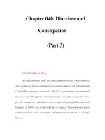
55 Growth and maturation after birth
Fig. 2.6 Conditions were extremely harsh for the people of the Netherlands exposed to the near-
famine conditions imposed by the Nazis in the winter of 1944/45. The effects of the poor
and unbalanced diet of women during pregnancy on their children and grandchildren have
been studied in detail. Photograph courtesy of the Dutch Institute for War Documentation.
circumstances this led to smaller, not larger fetuses although in other circum-
stances it did assist fetal growth. More recent studies from Southampton show that
the mix of dairy and meat products and the balance of protein, carbohydrate and
micronutrients such as folate can have important influences on the patterns of fetal
growth. The reality is that we actually know very little about what is the optimal
nutritional mix for a mother at different stages in pregnancy and in different cir-
cumstances. A major message of this book is that this is an area needing priority in
research. It is a topic we will return to in chapter 10.
Growth and maturation after birth
Afterbirth, growth continues in several distinct phases. In infancy, it is still largely
determined by the mother through either the supply of breast or bottle milk. While
maternal constraint has been removed, nutrient availability remains the dominant
56 Mother and fetus
regulating factor and the hormones primarily involved in regulating infant growth
are similar to those involved in prenatal growth – that is insulin and the insulin-like
growth factors (IGFs). But as the infant grows into childhood there is a gradual
switch in the hormonal regulation of growth. Somewhere between 6 and12 months
after birth, growth hormone (made by the pituitary gland) becomes the dominant
regulator ofgrowth andtakes over regulation of IGF secretion from the direct effects
of nutrition itself. The patterns of growth-hormone secretion between individuals
are relatively distinct. They are genetically determined to a degree and it would
appear that both maternal and paternal genetic factors are much more important
in determining postnatal than prenatal growth. This is why postnatal growth tends
to follow a given trajectory for an individual child and, if transiently knocked off
that trajectory by illness, the child generally returns to it. Hence final adult height
is closely correlated with parental height.
Growth in infancy and childhood remains exquisitely sensitive to environmental
factors. Infant diseases such as infection can reversibly impair growth and, if fre-
quent enough or severe enough, infant and childhood illness can lead to irreversible
stunting of the child. Emotional stresses can impact on growth-hormone secretion
and impair growth. And of course adequate nutrition is essential to normal growth.
Aspecial form of growth is called catch-up growth (the opposite being catch-
down growth). After an insult, when growth has been transiently impaired, there
may be a period of accelerated growth that returns growth to the original trajectory.
How this catch-up growth isregulated is poorly understood. Many children who are
born small due to a constrained intrauterine environment show catch-up growth in
infancy when relieved from the physiological and pathophysiological constraints in
utero. Some children who have grown excessively in utero, often because of maternal
diabetes, show catch-down (i.e. slower) growth after birth in order to reach their
genetically determined growth trajectory.
Relative growth in length and weight do not go in parallel. The change in relative
weight for length is reflected in measures such as ponderal index and body mass
index
21
,which are measures of weight for length. As the size of skeletal muscle in
childhood is generally proportional to bone mass and thus length, these measures
are somewhat crude measures of relative fatness. Relative fatness varies at birth –
low fatness being a major indicator of a sub-optimal fetal environment. Catch-up
in fatness and in length do not go in parallel, and the processes regulating these
growth patterns are not understood. Generally, fat gain occurs earlier than height
gain and the height gain may never be complete: children who are stunted as a
result of malnutrition when re-fed often end up with truncal obesity but are still
short.
21
Ponderal index is weight/height
3
and body mass index is weight/height
2
.
57 Growth and maturation after birth
Forreasons we do not fully understand, humans are the fattest of all species at
birth. Perhaps it is to ensure a fuel reserve to protect the high energy demands of
the disproportionately large human brain. This places extra demands on maternal
nutrient supply to the fetus in late gestation when the fetus is laying down fat. In
relative terms, this adiposity decreases over the first 2 to 3 years of life (perhaps
because it is used for brain metabolism) but then fat is again deposited during
childhood; from then on it is easy to get obese! This ‘adiposity rebound’, as it is
called, varies among individuals and populations, but as we will see later its timing
may have important consequences for subsequent health.
In late childhood, additional hormonal changes occur. The adrenal gland starts
to make, at least in humans, adrenal male-like hormones (androgens) and then the
gonads under hypothalamic control start to make either male or female hormones
according to the gender of the individual. This accelerates linear growth but at
the same time leads to development of the secondary sexual characteristics (breast
development, penile enlargement, pubic and axillary hair). Linear growth occurs at
special zones at the ends of the shafts of the long bones, called growth plates. These
are made of cartilage and at one side of the plate the cartilage cells multiply to form
columns, while at the otherend they convert from cartilage to bone, thus elongating
the shaft. As sex-steroid levels stay high over time, the growth-plate cartilage cells
convert into bone cells and the growth plate disappears; linear growth is then over.
This is not universal in all mammals, and some rodents continue to grow, albeit
slowly throughout life. Even after linear growth is over in humans, other tissues
continue to develop over some years, e.g. muscle continues to increase in bulk
by adding protein within the cells for some time. Psychosexual and psychosocial
maturation continues through adolescence.
Fatoradipose-tissueproportions also change greatly through development. Both
tissues have their cell number largely determined in fetal life, although recent evi-
dence questions this in relation to the regulation of fat-cell number. After birth
the relative amount of fat and muscle is largely a matter of how much protein
exists within a muscle cell and how much fat exists within a fat cell. Both are very
labile tissues and hormones such as insulin, growth hormone and cortisol regulate
whether fat is being laid down or mobilised, and whether protein is being laid down
in muscle and in the liver or being metabolised. The regulation of this partitioning
is essentially how metabolic homeostasis is maintained – that is how blood glucose
levels, plasma lipid levels etc. are regulated.
22
22
Insulin acts mainly on three tissues – liver, fat and muscle – to promote storage of fuels, and in every
cell type to promote glucose uptake for energy consumption. Insulin increases the conversion of glucose
into glycogen (the storage form of glucose in the liver), enhances fat deposition in fat and amino acid
incorporation into proteins in cells. Insulin’s actions are counteracted by other hormones so that blood-
glucose levels are maintained in a narrow range. When insulin deficiency occurs or insulin resistance is
58 Mother and fetus
In this chapter we have seen that development is an extremely dynamic process. It
is not simply determined by the genotype, and so does not occur solely in the form
of an encapsulated, hardwired developmental ‘programme’. Instead, the develop-
mental programme can be influenced by a range of environmental conditions and
these interactions can have long-term consequences for later health and disease. We
have to be extremely careful not to view body size and shape at birth as measures of
developmental outcome, especially in termsofthe underlying physiological func-
tion. The baby at birth has come a long way, and indeed may have arrived at the
same point down one of many paths. The detailed structure of its organs, their
function and the homeostatic processes that govern them, are obviously not visible
to the naked eye. But already the individual’s biological destiny is written in the
birth phenotype.
induced, diabetes ensues. Type 1 diabetes mellitus occurs when the pancreatic cells making insulin are
damaged by autoimmune processes. Type 2 diabetes occurs when the body’s cells become resistant to
insulin and the pancreas cannot keep making enough insulin to overcome the resistance. Insulin is a
hormone and it binds to receptors on the cell surface. In turn this stimulates a complex chain of events
within the cell that ultimately lead to changes in cell function. There are several ways in which insulin
resistance can be induced. One element is obesity, because insulin works less well when fat cells are
distended with fat. High fatty acid or lipid levels in blood also alter the function of insulin receptors and
can cause insulin resistance.
3
Fetal choices
In the previous chapter we described how the fetus responds to environmental
change to aid its survival. Many of these responses are transient and functional,
that is homeostatic, responses – for example the cessation of fetal breathing move-
ments to conserve oxygen. Others involve changes in developmental trajectories –
for example altered fetal growth to conserve nutrients. The latter often involve
irreversible developmental plastic changes and thus have potentially long-lasting
consequences. We will pursue this theme in this chapter and also introduce another
concept: namely that the fetus might make some adaptive responses not for imme-
diate survival advantage, but in expectation of future advantage after it is born, by
changing its developmental programme before it is born. This is the concept we
have termed predictive adaptive responses.
Fetal strategies
Fetal development is of course not an end in itself. In evolutionary terms, the
sole point of the process of development from embryo to adulthood is to achieve
competence to reproduce. The author Samuel Butler made the point well when he
phrased the aphorism that the ‘chicken is just the egg’s strategy for making another
egg’. The embryonic and fetal phases of development must be survived, the fetus
must attain independent lifeat birth and thechild must pass through puberty –these
are the necessary steps on the trail to reproductive competence. But as we have seen,
the birth phenotype can be highly affected by the environment of the developing
fetus. In turn this can have life-long phenotypic consequences, particularly because
many of the plastic changes made during development are irreversible.
Darwin realised that both sexual selection and natural selection gain importance
in evolutionary terms through reproduction, because only the selected members
of the species reproduce and thereby pass on their characteristics to the next gen-
eration. Placed in this context it is possible to understand that evolution might
59
60 Fetal choices
Metamorphosis
Terrestrial
Aquatic
larval gills
Fig. 3.1 The process of metamorphosis from the aquatic to the terrestrial forms in some amphibians
such as the Mexican axolotl (a form of salamander) can be triggered by external environ-
mental influences. Thyroxine applied to the water can do this experimentally. In the wild,
such processes (called neoteny in this case) show how developmental plasticity provides an
adaptive strategy to cope with environmental challenges, such as a pool drying up. Redrawn
from Principles of Development, Wolpert et al. (eds.). Oxford University Press, 2nd edn.
enable the selection of characteristics by which the embryo or fetus can make
strategic choices that would give the adult a greater chance of surviving the rigours
of postnatal life, allowing it to achieve reproductive competence. Of course these
strategic choices are not conscious, but the use of the word ‘choice’ is useful because
it implies that there may be costs as well as benefits to the option adopted.
This phenomenon is dramatically seen in amphibiansandsomereptiles. Tadpoles
of many species of frogs and toads develop in ponds that are at risk of drying up.
Forexample the spadefoot toad lays its eggs in rainwater puddles in the Texas
desert. Clearly if they are still tadpoles when the pond has evaporated, they die.
So to survive, these species have evolved the capacity to change the timing of their
metamorphosis into the mature form of toad when they sense the pond is drying
up. Other species of toad will hasten their metamorphosis if, as tadpoles, they sense
they are at higher risk of predation from dragonflies. There is a price to this strategy
however, in that the terrestrial forms have a reduced body size that may have a cost
later in life. These are examples of adaptive plasticity where there is an obvious
advantage to changing the rate of development.
In some salamanders these issues of environmentally induced metamorphosis
are even more dramatically demonstrated. Some such as the axolotl may stay in the
infantile (non-gilled form) throughout life, and only under situations where they
are forced to leave the pond do they undergo metamorphosis to the legged form.
Obviously the early environment is acting on the genotype to determine something
as striking as whether legs will form or not!
These examples show that the immature organism can indeed make adaptive
responses for immediate survival advantage that can also have life-long conse-
quences. The responses occur within the lifetime of the organism and, while
61 Embryo and fetal predictions of future environment
genetically determined (in that having the capacity to make the response must
have been selected), they are clearly purely environmentally induced. This is in
contrast to the rabbit’s ears or Darwin’s finches that we discussed in chapter 1,
where the change happens across generations and thus primarily operates through
classical Darwinian selection processes. Unlike amphibians and reptiles, mammals
in general appear to have a more limited repertoire of gross morphological change-
strategies that they can adopt. But as we shall see, developmental choices about
body size and body composition are made early in life in every generation in many
mammalian species, including Homo sapiens.
In chapter 1, we referred to the effect of season on the coat thickness of newborn
voles.Butmany of these mammalian choices aremuchmoresubtlethanjustexternal
appearance. They may involvebeingrelativelyresistanttoahormone such as insulin,
to changing the pattern of stress hormone release etc. The central feature is that
these are ‘strategic choices’ that can be made early in life by the fetus. While in
some cases, as for the timing of metamorphosis, they have immediate survival
advantage, in other situations such as the meadow vole’s coat thickness the choices
are made in anticipation – that is, in response to predictions of the nature of the
future environment, so that the organism will maximise its chances of survival in
its predicted future environment. Thus the key question becomes, ‘How can the
embryo or fetus predict its future environment?’
How can the embryo and fetus predict its future environment?
There are obvious differences between the environment of the mammalian
embryo/fetus (echidnas and platypuses excepted) and that of the larval forms of
insects and the egg-laying vertebrates.
1
While we can learn from the latter, we shall
restrict further consideration to mammalian species because our focus is to under-
stand this biology with reference to the human, and thusits role in the determination
of our health and disease.
As detailed in chapter 2, environmental ‘perception’ by the mammalian
embryo/fetus is largely dependent on the mother and the placenta. They act, in
series, as two transducers of information about the external environment to the
fetus. The crucial point is that the levels of nutrients passing into the fetal blood
from the placenta act as vital coded messages about the external environment,
which the growing fetus utilises to make strategic adaptive choices. It is intriguing
that such choices depend on the levels of nutrients themselves, rather than on a
special surrogate signal that has evolved for the purpose. Perhaps this represents an
economy in nature – what could be simpler than using nutrient levels to signal one
1
Intriguingly some live-bearing lizards and skinks also have a placenta-like structure by which nutrients are
transferred from mother to offspring, suggesting that placentas evolved more than once.
62 Fetal choices
of the most important aspects of the environment: nutrition
2
itself? But we will also
see that other non-nutrient signals, especially blood hormone levels, play a part in
environmental signalling. As we might expect, they can signal information about
many aspects of the environment, including the global nutritional environment in
which the mother is living.
The fetus responds to this maternally transduced environmental information,
with transient homeostatic responses as were described in chapter 2 or, under some
situations, by making some form of developmental plastic response. In turn this
response may have immediate adaptive value, like the decision to accelerate partu-
rition if the maternal environment is harmful (for example an infected uterus
3
).
Other responses, as we shall see, have less obvious immediate value but have clear
longer-term postnatal advantage, provided that the predicted future environment
matches the environment as transduced by the mother and placenta to the fetus.
So the fetus/embryo uses this environmental information in two ways – firstly, to
make immediate survival-related choices and secondly, to make long-term predic-
tions to maximise its advantage postnatally. The postnatal advantage can be defined
in Darwinian terms as choosing the best physiological trajectory for development
in the predicted postnatal environment to achieve maximal reproductive fitness.
This can be illustrated by reference to fetal nutrition and fetal stress. If the fetus is
exposed to a long period of reduced nutrient supply, it might expect to be delivered
into a world where nutrient supplies are low. Or if it experiences a high blood
glucose level constantly, it might anticipate being born into a carbohydrate-rich
environment. Or again, if its mother has continuously high blood cortisol levels,
then so will the fetus, and it might anticipate being born into a stressful envi-
ronment. None of this is an active decision-making process. It is the outcome of
evolutionary processes that have defined the capacity to adapt as a valuable aspect
of the survival of the species: indeedthese adaptive mechanisms are present in every
species examined. The processes could not occur if it were not for the phenomenon
of developmental plasticity. Otherwise there could be no long-term permanent dif-
ference in phenotype as a result of transient environmental change acting in fetal
life.
Thus how the fetus perceives its environment is critical both to its immediate
survival and also for its long-term adaptive advantage in the environment in which
2
It is important to re-emphasise what we mean by ‘fetal nutrition’. The fetus primarily uses substrates such
as glucose, amino acids and oxygen coming across the placenta from the mother. The fetus does swallow
amniotic fluid and can absorb some nutrients from this source but amniotic fluid is in part formed by
secretions from the fetal lung and fetal urine; there is no significant nutrient transfer from mother to fetus
through this route, although there may be some recycling! Fetal nutrition is thus the sum total of nutrients
the fetus receives from the placenta. This is not a directreflection of maternal nutrition. Nutrients do not
cross the placenta with equal efficiency and, as we discussed in the previous chapter, the placenta itself is
an active metabolic organ.
3
This is a common cause of premature labour.
63 When to choose
it anticipates living after birth. The mother and placenta act as transducers of
aspects of the environment to the fetus. As with any electronic or mechanical
transducer – for example a smoke sensor used in a fire-detection system – there
is a risk of malfunction. If the mother has diabetes mellitus that is not properly
controlled, she will have high blood glucose levels. These will be reflected in high
blood glucose levels in the fetus and the fetus will adapt in the expectation of high
nutrient availability postnatally. But the mother might very well be living in famine
conditions in the Horn of Africa. There would then be a gross mismatch between
fetal nutritional expectations and the reality of the environment in which the child
will live after birth. Similarly if the mother has heart disease, the fetus might adapt
in the expectation of being born into a low-oxygen environment when it is not –
a similar thing happens in the infant of a mother who smokes during pregnancy,
where her elevated blood carbon monoxide levels produce the same effect as having
low blood oxygen levels.
In these examples, the mother is incorrectly signalling information about the
environment to her fetus. Similar faulty signals can arise from the placenta, for if
it is abnormal, the transduction of the maternal/external environment to the fetus
will not be accurate. If the placenta has poor perfusion with maternal blood, then
the supply of oxygen and glucose to the fetus will be reduced. This will lead to
reduced fetal growth as an immediate adaptive survival response. It will also lead to
adaptive responses by the fetus in the expectation of being born into a nutritionally
deprived postnatal environment – it is the last of these responses that is predictive.
When to choose
While so far we have focused on the embryonic/fetal period, there is mounting
evidence that some predictive responses can be initiated even prior to this, perhaps
even as an egg before fertilisation. In rats in which nutrition was altered only for the
period prior to conception there were irreversible consequences for the progeny
throughout life (see chapter 5). There is increasing evidence that the conditions
in which in vitro fertilisation is conducted can have long-term consequences, at
least in farm animals.
4
The human correlates of this in modern reproductive tech-
nologies are poorly documented but there are data suggesting comparable phe-
nomena. The data show that environmental influences acting in the earliest period
of development can have echoes throughout life. Some of these might have imme-
diate adaptive value, although it is extraordinarily difficult to imagine how that can
be the case for the egg even before it is fertilised. Indeed it seems probable that
4
The alleged accelerated ageing of Dolly the sheep again suggests that the origin of the embryo (in this
case by a unique form of cloning) can have long-term consequences, although Dolly’s origin is so bizarre
(in biological terms) that we hesitate to use this as a definitive example of the point we are making.