Heart Failure - part 2 pdf
Bạn đang xem bản rút gọn của tài liệu. Xem và tải ngay bản đầy đủ của tài liệu tại đây (305.13 KB, 34 trang )
40. Masoudi FA, Rathore SS, Wang Y, et al. National
patterns of use and effectiveness of angiotensin-
converting enzyme inhibitors in older patients
with heart failure and left ventricular systolic
dysfunction. Circulation. 2004;110:724–731.
41. Smith NL, Chan JD, Rea TD, et al. Time trends in
the use of b-blockers and other pharmacotherapies
in older adults with congestive heart failure. Am
Heart J. 2004;148:710–717.
42. Masoudi FA, Gross CP, Wang Y, et al. Adoption
of spironolactone therapy for older patients with
heart failure and left ventricular systolic dysfunc-
tion in the United States, 1998–2001. Circulation.
2005;112:39–47.
CHAPTER 2 THE EPIDEMIC OF HEART FAILURE––––––19
This page intentionally left blank
Chapter 3
What Causes Heart Failure?
ALEXANDER R. LYON, MA, BM, BCH, MRCP/PHILIP A.
POOLE-WILSON, MD, FRCP, FMEDSCI
Introduction 21
Definition 21
Different Syndromes Referred to as Heart Failure and FUndamental Causes 22
Coronary Heart Disease—Acute Occlusion 24
Coronary Heart Disease—Left Ventricular Remodeling 25
Other Conditions Causing Reduced Coronary Blood Flow 28
Hypertension 28
Valve Disease 29
Primary Disease of Cardiac Muscle—the Cardiomyopathies 30
Hypertrophic Cardiomyopathy 35
Restrictive Cardiomyopathy 36
᭤ INTRODUCTION
Heart failure is a clinical entity diagnosed by doc-
tors. The key features of the syndrome are an
abnormality of the heart and the presence of
symptoms, typically, tiredness and shortness of
breath, which is worse on exercise. Heart failure
is common, becoming more common, can be eas-
ily diagnosed, is detectable, and effective treat-
ments are available. Death in heart failure occurs
most commonly as a result of a cardiac event such
as an arrhythmia (sudden death), ischemia of the
heart muscle (e.g., myocardial infarction, heart
attack), or decompensated heart failure. Thus the
natural history of heart failure begins and ends
with the heart (Fig. 3-1). But almost all of the clin-
ical characteristics of patients with heart failure
result from persistent stimulation of interacting
compensatory mechanisms not just in the heart
but in the peripheral circulation and body organs.
The clinical manifestations and pathophysiology
of heart failure should be considered as a multi-
system disease.
᭤ DEFINITION
The most widely quoted definition of heart fail-
ure is that heart failure is “A pathophysiological
state in which an abnormality of cardiac function
is responsible for the failure of the heart to
pump blood at a rate commensurate with the
requirements of the metabolising tissues.”
1
Other
early definitions have emphasized one or other
21
Copyright © 2007 by The McGraw-Hill Companies, Inc. Click here for terms of use.
physiological or biochemical abnormalities.
More recently, definitions have emphasized the
clinical nature of heart failure, for example, “A
clinical syndrome caused by an abnormality of
the heart and recognized by a characteristic pat-
tern of haemodynamic, renal, neural, and hor-
monal responses.”
2,3
The European Society of
Cardiology emphasized the need for three cri-
teria: typical symptoms and signs, an abnor-
mality of the heart, and preferably a response
to treatment.
4
More recently the American
College of Cardiology and the American Heart
Association stated “Heart failure is a complex
clinical syndrome that can result from any
structural or functional cardiac disorder that
impairs the ability of the ventricle to fill with or
eject blood.” A similar definition has been used
in major guidelines.
5
These recent definitions encompass the obvi-
ous central premise that a primary disease or
dysfunction of the heart is present. Cardiac failure
is a syndrome, not a specific disease. Management
should be targeted to treat the cause as well as
the spectrum of pathophysiology that comprises
the syndrome. Thus, it is logical to classify the
causes of cardiac failure based upon disease
pathology.
᭤ DIFFERENT SYNDROMES
REFERRED TO AS HEART FAILURE
TO AND FUNDAMENTAL CAUSES
A simple but clinically useful way to consider heart
failure is to first make the distinction between
acute heart failure, shock, and chronic heart fail-
ure (Table 3-1). Acute heart failure is synony-
mous with pulmonary edema and is a medical
emergency. The extreme symptom of breathless-
ness is closely related to the elevated left ventric-
ular pressure. Shock is a condition characterized
by a low systolic blood pressure (systolic pres-
sure <90 mm Hg), oliguria or anuria, and evidence
22––––––HEART FAILURE: A PRACTICAL APPROACH TO TREATMENT
Time
Mild
Moderate
Severe
Quality
of life
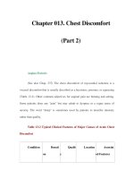
Loss of myocardium
Fall of BP—baroreceptors ergoreflexes
& chemoreflexes activated
Maintains hormone activation
Bacterial invasion
Immune & inflammatory response
Onset of cachexia
Hastens demise
Onset of
heart failure
Death
Sudden
death
Coronary
events
Progression
Figure 3-1 Progression of heart failure.
᭤ Table 3-1 Syndromes of heart failure
Entity Synonym or variant
Acute heart Pulmonary edema
failure
Circulatory Cardiogenic shock (poor
collapse peripheral perfusion, oliguria,
hypotension)
Chronic heart Untreated, overt, congestive,
failure undulating, treated,
compensated
of a constricted circulation such as cold periphery,
sweating, and mental confusion. Chronic heart fail-
ure is a condition where persistent damage to the
heart leads to a progressive state with persistent
symptoms. Many adjectives are added to the term
to emphasize one or other feature (Table 3-1).
The fundamental causes of heart failure are
easily stated and reflect the anatomical and phys-
iological features of the heart (Table 3-2). The most
common is myocardial disease. Myocardial dam-
age has traditionally been classified as due to one
or other manifestation of coronary heart disease
or as a cardiomyopathy (Table 3-3). Hypertension
is commonly associated with heart failure and
particularly with the progression of heart failure.
But hypertension is rarely the immediate or only
cause of heart failure. Patients with hypertension
often have coronary heart disease because
hypertension is an important risk factor causing
damage to the endothelium and promoting the
development of atherosclerosis. Such classifica-
tions focus on clinical characteristics. A different
approach is to consider the basic mechanisms of
heart failure but that has no clinical application at
present (Table 3-4).
Many words are added to the term heart fail-
ure (Table 3-5). These are often jargon. Forward
and backward failure reflects old ideas on the
pathophysiology of heart failure and should no
longer be used. Right and left heart failure usually
refer to pulmonary edema and breathlessness (left
heart failure) or evidence of fluid overload such as
raised venous pressure, enlarged liver, and
peripheral edema (right heart failure). This jargon
CHAPTER 3 WHAT CAUSES HEART FAILURE?–––––23
᭤ Table 3-2 General categories for causes
of heart failure
1. Myocardial disease
2. Valve disease
3. Pericardial disease
4. Congenital heart disease
5. Arrhythmias
᭤ Table 3-3 Myocardial causes of heart failure
Coronary artery
disease In all its many manifestations
Cardiomyopathy Dilated (DCM) - specific or
idiopathic (IDCM)
Hypertrophic (HCM or
HOCM or ASH)
Restrictive
ARVC
Hypertension
Drugs b-blockers
Calcium antagonists
Antiarrhythmics
Other or
unknown
DCM—dilated cardiomyopathy; IDCM—idiopathic dilated
cardiomyopathy; HCM—hypertropic cardiomyopathy;
HOCM—hypertropic obstructive cardiomyopathy; ASH—
asymmetric septal hypertrophy; ARVC—arrhythmic
right ventricular cardiomyopathy
᭤ Table 3-4 Fundamental abnormalities
in failing myocardium
1. Loss of muscle
2. Incoordinate contraction and abnormal
timing of contraction
3. Extracellular
Fibrosis, altered extracellular architecture,
shape and size of ventricle, slippage of
cells, fiber orientation
4. Cellular
Change of cell structure
Loss of intracellular matrix, hypertrophy,
hyperplasia
Change of cell function—systolic and/or
diastolic
Loss/aging of intrinsic repair mechanisms
Molecular
Calcium release and/or uptake
Response of contractile proteins to calcium
᭤ Table 3-5 Other terms used to describe
heart failure
1. Forward and backward heart failure
2. Right and left ventricular failure
3. Systolic and diastolic heart failure
4. High- and low-output heart failure
is largely nonsense, since the commonest cause of
right heart failure is left heart failure; fluid reten-
tion in chronic heart failure is a consequence of
retention of salt and water as a result of under-
perfusion of the kidney.
In recent years, a distinction has been made
between systolic and diastolic heart failure.
Diastolic heart failure is often referred to as
heart failure with preserved ventricular func-
tion. This distinction is the source of much dis-
cussion and controversy. In simple terms,
diastolic function exists when the heart remains
of a normal size and systolic heart failure exists
when the heart is enlarged. The old adage was
that “a big heart is a bad heart.” Diastolic heart
failure is common in the elderly and in the pres-
ence of myocardial ischemia and hypertension.
One further distinction is of clinical impor-
tance. There exists a group of conditions where
the cardiac output is greatly elevated in the pres-
ence of symptoms and signs identical to those
found in heart failure (Table 3-6). This is often
referred to as high-output heart failure but such
a phrase is misleading as the fundamental cause
is not the heart but other features of the circula-
tion or body systems. A better terminology is to
refer to these conditions as circulatory failure.
Diseases of any of the constitutive compo-
nent tissues of the heart and associated great
vessels can result in cardiac failure. The etiology
can be approached from a reductionist perspec-
tive, starting at the whole organ and tissue level,
and progressing to the cellular, subcellular, and
molecular causes (proteomic and genomic), or
vice versa from the expansionist perspective,
starting at the molecular level, and “expanding
up to the tissue and organ level.”
The prevalence of the different causes varies
depending upon gender, age, and geographical
region. In the Caucasian population of Western
Europe, the United States, and Australasia,
ischemic heart disease predominates, whereas
in the Afro-Caribbean population, hypertension
is the commonest cause. Chagas’ disease caused
by the parasite Trypanosoma cruzi is responsi-
ble for 20% of cardiac failure in South
America/Brazil,
6
but is only seen in returning
travelers and immigrants from this region in
European hospitals.
Coronary Heart Disease—Acute
Occlusion
Coronary heart disease, consequent to atheroscle-
rosis, is the commonest cause of heart failure in
Western populations, accounting for up to 70% of
cases.
7,8
The heart is critically dependent on a sup-
ply of oxygen from the coronary circulation; the
adenosine triphosphate (ATP) in heart muscle will
support about five beats. An acute coronary
occlusion causes diastolic contractile dysfunc-
tion within 6 seconds and systolic dysfunction
within 20 seconds. Intracellular acidosis develops
with the switch from aerobic to anaerobic metab-
olism, and the intracellular accumulation of phos-
phate from the breakdown of creatine phosphate
and ATP. Hydrogen and phosphate ions interfere
directly with the contractile proteins to promote
the formation of weak myofilament cross bridges.
The ATP depletion reduces sarcoplasmic reticu-
lum calcium ATPase and sodium-potassium
ATPase activity. The ATP-inhibited K
+
channel
opens, and triggers an efflux of potassium out of
the cell (within seconds), which is subsequently
amplified by reduced sodium-potassium ATPase
activity. This disrupts the ionic fluxes across the
sarcolemma and reduces the calcium removal
from the cytoplasm during diastole, depleting the
sarocoplasmic reticulum calcium stores and result-
ing in smaller systolic calcium transients. Lactate
accumulation causes mitochondrial damage and
24––––––HEART FAILURE: A PRACTICAL APPROACH TO TREATMENT
᭤ Table 3-6 Causes of circulatory failure
(high-output cardiac failure)
Anemia
Thyrotoxicosis
Beriberi
Arteriovenous fistula
Cirrhosis of the liver
Paget’s disease
Pregnancy
Renal cell carcinoma
disrupts action potential generation. The result is
cardiac tissue with abnormal electrical activity,
excitation-contraction coupling, and reduced
contractile tension. Total occlusion of the artery
leads to hemorrhagic necrosis of the myocardium
supplied by the artery, leading to irreversible
myocardial infarction. If the occluded coronary is
reopened after an initial delay of 30 minutes or
more, but before complete necrosis has devel-
oped, then the return of oxygen results in the
rapid production of free radicals within 2–4 min-
utes of reperfusion (reperfusion injury).
9
These
free radicals damage nucleic acids, cell mem-
branes, and intracellular proteins, initiating the
intracellular cascade via the p38 kinase and c-Jun
N-terminal kinase pathways, activation of the cas-
pase cascades, resulting in apoptosis and further
myocardial damage (Fig. 3-2).
The wave front of infarction starts at the endo-
cardial border and progresses to the epicardium
in areas of severe ischemia. The infarcted wall
becomes acutely dyskinetic (paradoxical outward
movement during systole), and ventricular dilata-
tion begins. This occurs within the constraints of
the pericardium, which reaches its limits of com-
pliance in the acute phase and exerts a constric-
tive effect on the acutely infarcted ventricle. The
increase in left ventricular diastolic pressure after
acute coronary occlusion in the dog angioplasty
model can be inhibited by prior removal of the
pericardium.
10
Coronary Heart Disease—Left
Ventricular Remodeling
Should the patient survive the acute episode of
myocardial infarction, a process of left ventricu-
lar remodeling is initiated, with further architec-
tural and structural changes to the ventricle
(Tables 3-7 and 3-8). The word was first used in
1982 so as to distinguish between extension of
an infarct, expansion of an infarct, and changes
in distant myocardium.
11,12
Remodeling occurs
in both the infarcted and remaining nonin-
farcted regions, further contributing to ventricu-
lar dysfunction. The extent of ventricular
dysfunction depends on the size and location of
the infarct, the presence of previous infarcts
elsewhere in the heart, the remaining coronary
supply with or without collaterals, and the
involvement of other cardiac structures, which
influence ventricular function such as the con-
ducting tissue, heart valves, and pericardium.
The region of necrosis involves damaged
myocytes and disruption of the extracellular
matrix. Loss of type I collagen fibers and intermy-
ocyte collagen struts occurs due to activation of
matrix metalloproteinases (1, 2, and 9 predomi-
nate in the heart), and is replaced by a deposi-
tion of collagen III and IV from fibroblasts,
stimulated by aldosterone and angiotensin II.
13,14
There is an overall increase in the myocardial
collagen content from 5% up to 25%, but it is
laid down in an irregular fashion, which dis-
rupts the fine myocardial architecture. This
allows myocyte slippage in the longitudinal
direction, leading with the loss of cells and vas-
culature to infarct thinning and expansion.
15,16
CHAPTER 3 WHAT CAUSES HEART FAILURE?–––––25
᭤ Table 3-7 Modish terms and concepts
in coronary heart disease
1. Stunning
2. Hibernation
3. Mummified myocardium
4. Stuttering ischemia
5. Preconditioning
6. Remodeling
7. Chronic ischemia
8. Ischemic cardiomyopathy
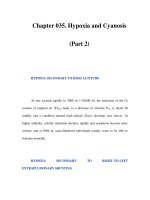
Seconds Minutes Minutes
0
10
20 30
40
50
60
5
30
60
Chest pain
ECG changes
Stunned myocardium
Cell necrosis
Total cell
necrosis
100
0
50
Normal (%)
Diastolic dysfunction
Systolic dysfunction
Loss of K
+
Acidosis
Figure 3-2 Timing of events after onset of
myocardial ischemia.
This is more extensive in areas with complete
absence of blood supply. The presence of collat-
erals, or late revascularization of the culprit ves-
sel, reduces infarct expansion. It is more evident
in anterior infarcts, and leads to an increase in
left ventricular circumference up to 25% during
the first week. This expansion alters the geome-
try of the left ventricle, with the normal ellipsoid
shape progressively replaced by a more spheri-
cal shape. Sphericity indices have been used to
quantify this change, based upon the ratio of the
observed biplane ventricular volume divided by
the volume of a theoretical ventricle with the
same biplane circumference but perfectly spher-
ical geometry. The normal human left ventricle
has a sphericity index of 0.66 at end diastole and
0.55 at end systole. After anterior myocardial
infarction, the sphericity index increases, with
the subsequent reduction in efficiency of blood
ejection from the chamber, higher filling pres-
sures, and reduced exercise capacity.
17
The infarction of one region of the left ven-
tricular wall requires the remaining myocardium
to compensate mechanically in order to maintain
adequate cardiac output. Eccentric hypertrophy
with sarcomeric replication in series occurs,
18
resulting in further increases in ventricular dimen-
sions and compliance. The increased wall stress
may stimulate the remaining noninfarcted
myocardium to hypertrophy in a concentric man-
ner, most commonly seen at the border zone of
the infarct. This process starts 1–2 months after the
initial infarction, and may progress for years
unless a terminal cardiac event intervenes.
Transient ischemia can produce temporary
reduction in contractile function, which is termed
myocardial stunning (Tables 3-7 and 3-8). A defin-
ition of stunned myocardium (stunning) is
mechanical dysfunction that persists after reperfu-
sion despite the absence of irreversible damage
and despite the restoration of normal or near-nor-
mal coronary flow.
19
The delayed recovery, from
a few hours up to several days, occurs despite
restoration of normal coronary flow in the
absence of irreversible damage. At a cellular level,
there is a transient increase in oxygen consump-
tion, despite continuous impairment of mechani-
cal function. This inefficient utilization of oxygen
may represent reduced myofilament calcium sen-
sitivity despite increases in cytosolic calcium lev-
els, possibly due to changes in myosin ATPase
activity. This is compounded by smaller degrees
of free radical production, including nitric oxide-
derived free radicals, which also contribute to the
dysfunction of myocardial stunning. Stunning can
occur in a variety of clinical settings. Early reper-
fusion after myocardial infarction, whether spon-
taneous or secondary to therapeutic thrombolysis
26––––––HEART FAILURE: A PRACTICAL APPROACH TO TREATMENT
᭤ Table 3-8 Ventricular dysfunction, stunning, hibernation, and clinical syndromes
Acute ventricular Immediate contractile failure (<2 minutes) Angioplasty (PCI)
dysfunction Stunning (approximately >2 minutes Stable or unstable angina
<15 minutes) but before any structural Prinzmetal’s angina
change with near-normal coronary flow Early thrombolysis
and dysfunction reversible Cardiac surgery
Chronic ventricular Early hibernation (hours but <3 months) Unstable angina
dysfunction or repetitive stunning Post-infarction
Silent ischemia
Chronic hibernation (>3 months) Stable angina
Dysfunction with reduced coronary blood Heart failure due to
flow but reversible coronary heart disease
Aortic stenosis
PCI—percutaneous coronary intervention
or primary angioplasty, may salvage ischemic but
noninfarcted myocardium within the territory of
the culprit vessel. This is of significant importance
clinically, as imaging may reveal large areas of
akinetic or dyskinetic myocardium in the early
post-infarct recovery period, but after allowing the
stunned myocardium to recover, the long-term
dysfunction may not be so severe, with the asso-
ciated prognostic implications.
20
During unstable
angina, and after exercise in patients with stable
but critical epicardial stenoses, regional wall
motion abnormalities have been demonstrated,
which recover with relief of angina and/or rest.
21
The recovery time is related to the duration of
angina on the treadmill or at rest, and may take
over 24 hours in severe cases. Myocardial stun-
ning is common after cardiac surgery requiring
cardioplegia and cardiopulmonary bypass, due to
the global myocardial ischemia generated with
cessation of coronary flow.
22
This setting demon-
strated that whilst inotropic agents can increase
contractile function of stunned myocardium, the
increase in oxygen consumption induced by
the inotropic stimulation is out of proportion to
the mechanical improvement. Sudden increases
in myocardial oxygen consumption, such as the
catecholamine surges seen in acute subarachnoid
hemorrhage and pheochromocytoma patients,
23,24
create a supply-demand mismatch and can cause
myocardial stunning.
Hibernating myocardium is another descrip-
tion of myocardial dysfunction, which has
become widespread.
25
The word was first used
by Diamond in 1978 when he commented,
“Reports of sometimes dramatic improvement in
segmental left ventricular function following coro-
nary bypass surgery, although not universal,
leaves the clear implication that ischemic nonin-
farcted myocardium can exist in a state of function
hibernation.”
26
But the term was popularized by
Rahimtoola in 1985 who described it thus “A state
of persistently impaired myocardial and left ven-
tricular dysfunction at rest due to reduced
coronary blood flow that can be partially or
completely restored to normal if the myocardial
oxygen supply/demand relationship is favorably
altered, either by improving blood flow and/or
reducing demand.”
27
Hibernation refers to viable
myocardium, which is exposed to chronic ischemia,
with hypocontractility, which is reversible on
restoration of normal blood flow. As implicated
by this definition, hibernation can only be diag-
nosed with absolute accuracy in retrospect after
revascularization has been performed. In contrast
to the pathology of acute occlusion described ear-
lier, mild-moderate ischemia results in transient
reduction of creatine phosphate and increase in
lactate production, but by 60–85 minutes these
return to near normal, and infarction does not
occur, despite persistent hypoperfusion. The sub-
sequent changes may represent an evolutionary
“protective” mechanism, as fetal cardiac gene
expression patterns are activated, and the chroni-
cally ischemic myocytes undergo structural cellu-
lar changes with sarcomere loss, increased
abundance of glycogen granules, rough endo-
plasmic reticulum and mitochondria, and an
increase in collagen strands.
28
These changes
occur over a timescale of days to weeks, and
with initially isolated functional hibernation,
progressing later to structural and functional
hibernation, which may be associated with wall
thinning.
29
The classical changes in left ventricular func-
tion caused by coronary artery disease and
described above occur within the region supplied
by the stenotic or occluded artery. Therefore,
regional wall motion abnormalities can be
explained by coronary disease. However, global
left ventricular dysfunction without regional vari-
ation can also be caused by coronary disease.
This is usually advanced three vessel disease, and
may be the result of infarction, hibernation,
and/or stunning. This often occurs in patients
without symptoms of angina, who present with
symptoms of cardiac failure. In a study of patients
with global left ventricular impairment (without a
history of ischemic heart disease [symptoms or
documented previous history]), 52% of patients
<72 years of age had coronary artery disease as
defined by at least one epicardial stenosis of
≥50%.
30
Furthermore milder stenoses, which are
not flow-limiting, may cause downstream myocar-
dial dysfunction through a variety of mechanisms
CHAPTER 3 WHAT CAUSES HEART FAILURE?–––––27
including cholesterol and thrombus embolism,
previous occlusion and recanalization, and as
regions initiating focal spasm.
Other Conditions Causing Reduced
Coronary Blood Flow
Whilst atherosclerosis is the commonest form of
coronary disease, many other conditions can
cause heart failure by reducing coronary blood
supply. These include congenital coronary anom-
alies, especially the interarterial anomalous left
coronary artery, coronary artery fistulae, the left
main stem arising from the pulmonary trunk,
and the stenosed “slit-like” left main orifice.
Coronary vasculitides, for example, periarteritis
nodosa, Kawasaki disease, systemic lupus ery-
thematosus (SLE), aortic dissection involving the
coronary ostia or aortic valve may all cause
myocardial dysfunction.
Hypertension
Hypertension is also a common cause of heart
failure, accounting for 14% cases in one U.K.
population-based study.
8
In the Framingham
study, a 20 mm Hg increase in systolic blood
pressure was associated with a 56% increased
risk for developing heart failure.
31
Advances in
primary care have led to a decrease in the inci-
dence with improved detection and treatment.
The majority of hypertensive patients have no
specific identifiable cause, so called “essential
hypertension,” which places an insidious after-
load strain on the heart through a variety of
mechanisms including sodium and water reten-
tion, arteriolar vasoconstriction, reduced vascular
compliance, faster reflection of the pulse wave
from stiffer small peripheral arteries, and activa-
tion of a range of neurohormonal systems. The
left ventricle demonstrates subtle abnormalities in
hypertensive patients even before hypertrophy
develops. These start with supranormal contrac-
tion with increased fractional shortening and
wall stress. The left ventricle hypertrophies in a
concentric manner to compensate, although
animal studies using gene knockout techniques
have revealed that left ventricular hypertrophy
(LVH) is not necessary for maintenance of ade-
quate cardiac output in the setting of increased
afterload.
32
The transcriptional changes bringing
about cardiac hypertrophy occur over different
timescales (Table 3-9). Therefore, pathological
hypertrophy should be viewed as the first stage
of hypertensive cardiac failure, although cardiac
output is maintained.
Many of the molecular cascades, which
induce hypertrophy, also cause myocyte apopto-
sis and lead to myocyte dysfunction. Angiotensin
II, endothelin, the gp130 signaling family, cal-
cineurin-mediated gene expression, stretch-
induced free radical production, and the three
subfamilies of the mitogen-activated protein
kinase family (ERK, JNK, and p38 kinase) are all
activated during development of ventricular
hypertrophy, and play roles in the transformation
from the hypertrophied but stable myocardium to
the irreversibly damaged and dysfunctional
myocardium of the failing heart.
33,34
Gap junction
remodeling also occurs between hypertrophied
cardiac myocytes, leading to increased dispersion
of electrical activity.
35,36
LVH causes reduced diastolic compliance,
longer isovolumic relaxation time, leading to
increased dependence on the atrial systole for
28––––––HEART FAILURE: A PRACTICAL APPROACH TO TREATMENT
᭤ Table 3-9 Cardiac hypertrophy—
transcriptional changes
30 minutes Immediate early genes c-fos,
c-jun, Erg-1, c-myc, Hsp70
6–12 hours b-MHC, skeletal a-actin
a-tropomyosin, ANP
Na/K ATPase
12–24 hours MLC-2, cardiac a-actin
>24 hours Increased RNA, increased
protein
Increased sarcomerogenesis
Increased cell size
b-MHC—b-myosin heavy chain; ANP—atrial natriuretic
peptide; ATPase—adenosine tri phosphatase; RNA—
ribonucleic acid
ventricular filling. Acute pulmonary edema is
often due to the inability to increase their end-
diastolic volume (preload reserve) in response
to increased preload or afterload, due to reduced
compliance and relaxation. Coronary vasodila-
tory capacity is reduced in LVH, and hyperten-
sives also develop atherosclerotic coronary
disease. As the hypertrophy progresses, myocar-
dial fiber shortening reduces, particularly in the
midwall, and hypertrophy allows total wall
shortening to be maintained despite this reduc-
tion in fiber shortening. Perivascular fibrosis
spreads through the myocardium inducing
myocyte necrosis as the capillary network is
destroyed, and apoptosis.
37
If the hypertension
remains poorly controlled, the hypertrophied
ventricle progressively dilates, the wall thins,
and the ventricle takes on the appearance and
mechanical characteristics of the dilated failing
ventricle with systolic dysfunction. The progno-
sis at this stage is very poor,
38
unless a significant
proportion of the ventricular dysfunction can be
accounted for by coexisting coronary disease
amenable to revascularization. This is a dynamic
process, and hypertensive cardiac failure is a
good example of a disease, which progresses
through various subtypes of cardiac failure
(hypertrophic, dilated, diastolic, systolic), expos-
ing the limitations of such classifications.
Valve Disease
Primary valvular disease accounts for 7% of car-
diac failure cases, and the majority involves dis-
ease of the left-sided cardiac valves. Incompetence
of the aortic and/or mitral valve results in a dilated
ventricular phenotype, to compensate for the
regurgitant volume by increasing stroke volume.
This requires the development of eccentric ven-
tricular hypertrophy to maintain the increased
ventricular output. Total ventricular muscle mass is
increased, although wall thickness may remain
within normal limits. Initially, the dilated ventricle
of the regurgitant valve can sustain the increased
ventricular ejection fraction required, provided
there are no coexisting threats to the myocardium,
for example, ischemic heart disease. However, the
chronic strain of this increased effort eventually
leads to the development of myocardial failure,
with changes in excitation-contraction coupling,
b-adrenoceptor expression and coupling, and
interstitial fibrosis.
39
The aim of medical manage-
ment is to predict this transformation in the natural
history of the individual’s valvular disease, in order
to time valve surgery optimally.
40,41
Acute, severe
regurgitation, such as that seen after papillary mus-
cle rupture or aggressive Staphylococcal aureus
endocarditis, cannot be tolerated and requires
emergency surgery. Lesser degrees of regurgita-
tion can be tolerated for long periods, particularly
with appropriate heart failure medication, and
providing arrhythmic complication do not inter-
vene. A combination of symptom development
and monitoring end-systolic diameter/volume is
the most effective strategy at present; although the
role of brain natriuretic peptide (BNP) monitoring
in this setting has yet to be defined. Type III mitral
regurgitation occurs secondary to left ventricular
dilation and dysfunction, due to annular enlarge-
ment, lateral displacement of the posterior papil-
lary muscles with resulting apical displacement of
the coaptation point of the mitral valve leaflets in a
tethered position.
42
These changes result in a cen-
tral regurgitant jet, and this should not be confused
with primary disease of the mitral valve leaflets
causing mitral regurgitation. Type III mitral regur-
gitation responds best to treatment of the left ven-
tricular failure, whereas the latter requires mitral
valve surgery.
Aortic stenosis results in a phenotype similar
to hypertensive cardiac failure, as both result in
increased afterload. LVH develops initially, via the
same mechanisms and with the predominant
problem of diastolic filling described above. If left
untreated, then the left ventricle also fails with
transformation to a dilated phenotype with a
reduced ejection fraction.
43
Aortic stenosis usually
occurs at the level of the valve cusps, and is most
commonly due to a congenital bicuspid valve in
the young and middle-aged adult (6% associated
with aortic coarctation), whereas atherosclerotic
plaque disease on the aortic surface of the cusps is
the commonest cause in the over 65 population.
CHAPTER 3 WHAT CAUSES HEART FAILURE?–––––29
With the increasing elderly population, this
degenerative aortic stenosis has become the com-
monest valvular disease in the Western world,
with between 2–9% of the population over
65 years affected.
44
Rheumatic stenosis of the aor-
tic valve is common in the developing world, and
always occurs in association with rheumatic mitral
disease. Rarely, aortic stenosis occurs at a
supravalvular level (Williams syndrome),
45
which
can be associated with coronary anomalies, or at
the subvalvular level in the left ventricular outflow
tract, usually in the form of a shelf of tissue
obstructing the outflow tract.
Mitral stenosis is predominantly due to
rheumatic heart disease after infection with a
Group A b-hemolytic Streptococcus pyogenes. It
is common in the developing world,
46
whereas
it is only seen in the surviving elderly and late
middle-aged populations in the developed
world. Mitral stenosis in isolation causes raised
left atrial pressure, pulmonary venous and arte-
rial hypertension, with development of right
ventricular failure and atrial fibrillation.
47
However, the stenotic valve is often also regurgi-
tant due to restricted leaflet movement, and the
inflammatory pannus of rheumatic disease may
extend down the chordae tendinea onto the
endocardial surface of the left ventricle, both
leading to left ventricular dysfunction.
48
These principles also apply to the pulmonary
and tricuspid valves, and right ventricular physi-
ology, although the etiology of right-sided
valvular disease is very different. Pulmonary
hypertension, infective endocarditis, especially
from intravenous drug abuse, and hospital-
acquired intravenous cannulae and indwelling
devices, carcinoid syndrome, rheumatic heart
disease, and congenital anomalies, for example,
pulmonary valve stenosis and Ebstein’s anomaly,
account for the majority.
Primary Disease of Cardiac
Muscle—the Cardiomyopathies
Primary disease of the cardiac muscle can present in
a number of guises, and previously, classification
has been based on the appearance and physiol-
ogy at echocardiography (ECG) and/or cardiac
catheterization, and pathological findings.
5
However, advances in molecular biology, and
specifically genotyping have resulted in a reeval-
uation of this classification.
49
The majority of
research on the disease has been presented under
the traditional classification, and we will discuss
the cardiomyopathies using the classical terms,
and then introduce the potential future molecular
classification.
In order to diagnose these conditions, it is
clearly essential to exclude other causative fac-
tors such as those discussed above. However,
multiple diseases can coexist and this requires
assessment of the time course of the disease as a
means to confirm the diagnosis. As alluded to
earlier, the presence of milder, non-flow-limiting
coronary disease, or a history of hypertension,
may complicate the clinical scenario.
There are three basic forms of functional
impairment that have been described:
1. dilated cardiomyopathy (DCM) (Table 3-10,
3-11, 3-12)
2. hypertrophic cardiomyopathy (HCM) (Table
3-13)
3. restrictive cardiomyopathy (RCM) (Table 3-14)
There are other forms of heart muscle dis-
ease, which extend this classification but are rare,
such as arrhythmogenic right ventricular dyspla-
sia, noncompacted left (or right) ventricle and
catecholomine-induced myocardial stunning.
DCM is a syndrome characterized by cardiac
enlargement and impaired systolic function of
one or both ventricles, in the setting of normal
coronary arteries, and absence of other struc-
tural or systemic causes (Table 3-10). The formal
diagnosis requires the left ventricle to be dilated
with the internal end-diastolic dimension
(LVEDD) >2.7 m
2
of body surface area and
either ejection fraction <45% or M-mode frac-
tional shortening <30%.
5
However, the normal
distribution of ventricular dimension across the
healthy population results in 1–2.5% of healthy
individuals fitting either of these parameters.
50
30––––––HEART FAILURE: A PRACTICAL APPROACH TO TREATMENT
CHAPTER 3 WHAT CAUSES HEART FAILURE?–––––31
᭤ Table 3-10 Causes of dilated cardiomyopathy
Familial cardiomyopathies
Genetic cardiomyopathies
Hypertension (eventually)
Infectious causes
Bacterial
Fungal
Parasitic (trypanosomiasis, toxoplasmosis,
schistosomiasis, trichinosis)
Rickettsial
Spirochetal
Viral (coxsackievirus, adenovirus, HIV)
Toxins
Alcohol
Cocaine
Heavy metals (cobalt, lead, mercury)
Carbon monoxide or hypoxia
Drugs
Chemotherapeutic agents (bleomysin,
doxorubicin, busulphan)
Antibiotics and antivirals (chloroquine,
zidovudine)
Antipsychotics
Metabolic disorders
Endocrine disease (diabetes mellitus)
Nutritional deficiencies (selenium, thiamine)
Storage disease (hemochromatosis, Refsum’s
disease, Fabry’s disease)
Autoimmune and collagen vascular disease
(sarcoidosis, SLE, rheumatoid arthritis,
Churg-Strauss syndrome)
Peripartum cardiomyopathy
Neuromuscular disorders (muscular dystrophies,
Friedreich’s ataxia)
HIV—human immunodeficiency virus; SLE—systemic
lupus erythematosus
᭤ Table 3-11 A classification of the genetic
causes of heart failure affecting
the cardiac myocyte
Sarcomeropathies
Cytoskeletalopathies
Laminopathies
Sarcoplasmic reticulopathies
Desmosomopathies
Mitochondrial diseases
᭤ Table 3-12 Genetic mutations linked to
dilated cardiomyopathy
Sarcomeric proteins:
b-Myosin heavy chain
a-Cardiac actin
Troponin T
a-Tropomyosin
Myosin binding protein C
Sarcoplasmic reticulum proteins:
Phospholamban
Titin/myofilament anchoring proteins:
Telethonin
Titin
Sarcolemma cytoskeleton:
Dystrophin
b-Sarcoglycan
d-Sarcoglycan
a-Dystrobrevin
Metavinculin
Emerin
Z-disk-associated proteins:
Muscle LIM protein
Desmosomal proteins
Desmoplakin
Desmin
Plakoglobin
Nuclear envelope proteins:
Lamin A/C
᭤ Table 3-13 Some genetic mutations
associated with hypertrophic
cardiomyopathy
Protein Gene
b-Myosin heavy chain MYH7
a-Myosin heavy chain MYH6
Essential myosin light chain MYL3
Regulatory myosin light chain MYL2
a-Cardiac actin ACTC
Cardiac troponin T TNNT2
Cardiac troponin I TNNI3
Cardiac troponin C TNNC1
a-Tropomyosin TPM1
Cardiac myosin binding protein C MYBPC3
Titin TTN
Muscle LIM protein CRP3
Phospholamban PLN
In addition, screening of families with affected
individuals frequently reveals asymptomatic
relatives with borderline normal ventricular
dimensions, which leads to difficulties in prog-
nostic and therapeutic advice.
About 10% of cases of congestive cardiac
failure in Western societies are due to DCM.
8
There are numerous causes of DCM (Table 3-10),
but in over 50% no underlying cause is found.
Whether these reflect unknown genetic muta-
tions, previous viral myocarditis, or toxin expo-
sure, with or without autoimmune destruction,
is not known, and it is possible that an environ-
mental insult unmasking a genetic weakness
may account for a large proportion.
Whatever the etiology, the final cardiac phe-
notype appears to follow a common pathological
pathway in response to myocardial damage. Some
cases result from the progressive deterioration in
ventricular muscle function with ongoing damage,
whereas others result from a single episode of
damage to which the ventricle responds by
remodeling and hypertrophy of the remaining
myocytes. The myocardium of DCM, whatever the
cause, is never normal. Usually there is dilatation
of all four chambers. The dilated left ventricle
becomes more spherical in shape, sometimes with
evidence of hypertrophy, though this is not a dom-
inant feature. Microscopically, myocyte orientation
is more tangential to the circumference, and indi-
vidual myocytes are elongated with an increased
cross-sectional area, but reduced intermyocyte
connections and gap junction formation. Together
with extensive areas of interstitial and perivascular
fibrosis, the result is a disorganized tissue with
abnormal contractile and relaxation properties,
and heterogeneous electrical conduction.
50
DCM
patients with interventricular conduction defects
have significantly worse systolic function, due in
part to ventricular incoordination, particularly if
total isovolumic time is increased, and a worse
prognosis.
51
However, the clinical course is highly
varied, and particularly in the group where a single
short-lived event is the sole cause, the prognosis
may be excellent.
Familial linkage of DCM may account for up
to 30% of cases. Mutations at 14 different chro-
mosomal loci have been described, affecting a
variety of proteins in the cardiomyocyte
52
(Tables 3-11 and 3-12). These proteins can
broadly be divided into sarcomeric/myofilament
proteins, titin and myofilament anchoring pro-
teins, Z-disk-associated proteins, sarcolemmal
cytoskeletal proteins, nuclear envelope proteins,
and intermediate proteins linking to the extracel-
lular matrix.
53
Familial DCM is genetically het-
erogeneous, and examples of all the Mendelian
modes of inheritance exist, and mitochondrial
inheritance has also been reported. In addition to
primary cardiac mutations, genetic variance of
other systems that influence development of car-
diac failure are also important. The polymor-
phisms of the angiotensin-converting enzyme
(ACE) gene have been well-characterized, and
the presence of the DD genotype is associated
32––––––HEART FAILURE: A PRACTICAL APPROACH TO TREATMENT
᭤ Table 3-14 Causes of restrictive
cardiomyopathy
1. Myocardial
Infiltrative
Amyloidosis
Sarcoidosis
Gaucher’s disease
Hurler’s disease
Fatty infiltration
Noninfiltrative
Idiopathic restrictive cardiomyopathy
Familial restrictive cardiomyopathy
Scleroderma
Pseudoxanthoma elasticum
Diabetic cardiomyopathy
Storage
Hemochromatosis
Fabry’s disease
Glycogen storage disease
2. Endomyocardial
Endomyocardial fibrosis
Hypereosinophilic syndrome
Carcinoid syndrome
Metastatic malignancy
Radiation
Drugs causing fibrotic endocarditis (serotonin,
methysergide, ergotamine, busulfan)
with higher plasma ACE levels and an adverse
prognostic outcome in DCM patients.
54
A subgroup of patients with genetic causes
has multisystem involvement, which may give
rise to recognizable syndromes, whose genetic
cause has been elucidated. Examples include
Duchenne’s muscular dystrophy, myotonic
dystrophy, facioscapulohumeral dystrophy,
Friedrich’s ataxia, Naxos disease, and Carvajal
syndrome (the last two representing the rare
cardiocutaneous syndromes).
Postviral myocarditis represents a spectrum of
patients, from those with classical fulminant viral
myocarditis, whose ventricular function is
observed to deteriorate during the course of their
illness, through to the majority, who present
with the features of DCM and a history of “recent
viral illness.”
55
The high background incidence of
symptom complexes resembling viral prodromes
in the general population added to the desire to
identify a source or cause on the part of the
patient may lead to significant overestimation of
this disorder. Initially serological evidence of
active viral infection was required, and can be
demonstrated in up to 33% of nonfamilial DCM.
56
However, the viral titres are unpredictable and
are dependent on the humoral immune system.
Improvement in detection of viral nucleic acids,
in particular by slot-blot probe hybridization and
polymerase chain reaction (PCR), has demon-
strated the persistence of viral particles in car-
diomyocytes after viral myocarditis in patients
who subsequently develop DCM.
57
Replicative
activity of these viral particles is not a necessity,
and their mere presence can induce DCM in
patients with activated immune systems. The
enteroviruses are the commonest culprit, and
myosin shares approximately 40% of its amino
acid sequence with the coxsackie B3 viral capsid
protein.
58
This provides a potent autoimmune
trigger, which may also occur in response to car-
diac protein epitopes exposed during membrane
disruption in the acute phases of viral myocardi-
tis. Interaction with the cellular and humoral
immune systems is critical, and some DCM
patients have abnormalities of the various com-
ponents of the immune system. Predisposition to
unregulated activation following the appropriate
viral trigger may unify the viral and immunologi-
cal hypotheses causing the continuing damage
and deterioration of the myocardium.
The worldwide human immunodeficiency
virus (HIV) epidemic has created a new category
of DCM. There are a variety of cardiac complica-
tions of HIV infection and its treatment, but left
ventricular enlargement and dysfunction has
been demonstrated in 15% of patients, with a
further 4% having isolated right ventricular
impairment.
59
DCM is strongly associated with
markers of advanced disease, including CD4
count of <100 cells/mL, and the presence of an
HIV-related encephalopathy. The underlying eti-
ology is multifactorial, including direct myocyte
infection by HIV, myocarditis secondary to
opportunistic infections, autoimmune cardiac
damage, nutritional deficiencies, and cardiotoxi-
city from both HIV therapy and illicit intravenous
drug abuse (if the cause of HIV infection).
A variety of toxins can damage the
myocardium. The degree of exposure, both in
dose and temporal course, together with the
potency of the toxin, will determine the level of
myocardial damage. Excess alcohol consumption
leads to a form of DCM, and is the underlying
cause in 3% cases.
60
Alcohol may cause myocar-
dial damage by various mechanisms. Ethanol and
its metabolites acetaldehyde and acetate have a
direct toxic effect of cardiomyocytes.
61
This can
cause an acute myocardial depression when
ingested in large quantities, raising blood ethanol
levels >1000 mg/L. Over the chronic course of
excess consumption, requiring >80 g/day for >5
years, ethanol impairs excitation-contraction cou-
pling, contractile protein and sarcolemmal func-
tion, with reduced myofibrillary protein synthesis.
Like other forms of DCM, the hearts of chronic
alcoholics with dilated ventricles show an excess
of collagen and interstitial fibrosis. Left ventricular
dilatation or dysfunction is detectable in up to
30% of chronic alcoholics, but unlike many of
the other causes of DCM, these changes are
reversible if abstinence is initiated early in the
course of the excess consumption. In addition to
the direct effects, alcohol can cause cardiac failure
CHAPTER 3 WHAT CAUSES HEART FAILURE?–––––33
through a variety of other means. Thiamine defi-
ciency associated with poor nutritional intake is
common in alcoholics and causes heart failure in
beriberi syndrome.
62
Alcohol predisposes to atrial
fibrillation and hypertension, both of which can
result in heart failure. Certain toxic substances can
be present in various alcoholic beverages. For
example, an outbreak of DCM in Canada was traced
to an excess of cobalt contaminating the brewing
of a popular beer.
63,64
Finally, chronic alcoholism
leads to profound cognitive impairments includ-
ing Korsakoff’s psychosis and dementia,
65
which
will result in alcoholics being less compliant with
their heart failure medication, whatever the cause.
Acute cocaine abuse can cause direct myocar-
dial depression, in addition to the ischemia
induced by coronary vasospasm.
66
There are
reports of DCM in chronic cocaine abusers, and
there is a synergistic toxic effect of taking cocaine
and alcohol together, with the cometabolite
cocaethylene also a potent dopamine reuptake
inhibitor, which is more lethal than cocaine alone
in animal models.
67
Various other drugs of abuse,
including amphetamines, 3,4-methylenedioxy-
methamphetamine (MDMA) (ecstasy), organic sol-
vents (toluene, kerosene, gasoline, acrylic paint
sprays), and organic nitrites (e.g., amyl nitrite) have
been associated with myocardial dysfunction and
heart failure.
A variety of prescribed drugs have cardiac
side effects, and in particular cardiotoxicity
resulting in a DCM phenotype.
68
The common-
est culprits are the anthracycline-derived
chemotherapy agents for a variety of solid and
hematological malignancies.
69
Soon after their
introduction, late cardiac failure was reported in
up to 30% patients who had received >500
mg/m
2
of doxorubicin (Adriamycin). Despite
limiting doses to <450 mg/m
2
and excluding
patients with preexisting cardiac dysfunction on
screening ECG, up to 3% of patients still develop
anthracycline-induced DCM. The presentation is
highly variable and although the cardiac failure
develops a median of 3 months after a dose of
anthracycline chemotherapy, case reports pre-
senting decades later are in the literature. The
mechanism of toxicity is uncertain, and may
involve free radical production and uncoupling
of mitochondrial ATP synthesis by doxorubicin
binding to cardiolipin in cardiac mitochondrial
membranes. Trastuzumab (Herceptin) is a novel
monoclonal antibody against erythroblastic
leukemia viral oncogene homolog 2 (ErbB2), a
coreceptor for neuroregulin signaling, and an
effective chemotherapeutic agent in the treat-
ment of breast cancer. It was initially introduced
as a second-line agent to anthracyclines, but 7%
of patients treated developed cardiomyopathy.
70
The protective mechanism of ErbB2 signaling in
the heart is still to be elucidated,
71
and trials of
trastuzumab monotherapy are also ongoing, but
it is accepted that it lowers the threshold of
anthracycline-induced cardiotoxicity. Likewise,
Paclitaxel also amplifies the effect of anthracy-
cline toxicity, up to 14% of patients receiving
dual chemotherapy in a trial for metastatic breast
cancer.
72
Radiation therapy can rarely cause late
ventricular systolic dysfunction, though modern
techniques including dose fractionation and
computerized blocking to reduce cardiac expo-
sure have now limited its incidence.
Peripartum cardiomyopathy is a form of
DCM, which must meet the following criteria:
(1) the development of cardiac failure in the last
4 weeks of pregnancy or within 5 months of
delivery, (2) absence of other causes of cardiac
failure, (3) absence of recognizable heart dis-
ease prior to the final 4 weeks of the pregnancy,
(4) DCM criteria for left ventricular dysfunc-
tion.
73
This definition attempts to exclude preex-
isting but undiagnosed DCM, which will
become symptomatic during pregnancy prior to
the final 4 weeks of the third trimester. There is
a varied course, with up to 50% cases reversible
on macroscopic imaging criteria. However, it is
more common in subsequent pregnancies, and is
associated with other complications of pregnancy
such as older maternal age, multiple pregnancy,
and preeclampsia.
Uncontrolled tachycardia, predominantly
atrial fibrillation, is now recognized as a cause
of left ventricular dilation and failure in a pattern
mimicking DCM. It has been called tachycar-
dia-induced cardiomyopathy, and generally
34––––––HEART FAILURE: A PRACTICAL APPROACH TO TREATMENT
requires heart rates of >120 beats/min to be
present for at least 3 months.
74
The continuous
excess metabolic demand creates an insidious
oxygen supply-demand mismatch, and the ven-
tricular dysfunction is reversible once adequate
rate control has been established. As the alter-
native clinical diagnosis is arrhythmia driven by
the primary DCM, the diagnosis can only be
made in retrospect after rate control has allowed
the ventricle to recover. However, is it likely that
this subtype of cardiac failure can be superim-
posed on other causes of left ventricular dys-
function, emphasizing the importance of rate
control in heart failure patients.
Hypertrophic Cardiomyopathy
Hypertrophic cardiomyopathy (HCM) is a rela-
tively common cardiac condition, affecting 1 in
500 of the general population.
75
It is characterized
by the presence of cardiac hypertrophy with a
hyperdynamic left ventricle, in the absence of the
other causes of LVH, systemic hypertension or aor-
tic stenosis, or of magnitude beyond that expected
by mild forms of these conditions. The hypertro-
phy is typically asymmetrical, although concentric
HCM does occur, and the usual diagnostic cutoff is
a wall thickness of ≥15 mm. However, genotype-
phenotype correlations demonstrate that almost
any wall thickness is compatible with the pres-
ence of an HCM mutation. The interventricular
septum is predominantly affected, but advances in
imaging technology have revealed a variety of
other forms including isolated midventricular, api-
cal, and right ventricular HCM. In addition to caus-
ing heart failure, many forms predispose to
malignant ventricular arrhythmias, and HCM is the
commonest cause of sudden death in the young
adult population.
76
In patients with significant septal or midven-
tricular hypertrophy, obstruction of the left ven-
tricular ejection may occur. This usually involves
the left ventricular outflow tract, and results
from the systolic anterior movement of the
mitral valves leaflets leading to midsystolic con-
tact with the septum. This causes a dynamic
obstruction, and may be persistent (detectable
in every cardiac cycle), labile (variable) or
latent, but provocable by exercise, standing,
postectopic response, the Valsalva maneuver, or
amyl nitrite inhalation. The presence or absence
of a gradient is important as it has prognostic
significance, although the magnitude of the
gradient does not.
77
In addition, the treatment
options are different in obstructive disease.
Mitral valve regurgitation, caused by the sys-
tolic anterior movement, concomitant mitral valve
prolapse, anterior mitral valve leaflet (AMVL)
damage from repeated traumatic contact with the
septum, and/or involvement of the papillary mus-
cles in the fibrotic disease process, also exacer-
bates heart failure in HCM patients.
The hypertrophied left ventricle is hyperdy-
namic with good systolic function early in disease,
often with generation of high intraventricular pres-
sures in obstructive disease. However, the left
ventricle in HCM has reduced compliance, and is
dependent on atrial systole for adequate filling.
These diastolic abnormalities are independent of
the degree or geometry of the hypertrophy, sug-
gesting a more widespread microscopic disease
process. The increased atrial pressures required
lead to atrial distension and enlargement, and
significant deterioration accompanies conversion
of rhythm to atrial fibrillation. Progressive myocar-
dial fibrosis from ischemia and myocyte degener-
ation eventually leads to wall thinning and
progressive dilatation of the left ventricle in a sub-
group of HCM patients, and if they survive free of
malignant arrhythmias, then they can convert
from a hypertrophic to a dilated left ventricular
phenotype with severe systolic dysfunction and
outflow obstruction disappears.
78
Apical HCM was first described in the
Japanese population in the 1970s, but is now
increasingly recognized globally.
79
It characteristi-
cally presents with chest pain in the young adult
with anterior T-wave inversion in addition to volt-
age criteria for LVH. Ventriculography reveals a
spade-shaped left ventricle at end diastole and the
diagnosis is confirmed by cardiac magnetic reso-
nance (MR). The prognosis with apical HCM is
better than other forms.
CHAPTER 3 WHAT CAUSES HEART FAILURE?–––––35
Fifty percent of patients have demonstrable
genetic causes (Table 3-13), which are pre-
donichantly transmitted in a Mendelian autoso-
mal dominant inheritance.
80
All the mutations
known to cause HCM affect 1 of 13 genes, all
which encode sarcomeric proteins involved in
the structure, regulation and contraction of the
thick and thin filaments. Over 200 different
mutations have been found, with varying
degrees of penetrance, even for the same muta-
tion within a family cohort. This genetic hetero-
geneity added to the clinical heterogeneity
ranging from benign to malignant forms with a
high rate of sudden death, varying ages of pre-
sentation, and symptom profiles leads unfortu-
nately to the current scenario where identifying
the mutant protein does not have sufficient pos-
itive or negative predictive power for prognosis.
The link between genotype and pathophysiol-
ogy is not clearly elucidated, and most cases
show significant myocyte disarray, with disrup-
tion of myocardial bundles into a characteristic
whorled pattern. Some myocytes are hypertro-
phied but otherwise appear normal, whereas
others have grossly disorganized intracellular
architecture. Extensive fibrosis is evident, and
abnormal intramural coronary arteries with wall
thickening and lumen reduction are present.
Restrictive Cardiomyopathy
The third group of cardiomyopathies is the restric-
tive cardiomyopathies (Table 3-13).
81,82
The main
feature of restrictive cardiomyopathy is abnormal
diastolic function. The ventricular walls become
rigid and noncompliant, usually due to either an
infiltrative or fibrotic pathology. This impedes
ventricular filling leading to high atrial pressures
with associated atrial distension, and the charac-
teristic tall, peaked “restrictive” E wave on trans-
mitral and/or transtricuspid Doppler ECG. It is
important to recognize that the term restrictive
when applied to transmitral filling represents
abnormally high atrial pressure with rapid early
filling, which may occur in many other conditions
causing heart failure, and the diagnosis of restric-
tive cardiomyopathy depends upon exclusion of
these other causes, certain characteristic features,
and identification of the cause. Depending upon
the underlying pathology, the ventricles are either
normal size or only slightly enlarged due to thick-
ening of the ventricular wall. This increased thick-
ness is due to infiltrative deposits or fibrosis, and
not myocyte hypertrophy. Systolic function may
initially appear normal, but usually deteriorates as
the disease advances.
The causes of restrictive cardiomyopathy
compromise a variety of conditions listed in
Table 3-13. The main causes vary according to
geographical location. In Europe, United States,
and Australasia, cardiac amyloid is the commonest
form of RCM whereas in the equatorial regions of
Africa, the Indian subcontinent, and parts of South
America, endomyocardial fibrosis (EMF) is one of
the commonest overall causes of cardiac failure.
The commonest causes will be briefly discussed.
Amyloidosis is a heterogeneous collection of
systemic disorders, which are characterized by
the extracellular deposition of autologous pro-
teins, which form twisted sheets of b-pleated fib-
rils in various tissues including the heart. A simple
stratification of these conditions is as follows:
1. A small excess in synthesis of a normal protein
results in slow deposition, which takes
decades to develop, for example, wild-type
transthyretin giving rise to “senile amyloidosis.”
2. A significant excess of a normal protein
results in rapid deposition and a faster clini-
cal time course presenting at a younger age.
Various mutations of transthyretin have been
described, and this form is familial amyloi-
dosis, inherited in an autosomal dominant
fashion, and is more common in the Afro-
Caribbean population.
3. A rapid synthesis of an abnormal protein, for
example, the immunoglobulin light chain
produced by the plasma cells of multiple
myeloma, traditionally referred to as amy-
loid light-chain (AL) amyloidosis.
4. The slower synthesis of an abnormal pro-
tein, such as the acute phase reactant Serum
amyloid A, which accumulates in chronic
inflammatory conditions such as rheumatoid
arthritis and tuberculosis.
36––––––HEART FAILURE: A PRACTICAL APPROACH TO TREATMENT
Sheets of the b-pleated fibrils deposit
between myocardial fibers and may occur in the
ventricles, atria, on the cardiac valves, and in the
aortic and coronary artery walls. The classical
presentation is with heart failure dominated by
right-sided findings, low voltage complexes on
the ECG, and a characteristic appearance on ECG
of granular, speckled, or sparkling myocardium,
which is thick, but does not thicken during
systole.
83
The thickening may be nonuniform
and resemble HCM, and the characteristic
restrictive transmitral filling pattern is present. In
the absence of systemic evidence of multiple
myeloma, endomyocardial biopsy may confirm
diagnosis demonstrating the presence of cardiac
amyloid on Congo red staining, and a typical
appearance on cardiac MR with a characteristic
pattern of global subendocardial late enhance-
ment coupled with abnormal myocardial and
blood-pool gadolinium kinetics has been
described.
84
Fabry’s disease, also known as angiokeratoma
corporis diffusum universale, has recently been
identified as a common cause of cardiomyopathy
with features of HCM and RCM.
85
It is an X-linked
disorder due to lysosomal a-galactosidase A defi-
ciency, leading to the intracellular accumulation
of glycosphingolipids (especially globo-triaosyl-
ceramide), which causes increased ventricular
wall thickness, restrictive diastolic filling, conduc-
tion tissue disease, and abnormalities of the mitral
valve.
86
Endomyocardial biopsy may reveal low
a-galactosidase A activity. The skin and kidneys
may also be involved.
Sarcoidosis is a granulomatous disorder of
unknown etiology, which may involve the
myocardium in up to 5% patients. This leads to
stiffening of the ventricular myocardium, con-
duction abnormalities, ventricular arrhythmias,
and rarely myocardial infarction due to coronary
involvement. The commonest cardiac problem
in sarcoidosis is right ventricular failure sec-
ondary to the diffuse pulmonary fibrosis seen in
advanced cases.
87
EMF occurs in equatorial regions as noted
earlier, with the highest incidence in Uganda,
Rwanda, and Nigeria.
81
There is extensive fibro-
sis, particularly affecting the endocardium, and
involves the inflow tracts of the right and/or left
ventricles, often involving the atrioventricular
valves and subvalvular apparatus. Both apices
are also frequently affected. Combined right and
left ventricular involvement occurs in 50% cases,
with 40% left-sided, and the remaining 10% right-
sided. Endocardial fibrosis acts as a substrate for
intracavity thrombus formation, leading to cavity
obliteration, pulmonary and systemic emboli.
The underlying etiology is not known, and the
presence of eosinophilia in some, but not all,
series has led to speculation of an eosinophil-trig-
gered damage, perhaps initiated by parasitic
infection. However, this has not been confirmed.
Eosinophils release a number of molecular medi-
ators, which are toxic to the myocardium, and
patients with marked eosinophilia may develop
endomyocardial involvement. This is most strik-
ing in patients with the hypereosinophilic syn-
drome (Löffler’s endocarditis), who have
persistent eosinophilia of >1500/mm
3
. The circu-
lating eosinophils invade the endocardium,
release their toxins (e.g., eosinophil cationic pro-
tein), and trigger an intense myocarditis and
endocarditis.
88
Mural thrombosis and fibrosis may
result, and coronary artery involvement may lead
to superimposed myocardial ischemia.
The above pathological categories are a useful
classification to approach the broad clinical entity
of cardiac failure. In the clinical scenario, patients
present with variable constellations of symptoms,
signs, and findings, and cardiac failure has been
divided into a variety of categories as described
above. Patients’ symptoms are highly subjective,
and are based on both cardiac and noncardiac
factors such as muscle tone, anemia, concomitant
respiratory or renal disease, cultural and society
issues, and there is no significant correlation
between symptom severity and objective mea-
surements of cardiac function, although there is
some value in prognostic determination.
The authors advocate the practical approach
of subclassification based upon the clinical time
course and etiology (Tables 3-1, 3-2, 3-3).
However, as molecular and genetic diagnostic
techniques improve and become more widely
available, newer classification may become
based on the particular protein, which is affected.
CHAPTER 3 WHAT CAUSES HEART FAILURE?–––––37
There is already a move to rename familial HCM
and DCM in this way, with the new categories
listed in Table 3-9. Finally the development of the
field of stem cell biology has revealed new
insights into cardiac physiology. The demonstra-
tion of resident cardiac stem cells with mitotic
activity in the adult human heart,
89
and the possi-
ble derivation of cardiac cells during repair from
circulating bone marrow-derived cells,
90,91
has
disbanded the view of the heart as a postmitotic
organ of fixed cell number. This dynamic
turnover appears to decrease with age, and we
believe that the development of cardiac failure is
in part due the overwhelming of the aging, inad-
equate repair processes by the insults of acquired
heart disease, which are far greater than those
experienced previously in evolution when the
repair systems were created and selected. The
supplementation of these reparative processes
with novel cellular and molecular therapies may
hopefully swing the balance away from cardiac
failure, whatever its label or cause.
᭤ REFERENCES
1. Martin WH, Berman WI, Buckey JC, et al. Effects
of active muscle mass size on cardiopulmonary
responses to exercise in congestive heart failure.
J Am Coll Cardiol. 2002;14:683–694.
2. Lee KS, Marwick TH, Cook SA, et al. Prognosis
of patients with left ventricular dysfunction, with
and without viable myocardium after myocar-
dial infarction: relative efficacy of medical ther-
apy and revascularization. Circulation. 1994;90:
2687–2694.
3. Mulligan IP, Fraser AG, Lewis MJ, et al. Effects of
enalapril on myocardial noradrenaline overflow
during exercise in patients with chronic heart
failure. Br Heart J. 1989;61:23–28.
4. Task FM, Swedberg K, Writing Committee, et al.
Guidelines for the diagnosis and treatment of
chronic heart failure: executive summary
(update 2005): the Task Force for the Diagnosis
and Treatment of Chronic Heart Failure of the
European Society of Cardiology. Eur Heart J.
2005;1;26(11):1115–1140.
5. Hunt SA, Abraham WT, Chin MH, et al. ACC/AHA
2005 Guideline Update for the Diagnosis and
Management of Chronic Heart Failure in the
Adult: a report of the American College of
Cardiology/ American Heart Association Task
Force on Practice Guidelines (Writing Committee
to Update the 2001 Guidelines for the Evaluation
and Management of Heart Failure): Developed
in Collaboration with the American College of
Chest Physicians and the International Society
for Heart and Lung Transplantation: Endorsed
by the Heart Rhythm Society. Circulation.
September 20, 2005;112(12):e154–e235.
6. Mendez GF, Cowie MR. The epidemiological
features of heart failure in developing countries:
a review of the literature. Int J Cardiol.
2001;80(2–3):213–9.
7. McDonagh TA, Morrison CE, Lawrence A, et al.
Symptomatic and asymptomatic left-ventricular
systolic dysfunction in an urban population.
Lancet. September 20, 1997;350(9081):829–33.
8. Cowie MR, Wood DA, Coats AJS, et al. Incidence
and aetiology of heart failure: a population-
based study. Eur Heart J. March 2, 1999;20(6):
421–8.
9. Zweier JL, Talukder MAH. The role of oxidants
and free radicals in reperfusion injury.
Cardiovasc Res. May 1, 2006;70(2):181–90.
10. Applegate RJ. Load dependence of left ventricu-
lar diastolic pressure-volume relations during
short-term coronary artery occlusion. Circulation.
February 1991;83(2):661–73.
11. Hutchins GM, Bulkley BH. Infarct expansion
versus extension: two different complications
of acute myocardial infarction. Am J Cardiol.
1982;65:1446–1450.
12. McKay RG, et al. Left ventricular remodeling
after myocardial infarction: a corollary to infarct
expansion. Circulation. 1986;74:693–702.
13. Kim HE, Dalal SS, Young E, et al. Disruption of
the myocardial extracellular matrix leads to car-
diac dysfunction. J Clin Invest. 2001;106(7):
857–66.
14. Johar S, Cave AC, Narayanapanicker A, et al.
Aldosterone mediates angiotensin II-induced inter-
stitial cardiac fibrosis via a Nox2-containing NADPH
oxidase. FASEB J. July 1, 2006;20(9):1546–8.
15. Olivetti G, Capasso JM, Sonnenblick EH, et al.
Side-to-side slippage of myocytes participates in
ventricular wall remodeling acutely after myocar-
dial infarction in rats. Circ Res. 1990;67: 23–34.
16. D’Armiento J. Matrix metalloproteinase disrup-
tion of the extracellular matrix and cardiac dys-
function. Trends Cardiovasc Med. April 2002;
12(3):97–101.
38––––––HEART FAILURE: A PRACTICAL APPROACH TO TREATMENT
17. Mitchell GF, Lamas GA, Vaughan DE, et al. Left ven-
tricular remodeling in the year after first anterior
myocardial infarction: a quantitative analysis of
contractile segment lengths and ventricular shape.
J Am Coll Cardiol. May 1992; 19(6):1136–44.
18. Kramer CM, Rogers WJ, Park CS, et al. Regional
myocyte hypertrophy parallels regional myocar-
dial dysfunction during post-infarct remodeling.
J Mol Cell Cardiol. September 1998;30(9):1773–8.
19. Bolli R, Marban E. Molecular and cellular mech-
anisms of myocardial stunning. Physiol Rev.
April 1, 1999;79(2):609–34.
20. Bolli R. Why myocardial stunning is clinically
important. Basic Res Cardiol. July 1998;93(3):
169–72.
21. Gerber BL, Wijns W, Vanoverschelde JL, et al.
Myocardial perfusion and oxygen consumption in
reperfused noninfarcted dysfunctional myocardium
after unstable angina : direct evidence for myocar-
dial stunning in humans. J Am Coll Cardiol.
December 1999;34(7):1939–46.
22. Anselmi A, Abbate A, Girola F, et al. Myocardial
ischemia, stunning, inflammation, and apoptosis
during cardiac surgery: a review of evidence. Eur
J Cardiothorac Surg. March 2004;25(3):304–11.
23. Kono T, Morita H, Kuroiwa T, et al. Left ventric-
ular wall motion abnormalities in patients with
subarachnoid hemorrhage: neurogenic stunned
myocardium. J Am Coll Cardiol. 1994;24(3):
636–40.
24. Takizawa M, Kobayakawa N, Uozumi H, et al. A
case of transient left ventricular ballooning
with pheochromocytoma, supporting patho-
genetic role of catecholamines in stress-induced
cardiomyopathy or takotsubo cardiomyopathy.
Int J Cardiol. January 2, 2007;114(1):E15–E17.
25. Vanoverschelde JL, Melin JA. The pathophysiol-
ogy of myocardial hibernation: current contro-
versies and future directions. Prog Cardiovasc
Dis. 2001;43:387–398.
26. Diamond GA, Forrester JS, deLuz PL, et al. Post-
extrasystolic potentiation of ischemic myocardium
by atrial stimulation. Am Heart J. 1978;95:
204–209.
27. Rahimtoola SH. A perspective on the three large
multicenter randomized clinical trials of coronary
bypass surgery for chronic stable angina.
Circulation. 1985;72:V123–V135.
28. Vanoverschelde JL, Melin JA. The pathophysiol-
ogy of myocardial hibernation: current contro-
versies and future directions. Prog Cardiovasc
Dis. March 2001;43:387–98.
29. Depre C, Kim SJ, John AS, et al. Program of cell
survival underlying human and experimental
hibernating myocardium. Circ Res. August 20,
2004;95(4):433–40.
30. Fox KF, Cowie MR, Wood DA, et al. Coronary
artery disease as the cause of incident heart failure
in the population. Eur Heart J. 2001;22:228–236.
31. Haider AW, Larson MG, Franklin SS, et al. Systolic
blood pressure, diastolic blood pressure, and
pulse pressure as predictors of risk for conges-
tive heart failure in the Framingham Heart Study.
Ann Intern Med. January 7, 2003; 138(1):10–6.
32. Esposito G, et al. Genetic alterations that inhibit
in vivo pressure-overload hypertrophy prevent
cardiac dysfunction despite increased wall
stress. Circulation. 2002;105:85–92.
33. Sugden PH. Signalling pathways in cardiac myocyte
hypertrophy. Ann Med. 2001;33:611–622.
34. Spragg DD, Leclercq C, Loghmani M, et al.
Regional alterations in protein expression in the
dyssynchronous failing heart. Circulation. 2003;
108:929–932.
35. Teunissen BE, Jongsma HJ, Bierhuizen MF.
Regulation of myocardial connexins during
hypertrophic remodelling. Eur Heart J. November
2004;25(22):1979–89.
36. Peters NS, Green CR, Poole-Wilson PA, et al.
Reduced content of connexin43 gap junctions in
ventricular myocardium from hypertrophied
and ischemic human hearts. Circulation.
September 1993;88(3):864–75.
37. Diez J, Gonzalez A, Lopez B, et al. Mechanisms
of disease: pathologic structural remodeling is
more than adaptive hypertrophy in hypertensive
heart disease. Nat Clin Pract Cardiovasc Med.
April 2005;2(4):209–16.
38. de Carvalho Frimm C, Soufen HN, Koike MK,
et al. The long-term outcome of patients with
hypertensive cardiomyopathy. J Hum Hypertens.
February 17, 2005;19(5):393–400.
39. Ishihara K, Zile MR, Kanazawa S, et al. Left ven-
tricular mechanics and myocyte function after
correction of experimental chronic mitral
regurgitation by combined mitral valve replace-
ment and preservation of the native mitral valve
apparatus. Circulation. 1992;86:II16–II25.
40. ACC/AHA 2006 Guidelines for the Management
of Patients With Valvular Heart Disease: A Report
of the American College of Cardiology/American
Heart Association Task Force on Practice
Guidelines (Writing Committee to Revise the
1998 Guidelines for the Management of Patients
CHAPTER 3 WHAT CAUSES HEART FAILURE?–––––39
With Valvular Heart Disease): Developed in
Collaboration With the Society of Cardiovascular
Anesthesiologists: Endorsed by the Society for
Cardiovascular Angiography and Interventions
and the Society of Thoracic Surgeons. Circulation.
August 1, 2006;114(5):E84–E231.
41. Borer JS, Bonow RO. Contemporary approach
to aortic and mitral regurgitation. Circulation.
November 18, 2003;108(20):2432–8.
42. Amigoni M, Meris A, Thune JJ, et al. Mitral regur-
gitation in myocardial infarction complicated by
heart failure, left ventricular dysfunction, or both:
prognostic significance and relation to ventricu-
lar size and function. Eur Heart J. February 1,
2007;28(3):326–33.
43. Otto CM. Valvular aortic stenosis: disease sever-
ity and timing of intervention. J Am Coll Cardiol.
June 6, 2006;47(11):2141–51.
44. Otto CM, Lind BK, Kitzman DW, et al. The car-
diovascular HS: association of aortic-valve scle-
rosis with cardiovascular mortality and morbidity
in the elderly. N Engl J Med. July 15, 1999;341(3):
142–7.
45. Eroglu AG, Babaoglu K, Oztunc F, et al.
Echocardiographic follow-up of children with
supravalvular aortic stenosis. Pediatr Cardiol.
November 2006;27(6):707–12.
46. Essop MR, Nkomo VT. Rheumatic and non-
rheumatic valvular heart disease: epidemiology,
management, and prevention in Africa.
Circulation. December 6, 2005;112(23):3584–91.
47. Chambers J. The clinical and diagnostic features
of mitral valve disease. Hosp Med. February 2001;
62(2):72–8.
48. Gaasch WH, Folland ED. Left ventricular func-
tion in rheumatic mitral stenosis. Eur Heart J.
July 1991;12(Suppl B):66–9.
49. Maron BJ, Towbin JA, Thiene G, et al.
Contemporary definitions and classification of
the cardiomyopathies: An American Heart
Association Scientific Statement From the Council
on Clinical Cardiology, Heart Failure and
Transplantation Committee; Quality of Care and
Outcomes Research and Functional Genomics
and Translational Biology Interdisciplinary Working
Groups; and Council on Epidemiology and
Prevention. Circulation. April 11, 2006;113(14):
1807–16.
50. Davies MJ, McKenna WJ. Dilated cardiomyopa-
thy: an introduction to pathology and pathogen-
esis. Br Heart J. 1994;72:S24.
51. Duncan AM, Francis DP, Henein MY, et al.
Limitation of cardiac output by total isovolumic
time during pharmacologic stress in patients
with dilated cardiomyopathy: activation-mediated
effects of left bundle branch block and coronary
artery disease. J Am Coll Cardiol. January 1,
2003;41(1):121–8.
52. Burkett EL, Hershberger RE. Clinical and genetic
issues in familial dilated cardiomyopathy. J Am
Coll Cardiol. April 5, 2005;45(7):969–81.
53. Kostin S, Hein S, Arnon E, et al. The cytoskeleton
and related proteins in the human failing heart.
Heart Fail Rev. October 2000;5(3):271–80.
54. Raynolds MV, Bristow MR, Bush EW, et al.
Angiotensin-converting enzyme DD genotype in
patients with ischaemic or idiopathic dilated car-
diomyopathy. Lancet. October 30, 1993;342 (8879):
1073–5.
55. Mason JW. Myocarditis and dilated cardiomy-
opathy: an inflammatory link. Cardiovasc Res.
October 15, 2003;60(1):5–10.
56. Caforio ALP, Mahon NJ, Tona F, et al. Circulating
cardiac autoantibodies in dilated cardiomyopa-
thy and myocarditis: pathogenetic and clinical
significance. Eur J Heart Fail. August 2002;4(4):
411–7.
57. Bowles NE, Richardson PJ, Olsen EG, et al.
Detection of Coxsackie-B-virus-specific RNA
sequences in myocardial biopsy samples from
patients with myocarditis and dilated cardiomy-
opathy. Lancet. May 17, 1986;1(8490):1120–3.
58. Cunningham MW, Antone SM, Gulizia JM, et al.
Cytotoxic and viral neutralizing antibodies
crossreact with streptococcal M protein,
enteroviruses, and human cardiac myosin.
Proc Natl Acad Sci. February 15, 1992;89(4):
1320–4.
59. Prendergast BD. HIV and cardiovascular medi-
cine. Heart. July 1, 2003;89(7):793–800.
60. Piano MR. Alcoholic cardiomyopathy: inci-
dence, clinical characteristics, and pathophysiol-
ogy. Chest. 2002;121(5):1638–50.
61. Zhang X, Li SY, Brown RA, et al. Ethanol and
acetaldehyde in alcoholic cardiomyopathy: from
bad to ugly en route to oxidative stress. Alcohol.
April 2004;32(3):175–86.
62. Naidoo DP. Beriberi heart disease in Durban: a
retrospective study. S Afr Med J. August 15,
1987;72(4):241–4.
63. Keen WW. Quebec beer-drinker’s cardiomyopa-
thy. JAMA. December 25, 1967;202(13):1145.
40––––––HEART FAILURE: A PRACTICAL APPROACH TO TREATMENT
64. Alexander CS. Cobalt-beer cardiomyopathy: a
clinical and pathologic study of twenty-eight
cases. Am J Med. October 1972;53(4):395–417.
65. Thomson AD, Marshall EJ. The natural history and
pathophysiology of Wernicke’s encephalopathy
and Korsakoff’s psychosis. Alcohol Alcohol.
March 1, 2006;41(2):151–8.
66. Lange RA, Hillis LD. Cardiovascular complica-
tions of cocaine use. N Engl J Med. August 2,
2001;345(5):351–8.
67. Hearn WL, Rose S, Wagner J, et al. Cocaethylene
is more potent than cocaine in mediating lethal-
ity. Pharmacol Biochem Behav. June 1991;39
(2):531–3.
68. Yeh ETH, Tong AT, Lenihan DJ, et al.
Cardiovascular complications of cancer therapy:
diagnosis, pathogenesis, and management.
Circulation. June 29,;109(25):3122–31.
69. Singal PK, Iliskovic N. Doxorubicin-induced car-
diomyopathy. N Engl J Med. September 24,
1998;339(13):900–5.
70. Smith KL, Dang C, Seidman AD. Cardiac dys-
function associated with trastuzumab. Expert
Opin Drug Saf. September 2006;5(5):619–29.
71. Crone SA, Zhao YY, Fan L, et al. ErbB2 is essen-
tial in the prevention of dilated cardiomyopathy.
Nat Med. May 2002;8(5):459–65.
72. Martin M, Lluch A, Ojeda B, et al. Paclitaxel plus
doxorubicin in metastatic breast cancer: prelim-
inary analysis of cardiotoxicity. Semin Oncol.
October 1997;24(5 Suppl 17):S17.
73. Sliwa K, Fett J, Elkayam U. Peripartum cardiomy-
opathy. Lancet. August 19, 2006;368(9536):
687–93.
74. Khasnis A, Jongnarangsin K, Abela G, et al.
Tachycardia-induced cardiomyopathy: a review
of literature. Pacing Clin Electrophysiol. July
2005;28(7):710–21.
75. Maron BJ, McKenna WJ, Danielson GK, et al.
American College of Cardiology/European
Society of Cardiology Clinical Expert Consensus
Document on Hypertrophic Cardiomyopathy. A
report of the American College of Cardiology
Foundation Task Force on Clinical Expert
Consensus Documents and the European
Society of Cardiology Committee for Practice
Guidelines. Eur Heart J. November 2003;24
(21):1965–91.
76. Ho CY, Seidman CE. A contemporary approach
to hypertrophic cardiomyopathy. Circulation.
June 20, 2006;113(24):E858–E862.
77. Maron MS, Olivolto I, Betocchis S, et al. Effect of
left ventricular outflow tract obstruction on clin-
ical outcome in hypertrophic cardiomyopathy.
N Engl J Med. 2003;348:295–303.
78. Cregler LL. Progression from hypertrophic car-
diomyopathy to dilated cardiomyopathy. J Natl
Med Assoc. 1989;81:820,824–826.
79. Sakamoto T. Apical hypertrophic cardiomyopa-
thy (apical hypertrophy): an overview. J Cardiol.
2001;37 (Suppl 1):161–78.
80. Richard P, Charron P, Carrier L, et al.
Hypertrophic cardiomyopathy: distribution of
disease genes, spectrum of mutations, and
implications for a molecular diagnosis strategy.
Circulation. May 6, 2003;107(17):2227–32.
81. Sliwa K, Damasceno A, Mayosi BM. Epidemiology
and etiology of cardiomyopathy in Africa.
Circulation. December 6, 2005;112(23):3577–83.
82. Hancock EW. Cardiomyopathy: differential
diagnosis of restrictive cardiomyopathy and
constrictive pericarditis. Heart. September 1,
2001;86(3):343–9.
83. Falk RH. Diagnosis and management of the car-
diac amyloidoses. Circulation. September 27,
2005;112(13):2047–60.
84. Maceira AM, Joshi J, Prasad SK, et al. Cardiovascular
magnetic resonance in cardiac amyloidosis.
Circulation. January 18, 2005;111(2):186–93.
85. Sachdev B, Takenaka T, Teraguchi H, et al.
Prevalence of Anderson-Fabry disease in male
patients with late onset hypertrophic cardiomyopa-
thy. Circulation. March 26, 2002;105(12):1407–11.
86. Pieroni M, Chimenti C, de Cobelli F, et al. Fabry’s
disease cardiomyopathy: echocardiographic
detection of endomyocardial glycosphingolipid
compartmentalization. J Am Coll Cardiol. April 18,
2006;47(8):1663–71.
87. Doughan AR, Williams BR. Cardiac sarcoidosis.
Heart. February 1, 2006;92(2):282–8.
88. Janin A. Eosinophilic myocarditis and fibrosis.
Hum Pathol. May 2005;36(5):592–3.
89. Beltrami AP, Barlucchi L, Torella D, et al. Adult
cardiac stem cells are multipotent and support
myocardial regeneration. Cell. September 19,
2003;114(6):763–76.
90. Quaini F, Urbanek K, Beltrami AP, et al. Chimerism
of the transplanted heart. N Engl J Med. January 3,
2002;346(1):5–15.
91. Orlic D, Kajstura J, Chimenti S, et al. Bone marrow
cells regenerate infarcted myocardium. Nature.
April 5, 2001;410(6829):701–5.
CHAPTER 3 WHAT CAUSES HEART FAILURE?–––––41
This page intentionally left blank
Chapter 4
Pathophysiology of Heart Failure
GARY S. FRANCIS, MD W. H./WILSON TANG, MD
Introduction 43
Adaptive Responses of the Myocardium in Heart Failure 44
Index Event—How Does Heart Failure Start? 44
Maladaptive Responses of the Myocardium in Heart Failure 45
How Adaptations in Heart Failure Go Wrong 45
The Frank-Starling Mechanism 45
Distribution of Cardiac Output and the Role of the Peripheral Vasculature 46
Ventricular Remodeling 47
Transition from Increased Cell Mass to Heart Failure 49
Altered Myocardial Energetic in Heart Failure 50
Other Peptides and Inflammatory Cytokines 50
Diastolic and Systolic Heart Failure 50
Summary 51
᭤ INTRODUCTION
Heart failure is defined differently by various
authors (see Chap. 1), but for the clinician, it is
fundamentally a complex clinical syndrome,
and not a stand-alone diagnosis. Like anemia or
renal failure, it has many causes and etiologies.
Although the pathophysiology is to some extent
dependent on the etiology, there are many
common features regardless of the underlying
cause and there are always some underlying
structural abnormalities. The clinical symptoms
include shortness of breath and fatigue, either at
rest or during exertion. In advanced cases, there
is usually evidence of salt and water retention.
This chapter will deal with pathophysiologic
mechanisms that contribute to the development
and progression of signs and symptoms of heart
failure. In general, heart failure implies structural
disease of the heart with functional conse-
quences to the circulation. It causes signs and
symptoms in patients, and can theoretically occur
from any form of heart disease. Hypertension,
coronary artery disease, valvular heart disease,
and cardiomyopathy are leading causes of heart
failure in the Western world (see Chap. 3). Heart
failure should be distinguished from circulatory
failure, which occurs when a component of the
circulation impedes circulatory homeostasis,
such as excessive circulating volume from acute
renal failure. In such cases, the heart itself may be
structurally and functionally normal, so that the
term “circulatory failure” may be preferred by
some. Heart failure may also occur in patients
43
Copyright © 2007 by The McGraw-Hill Companies, Inc. Click here for terms of use.