Báo cáo y học: "Low-concentration, continuous brachial plexus block in the management of Purple Glove Syndrome: a case report" pps
Bạn đang xem bản rút gọn của tài liệu. Xem và tải ngay bản đầy đủ của tài liệu tại đây (608.63 KB, 4 trang )
JOURNAL OF MEDICAL
CASE REPORTS
Singh et al. Journal of Medical Case Reports 2010, 4:48
/>Open Access
CASE REPORT
© 2010 Singh et al; licensee BioMed Central Ltd. This is an Open Access article distributed under the terms of the Creative Commons
Attribution License ( which permits unrestricted use, distribution, and reproduction in
any medium, provided the original work is properly cited.
Case report
Low-concentration, continuous brachial plexus
block in the management of Purple Glove
Syndrome: a case report
Georgene Singh*
1
, Verghese T Cherian
1
and Binu P Thomas
2
Abstract
Introduction: Purple Glove Syndrome is a devastating complication of intravenous phenytoin administration.
Adequate analgesia and preservation of limb movement for physiotherapy are the two essential components of
management.
Case presentation: A 26-year-old Tamil woman from India developed Purple Glove Syndrome after intravenous
administration of phenytoin. She was managed conservatively by limb elevation, physiotherapy and oral antibiotics. A
20G intravenous cannula was inserted into the sheath of her brachial plexus and a continuous infusion of bupivacaine
at a low concentration (0.1%) with fentanyl (2 μg/ml) at a rate of 1 to 2 ml/hr was given. She had adequate analgesia
with preserved motor function which helped in physiotherapy and functional recovery of the hand in a month.
Conclusion: A continuous blockade of the brachial plexus with a low concentration of bupivacaine and fentanyl helps
to alleviate the vasospasm and the pain while preserving the motor function for the patient to perform active
movements of the finger and hand.
Introduction
Intravenous administration of phenytoin can result in
soft tissue injury at the site of injection leading to oedema
and purplish-black discolouration of the hand. This is
known as the Purple Glove Syndrome (PGS). The man-
agement of PGS is mainly conservative, which includes
limb elevation and physiotherapy. Use of low concentra-
tion of local anaesthetic for brachial plexus block has the
added advantage of preserving motor function to facili-
tate physiotherapy in addition to providing adequate
analgesia and relief of vasospasm.
Case presentation
A 26-year-old Tamil woman from India presented with
an alleged history of generalized seizures. The emer-
gency-room physician administered 600 mg of pheny-
toin-sodium dissolved in 500 ml of normal saline through
a 20G cannula sited into a vein on the dorsum of her right
hand. Four hours later, the patient complained of pain at
the site of injection, which progressively became severe.
The fingers, hand and forearm were swollen and had a
purplish-black discolouration (Figure 1a). The radial
artery was palpable, albeit feeble, under the oedema. The
capillary refill under the nail bed was sluggish. The ultra-
sonic Doppler study of the arm showed normal flow
through the radial and the ulnar arteries but the veins
appeared collapsed.
The working diagnosis was that the patient had an isch-
emic hand, with the likelihood of progression to gan-
grene. This was possibly due to the extravasation of
phenytoin leading to PGS. Although a differential diagno-
sis of compartment syndrome and need for fasciotomy to
relieve the pressure was considered, it was decided to
manage conservatively.
The intravenous cannula was removed and the arm was
wrapped in a dry cotton-gauze dressing and kept elevated
to reduce the oedema. Since the pain was intense and not
relieved with non-steroidal anti-inflammatory drugs, the
stellate ganglion was blocked using 7 ml of 0.5% bupiva-
caine. This sympathetic blockade improved the capillary
refill and the mottled discolouration, and significantly
reduced the pain. However, it lasted for only three hours.
* Correspondence:
1
Department of Anaesthesiology, Christian Medical College, Vellore 632 004,
Tamil Nadu, India
Singh et al. Journal of Medical Case Reports 2010, 4:48
/>Page 2 of 4
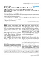
Therefore, it was planned to provide a continuous bra-
chial plexus block. A 20G intravenous cannula was
inserted into the interscalene grove 2.5 cm above the
clavicle and was directed distally to lie within the sheath
of the brachial plexus (Figure 2). A solution of 0.1% bupi-
vacaine with fentanyl (2 μg ml
-1
) was infused at a rate of
1-2 ml.hr
-1
using a Terumo TE-311 infusion pump. The
patient had adequate pain relief without any motor weak-
ness. The swelling and the discolouration continued to
improve with limb elevation, physiotherapy and oral anti-
biotics. As there was sufficient improvement, the cannula
was left in-situ for seven days to provide analgesia and aid
in physiotherapy. The patient was discharged from the
hospital after ten days with advice to continue physio-
therapy. By the end of a month, the blackish discoloura-
tion had disappeared and the range of movement of the
hand was nearly back to normal (Figure 1b).
Discussion
PGS, named for its distinctive discolouration and swell-
ing of the hands is a known complication of intravenous
administration of phenytoin-sodium [1]. It is character-
ized by intense pain, purplish black discolouration and
oedema at the site of injection which progresses to rest of
the limb [2]. The reported incidence among patients is 1-
7% after intravenous injection of phenytoin [1,3,4]. It
should be differentiated from extravasation of intrave-
nous fluid, injection site infection and intra-arterial injec-
tion. The persistence of the pain, blackish discolouration
and the oedema even after discontinuation of the intrave-
nous fluid and removal of the cannula differentiates it
from extravasation. The lack of purulent discharge or
pyrexia differentiates it from injection site infection.
Intra-arterial injection of a highly alkaline solution would
lead to arterial spasm, embolisation of insoluble drug
crystals, endothelial damage and vascular thrombosis
resulting in the absence of Doppler signal from the artery
[5].
Phenytoin is a weak acid and is insoluble in water [3].
However, injectable preparation is highly alkaline as it
contains 45% propylene glycol as the solvent and 10%
alcohol in water with sodium hydroxide to adjust the pH
to 12 [1]. The pathophysiology of PGS is poorly under-
stood [3]. It has been suggested that the alkaline drug pre-
cipitates upon contact with blood and leaks out of the
vein, from around the cannula, and into the interstitial
tissue [1]. This is likely to happen in a slow flowing
stream or if the cannula is kinked, leading to stasis.
Another possible mechanism could be that the highly
alkaline solution induces vasoconstriction of the vein
resulting in disruption of the endothelial-intercellular
junctions and seepage of the drug into the interstitial
space [1]. Extravasation of the highly albumin-bound (70-
90%) phenytoin increases the interstitial oncotic pressure
leading to oedema [1]. Propylene glycol with its high
osmolality causes necrosis of the tissue [1]. Although
20 G intravenous Cannula inserted into the interscalene
groove for continuous infusion of bupivacaine (0.1%) with
fentanyl (2 μg ml-1)
Figure 2 20 G intravenous Cannula inserted into the interscalene
groove for continuous infusion of bupivacaine (0.1%) with fenta-
nyl (2 μg ml-1).
Patient's right hand (1a) At the onset of Purple Glove Syn-
drome and (1b) One month later demonstrating complete
recovery of flexion
Figure 1 Patient's right hand (1a) At the onset of Purple Glove
Syndrome and (1b) One month later demonstrating complete re-
covery of flexion.
Singh et al. Journal of Medical Case Reports 2010, 4:48
/>Page 3 of 4
these explanations seem plausible, reports of PGS occur-
ring after the oral administration of phenytoin [6] also
suggest that the phenomenon may be due to phenytoin
itself and not directly due to the infusion [7].
Women and the elderly are said to have an increased
risk of PGS. Other factors associated with it include
peripheral vascular disease and diseases that weaken the
vascular and dermal integrity, use of intravenous cathe-
ters smaller than 20G and infusion of phenytoin at more
than 25 mg.ml
-1
[1,8].
Therefore, administration of phenytoin should be into a
free-flowing infusion line, through a large bore intrave-
nous catheter sited into a large vein of the forearm, in a
concentration of 10 mg.ml
-1
, and at a rate not exceeding
50 mg.min
-1
[9]. Any evidence of venous irritation such as
pain, oedema and erythema warrants immediate discon-
tinuation of the infusion and removal of the intravenous
catheter [1].
The diagnosis of PGS is based on the characteristic
clinical findings and a high index of suspicion when it
occurs after the administration of phenytoin. The man-
agement is mainly conservative (limb elevation, physio-
therapy, control of pain, reassurance to the patient) and
should be directed at minimizing the degree of soft tissue
damage [1]. The affected arm should not be used for
venepuncture or blood pressure measurement.
Arterioles, smaller arteries and peripheral veins are
normally under vasoconstrictor influence by the alpha
receptors. In addition to the vasoconstriction caused by
the highly alkaline solution, pain and anxiety also cause a
marked increase in arteriolar vasoconstriction mediated
by the sympathetic nervous system. This results in
increased resistance, reducing cutaneous perfusion. In
addition, there is an associated increase in vascular tone
which decreases the compliance of the venous system,
reducing its blood content and increasing the venous
pressure. Sympathetic blockade, by blocking the alpha
receptors, improves blood flow in vasospastic disorders
[10]. Stellate ganglion blockade has the advantage of
blocking the sympathetic innervation of the upper limb,
thus improving the perfusion and relieving the ischaemic
pain associated with vasospasm [11].
Because of tissue injury and ischaemia, PGS is very
painful. A low concentration local anaesthetic would
relieve the pain by preferentially blocking the Aδ and B
fibres [12]. Impulses in small fibres are blocked faster
than those in larger ones because of the amount of time
of drug diffusion and the length of the nerve to block
propagation of nerve impulses. In separate experiments,
it has been shown that nerve signals associated with both
beta fibres and A delta fibres are reduced at low concen-
tration of local anaesthetics [13]. Electrophysiologic stud-
ies have shown that bupivacaine diffuses relatively slowly
into fast conducting motor fibres at low concentrations
[14].
Selective sensory blockade, by preserving motor inner-
vation, allows the patient to move his or her fingers. This
helps in performing physiotherapy and improving venous
blood flow, both of which are crucial to recovery.
Infraclavicular approach to brachial plexus may also be
employed and a subcutaneously tunneled catheter may be
placed, especially if it is anticipated that the patient will
have difficulty in retaining the catheter without displace-
ment. Accurate placement may be confirmed using a
nerve stimulator or ultrasound guidance. In our case, as
even the tactile stimuli were excruciating, peripheral
nerve stimulator was not used.
Ropivacaine is another agent that may be considered
for low concentration brachial plexus blockade. The
degree of motor blockade produced by ropivacaine is less
than that of bupivacaine. So it is possible to produce a
more selective sensory blockade with ropivacaine. Fur-
thermore, if required, higher concentration of ropiva-
caine may be used with lesser risk of cardiotoxicity than
with bupivacaine [15].
Addition of fentanyl potentiates local anaesthetic
action via central opioid receptor mediated analgesia by
peripheral uptake of fentanyl to systemic circulation. Fen-
tanyl also acts directly on the peripheral neuronal cells as
the dorsal roots contain opioid binding sites. In addition,
because of the presence of bidirectional axonal transport
of opioid binding protein, fentanyl penetrates the nerve
membrane and acts at the dorsal horn [16]. Adjuvants
such as ketamine, alpha 2 adrenergic agonists have also
been used to potentiate the effects of local anaesthetics in
brachial plexus blocks [17].
Although brachial plexus blockade has been used for
this condition [18], to the best of our knowledge, the use
of low-dose bupivacaine (concentration and volume) that
has the added advantage of preserving the motor func-
tion of patients to perform active physiotherapy has not
been described before.
Conclusion
Intravenous administration of phenytoin should be
undertaken with care, ensuring a rate less than 50 mg per
minute [9], through a free-flowing infusion. The manage-
ment of PGS is primarily conservative. A continuous
blockade of the brachial plexus with a low concentration
of bupivacaine and fentanyl helps to alleviate the vasos-
pasm and the pain while preserving the motor function
for the patient to perform active movements of the finger
and hand.
Consent
Written informed consent was obtained from the patient
for publication of this case report and accompanying
Singh et al. Journal of Medical Case Reports 2010, 4:48
/>Page 4 of 4
images. A copy of the written consent is available for
review by the Editor-in-Chief of this journal.
Competing interests
The authors declare that they have no competing interests.
Authors' contributions
GS treated the patient, documented the progress and outcome and prepared
the initial manuscript. VTC performed the placement of the cannula, and was a
major contributor in editing and revising the manuscript. BPT was the primary
physician under whom the patient was admitted, supervised the hand therapy
and further management of the patient and reviewed the manuscript. All
authors read and approved the final manuscript.
Author Details
1
Department of Anaesthesiology, Christian Medical College, Vellore 632 004,
Tamil Nadu, India and
2
Dr Paul Brand Centre for Hand Surgery, Christian Medical College, Vellore 632
004, Tamil Nadu, India
References
1. Snelson C, Dieckman B: Recognizing and managing purple glove
syndrome. Crit Care Nurse 2000, 20(3):54-61.
2. Bhattacharjee P, Glusac EJ: Early histopathologic changes in purple
glove syndrome. J Cutan Pathol 2004, 31(7):513-515.
3. Edwards JJ, Bosek V: Extravasation injury of the upper extremity by
intravenous phenytoin. Anesth Analg 2002, 94(3):672-673.
4. O'Brien TJ, Cascino GD, So EL, Hanna DR: Incidence and clinical
consequence of the purple glove syndrome in patients receiving
intravenous phenytoin. Neurology 1998, 51(4):1034-1039.
5. Righini M, Angellillo-Scherrer A, Gueddi S, Le Gal G, Bounameaux H:
Management of severe ischemia of the hand following intra-arterial
injection. Thromb Haemost 2005, 94(1):219-221.
6. Yoshikawa H, Abe T, Oda Y: Purple glove syndrome caused by oral
administration of phenytoin. J Child Neurol 2000, 15(11):762.
7. Burneo JG, Anandan JV, Barkley GL: A prospective study of the incidence
of the purple glove syndrome. Epilepsia 2001, 42(9):1156-1159.
8. Spengler RF, Arrowsmith JB, Kilarski DJ, Buchanan C, Von Behren L,
Graham DR: Severe soft-tissue injury following intravenous infusion of
phenytoin. Patient and drug administration risk factors. Arch Intern Med
1988, 148(6):1329-1333.
9. Joint Formulary Committee: British National Formulary. London 1999.
10. Breivik H, Cousins MJ, Lofstrom JB: Sympathetic Neural Blockade of
Upper and Lower Extremity. In Neural Blockade in Clinical Anesthesia and
Management of Pain 3rd edition. Edited by: Cousins MJ, Bridenbaugh PO.
Philadelphia: Lippincott; 1998:411-445.
11. Elias M: Cervical sympathetic and stellate ganglion blocks. Pain
Physician 2000, 3(3):294-304.
12. Strickartz GR:
Neural Physiology and local anesthesia action in Neural
Blockade in Clinical Anaesthesia and management of pain. In Neural
Blockade in Clinical Anesthesia and Management of Pain 3rd edition. Edited
by: Cousins MJ, Bridenbaugh PO. Philadelphia: Lippincott; 1998:35-54.
13. Heavner JE, deJong R: Lidocaine blocking concentration for B- and C-
nerve fibers. Anesthesiology 1974, 40:228.
14. Rosenberg PH, Heinonen E: Differential sensitivity of A and C nerve
fibres to long acting amide local anaesthetics. Br J Anaesth 1983,
55(2):163-167.
15. McClure JH: Ropivacaine. Br J Anaesth 1996, 76:300.
16. Nishikawa K, Kanaya N, Nakayama M, Igarashi M, Tsunoda K, Namiki A:
Fentalyl Improves Analgesia but Prolongs the Onset of Axillary Brachial
Plexus Block by Peripheral Mechanism. Anesth Analg 2000, 91:384-387.
17. Klein SM, Nielsen KC: Brachial plexus blocks: infusions and other
mechanisms to provide prolonged analgesia. Curr Opin Anaesthesiol
2003, 16(4):393-399.
18. Mahajan RP, Batra YK, Rajeev S: Intravenous phenytoin and
percutaneous arterial cannulation: the purple-glove syndrome. Eur J
Anaesthesiol 2007, 24(10):900-901.
doi: 10.1186/1752-1947-4-48
Cite this article as: Singh et al., Low-concentration, continuous brachial
plexus block in the management of Purple Glove Syndrome: a case report
Journal of Medical Case Reports 2010, 4:48
Received: 7 January 2009 Accepted: 10 February 2010
Published: 10 February 2010
This article is available from: 2010 Singh et al; licensee BioMed Central Ltd. This is an Open Access article distributed under the terms of the Creative Commons Attri bution License ( s/by/2.0), which permits unrestricted use, distribution, and reproduction in any medium, provided the original work isproperly cited.Journal of Medical Case Reports 2010, 4:48