Báo cáo y học: "Midgut pain due to an intussuscepting terminal ileal lipoma: a case report" pps
Bạn đang xem bản rút gọn của tài liệu. Xem và tải ngay bản đầy đủ của tài liệu tại đây (436.54 KB, 4 trang )
CAS E REP O R T Open Access
Midgut pain due to an intussuscepting terminal
ileal lipoma: a case report
Noormuhammad O Abbasakoor
1
, Dara O Kavanagh
1
, Diarmaid C Moran
1
, Barbara Ryan
2
, Paul C Neary
1*
Abstract
Introduction: The occurrence of intussusception in adults is rare. The condition is found in 1 in 1300 abdominal
operations and 1 in 100 patients operated for intestinal obstruction. The child to adult ratio is 20:1.
Case presentation: A 52-year-old Irish Caucasian woman was investigated for a 3-month history of intermittent
episodes of colicky midgut pain and associated constipation. Ileocolonoscopy revealed a pedunculated lesion in
the terminal ileum prolapsing into the caecum. Computed tomography confirmed a smooth-walled,
nonobstructing, low density intramural lesion in the terminal ileum with secondary intussusception. A laparoscopic
small bowel resection was performed. Histology revealed a large pedunculated polypoidal mass measuring 4 × 2.5
× 2 cm consiste nt with a submucosal lipoma. She had complete resolution of her symptoms and rema ined well at
12-month follow-u p.
Conclusion: This case highlights an unusual cause of incomplete small bowel obstruction successfully treated
through interdisciplinary cooperation. Ileal lipomas are not typically amenable to endoscopic removal and require
resection. This can be successfully achieved via a laparoscopic approach with early restoration of premorbid
functioning.
Introduction
Neoplasms of the small intesti nes are rare [1]. Gastroin-
testinal lipo mas are benign tumors that can occur in the
small bowel but occur most c ommonly in the col on.
The majority are asymptomatic and are detected inci-
dentally on abdominal imaging. Removal is warranted if
tissue diagnosis is deemed essential or if severe sympto-
matology, such as pain or bleeding, exists [2].
We report a case of terminal ileal lipoma causing
intermittent intussusception in a 52-year-old woman.
The lipoma was diagnosed at ileocolonoscopy and suc-
cessfully removed through laparoscopy. A r eview of t he
literature on small bowel intussception and gastrointest-
inal (GI) lipomas is also presented in this report.
Case presentation
A 52-year-old Irish Caucasian woman presented with a
three-month histor y of int ermittent central abdominal
pain and constipation. She did not describe gastrointest-
inal bleeding or weight loss. She previously underwent a
transabdominal hysterectomy for men orrhagia. Her phy-
sical e xamination was unremarkable. Initial investiga-
tions, such as blood tests, abdomen ultrasound and
gastroscopy were unremarkable. Ileocolonoscopy
revealed a pedunculated terminal ileal lesion prolapsing
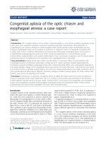
into her caecum. Computed tomography (CT) of her
abdomen and pelvis demonstrated a smooth-walled,
low-density, intramural lesion in the terminal ileum. It
measured 3.2 × 1.6 cm. The ileum at the proximal end
of the lesion was mildly dilated with a centrally placed
narrowed channel of contrast, which was consistent
with an in tussusception possibly secon dary to an intra-
mural lipoma. There was no evidence of obstruction
(Figure 1).
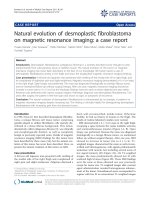
She underwent an elective laparoscopic small bowel
resection and stapled functional end-to-end anasto-
moses. On macroscopy the lesion appeared as a larg e
pedunculated polypoid mass measuring 4 × 2.5 × 2 cm
with focal mucosal ulceration (Figure 2). Microscopy
revealed a submucosal lipoma with blunting of the over-
lying mucosal villi and pyloric gland metaplasia. She
made an uneventful recovery and was discharged home
on the fourth postoperative day. She returned to work
* Correspondence:
1
Division of Colorectal Surgery, Adelaide and Meath Incorporating the
National Children’s Hospital, Tallaght, Dublin 24, Ireland
Abbasakoor et al. Journal of Medical Case Reports 2010, 4:51
/>JOURNAL OF MEDICAL
CASE REPORTS
© 2010 Abbasakoor et al; licensee BioMed Central Ltd. This is an Open Access arti cle distributed under the terms of the Creative
Commons Attribution License (ht tp://creativecommons.org/licenses/by/2.0), which permits unrestricted use, distri bution, and
reproduction in any medium, provided the original work is properly cited.
Figure 1 Contrast-enhanced computed tomography scan of the abdomen demonstrates a smooth-walled, low-density intramural
lesion. It measures 3.2 × 1.6 cm. The ileum at the proximal end of the lesion is mildly dilated with a centrally placed narrowed channel of
contrast consistent with an intussusception.
Figure 2 Macroscopic view of a large pedunculated polypoid mass arising from the luminal surface of the ileal resection specimen.
Appearances are consistent with a lipoma.
Abbasakoor et al. Journal of Medical Case Reports 2010, 4:51
/>Page 2 of 4
on the 12
th
postoperative day. She remained free of
symptoms at three-month follow-up.
Discussion
Lipomas are benign tumors of mesenchymal origin.
They are the second most common benign tumors in
the small intestine and account for 10% of all benign
gastrointestinal tumors and 5% of all gastrointestinal
tumors. They are predominantly submucosal and pro-
trude into the lumen [2]. Occasionally, they arise in the
serosa. Gastrointestinal lipomas are most commonly
located in the colon (65% to 75%, especially on the right
side), small bowel (20% to 25%), and occasional ly in the
foregut (<5%) [2]. Lipomas are largely asymptomatic.
Major presentin g features are intestinal obstruction and
hemorrhage [3].
Intussusception in adults is a rare entity that it is gen-
erally caused by definable intraluminal pathology [4].
Diagnosis can be challe nging. Intussusception is classi-
fied according to its gastrointestinallocation: enteric,
ileocaecal, or colonic [4]. In il eocaecal intussusceptio ns,
the ileocaecal valve acts as the lead point. The ileum
(’intussusceptum’) telescopes into the colon (’intussusci-
piens’) through the ileocaecal valve [5,6]. Intussusception
leads to the development of venous and lymphatic con-
gestion, which results in intestinal edema. If not treated
promptly, the arterial blood supply t o the bowel will b e
compromised, thus leading to ischaemia, perforation
and peritonitis [4]. Only 5% of all intussusceptions
occur in adults [7]. In 90% of these cases a predisp osing
lesion is identified [7]. This is contrary to intussuscep-
tion in the pediatric population where an organic lesion
is found in only 10% of documented cases [3]. In adults,
it is important to differentiate between small bowel and
colonic intussusception. I n 63% of cases of small bowel
intussusceptions, a benign underlying lesion can be
found. Meanwhile, a malignant etiology has to be
expected in 58% of cases of large bowel intussusceptions
[8].
Lipomas can be diagnosed through conventional endo-
scopy, capsule endoscopy, barium studies and, most
importantly, CT. Typical endoscopic features are
smooth, yellowish surface with pedunculated or sessile
base, as seen in this case. Other endoscopic characteris-
tics are the “ cushion sign” and “nake d fat sign” [2]. CT
usually reveals a smooth, well-demarcated sausage-
shaped mass. It may also reveal associated intussuscep-
tion if present [5]. Capsule endoscopy and digital bal-
loon endoscopy are newer means for diagnosing lipomas
and are particularly helpful in cases involving small
bowel lipomas [2]. Associated intussusception can be
confirmed on contrast enema (’ crescent sign’), CT and
magnetic resonance imaging (MRI). Multislice CT facili-
tates the assessment of vascular supply to the affected
bowel loop in cases of intussusception where imp ending
ischemia is suspected [4].
The treatment for lipomas depends on the clinical
manifestations. Indications for their removal include
intestinal obstruction, hemorrhage and malignant poten-
tial [4]. There is a theoretical risk of sarcomatous
change but this has rarely been documented in the lit-
erature [1]. Endoscopic removal is possible but poten-
tially complicated. In view of the submucosal location,
there is an inherent risk of perforation [9]. Furthermore,
lipomas have high water con tent, which mean s a large
amount of cautery is necessary to achieve effective
hemostasis [9]. Surgery can be performed through
laparoscopy or via an open approach. The type of resec-
tion and anastomosis depends on the location, bowel
wall integrity, and vascular supply of the lipoma [6].
Elective laparoscopic resection of lipomas is the treat-
ment of choice with the concomitant benefits of laparo-
scopic surg ery, such as shorter duration of hospital stay,
less postoperative pain, early restoration of (GI) function
and good cosmesis [6].
Conclusion
In this case, we illustrate the importance of a thorough
interdisciplinary evaluation of patients w ith midgut
abdominal pain. It highlights the diagnostic values of
CT scanning and completed ileocolonoscopy. Despite
preoperative localization, laparoscopy facilitates a thor-
ough evaluation of the intraperitoneal contents and
therapeutic resection of the affected segment. This
report confirms the recognized benefits of laparoscopic
surgery with associa ted e arly return to pr emorbid func-
tioning. In patients with persistent episodes of incom-
plete intestinal obstruction, aty pical causes, such as the
etiology we describe here, should be considered.
Consent
Written i nformed consent was obtained from our
patient for publication of this case report and any
accompanying images.
Author details
1
Division of Colorectal Surgery, Adelaide and Meath Incorporating the
National Children’s Hospital, Tallaght, Dublin 24, Ireland.
2
Department of
Gastroenterology, Adelaide and Meath Incorporating the National Children’s
Hospital, Tallaght, Dublin 24, Ireland.
Authors’ contributions
NOA contributed in collecting the requisite literature and wrote the case
report. DOK also collected the requisite literature and reviewed the literature.
DCM also contributed in collecting the requisite literature. BR and PCN were
involved in the diagnosis of our patient. PCN also performed the surgery. All
authors read and approved the final manuscript.
Competing interests
The authors declare that they have no competing interests.
Abbasakoor et al. Journal of Medical Case Reports 2010, 4:51
/>Page 3 of 4
Received: 19 September 2009
Accepted: 11 February 2010 Published: 11 February 2010
References
1. Rathore MA, Andrabi SI, Mansha M: Adult intussusception: a surgical
dilemma. J Ayub Med Coll Abbottabad 2006, 18(3):3-6.
2. Chou JW, Feng CL, Lai HC, Tsai CC, Chen SH, Hsu CH, Cheng KS, Peng CY,
Chung PK: Obscure gastrointestinal bleeding caused by small bowel
lipoma. Inter Med 2008, 47:1601-1603.
3. Balik AA, Ozturk G, Aydinli B, Alper F, Gumus H, Yildirgan MI, Basoglu M:
Intussusception in adults. Acta Chir Belg 2006, 106(4):409-412.
4. Lin HH, Chan DC, Yu CY, Chao YC, Hsieh TY: Is this a lipoma?. Am J Med
2008, 121(1):21-23.
5. Michael A, Dourakis S, Papanikolaou I: Ileocaecal intussusception in an
adult caused by a lipoma of the terminal ileum. Ann Gastroenterol 2001,
14(1):56-59.
6. Takaaki T, Matsui N, Hiroshi K, Takemoto Y, Oka K, Seyama A, Morita T:
Laparoscopic resection of an ileal lipoma: report of a case. Surg Today
2006, 36:1007-1011.
7. Meshikhes AW, Al-Momen SA, Al Talaq FT, Al-Jaroof AH: Adult
intussusception caused by a lipoma in the small bowel: report of a case.
Surg Today 2005, 35(2):161-165.
8. Oyen TL, Wolthuis AM, Tollens T, Aelvoet C, Vanrijkel JP: Ileo-ileal
intussusception secondary to a lipoma: a literature review. Acta Chir Belg
2007, 107:60-63.
9. Yoshimura H, Murata K, Takase K, Nakano T, Tameda Y: A case of lipoma of
the terminal ileum treated by endoscopic removal. Gastrointestinal Endosc
1997, 46(5):461-463.
doi:10.1186/1752-1947-4-51
Cite this article as: Abbasakoor et al.: Midgut pain due to an
intussuscepting terminal ileal lipoma: a case report. Journal of Medical
Case Reports 2010 4:51.
Submit your next manuscript to BioMed Central
and take full advantage of:
• Convenient online submission
• Thorough peer review
• No space constraints or color figure charges
• Immediate publication on acceptance
• Inclusion in PubMed, CAS, Scopus and Google Scholar
• Research which is freely available for redistribution
Submit your manuscript at
www.biomedcentral.com/submit
Abbasakoor et al. Journal of Medical Case Reports 2010, 4:51
/>Page 4 of 4