Báo cáo y học: "Spinal cord stimulation as a treatment for refractory neuropathic pain in tethered cord syndrome: a case report ppsx
Bạn đang xem bản rút gọn của tài liệu. Xem và tải ngay bản đầy đủ của tài liệu tại đây (441.04 KB, 4 trang )
CAS E REP O R T Open Access
Spinal cord stimulation as a treatment for
refractory neuropathic pain in tethered cord
syndrome: a case report
Maarten Moens
1*
, Ann De Smedt
2
, Jan D’Haese
3
, Steven Droogmans
4
, Cristo Chaskis
5
Abstract
Introduction: The spinal cord is a target for many neurosurgical procedures used to treat chronic severe pain.
Neuromodulation and neuroablation are surgical techniques based on well-known specific anatomical structures.
However, anatomical and electrophysical changes related to the tethered spinal cord make it more difficult to use
these pro cedures.
Case presentation: We report the case of a 37-year-old Caucasian woman who had several surgical interventions
for tethered cord syndrome. These interventions resulted in severe neuropathic pain in her lower back and right
leg. This pain was treated by spinal cord stimulation using intra-operative sensory mapping, which allowed the
cord’s optimal placement in a more caudal position.
Conclusion: The low-voltage and more caudally placed electrodes are specific features of this treatment of
tethered cord syndrome.
Introduction
Tethered cord syndrome (TCS) is a clinical condition
caused by prolonged stretching of the lower part of the
spinal cord, especially the conus terminalis. It results in
the abnormal attac hment of t he spinal co rd to its sur-
rounding tissues. Its clinical manifestations include
backache and leg pain (especially with flexion), bowel
and bladder dysfunction, lower limb weakness, sensory
changes, gait abnormalities, and musculoskeletal defor-
mities of the feet and the spine [1-3]. Primary or conge-
nital causes of TCS can be explained by abnormal
secondary neurulation and disorders that are of caudal
eminence. On the other hand, acquired causes such as
infection, tumor or scars can also lead to tethering [1,3].
The development or progression of symptoms often
call for an untethering operation, which involves abnor-
mal anatomy and associated entities like lipomas, myelo-
meningoceles, lipomyelomeningoceles, dermal sinus and
spina bifida occulta [3,4].
PainisaverycommonsymptomofTCS.Thepain
worsens with flexion or vigorous physical activity. It
affects the lower back, the perineum and/or the legs.
Among all the symptoms, however, pain is the one most
likely to be improved by surgery, involving a success
rate of up to 75% in the adult patients [3].
Unfortunately, complex post-operative pain syndromes
are difficult to treat with pharmaco logical and interven-
tional pain treatments.
One of the more invasive, but effective, treatments for
chronic neuropathic pain is neurostimulation. This
treatment is based on creating paresthesias due to elec-
trical stimulation in the affected and painful area.
Anatomical changes in the spinal cord, for example,
tethered cord syndrome, may influence the exact level
and location of electrode implantation.
Case presentation
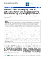
We report the case of a 37-year-old Caucasian woman
(Figure 1) with a history of several surgical interventions
for untethering her spinal cord after undergoing a resec-
tion of a sacral lipomyelomeningocele at the age of 23.
Our patient had a spa stic bladder that had rec overed
partly. Despite these surgeries, however, she has suffered
many years of severe chronic pain with heavy burning,
* Correspondence:
1
Department of Neurosurgery, UZ Brussel, Laarbeeklaan, Brussels, 1090,
Belgium
Moens et al. Journal of Medical Case Reports 2010, 4:74
/>JOURNAL OF MEDICAL
CASE REPORTS
© 2010 Moens et al; licensee BioMed Central Ltd. This is an Open Access article distributed under the terms of the Creative Commons
Attribu tion License (h ttp://creativecommons.org/licenses/by/2.0), which permits unrestricted use, distribution, and reproduction in
any medium, provided the original work is prope rly cite d.
dysesthesia and hyperalgesia at her buttocks and her
right posterior thigh.
A neurological examination revealed that our patient
had no neurological problems besides the sensory deficit
at her buttocks and leg and he r hyper-reflexive neuro-
genic bladder problem. She scored 7 on the DN4 ques-
tionnaire for indicating neuropathic pain [5]. Her
treatment with c arbamazepine, pregabalin, gabapentin
and fentanyl was not effective . Her other treatment with
physiotherapy and psychological guidance neither chan-
ged nor reduce her pain, and only high-frequency trans-
cutaneous electrical nerve stimulation (TENS) partly
diminished her pain.
Subsequently, a spinal cord stimulator (SCS) was sur-
gically placed when our patient was under epidural
anesthesia. An epidural catheter was inserted at levels
L2 to L3. Our patient was injected with a loading dose
of 0.5% ropivacaine with 0.5 μg/ml sufenta. She was also
administered with top-up doses of 4 ml 0.5% ropiva-
caine to reach a segmental sensory block. We also per-
formedamid-lineflavectomyatlevelsT10toT11and
orthodromically in serted a Specify 565 electrode (Med-
tronic Inc., Minneapolis, Minnesota) at levels T9 to T10
and T11-T12 (retrograde). Using intra-operative stimu-
lation, we performed a mapping of our patient’ssensory
responses to epidural stimulation, all the while searching
for the best level of stimulation. At the higher levels (T9
to T10) we noted paresthesias to her loins, abdominal
wall and ant erior part o f the upper leg but not to her
buttock or posterior thigh. Parest hesias were only
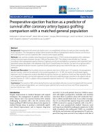
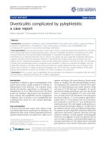
achieved by stimulation at level T12. The refore, the
Specify 565 electrode was centered at level T12 for defi-
nitive implantation (Figure 2).
Following the operation, the intensity of the pain she
felt improved from 9 pre-operative to 2 post-operative
onthevisualanaloguescale(VAS).Shewasableto
reduce her painkillers substantially and required a daily
dose of just 500 mg of paracetamol. Her definitive SCS
parame ters were from 0.1 to 0.2 V for amplitude, 60 Hz
for frequency, and 240 μsec for pulse width.
Discussion
The effectiveness of SCS in patients with chronic
intractable neuropathic pain is well-known and compre-
hensively described [6].
According to the authors’ expertise and preference,
the placement of surgical plate electrodes is the choice
of implantation. This placeme nt offers a broader stimu-
lation pattern, lower stimulation requirements, better
long-term e ffectivene ss, and lower migration rate. Such
are the technical advantag es of plate electrodes as com-
pared with percutaneously implanted electrodes [7-9].
Intra-operative stimulation is the cornerstone of any
successful procedure. Patients should be able to perceive
stimulation in areas where they feel pain. Patients,
therefore, must be awake, feel comfortable without any
pain, and fully cooperative to report this to the implant
team during the placement of electrodes [10,11]. Epi-
dural anesthesia is the technique of choice wh en using
minimally invasive flavectomy, because it provides better
hemodynamic stability. Compared to subarachnoid
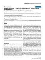
Figure 1 T2-weighted sagittal magnetic resonance imaging of
the lower spinal cord. Green arrow points to elongated spinal
cord.
Figure 2 Corona l view of computed tomograp hy scan of the
lower dorsal spine. Green arrow points to Specify 565 electrode at
levels D11 and D12.
Moens et al. Journal of Medical Case Reports 2010, 4:74
/>Page 2 of 4
anesthesia, epidural anesthesia a lso promotes the po ssi-
bility of extending intra-operative anesthesia through
the epidural catheter without any meningeal puncture
[12]. With this type of anesthesia patients can feel par-
esthesias during intra-operative stimulation because
local epidural anesthetic acts mostly at the nerve roots
level and do not completely block the spinal cord [12].
Technically, the coverage of plate electrodes is limited
to 5 levels (2 to 3 levels in orthodromical direction and
2 levels in retrograde sense) depending on the type of
electrode. Therefore, guidelines to direct the current
flow more precisely and to stimulate the desired body
areas are necessary. Barolat et al.(1991)publisheddata
on spine levels of cathode in connection with specific
body areas. They concluded that areas difficult to stimu-
late are the neck, the lower back, and the perineum. For
stimulation of the buttock, electrodes can be placed in a
range of T10 to L1 with corresponding stimulation pat-
tern at levels T11 to T12. It must be activated first on
the posterior leg fibers, then on the posterior thigh, and
lastly on the buttock [13].
In our daily practice, we usually place the electrodes at
levels T9 to T10 in order to stimulate our patient’ s
whole leg and lower back. But in this case, because of
the stretching of the lower part of our patient’sspinal
cord, we performed a sensory mapping of the spinal
cord. As expected, our patient felt the paresthesias in
the examined regions when they were stimulated one
level below the usual level but in the normal range as
described by Barolat et al. (1991).
We may hypothesize that the anatomical stretching in
TCS is more extended than the electrophysiol ogical and
functional tensions. In vitro, experiments showed that
maximal cord elongation occurs at the lumbosacral
region, s ome elongation at the thoracic area, and mini-
mal to none at all at the cervical region [14]. Electro-
physiological testing in more patients should b e
undertaken in order to generalize this hypothesis.
Another point that was observable in our patient’s
case is the low voltage used to obtain excellent pain
relief. This can also be explai ned by the anatomy of the
spinal cord in TCS. Due to the attachments or scars, the
spinal cord is no w placed at a more posterior place and
thus in closer contact with the dura mater. It is proven
that the voltage needed for the recruitment of nerve
fibers, and thus the perception threshold of paresthesia,
is related with the distance between the electrode and
the spinal cord [15].
The low voltage needed to relieve pain lowers energy
consumption and favors a longer battery life. In the end it
benefits the cost effectiveness of SCS in patients with TCS.
The implantation of a spinal cord stimulat or in a
patient with TCS as an effective treatment for refract ory
chronic neuropathic pain has not been described
previously. Despite the anatomical abnormality of the
spinal cord in TCS, neuromodulation is an effective
therapeutic option to achieve pain relief.
Depending on the severity of the tethered cord, the
electrode must b e implanted more caudally than in
cases involving normal spinal cord. In our opinion, this
and its low voltage requirement are the two main ele-
ments for the successful treatment of TCS using
neuromodulation.
Conclusion
We reported for the first time a case of sensory map-
ping for S CS in the treatment of neuropathic pain in
TCS. We successfully implanted the epidural electrode
in a more caudal position than usual, while using a
lower voltage to obtain the best response.
Consent
Written info rmed consent was obtained from our
patient for publication of this case report and any
accompanying images. A copy of the written consent is
available for review by the Editor-in-Chief of this
journal.
Abbreviations
SCS: spinal cord stimulation; TCS: tethered cord syndrome; TENS:
transcutaneous electrical nerve stimulation; VAS: visual analogue scale.
Acknowledgements
Special thanks to Professor Gabriel Moens for his editorial advice. Maarten
Moens is Clinical Investigator of The Research Foundation in Flanders,
Belgium. Steven Droogmans is an aspirant of The Research Foundation in
Flanders, Belgium.
Author details
1
Department of Neurosurgery, UZ Brussel, Laarbeeklaan, Brussels, 1090,
Belgium.
2
Department of Neurology, UZ Brussel, Laarbeeklaan, Brussels, 1090,
Belgium.
3
Department of Anesthesiology, UZ Brussel, Laarbeeklaan, Brussels,
1090, Belgium.
4
Department of Cardiology, UZ Brussel, Laarbeeklaan, Brussels,
1090, Belgium.
5
Department of Neurosurgery, CHU de Charleroi, Boulevard
Paul Janson, Charleroi, 6000, Belgium.
Authors’ contributions
MM performed the implantation, analyzed data of our patient and drafted
the manuscript. ADS examined our patient and was the major contributor in
writing the manuscript. JDH performed the intra-operative testing and was a
major contributor in writing the manuscript. SD was involved in the critical
revision of the manuscript for important intellectual content. CC involved in
supervision. All authors read and approved the final manuscript.
Competing interests
The authors declare that they have no competing interests.
Received: 4 November 2009 Accepted: 25 February 2010
Published: 25 February 2010
References
1. Agarwalla PK, Dunn IF, Scott RM, Smith ER: Tethered cord syndrome.
Neurosurg Clin N Am 2007, 18:531-547.
2. Yamada S, Won DJ, Pezeshkpour G, Yamada BS, Yamada SM, Siddiqi J,
Zouros A, Colohan AR: Pathophysiology of tethered cord syndrome and
similar complex disorders. Neurosurg Focus 2007, 23:1-10.
Moens et al. Journal of Medical Case Reports 2010, 4:74
/>Page 3 of 4
3. Lew SM, Kothbauer KF: Tethered cord syndrome: an updated review.
Pediatr Neurosurg 2007, 43:236-248.
4. Steinbok P, Garton HJ, Gupta N: Occult tethered cord syndrome: a survey
of practice patterns. J Neurosurg 2006, 104:309-313.
5. Bouhassira D, Attal N, Alchaar H, Boureau F, Brochet B, Bruxelle J, Cunin G,
Fermanian J, Ginies P, Grun-Overdyking A, Jafari-Schluep H, Lantéri-Minet M,
Laurent B, Mick G, Serrie A, Valade D, Vicaut E: Comparison of pain
syndromes associated with nervous or somatic lesions and development
of a new neuropathic pain diagnostic questionnaire (DN4). Pain 2005,
114:29-36.
6. Cruccu G: Treatment of painful neuropathy. Curr Opin Neurol 2007,
20:531-535.
7. Villavicencio AT, Leveque JC, Rubin L, Bulsara K, Gorecki JP: Laminectomy
versus percutaneous electrode placement for spinal cord stimulation.
Neurosurg 2000, 46:399-405.
8. Racz GB, McCarron RF, Talboys P: Percutaneous dorsal column stimulator
for chronic pain control. Spine 1989, 14:1-4.
9. North RB, Kidd DH, Olin JC, Sieracki JM: Spinal cord stimulation electrode
design: prospective, randomized, controlled trial comparing
percutaneous and laminectomy electrodes-part I: technical outcomes.
Neurosurg 2002, 51:381-389.
10. Vangeneugden J: Implantation of surgical electrodes for spinal cord
stimulation: classical midline laminotomy technique versus minimal
invasive unilateral technique combined with spinal anaesthesia. Acta
Neurochir Suppl 2007, 97:111-114.
11. Lind G, Meyerson BA, Winter J, Linderoth B: Implantation of laminotomy
electrodes for spinal cord stimulation in spinal anesthesia with
intraoperative dorsal column activation. Neurosurg 2003, 53:1150-1153.
12. García-Pérez ML, Badenes R, García-March G, Bordes V, Belda FJ: Epidural
anesthesia for laminectomy lead placement in spinal cord stimulation.
Anesth Analg 2007, 105:1458-1461.
13. Barolat G, Zeme S, Ketcik B: Multifactorial analysis of epidural spinal cord
stimulation. Stereotact Funct Neurosurg 1991, 56:77-103.
14. Sarwar M, Crelin ES, Kier EL, Virapongse C: Experimental cord stretchability
and the tethered cord syndrome. AJNR Am J Neuroradiol 1983, 4:641-643.
15. Alo KM, Holsheimer J: New trends in neuromodulation for the
management of neuropathic pain. Neurosurgery 2002, 50:690-703.
doi:10.1186/1752-1947-4-74
Cite this article as: Moens et al.: Spinal cord stimulation as a treatment
for refractory neuropathic pain in tethered cord syndrome: a case
report. Journal of Medical Case Reports 2010 4:74.
Submit your next manuscript to BioMed Central
and take full advantage of:
• Convenient online submission
• Thorough peer review
• No space constraints or color figure charges
• Immediate publication on acceptance
• Inclusion in PubMed, CAS, Scopus and Google Scholar
• Research which is freely available for redistribution
Submit your manuscript at
www.biomedcentral.com/submit
Moens et al. Journal of Medical Case Reports 2010, 4:74
/>Page 4 of 4