Báo cáo y học: "Endoscopic management of biliary fascioliasis: a case report" ppt
Bạn đang xem bản rút gọn của tài liệu. Xem và tải ngay bản đầy đủ của tài liệu tại đây (867.55 KB, 4 trang )
CAS E REP O R T Open Access
Endoscopic management of biliary fascioliasis:
a case report
Rajan F Ezzat
*
, Taha A Karboli, Kalandar A Kasnazani, Adnan MH Hamawandi
Abstract
Introduction: Fasciola hepatica, an endemic parasite common in Iraq and its neighboring countries, is a very rare
cause of cholestasis worldwide. Humans can become definitive hosts of this parasite through their ingestion of a
contaminated water plant, for example, contaminated watercress. Symptoms of cholestasis may appear suddenly
and, in some cases, are preceded by long periods of fever, eosinophilia, and vague gastrointestinal symptoms. Here
we report the case of a woman with a sudden onset of symptoms of cholangitis. Her infection was proved by
endoscopic retrograde cholangiography to be due to Fasciola hepatica infestation.
Case presentation: A 38-year-old Kurdish woman from the northern region of Iraq presented with fever, right
upper quadrant abdominal pain, and jaundice. An examination of the patient revealed elevated total serum
bilirubin and liver enzymes. An ultrasonography also showed a dilatation of her common bile duct. During
endoscopic retrograde cholangiopancreatography, a filling defect was identified in her common bile duct. After
sphincterotomy and balloon extraction, one live Fasiola hepatica was extracted and physically removed.
Conclusion: Fasciola hepatica should be a part of the differential diagnosis of common bile duct obstruction.
When end oscopic retrograde cholangiopancreatography is available, the disease can be easily diagnosed and
treated.
Introduction
Fasciola hepatica (FH) is a leaf-shaped trematode t hat
usually attacks cattle and sheep. Humans can become
accidental hosts through drinking contaminated water
or ingesting raw green vegetables contaminated with
encysted metacercariae. The bacteria’ s larva penetrate s
the int estinal wall to enter the peritoneal cavity. It then
usuallypassesthroughthelivercapsuleandhepatictis-
sues where, after it becomes an adult, finally invades the
biliary tract [1]. The greatest number of infected people
has been reported in Bolivia, China, Ecuador, Egypt,
France, Iran, Peru, and Portugal. In Iraq, Lebanon, Mor-
occo, Tunisia and Yemen fewer than 100 cases have
been documented so far, implying that the problem has
probably not yet received enough attention in these
countries.
Infestation with FH h as two distinct clinic al phases:
one corresp onding to the hepatic migratory phase of the
life cycle of the flukes, and the other corresponding to
the presence o f the parasites in their final location in
the bile ducts. FH infestation may be suspected in
patients who exhibit tender hepatomegaly, fever, and
eosonophilia. Adult flukes can cause obstructive jaun-
dice or make a patient vulnerable to cholelithiasis [2].
Here we report a case of FH infestation that was
diagnosed endoscopically and treated by endoscopic
extraction along with antiparasitic medication.
Case presentation
A 38-year-old Kurdish woman from the northern region
of Iraq presented with fever, right upper quadrant pain,
and jaundice for three days. She had two cesarean sec-
tions in 1995 and 2003 and an appendice ctomy three
years prior to presentation. Six months prior to presen-
tation, she underwent laproscopic cholecystectomy, in
which no parasites were found in her gall bladder.
Her physical examination revealed jaundice, scars of
previous surgical procedures, and right subcostal tender-
ness without hepatomegaly. Her laboratory investigations
revealed the following results: hematocrit, 30%; white
blood cell count, 6700/cmm;eosonophil, 15%: platelets,
* Correspondence:
Kurdistan Gastrointestinal Center, Sulaimanyah Teaching Hospital,
Sulaimanyah, Iraq
Ezzat et al. Journal of Medical Case Reports 2010, 4:83
/>JOURNAL OF MEDICAL
CASE REPORTS
© 2010 Ezzat et al; licensee BioMed Central Ltd. This is an Open Access article distributed under the terms of the Creative Commons
Attribution License ( which permits unrestricted use, distribution, and reproduction in
any medium, provide d the original work is properly cited.
169000/cmm; erythrocyte sedimentation rate (ESR), 45
mm/1st hour; alanine aminotransferase, 74 IU/L;
aspartate aminotransferase 93 IU/L; gamma glutamyl
transferase, 319 IU/L; alkaline phosphatase, 266 IU/L;
and total serum bilirubin, 2.2 mg/dl. Results of her ultra-
sonography revealed normal liver parenchyma, removed
gall bladder, normal intrahepatic bile ducts and dilated
common bile duct (12 mm). Her stool examination tested
negative for ova.
Endoscopic retrograde cholangiopancreatography
(ERCP) was done to test for extrahepatic cholestasis.
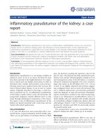
This revealed dilated common bile duct (with a
diameter of 11 mm) and a filling defect in her common
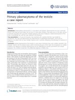
bile duct (Figure 1). During balloon extraction after her
endoscopic sphincterotomy, one live FH was for ced
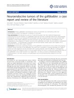
through her choledocus (Figure 2) and into the lumen
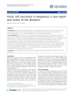
ofherduodenum(Figures3and4)andthenphysically
removed by biopsy forceps (Figure 5).
We prescribed our patient with a single morning dose
of 10 mg/kg of Tricalbendazole [3]. On her follow-up
examination, after four weeks, her liver enzymes had
returned to normal. Another ultrasonography revealed
that the size of her bile duct was normal.
Discussion
A variety of liver flukes, including Fasciola hepatica,
may c olonize the biliary tree of humans where they lay
their eggs. These eggs can give rise to the formation of
gallstones by serving as nidus for them. Living or dead
worms may occlude the bile ducts, thus causing
obstruction and sometimes cholangitis. Fascioliasis is
primarily a common disease of livestock animals such as
cattle and sheep, with humans serving occasionally as
accidental hosts.
Two stages have been described in human fascioliasis:
an acute phase, which coincides with h epatic invasion,
and a chronic phase, which develops due to the pre-
sence of flukes in the bile ducts [4]. The metacercariae
for these parasites encyst on freshwater plants, such as
wild watercress. Human consumption of aquatic plants
harvested from contaminated areas can lead to infection.
Subsequently, the developing larv ae penetrate the gut
wall and enter the peritoneal cavity. After a period of
migration for six to nine weeks, the flukes penetrat e the
Figure 1 Cholangiogram revealing a dilated common bile duct.
Figure 2 Fasciola coming out from the choledocus.
Figure 3 Fasciola in the duodenum.
Ezzat et al. Journal of Medical Case Reports 2010, 4:83
/>Page 2 of 4
capsule of the liver and mature in the biliary tree and
begin to pass their eggs. In the acute phase of the dis-
ease, our patient may have prolonged fever, right upper
quadrant pain, liver enlargement and eosinophilia that
can be easily misdiagnosed. These symptoms abate
when the chronic phase is reached. Once the flukes
enter the bile ducts, they may cause symptoms due to
cholestasis and cholangitis.
Although the definitive diagnosis for fascioliasis can be
made by detecting the eggs of the parasite in the stool
or duodenal aspirates, egg detection rate is not high
because of the low egg production rate of the parasite.
Immunoserological tests thus become the basis for the
diagnosis of fasc ioliasis, especially during its early stages
or in ectopic infections, but an enzyme-linked immuno-
sorbent assay (ELISA) test provides more ra pid and reli-
able results. Although some parts of Iraq are endemic
areas for human fascioliasis, we did not immediately
arrive at this diagnosis in the case of our patient. We
did not come up with a diagnosis before the parasit e
reached a full term (ERCP) because Fasciola hepatica is
still considered as a very rare cause of biliary obstruc-
tion. As expected, sphincterotomy and balloon extrac-
tion rapidly alleviated our patient’s symptoms.
Unlike patients with other liver flukes, therapeutic fail-
ureiscommoninpatientswithFasciola hepatica trea-
ted with praziquantel. Bithionol or triclabendazole
remains the treatment of choice for this parasitic infec-
tion. The use of bithionol, with a recommended dose of
30 to 50 mg/kg every other day for 10 to 15 doses or
repeated doses has resulted in the cure of acute and
prolonged fasci oliasis. Triclabendazole, another effective
and safe drug for fascioliasis, has been found to eradi-
cate the parasite with a single oral dose of 10 mg/kg [4].
Our patient was treated with a single 10 mg/kg oral
dose of triclabendazole.
We report this case because fascioliasis should be kept
in mind in the treatment of patients with cholestasis
and preceding vague gastrointestinal symptoms, espe-
cially in endemic areas of the world.
Conclusion
Chronic biliary fascioliasis may be asymptomatic or it
may present with biliary obstruction, cholangitis, or por-
tal fibrosis [5]. Our patient presented with cholangitis,
and FH was not suggested during the investigations
prior to ERCP when her condition was being diagnosed
and treated. However, serological tests can help in arriv-
ing at the correct diagnosis [2], although such tests are
not available in Kurdistan.
The technique of endoscopic sphincterotomy was initially
introduced to treat common bile duct stones. The indica-
tions have been expanded to include other biliary disorders.
Currently, this method is considered as the optimal
approach in treating biliary parasitosis including biliary fas-
cioliasis, biliary ascariasis, and biliary hydatid disease [6-8].
Previous reports have noted success with the combination
of ERCP and sphincterotomy for extracting FH from the
biliary tree [6,9,10]. This case report emphasizes the inclu-
sion of FH in the differential diagnosis for symptoms of
right upper qu adrant pain and co mmon bile duct dilata-
tion, particularly when ERCP management is a menable.
Figure 4 Fasciola swimming in the duodenum.
Figure 5 Fasciola hepatica in vitro.
Ezzat et al. Journal of Medical Case Reports 2010, 4:83
/>Page 3 of 4
Consent
Written informed consent was obtained from our
patient for publication of this case report and any
accompanying images. A copy of the written consent is
available for review by t he Editor-in-Chief of this
journal.
Authors’ contributions
RE collected, analyzed and interpreted our patient’s data, and assisted in the
therapeutic Endoscopy of our patient. TK performed the endoscopy and
assisted in interpretating our patient’s data. KK assisted in the therapeutic
endoscopy and analyzed our patient’s data. AH assisted in analyzing and
collecting the patients’ data. All authors read and approved the final
manuscript.
Competing interests
The authors declare that they have no competing interests.
Received: 31 December 2008
Accepted: 6 March 2010 Published: 6 March 2010
References
1. Adel AFM: Trematodes and other flukes. Principles and Practice of Infectious
Diseases Philadelphia: Churchill LivingstoneMandell GL, Bennet JE, Dolin R ,
5 2000, 2954-2956.
2. Marsden PD: Parasitic disease of the liver. Diseases of the Liver
Philadelphia: Lippincott William and WilkinsShiff 1999, 1078-1088.
3. Barrett-Conner E: Fluke infections. Infectious Diseases and Medical
Microbiology Philadelphia: WB SaundersBraude AI , 2 1986, 979-982.
4. Lopez VR, Dominguez CA, Garron C: Successful treatment of human
fascioliasis with tricalbendazole. Eur J Clin Microbiol Infect Dis 1999,
18(7):525-526.
5. Dias LM, Silva R, Viana HL, Palhinhas M, Viana RL: Biliary fascioliasis:
diagnosis, treatment and follow-up by ERCP. Gastrointest Endosc 1996,
43:616-620.
6. Veerappan A, Siegel JH, Podany J, Prudente R: Fasciola hepatica
pancreatitis: endoscopic extraction of live parasite. Gastroinest Endosc
1991, 37:437-435.
7. Khuroo MS, Zargar SA, Mahajan R: Hepatobiliary and pancreatic ascariasis
In India. Lancet 1990, 335 :1503-1506.
8. Alkarawi MA, Yasawy MI, Mohamed A: Endoscopic management of biliary
hydatid disease: report of six cases. Endoscopy 1991, 23:278-281.
9. Ozer B, Serin E, Gümürdülü Y, Gür G, Yilmaz U, Boyacioğlu S: Endoscopic
extraction of living fasciola hepatica: case report and review of
literature. Turk J Gastroenterol 2003, 14(1):74-77.
10. Bafandeh Y, Daghestani D, Rad S: Biliary tract obstruction due to fasciola
hepatica managed by ERCP. IJMS 2003, 28(1):43-45.
doi:10.1186/1752-1947-4-83
Cite this article as: Ezzat et al.: Endoscopic management of biliary
fascioliasis: a case report. Jo urnal of Medical Case Reports 2010 4:83.
Submit your next manuscript to BioMed Central
and take full advantage of:
• Convenient online submission
• Thorough peer review
• No space constraints or color figure charges
• Immediate publication on acceptance
• Inclusion in PubMed, CAS, Scopus and Google Scholar
• Research which is freely available for redistribution
Submit your manuscript at
www.biomedcentral.com/submit
Ezzat et al. Journal of Medical Case Reports 2010, 4:83
/>Page 4 of 4