Báo cáo y học: "Twin fetus in fetu in a child: a case report and review of the literature" pptx
Bạn đang xem bản rút gọn của tài liệu. Xem và tải ngay bản đầy đủ của tài liệu tại đây (988.3 KB, 7 trang )
CAS E REP O R T Open Access
Twin fetus in fetu in a child: a case report and
review of the literature
Ajay N Gangopadhyay
1*
, Arvind Srivastava
2
, Punit Srivastava
1
, Dinesh K Gupta
1
, Shiv P Sharma
1
, Vijayendra Kumar
1
Abstract
Introduction: Fetus in fetu is an extremely rare condition wherein a malformed fetus is found in the abdomen of
its twin. This entity is differentiated from teratoma by its embryological origin, its unusual location in the
retroperitoneal space, and the presence of vertebral organization with limb buds and well-developed organ
systems. The literature cites less than 100 cases worldwide of twin fetus in fetu.
Case presentation: A two-and-a-half-month-old Asian Indian baby boy had two malformed fetuses in his
abdomen. The pre-operative diagnosis was made by performing an ultrasound and a 64-slice computer
tomography scan of the baby’s abdomen. Two fetoid-like masses were successfully excised from the
retroperitoneal area of his abdomen. A macroscopic examination, an X-ray of the specimen after operation, and
the histological fe atures observed were suggestive of twin fetus in fetu.
Conclusion: Fetus in fetu is an extreme ly rare condition. Before any operation is carried out on a patient, imaging
studies should first be conducted to differentiate this condition from teratoma. Surgical excision is a curative
procedure, and a macroscopic examination of the sac should be done after twin or multiple fetus in fetu are
excised.
Introduction
Fetus in fetu (FIF) is a rare condition associated with
abnormal embryogenesis in a diamniotic, monochorionic
pregnancy, wherein a vertebrate fetus is enclosed within
the body of another normally developing fetus [1]. The
FIF complex is characteristically composed of a fibrous
membrane (equivalent to the chorioamniotic complex)
that contains some fluids (equivalent to the amniotic
fluid) and a fetus suspended by a co rd or pedicle. In the
uterus, the growth of an FIF initially parallels that of its
twin, but stops abruptly because of eith er the vascular
dominance of the host twin or an inherent defect in the
parasitic twin [2]. FIF is mostly anencephalic, but in
almost all cases its vertebral column and limbs are pre-
sent (91% and 82.5%, respectively). At the same time its
lower limbs are more developed than the upper limbs.
An FIF is rarely found in the central nervous system,
gastrointestinal tract, retroperitoneum, vessels or geni-
tourinary tract of its host twin. It is found even more
rarely in the lungs, adrenal glands, pancreas, spleen or
lymph nodes [3]. Even without performing an operation
to remove the parasitic twin, the existence of the condi-
tion can be diagnosed through ultrasonography, plain
X-ray and a computed tomography (CT) scan of the
host’s abdomen. T he surgical removal of the twin fetus
is the treatment of choice.
In most cases of FIF, only one fetus exists inside th e
baby. Only in extremely rare cases are multiple fetuses
found.
Case presentation
A two-and-a-half-month-old, first-born, Asian Indian
baby boy was admitted to the dep artment of Pediatric
surgery, S.S hospital, BHU, due to recurrent episodes of
vomiting and abdominal distension since he was one
month old. Upon examination of the baby’ sabdomen
we discovered that a smooth, firm and non-tender mass
was present in the left half of his abdomen. Conven-
tional X-ray of the abdomen showed a soft tissue mass
with a vertebrae-like column (Figure 1). An ultrasound
of the baby’s abdomen showed a large, encysted, hypere-
choic and calcified heterogenous complex mass. A 64-
slice CT scan of his abdomen revealed a soft tissue mass
* Correspondence:
1
Department of Pediatric Surgery, Institute of Medical Sciences, Banaras
Hindu University, Varanasi, India
Gangopadhyay et al. Journal of Medical Case Reports 2010, 4:96
/>JOURNAL OF MEDICAL
CASE REPORTS
© 2010 Gangopadhyay et al; licensee BioMed Central Ltd. This is an Open Access a rticle distributed under the terms of the Creative
Commons Attribu tion License ( which permits unrestricted use, distribution, and
reproduction in any medium, provided the origina l work is properly cited.
that had a bony outline resembling a fetus (Figures 2
and 3). Interestingly, we found nothing significant in the
baby’s family history.
We performed an elective laparotomy after correcting
the baby’s fluid and electrolyte levels. We then found a
well-encapsulated cystic retroperitoneal mass that was
displacing his spleen, transverse colon and pancrea s.
This displacement presented laterally and caudally
toward his cephaloid and left kidney (Figure 4). The
mass had a separate blood supply connected to the
baby’s abdominal aorta just below his left renal arte ry.
We mobilized, without complication, his left colon, pan-
creas, duodenum and small bowel, after which we were
able to excise the mass completely.
The sac contained two miniature fetuses connected to
each other by a cord-like structure at the umbilicus. The
miniature fetuses had a well-defined foot, skin with hairs,
a convex and pliable skull bone, and other undifferen-
tiated tissues (Figure 5). A radiograph of the specimen
showed cranial bones and long bones with vertebral col-
umns (Figure 6). We then performed a macroscopic
pathological examination, from which we were able to
note that the mass measured 20 × 8 × 5 cm. It was also
composed of a head with hair, a trunk, and rudimentary
limbs connected by cord-like structures. The mass corre-
sponded to an incompletely developed twin fetus.
A microscopic exam ination showed that the under de-
veloped twin had mature embryonic tissues containing
Figure 1 Plain X-ray of the vertical calcification on the left side of the abdomen.
Gangopadhyay et al. Journal of Medical Case Reports 2010, 4:96
/>Page 2 of 7
elements of the three germinative layers. Skin, a verteb-
ral column, germinative buds of limbs, central nervous
tissue (encephalus and coroidal plexus), a stomach,
small and large bowels, pancreas, adrenal glands, k id-
neys, upper and lower airways, cardiac striate d muscles,
and lymphoid tissue-like spleen were found. The histo-
pathological study of the specimen supported the con-
clusion that the previously imaged calcifications could
be assumed to be the skull and bony constituents of the
vertebral axis, some parts of the skull, and bony consti-
tuents of the rudimentary limbs.
Our patient recovered well after the surgery and was
discharged. To rule out any recurrence he was followed
up through clinical examination, plain abdominal X-ray
examination, abdominal ultrasound, and serum alpha-
fet oprotein (AFP). We were unable to detect any recur-
rence of his previous symptoms one year after the
operation.
Discussion
The t erm “fetus i n fetu” was first used by Johann Frie-
drich Meckel during the late 18th cent ury [4]. Subse-
quently, Willi s described it as a rare condition where a
malformed parasitic twin resides in the body of its host,
usually in the host’s abdominal cavity [5]. The con dition
represents an aberration of monozygotic diamniotic
twinning where the unequal division of the totipotent
inner cell mass of the developing blastocyst leads to the
inclusion of a smaller cell mass within a maturing sib-
ling embryo.
This rare pathology occurs only once every 500,000
births [6]. Fewer than 100 cases worldwide have been
reported [7]. The literature rarely describes multiple or
twin FIF. The majority of cases of FIF occur during
infancy, with the oldest reported case b eing that of a
47-year-old man [1]. Thakral et al. report ed that FIF
occurs equally among t he male and female populations
[8]. In 70% of reported cases, the chie f presenting com-
plaint is an abdominal mass [9]. The mass is predomi-
nantly retroperitoneal in 80% of cases [5], while
reported uncommon sites are the oral cavity [4], the
sacrococcygeal region [10] and the scrotum [7].
The presence of a vertebral column in the FIF is a n
important feature that distinguishes it from a t eratoma.
The clear identification of a verterbal column shows
that fetal development of the included twin had
advanced at least beyond the primitive streak stage (12
to 15 days of gestation) to a notochord, which is the
precursor of the vertebral column [1-3,8]. FIF generally
occurs singly . Multiple masses have been found in only
a few instances. Our patient exemplifies the occurrence
of FIF as a partially developed twin fetus [11]. The mass
we found in our patient was enveloped by a sac that
contained a second mass, which was suggestive of a
twin FIF. There are instances wher e no symptoms at all
occur. In some cases, however, symptoms pre sent as an
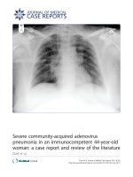
Figure 2 Abdominal computed tomography of the fetus with a large encapsulated peritoneal cavity mass and mature vertebral
skeleton.
Gangopadhyay et al. Journal of Medical Case Reports 2010, 4:96
/>Page 3 of 7
effect of the mass, such as abdominal distension, feeding
difficulties, emesis, jaundice and dyspnea [2, 11]. In our
case, our patient presented with distension of the abdo-
men and recurrent vomiting.
The pre-operative diagnosis of FIF depends on its
related radiological findings. Plain abdominal X-ray
examination may prove helpful, as up to half of reported
cases show the presence of a vertebral column and axial
skeleton [1], which was also the case for our patient.
Meanwhile, Hoeffel et al. [1] discussed the inability of
radiographic examination to visualize the vertebral axis
of the FIF. This inability to visualize the vertebral axi s
when a patient is examined through a CT sca n, how-
ever, should not lead to diagnostic exclusion because an
under-developed and markedly dysplas tic spinal column
may have prevented identification of the patholo gy at
imaging.
Sonographic findings are usually those of a complex
cystic mass with ill-defined s olid internal components.
Imaging continues to play an important role in diagnos-
ing FIF. CT and MRI have been proven to be very help-
ful in sugge sting a pre- operative diagnosis [11]. In o ur
Figure 3 A 64-slice computed tomography scan of the bony outline of the fetus in fetu.
Gangopadhyay et al. Journal of Medical Case Reports 2010, 4:96
/>Page 4 of 7
case, diagnosis was made pre-operatively through a CT
scan; nevertheless abdominal ultrasound cannot be
ignored in the initial evaluation of the anatomy of FIF.
The twin fetus is typically suspended by a pedicle within a
complete sac that contains fluid or sebaceous material.
There is no placenta or chorionic villi at the point of
attachment to the host [12]. In our case, our patient’stwin
was present within the sac in his left retroperitoneum.
The twin was also found suspended by a vascular pedicle
to its host’s abdominal aorta, and multiple vascular attach-
ments to the surrou nding bowel were noted. It is impor-
tant to note that the presence of a twin fetus in the host’s
abdomen is extremely rare.
Although the prognosis for FIF is more favorable than
for cystic teratoma, the presence of immature eleme nts
nevertheless indicates the need for close clinical,
Figure 4 Intra-operative picture of the fetus in fetu enveloped by a sac.
Figure 5 Twin fetus in fetu connected by a cord-like structure.
Gangopadhyay et al. Journal of Medical Case Reports 2010, 4:96
/>Page 5 of 7
radio logical and serological (AFP) follow-up [6]. Despite
the AFP levels before and after surgery remaining at
normal values, a possible recurrence of a malignant tera-
toma after FIF resection must best be kept in mind.
This was the reason why we continued to monitor the
serial tumor marker levels of our patient, while also
conducting cross-sectional imaging follow-up examina-
tions [1].
Conclusion
Alhtough it is rarely the conclusive diagnosis, FIF should
still be considered in a child presenting with progres-
sively increasing abdominal swelling and vomiting.
Although definitive diagnosis is best made using CT and
MRI techniques, plain X-rays and ultrasonography can
still be useful in the initial work-up pri or to surgery.
Post-operative X-ray e xamination of a specimen from
the mass can ultimate ly confirm the diagnosis of FIF.
The mass, however, should still be examined for the
occurrence of multiple fetus even after it has already
been excised.
Consent
Written informed consent was obtained from the par-
ents of our patient for publication of this case report
and any accompanying images. A copy of the written
consent is available for review by the Editor-in-Chief of
this journal.`
Author details
1
Department of Pediatric Surgery, Institute of Medical Sciences, Banaras
Hindu University, Varanasi, India.
2
Department of Radiodiagnosis, Institute of
Medical Sciences, Banaras Hindu University, Varanasi, India.
Authors’ contributions
ANG, SPS and PS operated on our patient and reviewed the literature. DKG
and VK were the main moderators of the manuscript. All authors read and
approved the final manuscript.
Competing interests
The authors declare that they have no competing interests.
Received: 22 October 2009 Accepted: 25 March 2010
Published: 25 March 2010
References
1. Hoeffel CC, Nguyen KQ, Phan HT, Truong NH, Nguyen TS, Tran TT, Fornes P:
Fetus in fetu: a case report and literature review. Pediatrics 2000,
105:1335-1344.
2. Patankar T, Fatterpekar GM, Prasad S, Maniyar A, Mukherji SK: Fetus in fetu:
CT appearance–report of two cases. Radiology 2000, 214:735-737.
3. Magnus KG, Millar AJ, Sinclair-Smith CC, Rode H: Intrahepatic fetus-in-fetu:
a case report and review of the literature. J Pediatr Surg 1999,
34:1861-1864.
4. Senyüz OF, Rizalar R, Celayir S, Oz F: Fetus in fetu or giant epignathus
protruding from the mouth. J Pediatr Surg 1992, 27:1493-1495.
5. Willis RA: The borderland of embryology and pathology. Washington, DC:
Butterworths, 2 1962, 442-462.
6. Hopkin KL, Dickson PK, Ball TI, Ricketts RR, O’Shea PA, Abramovosky CR:
Fetus in fetu with malignant recurrence. J Pediatr Surg 1997,
32:1476-1479.
7. Kakizoe T, Tahara M: Fetus in fetu located in the scrotal sac of a newborn
infant: a case report. J Urol 1972, 107:506-508.
8. Thakral CL, Maji DC, Sajwani MJ: Fetus in fetu: a case report and review of
literature. J Pediatr Surg 1998, 33:1432-1434.
Figure 6 Plain X-ray of the fetal specimen with a vertebral column.
Gangopadhyay et al. Journal of Medical Case Reports 2010, 4:96
/>Page 6 of 7
9. Knox JS, Webb AJ: The clinical features and treatment of fetus in fetu:
two case reports and review of literature. J Pediatr Surg 1975, 10:483-489.
10. Sanal M, Kucukcelebi A, Abasiyanik F, Erdogan S, Kocabasoglu U: Fetus in
fetu and cystic rectal duplication in a newborn. Eur J Pediatr Surg 1997,
7:120-121.
11. Luzzato C, Talenti E, Tregnaghi A, Fabris S, Scapinello A, Guglielmi M:
Double fetus in fetu: diagnostic imaging. Pediatr Radiol 1994, 24:602-603.
12. Chua JH, Chui CH, Sai Prasad TR, Jabcobsen AS, Meenakshi A, Hwang WS:
Fetus-in-fetu in the pelvis: report of a case and literature review. Ann
Acad Med (Singapore) 2005, 34:646-649.
doi:10.1186/1752-1947-4-96
Cite this article as: Gangopadhyay et al.: Twin fetus in fetu in a child: a
case report and review of the literature. Journal of Medical Case Reports
2010 4:96.
Submit your next manuscript to BioMed Central
and take full advantage of:
• Convenient online submission
• Thorough peer review
• No space constraints or color figure charges
• Immediate publication on acceptance
• Inclusion in PubMed, CAS, Scopus and Google Scholar
• Research which is freely available for redistribution
Submit your manuscript at
www.biomedcentral.com/submit
Gangopadhyay et al. Journal of Medical Case Reports 2010, 4:96
/>Page 7 of 7