Báo cáo y học: "Non-Hodgkin''''s lymphoma presenting as a primary bladder tumor: a case report" ppsx
Bạn đang xem bản rút gọn của tài liệu. Xem và tải ngay bản đầy đủ của tài liệu tại đây (634.41 KB, 5 trang )
JOURNAL OF MEDICAL
CASE REPORTS
Díaz-Peromingo et al. Journal of Medical Case Reports 2010, 4:114
/>Open Access
CASE REPORT
BioMed Central
© 2010 Díaz-Peromingo et al; licensee BioMed Central Ltd. This is an Open Access article distributed under the terms of the Creative
Commons Attribution License ( which permits unrestricted use, distribution, and repro-
duction in any medium, provided the original work is properly cited.
Case report
Non-Hodgkin's lymphoma presenting as a primary
bladder tumor: a case report
José A Díaz-Peromingo*
1
, Javier Tato-Rodríguez
2
, Paula M Pesqueira-Fontán
1
, Sonia Molinos-Castro
1
, María C Gayol-
Fernández
1
and Juliusz P Struzik
2
Abstract
Introduction: Primary lymphoma of the bladder represents 0.2% of all bladder malignancies. Secondary involvement
of the bladder by malignant lymphoma occurs in 10% to 50% of cases. Most lymphomas of the bladder are non-
Hodgkin's lymphomas of the B-cell type, with preponderance among women. The impact of positron emission
tomography (PET) on tumor staging has recently become very important due to its use in the study of diagnosis
extension and individual therapy design.
Case presentation: We report the case of a 79-year-old Caucasian man with intermittent haematuria as the
presenting symptom of non-Hodgkin's lymphoma of the bladder. He was first diagnosed with primary lymphoma of
the bladder using the current staging method, but a positron emission tomography study subsequently revealed that
he instead had a secondary involvement of the bladder.
Conclusion: The staging of non-Hodgkin's lymphomas, which is useful in order to plan accurate therapy, has been
changing since the introduction of positron emission tomography scanning. Primary lymphomas of the bladder,
although very rare, may be even more uncommon when this imaging technique is used to assess the extension of the
disease. Although the interpretation of this technique has some limitations that should be taken into account, the
extensive use of positron emission tomography should nonetheless help improve the diagnosis of this disease.
Introduction
Most tumors of the bladder are derived from the epithe-
lium. Non-epithelial tumors are extremely rare. Among
these, leiomyosarcomas are the most common in adults
and rhabdomyosarcomas are the most common in chil-
dren [1]. Metastatic tumors more frequently affect the
bladder neck and the deep trigone and represent approxi-
mately 15% of all known bladder malignancies [2].
Meanwhile, primary lymphomas of the bladder are very
rare and were first described by Eve in 1885 [3]. They rep-
resent 0.2% of all known bladder cancers [4]. Secondary
bladder involvement is reported in 10% to 50% of cases,
with a maximum incidence between the fourth and sixth
decades of life [5,6]. Most lymphomas of the bladder are
low-grade non-Hodgkin's lymphomas (NHL) of the B-cell
type [1,7]. Women are affected more frequently than men
[4,8].
The most frequent symptoms of bladder lymphomas
are gross haematuria followed by concomitant urinary
tract infection, dysuria and increased urinary frequency
[9]. Other complications like hydronephrosis, fistulas or
involvement of the entire bladder are very rare [10].
Usually, the diagnosis of primary lymphoma of the
bladder is one of exclusion. It is made in the absence of
any other nodal or extranodal involvement after biopsy
with immunohistochemical study, and after a negative
study of disease extension, which includes bone marrow
biopsy and computed tomography (CT) [11]. This
approach is changing with the introduction of positron
emission tomography (PET) to assess for other nodal or
extranodal involvement when a possible primary lym-
phoma of the bladder is suspected. In this case report, we
present a case of primary lymphoma of the bladder in
which PET scanning changed the diagnosis to extended
NHL. We review the literature focusing on the use of PET
in the assessment of tumor extension.
* Correspondence:
1
Department of Internal Medicine, Hospital da Barbanza, Oleiros, Riveira,
15993, Spain
Full list of author information is available at the end of the article
Díaz-Peromingo et al. Journal of Medical Case Reports 2010, 4:114
/>Page 2 of 5
Case presentation
A 79-year-old Caucasian, Spanish man was admitted to
our hospital because of intermittent painful gross haema-
turia lasting for seven days. He smoked 20 cigarettes per
day and reported having an appendectomy 40 years prior
to presentation. He had worked as a seaman in his youth
and was currently retired. He took no medications.
Results of his general examination were normal. His
rectal examination revealed an enlarged prostate, grade I/
IV, with no masses or nodules. Results of his analytical
studies including serum chemistry, prostate specific anti-
gen (PSA), and coagulation studies were normal. A mild
anaemia (hemoglobin = 11.3 g/L) with a normal mean
corpuscular volume was reported. His erythrocyte sedi-
mentation rate (ESR) was 67 mm per hour. An ultrasono-
graphic study of his abdomen had normal results, except
for an irregular border of the right lateral wall of his blad-
der.
Meanwhile, his intermittent gross haematuria contin-
ued, sometimes with associated blood clots. A cystoscopy
procedure was performed and an irregular mass affecting
his right antero-lateral wall was found. Biopsy revealed a
diffuse, large B-cell lymphoma with the following immu-
nohistochemical findings: CD20-, BCL6-, and DC10-pos-
itive. Meanwhile, his MIB1 had a high proliferation level.
CT scans of his chest, abdomen and pelvis were per-
formed and showed neither enlarged nodes nor liver or
spleen involvement. No metastatic lesions were likewise
found. His peripheral blood smear test was normal, as
were his direct and indirect Coombs tests, serum protein
counts, and plasma serum immunoglobulins. His β2-
microglobulin was 2.79 mg/dL (normal range = <0.27
mg/dL).
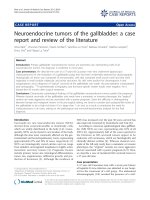
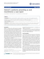
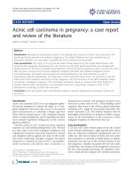
Results of a whole body PET study of our patient (Fig-
ures 1 and 2) revealed two nodes with increased metabo-
lism in the left part of his neck, and another area close to
his left supraclavicular space, which was suggestive of
nodal involvement. An enlarged left mediastinal lymph
node was also found on our patient. His left suprarenal
gland showed hypermetabolism. His abdomen appeared
to have multiple lymph node infiltrates affecting his lum-
bar region in particular, both his iliac lymphatic chains,
and those close to his bladder with associated hyperme-
tabolism of his bladder walls. Results of his bone marrow
biopsy were also normal.
Our patient was started on a treatment with CNOP
(cyclophosphamide, mitoxantrone, vincristine and pred-
nisolone) and monoclonal antibodies anti-CD20. He
showed good tolerance and initial response to this treat-
ment.
Conclusion
Our patient showed no abnormalities on the CT study.
Nevertheless, significant nodal and extranodal (suprare-
nal gland) extension of his disease was discovered upon
performing a PET study.
PET with 2- [fluorine 18] fluoro-2-deoxy-D-glucose
(FDG) is increasingly being used in combination with CT
to evaluate thoracic and abdominopelvic malignancies
[12]. A common systemic malignancy involving the pelvis
is NHL. Whole-body PET is useful in the detection of a
wide variety of both primary and metastatic malignancies
because of the high glycolytic rate that the malignant tis-
sue presents. The presence of FDG uptake in benign
inflammatory conditions may limit the specificity of PET.
Sensitivity for the detection of malignant lesions is about
97% and the positive predictive value is 94%. This tech-
nique is promising both in determining the nature of a
localized lesion, as well as in defining the systemic extent
of a malignant disease [13].
A number of studies have shown FDG-PET to be useful
and in fact superior to CT for primary staging and assess-
ment of disease extension in both Hodgkin's disease and
NHL. The technique is reported to have sensitivities of
82% to 99% and specificities of 99% to 100% [14].
Although data regarding the use of in-line FDG-PET-CT
systems in evaluating lymphoma are inconclusive, pre-
liminary results appear to indicate that this technique is
useful when a guided biopsy procedure is needed [15]. In
one study that compared the diagnostic performance of
PET alone, CT alone, and fused images for restaging or
follow-up of patients with malignant lymphoma, 50
patients with NHL were included. In this study, the inter-
pretation of PET alone (sensitivity = 86.1%, specificity =
99.4%, accuracy = 91.0%), and fused images (98.0%,
99.4%, and 99.2%, respectively) yielded significantly bet-
ter diagnostic performance than CT alone (59.4%, 96.1%,
91.0%; P < 0.001). In particular, findings in cervical, supr-
aclavicular and extranodal regions were more accurately
identified using PET (P < 0.05) [16]. With regard to stag-
ing, FDG-PET is more sensitive and specific than conven-
tional staging methods in FDG avid lymphomas such as
Hodgkin's lymphoma and most aggressive NHLs.
In assessing a patient's response to therapy, FDG-PET
at the end of treatment seems to aid considerably in dif-
ferentiating between residual masses with and without
residual lymphoma. Concerning treatment planning,
meanwhile, in the context of a combined-modality ther-
apy, radiotherapy for lymphomas is moving towards more
conformal techniques to reduce the irradiated volume
and to include only the macroscopic lymphoma. In this
context, accurate imaging is essential, and FDG-PET in
combination with CT scan is increasingly being used.
The availability of PET and CT scanners suited for virtual
simulation has aided in this process [17].
The limitations of FDG-PET in detecting lymphomas
have included variable FDG uptake in low-grade lympho-
mas; physiologic activity in muscles, bone marrow, bow-
Díaz-Peromingo et al. Journal of Medical Case Reports 2010, 4:114
/>Page 3 of 5
Figure 1 Coronal view shows multiple areas of increased metabolism affecting the neck, mediastinum, left suprarenal gland, abdominal
lymph nodes and the bladder.
Díaz-Peromingo et al. Journal of Medical Case Reports 2010, 4:114
/>Page 4 of 5
els, and the urinary system; and FDG uptake in
inflammatory or infectious processes, any of which may
mask or mimic tumor signals [18]. Another limitation in
the analysis of the pelvis and the urinary tract is the phys-
iological excretion of radiotracers [19].
This case suggests the need for extensive lymphoma
staging, and especially the need for PET implementation,
in order to make an accurate diagnosis of the extension of
the disease and to properly design a course of treatment.
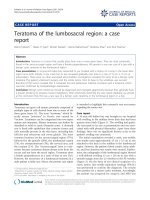
Figure 2 This image shows the PET findings in a three-dimensional projection view.
Díaz-Peromingo et al. Journal of Medical Case Reports 2010, 4:114
/>Page 5 of 5
Consent
Written informed consent was obtained from our patient
for publication of this case report and any accompanying
images. A copy of the written consent is available for
review by the Editor-in-Chief of this journal.
Competing interests
The authors declare that they have no competing interests.
Authors' contributions
JADP, JTR and PPF analyzed patient data on aematological disease and PET
interpretation. SMC, MCGF and JPS reviewed the literature related to the clini-
cal case. JADP, JTR and PPF were major contributors in writing the manuscript.
JPS provided help in translating the manuscript into English. All authors read
and approved the final manuscript.
Author Details
1
Department of Internal Medicine, Hospital da Barbanza, Oleiros, Riveira, 15993,
Spain and
2
Department of Urology, Hospital da Barbanza, Oleiros, Riveira,
15993, Spain
References
1. Mourad WA, Khalil S, Radwi A, Peracha A, Ezzat A: Primary T-cell
lymphoma of the urinary bladder. Am J Surg Pathol 1998, 22:373-377.
2. Bates AW, Baithun SI: Secondary neoplasms of the bladder are
histological mimics of non-transitional cell primary tumors:
clinicopathological and histological features of 282 cases. Histopathol
2000, 36:32-40.
3. Jacobs A, Symington T: Primary lymphosarcoma of urinary bladder. Br J
Urol 1953, 25:119-126.
4. Kuhara H, Tamura Z, Suchi T, Hattori R, Kinukawa T: Primary malignant
lymphoma of the urinary bladder: a case report. Acta Pathol Jpn 1990,
40:764-769.
5. Aigen AB, Phillips M: Primary malignant lymphoma of urinary bladder.
Urol 1986, 28:235-237.
6. Downs TM, Kibel AS, De Wolf WC: Primary lymphoma of the bladder: a
unique cystoscopic appearance. Urol 1997, 49:276-278.
7. Fernández Aceñero MJ, Martín Rodilla C, López García-Asenjo J, Coca
Menchero S, Sanz Esponera J: Primary malignant lymphoma of the
bladder: report of 3 cases. Pathol Res Pract 1996, 192:160-163.
8. Freeman C, Berg JW, Cutler SJ: Occurrence and prognosis of extranodal
lymphomas. Cancer 1972, 29:252-260.
9. Santino AM, Shumaker EJ, Garces J: Primary malignant lymphoma of the
bladder. J Urol 1970, 103:310-313.
10. Arda K, Ozdemir G, Günes¸ Z, Ozdemir H: Primary malignant lymphoma of
the bladder: a case report and review of the literature. Int Urol Nephrol
1997, 29:319-322.
11. Evans DA, Moore AT: The first case of vesicovaginal fistula in a patient
with primary lymphoma of the bladder: a case report. J Med Case
Reports 2007, 27:1-105.
12. Subhas N, Patel PV, Pannu HK, Jacene HA, Fishman EK, Wahl RL: Imaging
of pelvic malignancies with in-line FDG PET-CT: case examples and
common pitgalls of FDG PET. RadioGraphics 2005, 25:1031-1043.
13. Hoh CK, Hawkins RA, Glaspy JA, Dahlbom M, Tse NY, Hoffman EJ,
Schiepers C, Choi Y, Rege S, Nitzsche E: Cancer detection with whole-
body PET using 2-[18F] fluoro-2-deosy-D-glucose. J Comput Assist
Tomogr 1993, 17:582-589.
14. Schiepers C, Filmont JE, CZernin J: PET for staging of Hodgkin's disease
and non-Hodgkin lymphoma. Eur J Nucl Med Mol Imaging 2003,
30(Suppl 1):S82-S88.
15. Schoder H, Larson SM, Yeung HW: PET/CT in oncology: integration into
clinical management of lymphoma, melanoma, and gastrointestinal
malignancies. J Nucl Med 2004, 45(Ssuppl 1):S72-S81.
16. Nogami M, Nakamoto Y, Sakamoto S, Fukushima K, Okada T, Saga T,
Higashi T, Senda M, Matsui T, Sugimura K: Diagnostic performance of CT,
PET, side-by-side, and fused image interpretations for restaging of
non-Hodgkin lymphoma. Ann Nucl Med 2007, 21:189-196.
17. Specht L: 2-[18F]fluoro-2-deoxyglucose positron-emission
tomography in staging, response evaluation, and treatment planning
of lymphomas. Semin Radiat Oncol 2007, 17:190-197.
18. Barrington SF, O'Doherty MJ: Limitations of PET for imaging lymphoma.
Eur J Nucl Med Mol Imaging 2003, 30(Suppl 1):S117-S127.
19. Mantzarides M, Papathanassiou D, Bonardel G, Soret M, Gontier E,
Foehrenbach H: High-grade lymphoma of the bladder visualized on
PET. Clin Nucl Med 2005, 30:478-480.
doi: 10.1186/1752-1947-4-114
Cite this article as: Díaz-Peromingo et al., Non-Hodgkin's lymphoma pre-
senting as a primary bladder tumor: a case report Journal of Medical Case
Reports 2010, 4:114
Received: 28 October 2008 Accepted: 26 April 2010
Published: 26 April 2010
This article is available from: 2010 Díaz-Peromingo et al; licensee BioMed Central Ltd. This is an Open Access article distributed under the terms of the Creative Commons Attribution License ( ), which permits unrestricted use, distribution, and reproduction in any medium, provided the original work is properly cited.Journal of Medical Case Reports 2010, 4:114