Báo cáo y học: "Treatment of eccrine porocarcinoma with metastasis to the parotid gland using intensity-modulated radiation therapy: a case report" pptx
Bạn đang xem bản rút gọn của tài liệu. Xem và tải ngay bản đầy đủ của tài liệu tại đây (1.3 MB, 5 trang )
JOURNAL OF MEDICAL
CASE REPORTS
Zeidan et al. Journal of Medical Case Reports 2010, 4:147
/>Open Access
CASE REPORT
BioMed Central
© 2010 Zeidan et al; licensee BioMed Central Ltd. This is an Open Access article distributed under the terms of the Creative Commons
Attribution License ( which permits unrestricted use, distribution, and reproduction in
any medium, provided the original work is properly cited.
Case report
Treatment of eccrine porocarcinoma with
metastasis to the parotid gland using
intensity-modulated radiation therapy: a case
report
Youssef H Zeidan*
1
, A Jason Zauls
1
, Masha Bilic
2
, Eric J Lentsch
3
and Anand K Sharma
1
Abstract
Introduction: Cutaneous eccrine porocarcinomas are uncommon malignant tumors of the sweat gland.
Case Presentation: A 76-year-old Caucasian man presented to our hospital with a left temporal mass. We describe a
case of eccrine porocarcinoma with metastasis to the parotid gland with special emphasis on the role of surgical
resection and adjuvant radiation therapy.
Conclusion: Besides surgical resection, little is known about the role of adjuvant therapy in managing eccrine
porocarcinomas. Radiation therapy should be considered within a multidisciplinary approach in patients with primary
or recurrent eccrine porocarcinomas.
Introduction
Eccrine porocarcinoma (EPC) is a rare malignant tumor
of the sweat gland that accounts for 0.005% of all skin
cancers [1]. Due to its low incidence, current knowledge
about EPC is limited to case reports, with only 250 cases
having been reported worldwide. Common EPC lesions
occur on the lower extremities (50%) followed by the
trunk (24%) or the head and neck (24%), with more than
50% of cases occurring in men [2]. EPC has also been
reported to involve the vulva [3], penis [4] and upper
extremities [5]. Although the age at presentation ranges
from 19 to 90 years [6], EPC tends to affect the elderly, at
an average age of 68. Surgical resection remains the gold
standard for treatment. There is a 17% incidence of local
recurrence and an 11% incidence of distant metastasis
[7], which indicates that there is a role for adjuvant ther-
apy.
We report the case of a patient with cutaneous EPC
with secondary metastasis to the parotid gland who was
treated with intensity-modulated radiation therapy after
surgical resection.
Case Presentation
A 76-year-old Caucasian man presented with a long his-
tory of a left temporal lesion that had progressively
enlarged only recently. Initial surgical excision yielded a
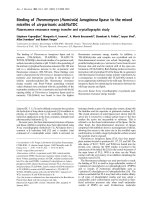
mass measuring 2.5 × 2.2 × 1 cm. Histological examina-
tion revealed tumor islands infiltrating the dermis and
connecting to the epidermis with a lobulated morphology
(Figure 1). Since the deep resection margin was positive
for malignant cells, a re-excision was performed and neg-
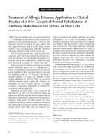
ative margins were verified microscopically. Eight months
later, our patient presented with a 2 cm firm mass overly-
ing the left parotid gland with minimal mobility on physi-
cal examination (Figure 2). Whole body imaging in the
form of a positron emission tomography (PET) scan
showed increased uptake in the left parotid gland, which
was consistent with metastasis. Our patient underwent
left parotidectomy with facial nerve preservation and cer-
vical lymphadenectomy. Surgical pathology specimens
revealed a moderately differentiated carcinoma with
growth pattern and morphological features consistent
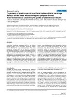
with porocarcinoma. Microscopically, nests of polygonal
malignant cells were seen infiltrating the papillary and
reticular dermis (Figure 3). A surrounding dense fibrous
* Correspondence:
1
Department of Radiation Oncology, Medical University of South Carolina,
Charleston, SC, USA
Full list of author information is available at the end of the article
Zeidan et al. Journal of Medical Case Reports 2010, 4:147
/>Page 2 of 5
stroma could be visualized (Figure 3). The cervical lymph
nodes (14 in total) were negative.
Given the aggressive behavior of EPC described in the
literature and evidence for the effective use of radiation
therapy [8], our patient was offered intensity-modulated
radiation therapy (IMRT). After initial consultation at the
Head and Neck Radiation Oncology Clinic of the Medical
University of South Carolina (MUSC) Hollings Cancer
Center, a consent form was obtained. Treatment planning
consisted of a contrasted head and computed tomogra-
phy (CT) scan of the neck with our patient lying supine,
using a head and neck thermoplast mask with bite block
immobilization. Thin (3 mm) axial images were imported
into the ADAC Pinnacle planning system (ADAC Labora-
tories, Milpitas, CA, USA). In this case, IMRT was
designed using an inverse-planning algorithm.
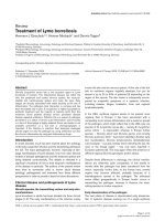
The six-beam heterogeneous plan entailed delivering a
total of 60 Gy in 30 treatment fractions over six weeks. A
CT scan with isodose distributions and a dose-volume
histogram for the final treatment plan are shown in Fig-
ure 4. Particular attention was directed to regions of
interest such as the left inner ear and optic nerve, to min-
imize radiation exposure well below the reported toler-
ance doses of 40 Gy and 50 Gy, respectively [9].
Weekly on-site treatment visits were documented dur-
ing the treatment phase of our patient, followed by sched-
uled three-month follow-up visits. Our patient tolerated
the radiation treatment well, and developed only mild
xerostomia and mild paresthesias. Further follow-up
revealed that our patient remains disease-free 10 months
after completion of the treatment course.
Discussion
Eccrine porocarcinoma is challenging to diagnose based
on clinical presentation alone, and histopathological
examination is almost always required. Typically, a
Figure 1 Photomicrograph of the initial left temporal mass show-
ing islands of polygonal tumor cells invading the dermis (hema-
toxylin and eosin, 10×).
Figure 2 Macroscopic view showing a new left parotid lesion and
a scar at the previous frontotemporal lesion.
Figure 3 Photomicrographs of surgical pathology specimens. (A-
B) Photomicrograph of left parotid mass showing tumor cells of similar
cytology to the original temporal mass infiltrating the parotid gland.
Ductal structures characteristic of eccrine porocarcinoma are seen (he-
matoxylin and eosin, 4× to 20×).
Zeidan et al. Journal of Medical Case Reports 2010, 4:147
/>Page 3 of 5
patient presents with an erythematous papule with a
recent change in size, bleeding or itching. The differential
diagnosis includes squamous cell carcinoma (SCC), basal
cell carcinoma, Paget's disease and metastatic cancer.
Positive staining for periodic acid-Schiff (PAS), carcino-
embryonic antigen (CEA) or angiotensin type 1 receptor
can aid in making the diagnosis [10,11]. Although the eti-
ology remains unknown, it has been suggested that EPC
arises from the malignant transformation of eccrine
poroma. Interestingly, an association has been proposed
Figure 4 (A) Axial, saggital and coronal CT images showing the final intensity-modulated radiation therapy (IMRT) plan and isodose distri-
butions around the tumor bed. Gross tumor volume (GTV) is outlined from contrast enhanced planning CT scan. Isodose lines of decreasing ener-
gies radiate out from the center of the tumor bed. The tumor is completely contained by the 95% isodose blue line (57 Gy). (B) Cumulative dose-
volume histogram (DVH) of the IMRT plan. The curves illustrate the dose distribution for the clinical target volume (CTV: defined as GTV +0.5 cm), man-
dible, left parotid, optic nerves, optic chiasm and a cervical lymph node.
Zeidan et al. Journal of Medical Case Reports 2010, 4:147
/>Page 4 of 5
between EPC and the immuncompromised states such as
human immunodeficiency virus (HIV), diabetes and
organ transplantation [12].
Wide local excision [7] and Mohs surgery [13] are
widely accepted treatment modalities for primary EPC.
Surgical excision has a cure rate of 70-80% and a local
recurrence rate of 20%. Excellent outcomes have been
reported following Moh's surgery, with patients in remis-
sion after five years of follow-up.
Based on the literature, the role of chemotherapy in the
treatment of EPC remains unclear. Orphan cases with
good responses to 5-fluorouracil [14], thiotepa and
Cytoxan (cyclophosphamide) [15] have been reported.
However, other studies have described cases showing no
clinical response to chemotherapy [16]. In one report,
four cases of pediatric EPC were treated with a combina-
tion of 5-fluorouracil, doxorubicin and cyclophosph-
amide. No response was observed after one year of
therapy [17]. In general, chemotherapy, if considered at
all, is reserved for metastatic EPC.
On the other hand, the role of radiation therapy in EPC
seems to have changed over the years from radioresis-
tance in earlier reports [15] to good local control in more
recent studies [16]. Perhaps the changes in these anec-
dotal reports reflect the evolution of technological
advances in radiation therapy over time. Combinations of
photons and electrons were used in several cases. Table 1
summarizes reported cases and outcomes of EPC involv-
ing radiation therapy.
Until further studies on EPC are conducted, one can
extrapolate important lessons from clinical experience
with the more common SCC of the head and neck. The
parotid gland is a common site for metastasis of cutane-
ous tumors of the scalp, frontotemporal and periauricular
regions. Poor outcomes have been associated with
parotid disease, with a two-year survival of only 74%. Sur-
vival of patients drops further to 67% when parotid dis-
ease is concurrent with neck disease [18]. Patients who
require parotidectomy are more likely to have recurrent
lesions. Given the importance of parotid involvement,
some authorities have advocated modifying the current
American Joint Commission on Cancer (AJCC) staging
for metastatic cutaneous carcinoma in order to distin-
guish parotid from neck diseases [19]. Post-operative
radiation therapy has emerged as an integral part of the
care for aggressive cutaneous carcinoma patients with
parotid invasion. A landmark study by Taylor et al. dem-
onstrated that patients treated with post-operative radia-
tion therapy had 89% local disease control, compared to
63% for those treated with surgery alone and 46% for
those treated with radiation alone [8]. In a more recent
study, Weber and colleagues further confirmed the value
of a dual approach of surgery and radiation therapy [20].
Conclusion
More than 45 years after its original description by
Pinkus and Mehregan [21], guidelines for staging and
treatment EPC are still lacking. The current report
describes the presentation and management of an inter-
esting case of cutaneous EPC with metastasis to the
parotid gland. Although the short follow-up period is a
limitation, to the best of our knowledge there has only
been one previously reported case akin to ours [22].
Because of its aggressive potential for metastatic spread, a
multidisciplinary approach involving surgery, pathology
and radiation therapy should be considered in the man-
agement of EPC.
Consent
Written informed consent was obtained from the patient
for publication of this case report and any accompanying
Table 1: Summary of reported eccrine porocarcinoma cases involving adjuvant radiation therapy.
Location XRT Outcome
(Remission)
Ref
Hand 70 Gy to tumor and 50 Gy to
regional LNs
24 months [23]
Nose 71.6 Gy to tumor and 50 Gy to
supraclavicular LNs
35 months [22]
Parotid 70 Gy to parotid and 46 Gy to
lower cervical LNs
27 months [22]
Scalp and cervical LNs 76.4 Gy to scalp and 76.5 Gy to
posterior cervical LNs
Death at 6 weeks [22]
Inguinal and para-aortic LNs 50.4 Gy 8 months [24]
Left vulva 50.4 Gy to left pelvis and 60 Gy
to left inguinal LNs
19 months [3]
LNs: lymph nodes; XRT: radiation therapy.
Zeidan et al. Journal of Medical Case Reports 2010, 4:147
/>Page 5 of 5
images. A copy of the written consent is available for
review by the Editor-in-Chief of this journal.
Abbreviations
AJCC: American Joint Commission on Cancer; CEA: carcinoembryonic antigen;
CT: computed tomography; EPC: eccrine porocarcinoma; HIV: human immu-
nodeficiency virus; IMRT: intensity-modulated radiation therapy; PAS: periodic
acid-Schiff; PET: positron emission tomography; SCC: squamous cell carci-
noma.
Competing interests
The authors declare that they have no competing interests.
Authors' contributions
YHZ collected the data regarding our patient, prepared the manuscript, and
also researched the related literature. AJZ assisted in interpreting the data from
our patient. AKS and EJL supervised YHZ and AJZ and were major contributors
in writing the manuscript. MB provided the pathology figures and their leg-
ends. All the authors read and approved the final manuscript.
Author Details
1
Department of Radiation Oncology, Medical University of South Carolina,
Charleston, SC, USA,
2
Department of Pathology, Medical University of South
Carolina, Charleston, SC, USA and
3
Department of Otolaryngology, Medical
University of South Carolina, Charleston, SC, USA
References
1. Cowden A, Dans M, Militello G, Junkins-Hopkins J, Van Voorhees AS:
Eccrine porocarcinoma arising in two African American patients:
distinct presentations both treated with Mohs micrographic surgery.
Int J Dermatol 2006, 45(2):146-150.
2. Sosnowski J, Stetter-Neel C, Cole D, Durham JP, Mawhinney MG: Protein
kinase C mediated anti-proliferative glucocorticoid-sphinganine
synergism in cultured Pollard III prostate tumor cells. J Urol 1997,
158(1):269-274.
3. Katsanis WA, Doering DL, Bosscher JR, O'Connor DM: Vulvar eccrine
porocarcinoma. Gynecol Oncol 1996, 62(3):396-399.
4. Grayson W, Loubser JS: Eccrine porocarcinoma of the penis. J Urol 2003,
169(2):611-612.
5. Mehregan AH, Hashimoto K, Rahbari H: Eccrine adenocarcinoma. A
clinicopathologic study of 35 cases. Arch Dermatol 1983,
119(2):104-114.
6. Goedde TA, Bumpers H, Fiscella J, Rao U, Karakousis CP: Eccrine
porocarcinoma. J Surg Oncol 1994, 55(4):261-264.
7. Robson A, Greene J, Ansari N, Kim B, Seed PT, McKee PH, Calonje E:
Eccrine porocarcinoma (malignant eccrine poroma): a
clinicopathologic study of 69 cases. Am J Surg Pathol 2001,
25(6):710-720.
8. Taylor BW Jr, Brant TA, Mendenhall NP, Mendenhall WM, Cassisi NJ,
Stringer SP, Million RR: Carcinoma of the skin metastatic to parotid area
lymph nodes. Head Neck 1991, 13(5):427-433.
9. Emami B, Lyman J, Brown A, Coia L, Goitein M, Munzenrider JE, Shank B,
Solin LJ, Wesson M: Tolerance of normal tissue to therapeutic
irradiation. Int J Radiat Oncol Biol Phys 1991, 21(1):109-122.
10. Hara K, Kamiya S: Pigmented eccrine porocarcinoma: a mimic of
malignant melanoma. Histopathology 1995, 27(1):86-88.
11. Arslan E, Tatar C, Aksoy A, Tutuncu N: De novo malignant eccrine poroma
of the nose: a review of the midface as a location. Plast Reconstr Surg
2004, 113(7):2227-2229.
12. Mahomed F, Blok J, Grayson W: The squamous variant of eccrine
porocarcinoma: a clinicopathological study of 21 cases. J Clin Pathol
2008, 61(3):361-365.
13. Wittenberg GP, Robertson DB, Solomon AR, Washington CV: Eccrine
porocarcinoma treated with Mohs micrographic surgery: A report of
five cases. Dermatol Surg 1999, 25(11):911-913.
14. Swanson JD Jr, Pazdur R, Sykes E: Metastatic sweat gland carcinoma:
response to 5-fluorouracil infusion. J Surg Oncol 1989, 42(1):69-72.
15. el-Domeiri AA, Brasfield RD, Huvos AG, Strong EW: Sweat gland
carcinoma: a clinico-pathologic study of 83 patients. Ann Surg 1971,
173(2):270-274.
16. Shiohara J, Koga H, Uhara H, Takata M, Saida T: Eccrine porocarcinoma:
clinical and pathological studies of 12 cases. J Dermatol 2007,
34(8):516-522.
17. Chow CW, Campbell PE, Burry AF: Sweat gland carcinomas in children.
Cancer 1984, 53(5):1222-1227.
18. Khurana VG, Mentis DH, O'Brien CJ, Hurst TL, Stevens GN, Packham NA:
Parotid and neck metastases from cutaneous squamous cell carcinoma
of the head and neck. Am J Surg 1995, 170(5):446-450.
19. O'Brien CJ, McNeil EB, McMahon JD, Pathak I, Lauer CS, Jackson MA:
Significance of clinical stage, extent of surgery, and pathologic
findings in metastatic cutaneous squamous carcinoma of the parotid
gland. Head Neck 2002, 24(5):417-422.
20. Lai SY, Weinstein GS, Chalian AA, Rosenthal DI, Weber RS: Parotidectomy
in the treatment of aggressive cutaneous malignancies. Arch
Otolaryngol Head Neck Surg 2002, 128(5):521-526.
21. Pinkus H, Mehregan AH: Epidermotropic eccrine carcinoma. A case
combining features of eccrine poroma and Paget's dermatosis. Arch
Dermatol 1963, 88:597-606.
22. Harari PM, Shimm DS, Bangert JL, Cassady JR: The role of radiotherapy in
the treatment of malignant sweat gland neoplasms. Cancer 1990,
65(8):1737-1740.
23. DaSilva MF, Terek R, Weiss AP: Malignant eccrine poroma of the hand: a
case report. J Hand Surg Am 1997, 22(3):511-514.
24. Yamashita H, Kadono T, Tamaki K, Nakagawa K: Interesting response to
concurrent chemoradiation in metastatic eccrine porocarcinoma. J
Dermatol 2008, 35(9):606-607.
doi: 10.1186/1752-1947-4-147
Cite this article as: Zeidan et al., Treatment of eccrine porocarcinoma with
metastasis to the parotid gland using intensity-modulated radiation therapy:
a case report Journal of Medical Case Reports 2010, 4:147
Received: 28 September 2009 Accepted: 22 May 2010
Published: 22 May 2010
This article is available from: 2010 Zeidan et al; licensee BioMed Central Ltd. This is an Open Access article distributed under the terms of the Creative Commons Attribution License ( ), which permits unrestricted use, distribution, and reproduction in any medium, provided the original work is properly cited.Journal of Medical Case Reports 2010, 4:147