Báo cáo y học: "Compressive stenosis of the left hepatic vein as a pathogenesis of postresectional liver failure: a case report" potx
Bạn đang xem bản rút gọn của tài liệu. Xem và tải ngay bản đầy đủ của tài liệu tại đây (762.71 KB, 5 trang )
JOURNAL OF MEDICAL
CASE REPORTS
Ninomiya and Ikeda Journal of Medical Case Reports 2010, 4:163
/>Open Access
CASE REPORT
BioMed Central
© 2010 Ninomiya and Ikeda; licensee BioMed Central Ltd. This is an Open Access article distributed under the terms of the Creative
Commons Attribution License ( which permits unrestricted use, distribution, and repro-
duction in any medium, provided the original work is properly cited.
Case report
Compressive stenosis of the left hepatic vein as a
pathogenesis of postresectional liver failure: a case
report
Mizuki Ninomiya*
1,2
and Tetsuo Ikeda
1
Abstract
Introduction: Postresectional liver failure (PLF) is a devastating and fatal complication of major hepatic resection, and
we do not have a full understanding of the pathogenic mechanisms involved. No reliable treatment other than liver
transplantation currently exists for PLF.
Case presentation: A 46-year-old Japanese man experienced PLF after an extended right hepatectomy for liver
malignancy. Seven months after surgery, the patient's Model for End-Stage Liver Disease (MELD) score had reached 23.
Doppler ultrasound study and three-dimensional computed tomography images showed a stenosed left hepatic vein
compressed by surrounding hypertrophied hepatic parenchyma. Transluminal balloon angioplasty and stent
placement therapy were conducted eight months after surgery. The pressure gradient between the hepatic vein and
right atrium decreased from 13 to 3 mmHg after stent placement. Thereafter, the patient recovered.
Conclusion: Hepatic venous compression by surrounding hypertrophied hepatic parenchyma might, at least in part,
be associated with the occurrence of PLF. Surgeons should bear this possibility in mind when confronted with cases of
PLF, as early diagnosis and stent placement improves patients' chances of recovery.
Introduction
Hepatic resection is the preferred treatment for hepatic
malignancies like hepatocellular carcinoma or colorectal
liver metastases. Although major hepatic resection is now
accomplished with mortality rates of less than 5% in high
volume centers, post-resectional liver failure (PLF)
remains a potentially devastating complication and often
proves fatal [1]. The risk of PLF increases as the amount
of resected liver parenchyma increases. In general, the
relative remnant liver volume (RLV) ratio, defined as the
percentage remaining liver volume compared with stan-
dard liver volume, is regarded as a reliable parameter in
the prediction of PLF. In normal livers, an RLV ratio
below 25% has been reported to be a strong predictor of
serious hepatic dysfunction following liver resection
[2,3]. Unfortunately, no reliable disease-specific therapy
exists for PLF, with the exception of liver transplantation
in some limited cases, and PLF mortality rates are
between 60 and 80% [4,5]. Understanding the exact
pathophysiology of PLF would enable us to establish
effective treatments other than liver transplantation.
Here we present a case of PLF that was successfully
treated by hepatic venous stent placement therapy. We
also discuss the possible pathogenesis of PLF.
Case presentation
A 46-year-old Japanese man was referred to our hospital
with intrahepatic cholangiocellular carcinoma. The
tumor was 3 cm in diameter and located at segment 8,
between the root of the middle and right hepatic veins.
Preoperative liver function tests were normal, and preop-
erative blood work was negative for hepatitis virus mark-
ers. The patient underwent an extended right
hepatectomy without resection of the extrahepatic bile
duct. The patient's middle hepatic vein was divided
approximately 2 cm upstream from the root of the middle
and left hepatic veins.
The patient's postoperative course was uneventful until
day 10, when a gradual increase in serum bilirubin levels
and ascites formation were noted. Although obstructive
* Correspondence:
1
Department of Surgery, Oita Prefectural Hospital, Bunyo, Oita, 870-8511,
Japan
Full list of author information is available at the end of the article
Ninomiya and Ikeda Journal of Medical Case Reports 2010, 4:163
/>Page 2 of 5
jaundice and portal thrombosis were excluded by ultra-
sound and computed tomography (CT) scans, the
patients continued to deteriorate, displaying hyperbiliru-
binemia, coagulopathy and refractory ascites (Table 1).
Neither circulatory disturbance nor hepatic encephalopa-
thy were seen during this period. The patient's Model for
End-Stage Liver Disease (MELD) score also increased
gradually, and reached 23 seven months after the initial
hepatic surgery. MELD scores between 20 and 29 are
associated with 3-month mortality rates of over 75%.
Repeated abdominal paracentesis were necessary to treat
his refractory ascites, which amounted to more than
10,000 ml per week.
In order to rule out an insufficient hepatic regeneration,
the patient's liver volume was assessed by CT-volumetry
using previously described methods [6]. Seven months
after surger, his liver volume was 1290 ml, almost identi-
cal to the standard liver volume (1222 ml) which was cal-
culated using Urata's formula [7]. Nevertheless, serum
levels of hepatocyte growth factor were elevated to 1.61
ng/ml, signifying that the liver was still being stimulated
to regenerate. Serum hyaluronic acid levels were also ele-
vated to 3030 ng/ml, suggesting that sinusoidal endothe-
lial cell function was severely disturbed [8]. An
assessment of the patient's hepatic circulation by spectral
Doppler ultrasound revealed a good hepatic arterial flow,
but a relatively weak intrahepatic portal signal with no
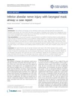
diastolic flow. The Doppler waveform of hepatic venous
flow was monophasic, suggesting the presence of hepatic
venous stenosis (Figure 1). Because axial CT images only
were not conclusive regarding the presence of hepatic
vein stenosis, multiplanar reconstruction (MPR) images
and three-dimensional CT images were also assessed, and
the stenosis at the root of the left hepatic vein was clearly
visualized (Figure 2).
To further confirm the presence of hepatic venous
stenosis, the pressure gradient between the left hepatic
vein and right atrium was measured. Hepatic venous
catheterization was performed through the jugular vein.
The mean hepatic venous pressure was 15.6 mmHg, and
the pressure abruptly decreased when the catheter was
pulled through the assumed stenotic point into the right
atrium. The pressure gradient between the left hepatic
vein right atrium was 13.2 mmHg. The normal hepatic
venous pressure gradient lies between 1 and 2 mmHg [9].
Thus, we confirmed the presence of hepatic venous
stenosis morphologically and functionally.
Written informed consent was obtained from the
patient for endovascular treatment. As an initial treat-
ment option, hepatic venous balloon dilation was per-
formed eight months after surgery. A transluminal
angioplasty catheter (Synergy Balloon Catheter, Boston
Scientific, Tokyo) with a balloon diameter of 10 mm and a
length of 40 mm was used for venous dilatation.
Table 1: Parameters of liver function before and after operation
Preoperative Postoperative
1 month 4 months 7 months
Total bilirubin (mg/dl) 1.2 9.8 16.5 30.1
Albumin (g/dl) 4.4 3.2 2.8 2.3
Prothorombin time (%) 112 89 81 68
AST (U/dl) 30 48 46 51
MELD score - 13 14 23
AST, aspartate aminotransferase; MELD, model for end-stage liver disease
Figure 1 Spectral Doppler ultrasound study and contrast-en-
hanced computed tomography (CT) images at 7 months after sur-
gery.
Ninomiya and Ikeda Journal of Medical Case Reports 2010, 4:163
/>Page 3 of 5
Although the pressure gradient across the left hepatic
vein and right atrium decreased from 13 to 4 mmHg
immediately after intervention, the effect was temporary.
Ten minutes after balloon dilatation, the pressure gradi-
ent had increased to 10 mmHg. Although serum bilirubin
levels decreased unexpectedly to near the normal range,
refractory ascites remained. Therefore, the patient under-
went endovascular stent placement therapy seven weeks
later.
A self-expandable metallic stent (Luminexx, BARD,
Covington, GA) with a diameter of 8 mm and a length of
40 mm was placed into the left hepatic vein via the inter-
nal jugular vein. Because the large umbilical fissure vein
was bifurcated just behind the stenosis, another metallic
stent with a diameter of 8 mm and a length of 2 mm was
inserted into it using a stent-in-stent technique (Figure 3).
At the same time, in order to relieve the symptom of
lower leg edema, the retrohepatic inferior vena cava was
also dilated with a metallic stent (Spiral Z stent, Wilson-
Cook Inc., Winston-Salem, MA) with a diameter of 20
mm and a length of 60 mm. The pressure gradient across
the left hepatic vein and right atrium decreased to 3
mmHg just after stent placement, which was maintained
even after 10 minutes. A Doppler ultrasound study three
weeks after stent placement showed an increased hepatic
venous flow velocity and a pulsatile venous waveform
(Figure 4). Along with the normalized bilirubin level, the
patient's ascitic fluid volume also decreased favorably
after the stent placement. Thus, the patient had recov-
ered from his PLF.
Discussion
Although many factors have been proposed to be associ-
ated with increased risk of PLF, including inadequate
hepatic regeneration, pre-existing cirrhosis and pro-
longed liver ischemia during resection, the critical factor
is believed to be an insufficient remnant liver mass [3].
Despite this, the mechanisms of PLF in the majority of
clinical cases are thought to be multifactorial, not due
purely to small remnant liver volume.
An adequate hepatic venous drainage is a prerequisite
in the recovery process of a damaged liver and its signifi-
cance is magnified in the case of extended right hepatec-
tomy or trisegmentectomy, whereby the left hepatic vein
is the only conduit to drain the entire remaining liver. It
seemed that in our case, the root of the left hepatic vein
was compressed by hypertrophied liver parenchyma sub-
sequent to a vigorous regenerative response. Although
reports of postoperative hepatic venous stenosis are com-
mon, most of these are stenosis after liver transplanta-
tion, whereby hepatic veins are more prone to mechanical
stenosis than in the case of hepatectomy. This is due to
suture anastomosis and to the possibility of venous kink-
ing associated with graft dislocation [10]. It could be
inferred that hepatic venous compression by surrounding
hypertrophied liver parenchyma might have been over-
Figure 3 Hepatic venography after stent placement.
Figure 4 The Doppler ultrasound images before (A, C) and after
(B, D) the stent placement therapy.
Figure 2 Images from computed tomography (CT) data delineat-
ing the stenosis of hepatic vein.
Ninomiya and Ikeda Journal of Medical Case Reports 2010, 4:163
/>Page 4 of 5
looked as the pathogenesis of PLF in previous cases. The
diagnosis of hepatic venous stenosis by means of ordinary
CT images seemed to be much more difficult than
expected, because a single axial CT image could not
clearly depict the outline of the hepatic vein with caudal
inclination. Meanwhile, with the spread of liver trans-
plantation, the usefulness of Doppler ultrasound for the
diagnosis of hepatic venous stenosis had been calrified.
Ko et al. reported that a persistent monophasic wave pat-
tern on Doppler ultrasound images suggested, but did not
conclusively indicate, hepatic venous stenosis after liver
transplantation [11]. Therefore, when hepatic venous
stenosis is suspected as a cause of PLF, a screening Dop-
pler ultrasound study should be used to assist in making a
definitive diagnosis, taking into consideration other stud-
ies such as three-dimensional CT imaging, hepatic
venography and measurement of the pressure gradient
across the stenosis.
Figure 5 diagrams the probable mechanism of PLF in
our case. Outflow disturbance of the liver had led to a
microcirculatory disturbance, and subsequently to a
decrease in the functional hepatocyte volume. In order to
meet the metabolic demand under these circumstances,
the liver might have been stimulated chronically to regen-
erate, as represented by elevated hepatocyte growth fac-
tor levels in the serum. Hepatic regeneration is
accompanied by a complex remodeling of the hepatic tis-
sue with a transient breakdown of the lobular architec-
ture. As we reported previously, the more the liver is
stimulated to regenerate, the greater the derangement of
lobular architecture and consequently hyperbilirubine-
mia and microcirculatory disturbances [12]. Such a
vicious circle for PLF might have been disconnected by
hepatic venous stent placement, ameliorating microcir-
culatory disturbances.
Transluminal balloon angioplasty and stent placement
therapy are currently the preferred mode of treatment for
hepatic venous stenosis. According to the treatment
results reported by Ko et al., hepatic venous stenoses
after liver transplantation were treated favorably by bal-
loon angioplasty, although repeat angioplasty was neces-
sary against restenosis [13]. The main cause of
anastomotic stenosis after liver transplantation is thought
to be a fibrosis or intimal hyperplasia around the anasto-
motic site. However, stenosis after partial hepatectomy
seems to be attributable, at least in part, to compression
by surrounding hypertrophied parenchyma. Therefore, as
in the present case, the treatment effects of balloon
angioplasty for compressed stenosis are temporary and
limited. From our experience of the present case, we
thought that stent placement would be a preferable treat-
ment measure for stenosis caused by extrinsic compres-
sion.
Conclusions
Hepatic venous compression by surrounding hypertro-
phied hepatic parenchyma might, at least in part, be asso-
ciated with the occurrence of PLF. Recognition of hepatic
venous compression as one of the pathogenic mecha-
nisms of PLF may help to establish an adequate mode of
treatment, such as stent placement therapy, and improve
the prognosis of patients without requiring liver trans-
plantation.
Consent
Written informed consent was obtained from the
patient's family for the publication of this case report and
any accompanying images. A copy of the written consent
is available for review by the Editor-in-Chief of this jour-
nal.
Abbreviations
CT: computed tomography; MELD: Model for End-Stage Liver Disease; MPR:
multiplanar reconstruction; PLF: postresectional liver failure; RLV: relative rem-
nant liver volume.
Competing interests
The authors declare that they have no competing interests.
Authors' contributions
MN interpreted the patient's data, devised the therapeutic plan, and wrote the
manuscript. TI helped in planning the therapeutic plan and drafting the manu-
script. All authors read and approved the final manuscript.
Acknowledgements
The technical assistance of Eiji Komatsu and Toru Maeda (Department of Radi-
ology, Oita Prefectural Hospital) are gratefully acknowledged.
Author Details
1
Department of Surgery, Oita Prefectural Hospital, Bunyo, Oita, 870-8511, Japan
and
2
Department of Surgery, National Hospital Organization Fukuoka Higashi
Medical Center, Koga, 811-3195, Japan
Figure 5 The possible mechanism of postresectional liver failure
in the present case.
Ninomiya and Ikeda Journal of Medical Case Reports 2010, 4:163
/>Page 5 of 5
References
1. Jarnagin WR, Gonen M, Fong Y, DeMatteo RP, Ben-Porat L, Little S, Corvera
C, Weber S, Blumgart LH: Improvement in perioperative outcome after
hepatic resection: analysis of 1,803 consecutive cases over the past
decade. Ann Surg 2002, 236:397-406.
2. Schindl MJ, Redhead DN, Fearon KC, Garden OJ, Wigmore SJ: The value of
residual liver volume as a predictor of hepatic dysfunction and
infection after major liver resection. Gut 2005, 54:289-296.
3. Broek MA Van den, Damink SW, Dejong CH, Lang H, Malago M, Jalan R,
Saner FH: Liver failure after partial hepatic resection: definition,
pathophysiology, risk factors and treatment. Liver Int 2008, 28:767-780.
4. Otsuka Y, Duffy JP, Saab S, Farmer DG, Ghobrial RM, Hiatt JR, Busuttil RW:
Postresection hepatic failure: successful treatment with liver
transplantation. Liver Transpl 2007, 13:672-679.
5. Balzan S, Belghiti J, Farges O, Ogata S, Sauvanet A, Delefosse D, Durand F:
The "50-50 criteria" on postoperative day 5: an accurate predictor of
liver failure and death after hepatectomy. Ann Surg 2005, 242:824-828.
6. Hiroshige S, Shimada M, Harada N, Shiotani S, Ninomiya M, Minagawa R,
Soejima Y, Suehiro T, Honda H, Hashizume M, Sugimachi K: Accurate
preoperative estimation of liver-graft volumetry using three-
dimensional computed tomography. Transplantation 2003,
75:1561-1564.
7. Urata K, Kawasaki S, Matsunami H, Hashikura Y, Ikegami T, Ishizone S,
Momose Y, Komiyama A, Makuuchi M: Calculation of child and adult
standard liver volume for liver transplantation. Hepatology 1995,
21:1317-1321.
8. Mizuguchi T, Katsuramaki T, Nobuoka T, Kawamoto M, Oshima H,
Kawasaki H, Kikuchi H, Shibata C, Hirata K: Serum hyaluronate level for
predicting subclinical liver dysfunction after hepatectomy. World J Surg
2004, 28:971-976.
9. Kumar A, Sharma P, Sarin SK: Hepatic venous pressure gradient
measurement: time to learn! Indian J Gastroenterol 2008, 27:74-80.
10. Shin JH, Sung KB, Yoon HK, Ko GY, Kim KW, Lee SG, Hwang S, Ahn CS, Kim
KH, Moon DB, Song HY, Ha TY: Endovascular stent placement for
interposed middle hepatic vein graft occlusion after living-donor liver
transplantation using right-lobe graft. Liver Transpl 2006, 12:269-276.
11. Ko EY, Kim TK, Kim PN, Kim AY, Ha HK, Lee MG: Hepatic vein stenosis after
living donor liver transplantation: evaluation with Doppler US.
Radiology 2003, 229:806-810.
12. Ninomiya M, Shimada M, Terashi T, Ijichi H, Yonemura Y, Harada N,
Soejima Y, Suehiro T, Maehara Y: Sustained spatial disturbance of bile
canalicular networks during regeneration of the steatotic rat liver.
Transplantation 2004, 77:373-379.
13. Ko GY, Sung KB, Yoon HK, Kim JH, Song HY, Seo TS, Lee SG: Endovascular
treatment of hepatic venous outflow obstruction after living-donor
liver transplantation. J Vasc Interv Radiol 2002, 13:591-599.
doi: 10.1186/1752-1947-4-163
Cite this article as: Ninomiya and Ikeda, Compressive stenosis of the left
hepatic vein as a pathogenesis of postresectional liver failure: a case report
Journal of Medical Case Reports 2010, 4:163
Received: 21 October 2009 Accepted: 28 May 2010
Published: 28 May 2010
This article is available from: 2010 Ninomiya and Ikeda; licensee BioMed Central Ltd. This is an Open Access article distributed under the terms of the Creative Commons Attribution License ( ), which permits unrestricted use, distribution, and reproduction in any medium, provided the original work is properly cited.Journal of Medical Case Reports 2010, 4:163