Báo cáo y học: "Occult gallbladder carcinoma presenting as a primary ovarian tumor in two women: two case reports and a review of the literature" pot
Bạn đang xem bản rút gọn của tài liệu. Xem và tải ngay bản đầy đủ của tài liệu tại đây (2.26 MB, 7 trang )
CASE REPO R T Open Access
Occult gallbladder carcinoma presenting as a
primary ovarian tumor in two women: two case
reports and a review of the literature
Yashwant Kumar
1*
, Alka Chahal
3
, Monika Garg
3
, Anjali Bhutani
2
Abstract
Introduction: The ovary is a common site of metastasis from various organs. However, little is known about
gallbladder carcinoma metastasizing to the ovaries and presenting as a primary ovarian tumor.
Case presentation: We report two cases of a metastatic gallbladder carcinoma which mimicked a primary
ovarian tumor in a 35-year-old and a 62-year-old North Indian woman. Clini cally, both our patients presented
with abdominal masses without obvious signs and symptoms related to gallbladder carcinoma. Radiology
suggested the possibility of a primary ovarian tu mor with chronic cholecystiti s and cholelithiasis. The gross
features also mimicked a primary malignant ovarian tumor in the first case and a benign mucinous neoplasm in
the second case. Exact diagnoses could only be made after thoro ugh sampling from both the ovaries and
gallbladder.
Conclusions: Gallbladder carcinoma with metastasis to the ovaries can mimic both malignant and benign
primary ovarian tumors. Extensive cysti c change in the ovary due to metastasis from gallbladder carcinoma has
rarely been reported. A high index of suspicion and thorough sampling are essential to avoid misdiagnosis in
such cases.
Introduction
Ovary is a relatively frequent site of metastasis from var-
ious organs especially pancreas and gastrointestinal
tract. Rarely, the metastasis may precede detection of
the primary site and may present as an ovarian tumor
[1]. Metastasis from gallbladdertoovaries,though
known, is rare with only few reports available in the
English literature [2-9]. Some of these were initially mis-
diagnosed as a primary ovarian tumor. Lack of aware-
ness or limited information may be the reasons for
incorrect diagnosis in these cases. Therefore the unique
features of occult gallbladder cancer going to ovary need
to be explored and reported. Here we describe two such
cases that were missed on initial examination. A review
of literatur e has been carried out to search for the most
important features which will aid in arriving at a correct
diagnosis.
Case presentation
Case 1
Clinical findings
A 35-year-old North Indian woman presented with
abdominal pain and discomfort with loss of appetite and
indigestion for one month. Systemic examinatio n
revealed abdominal distension and slight tenderness in
her right hypochondrium a long with palpable bilateral
adnexal masses. There was no icterus, but mild elevation
of serum bilirubin with normal liver enzyme levels. An
ultrasound examination of her abdomen showed a diffu-
sely thickened gallbladder with multiple calculi and
bilateral large, solid-cystic adne xal masses suggestive of
a primary ovarian malignancy with chronic cholecystitis
and cholelithiasis. Her serum tumor marker CA-125
was raised (267.4 U/mL, reference range 0-36 U/mL).
Our patient underwent total abdominal hysterectomy
and bilateral salpingo-oophorectomy with cholecystec-
tomy. On exploration during surgery the gallbladder was
found to be inflamed and ad herent to part of omentum,
* Correspondence:
1
The Pine, Near Ashiana Regency, Chhota Shimla, Shimla -171002, India
Kumar et al. Journal of Medical Case Reports 2010, 4:202
/>JOURNAL OF MEDICAL
CASE REPORTS
© 2010 Kumar et al; licensee BioMed Central Ltd. This is an Open Access article distributed under the terms of the Creative Commons
Attribution Lic ense (http://creativec ommons.org/licenses/by/2.0), which permits unrestricted use, distri bution, and reproduction in
any medium, provided the original work is properly cited.
therefore extended omentectomy was p erformed with
removal of pelvic and retro-pancreatic lymph nodes.
Histopathology findings
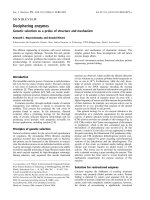
Both her right a nd left ovari es were enlarged and mea-
sured17×8×5cmand16×7×5cm,respectively.
External surface of both was nodular (Figure 1a) and sli-
cing revealed the par enchyma al most complet ely
replaced by a tumor with involvement of hilum as well.
The cut surface was multinodular and had a variegated
appearance with both solid and cystic areas. Solid areas
were well demarcated, soft to f irm and pale-yello w in
color. The cystic spaces were filled with mucinous mate-
rial (Figure 1b). Bilateral fallopian tubes, uterus and cer-
vix were normal.
Both the masses showed a similar m orphology on
microscopy. Solid areas were composed of irregular
glands and nests infiltrating the loose stroma (Figure
1c). The tumor was reaching up to capsule and
encroaching upon the surface. The glands were lined by
large pleomorphic cells exhibiting high grade nuclear
atypia. Cystic areas showed dilated spaces lined by
malignant cells (Figure 1d). Bizarre tumor giant cells,
occasional signet ring cells and atypical mitotic figures
were noted. Large areas of infarction and necrosis were
also seen. Normal ov arian stroma was identified in one
of the sections only.
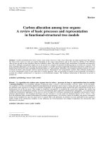
The gallbladder had a gangrenous appearance with dif-
fusely hemorrhagic an d thickened wall covered with
slough on both the serosal as well as mucosal aspect (Fig-
ure 2a). The lumen contained multiple mixed stones.
Besides extensive necrosis and hemorrhage, sections
from viable areas showed an invasive adenocarcinoma
with transmural involvement of the wall and overlying
dysplastic epithelium (Figure 2b). Perineural invasion was
also noted. The omentum and retro-pancreatic lymph
nodes showed tumor metastasis in the form of pools of
mucin infiltrating and dissecting the native tissue. The
tumor cells were found to be floating within the mucin
and many of them had a signet ring appearance.
Case 2
Clinical findings
A 62-year-old woman from a Northern part of India
presented with complaints of pain and swelling in the
abdomen and generalized weakness for a duration of
four months. Routine biochemistry including liver func-
tion tests and hematologi cal parameters were normal. A
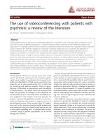
computed tomography (CT) scan of her abdomen
showed two large masses arising from pelvis on either
sid e of the uterus. The masses were reaching up to epi-
gastrium and displacing gut loops anteriorly and
towards right side. Both of them wer e largely cystic with
well defined walls (Figure 3). Her gallbladder contained
multiple stones and wall in the fundic region was thick-
ened resembling calcification. There was no ascitis or
pleural effusion and CA-125 was raised (148.2 U/mL).
Radiological impression was cholelithiasis and bilateral
ovarian tumor of benign nature. However, considering
theageofourpatient,sizeofthemassesandraised
CA-125 it was thought to be an ovarian malignancy and
exploratory laparotomy was done for total abdominal
hysterectomy with bilateral salpingo-oophorectomy and
cholecystectomy. Intra-operative findings revealed bilat-
eral cystic ovarian masses and a hard and solid gallblad-
der mass firmly adherent to surrounding tissue.
Omental nodules were also noted and removed.
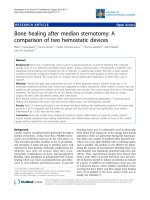
Figure 1 (A) Capsular surface of bilateral ovarian masses.Note
the smooth looking but nodular outer surface. Also note size of
both the masses compared to uterus. (B) Cut surface of a solid
cystic growth with solid grey-white areas present in the form of
nodular deposits. (C) On microscopy tumor glands were forming
glands of variable size and shape. (D) Tumor tissue represented by
large cystic spaces lined by flattened epithelium. Smaller glands are
also present in between.
Figure 2 (A) Diffusely hemorrhagic and ulcerated gallbladder
mucosa. No growth is apparent. (B) An invasive adenocarcinoma
with dysplastic overlying epithelium.
Kumar et al. Journal of Medical Case Reports 2010, 4:202
/>Page 2 of 7
Histopathology findings
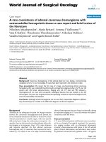
Bilateral ovarian masses were well encapsulated with
right mass measuring 20 × 18 × 11 cm and left 18 ×
13 × 10 cm. Capsular surfac e of both revealed evenly
distribute d multiple tiny pinhead size excrescences (Fig-
ure 4a). Cut surface revealed multiloculated cystic
tumor filled with thick and solidified gelatinous material
as well as dull colored fluid (Figure 4b). The septae
were papery thin, at places forming s mall cysts giving a
spongy appearance. No solid areas were found in either
of the masses even on serial slicing except two very
small 0.5 cm diameter, subcapsular grey-white nodules.
Her uterus showed an incidental 1.5 cm intra-mural
leiomyoma in the fundic region. Her cervix, bilateral
fallopian tubes and ovarian pedicles were normal.
On microscopy cystic spaces were lined by flattened
epithel ium and filled with acellular material (Figure 5a).
On low power examination lining epithelium was flat-
tened to columnar and appeared bland without any stra-
tification or multilayering. Therefore the possibility of
benign mucinous cystadenoma was initially proposed.
The additional sections however revealed marked atypia
of the lining epithelium. Two ou t of 23 sections taken
from small subcapsular nodules showed atypically prolif-
erating mucinous epithelium (Figure 5b). Few papillae
were also seen lined by epithelial cells with marked aty-
pia. Intervening stroma was scanty but few foci of infil-
tration by irregular shaped glands were identified
Figure 3 CT scan of abdomen showing two large cystic masses
arising from pelvis.
Figure 4 (A ) Well encapsulated left ovarian mass.Notetiny
pinhead size excrescences on the surface (arrow). (B) The cut
surface resembling a multiloculated benign cystic tumor.
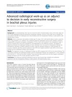
Figure 5 (A) Large cystic spaces lined by flattened epithelium
and filled with acellular material. (B) Malignant tumor glands
with back to back arrangement. Note marked atypia of cells within
papillae (inset). (C) Irregular shaped glands within the desmoplastic
stroma. (D) Surface implants.
Figure 6 (A) Thickened gallbladder wall with a fragmented
stone. (B) Well formed tumor glands within a desmoplastic stroma.
The glands are lined by columnar cells with basally placed nuclei.
Kumar et al. Journal of Medical Case Reports 2010, 4:202
/>Page 3 of 7
(Figure 5c). Tiny excrescences present on the capsular
surface showed tumor gland deposits (Figure 5d) sup-
porting the possibility of a metastatic tumor. Uninvolved
ova rian parenchyma was fibrous and contained hemosi-
derin laden and foamy macrophages.
The serosal surface of gallbladder was smooth and
shiny. The lumen was impacted with a 1.3 cm dia-
meter cholesterol stone. In the body region mucosa
was ulcerated with variably thickened wall (Figure 6a).
Microscopy showed a moderate ly differentiated adeno-
carcinoma (Figure 6b). Omental nodules showed meta-
static tumor d eposits with a similar morphology as in
case 1.
Discussion
The incidence of ovarian metastasis from different
organs is nearly five to 15% [7]. Although a figure of 6%
cases of gallbladder carcinoma with metastasis to ovary
has been quoted by Albores-Saavedra [10], a description
of only 19 such cases could be found in the literature
(Table 1) [2-9]. Of these, eight cases presented with
ovarian masses [2,3,5,6,8,9] and clinico-radiological find-
ings in five mimicked a primary ovarian tumor [2,3,8].
With a pre-operative radiological investigation, diagnosis
could not be established in four cases [4-7] and few
were misdiagnosed as primary ovarian tumor even on
histology [6,7].
Table 1 A summary of reported cases of gallbladder carcinoma with ovarian metastasis.
Author No.
of
cases
Age
(yrs)
Clinical
presentation
Detection
of primary/
secondary
Laterality Size (cm) Histopathology of ovary
Gross Micro
Khunamornpong
et al.[2]
8 47-83 Pelvic mass,
abdominal
distension,
vaginal
bleeding,
hematochezia
n = 1 each
abdominal
pain, unknown
n = 2 each
Primary first
n=3
Simultaneous
n=5
Bilateral 0.5-16.5 Smooth external surface in
majority, cut surface
predominantly solid-cystic
or solid in some, cyst
content mucoid in majority
All except 1 were
recognized as metastatic
tumors; initially diagnosis
was not appreciated in 1
case. All had foci
indistinguishable from
primary surface epithelial
neoplasms
Young and Scully
[3]
5 33-72 Abdominal
pain
n=4
Pelvic mass
n=1
Primary first
n=1
Simultaneous
n=3
Ovarian first
n=1
Bilateral 2.5-13 Lobulated external surface.
Cut surface in all except 1
was nodular and solid
Half of them were difficult
to diagnose and simulated
primary ovarian neoplasm
Ayhan et al.[4] 1 33 Abdominal
pain
Simultaneous Unilateral 3 - -
Miyagui et al.[5] 1 43 Confusion Simultaneous Bilateral 17 and 19 Cut surface compact
intermingled with cystic
areas containing yellow
gelatinous fluid
Ovarian architecture
entirely replaced
neoplastic cells disposed
in alveolar and trabecular
patterns. Mucin & signet
ring cells
Jain et al.[6] 1 45 Pelvic mass Simultaneous Bilateral - - Malignant cystic deposits
Jarvi et al.[7] 1 82 Abdominal
pain
Simultaneous Bilateral - Solid cystic masses with
focally roughened surfaces
Bilateral benign serous
cystadenoma with
deposits of metastatic
adenocarcinoma
Taranto et al.[8] 1 52 Pelvic mass Primary first Bilateral 15 - Difficult to distinguish
from a primary mucinous
adenocarcinoma of the
ovary even on histology
Majumdar et al.
[9]
1 38 Abdominal
pain
and distension
Simultaneous Bilateral 13 and 8 - Papillary pattern, cystic
spaces, extracellular mucin,
surface implants
Kumar et al.
(present study)
235
62
Abdominal
pain
Abdominal
pain and
distension
Simultaneous Bilateral 17 and 18
20 and 18
Case 1: Solid cystic masses
and gangrenous
gallbladder
Case 2: Entirely cystic,
multiloculated ovarian
masses filled with thick and
thin mucin
Nodular growth with
infiltrative pattern.
Presence of surface
deposits, cellular atypia,
and infiltrative pattern
Kumar et al. Journal of Medical Case Reports 2010, 4:202
/>Page 4 of 7
Similar to the present report, a majority of such patients
had non-specific abdominal or pelvic symptoms (pain, dis-
tension, or mass). Jaundice or other symptoms related to
gallbladder carcinoma were observed in only few cases
[2,6,9]. Radiological features of malignancy were masked
by chronic cholecystitis or cholelithiasis. Serological mar-
kers such as alkaline phosphatase, CA19-9, CEA, and CA-
125 were found to be variable at the time of met astases
[2-4,6-9]. In both our patients CA-125 levels were raised,
however CA19-9 was not assessed. A variable clinical pre-
sentation, radiology and serum markers make the appro-
priate histological diagnosis mandatory [3,11-13].
The morphological features, on histology of metasta-
sis, may mimic not only malignant but also a benign
ovarian tumor as observed in our patients. In the first
case, the gallbladder was gangrenous and no obvious
growth was apparent on gross examination. Microscopi-
cally, only a few tumor glands were noticed in one of
the sections tak en from the gallbladder. The origin of
these glands could not be traced from these initial sec-
tions. The gallbladder therefore was re-grossed. Repeat
sections taken revealed a tumor diffusely involving the
gallbladder wall with overlying dysplastic epithelium.
This along with a bilateral tumor, multinod ularity, infil-
trative pattern and presence of uninvolved tissue sup-
ported the possibility of a metastatic carcinoma rather
than a primary malignancy in the ovaries.
The second case showed a full-fledged gallbladder
malignancy. The ovarian masses, however, were comple-
tely cystic with no solid areas. The initial sections sug-
gested possibility of a benign mucinous tumor.
However, presence of focal atypia in the lining epithe-
lium and a high index of suspicion, in view of presence
of a gallbladder malignancy led to re-examination of the
specimen. Tiny pinhead size elevations over the capsule
(Figure 3b) and subcapsular nodules identified on sec-
ond look revealed malignant glands, which supported
the possibility of a metastatic tumor.
Table 2 Pathological features differentiating a secondary from primary ovarian tumor [2,11,14,15]
Pathological features Secondary Primary
Gross
Bilaterality ✓
Surface implants ✓
Multinodular growth ✓
Size > 10 cm ✓✓
Smooth tumor surface ✓
Mural nodule ✓
Micro
Surface implants in the form of irregular/dilated/cystic/angulated/tubular glands/cell nests or single tumor cells within a
desmoplastic/hyalinized stroma
✓
Infiltrative pattern (disorderly penetration of the stroma by small glands, tubules, or single cells, including signet-ring cells,
usually within a desmoplastic stroma)
✓
Growth in the ovarian hilum ✓
Foci of uninvolved ovarian tissue ✓
Mucin without epithelial cells on the tumor surface or the residual ovarian surface ✓
A predominantly cystic gross appearance with only few solid necrotic or hemorrhagic areas ✓✓
Grossly mucinous cyst contents ✓✓
Areas of a cribriform, villous, or solid growth ✓✓
Microscopic mucin extravasation into the stroma ✓✓
Benign or borderline-appearing areas (either with atypia only or with intraepithelial carcinoma) ✓✓
Focal endometrioid-like appearance ✓✓
Microscopic cysts, generally > 2 mm ✓✓
“Expansile” invasive pattern (sharply demarcated, multicystic or labyrinthine spaces lined by malignant-appearing epithelial
cells, with minimal or no recognizable intervening stroma, in an area exceeding 10 mm and at least 3 mm in any single
dimension)
✓
A complex papillary epithelial growth (branching papillae with epithelial stratification and little or no stromal support) ✓
Intraluminal necrotic material (tumor cell karyorrhectic nuclear fragments, neutrophils, and acellular debris) in gland-cyst
lumens
✓
Immunohistochemistry
CK-7 ✓✓
CK-20 ✓✓
Dpc4 ✓✓
Kumar et al. Journal of Medical Case Reports 2010, 4:202
/>Page 5 of 7
In the literature a variety of features have been
emphasized (Table 2) that may help to differentiate
metastasis from a primary ovarian tumor [2,11,14].
Amongst these, the bilaterality, surface implants, multi-
nodularity, infiltrative pattern, foci of uninvolved ovarian
tissue, growth in the ovarian hilum, mucin without
epithelial cells on the tumor surface and presence of sig-
net ring cells are the most important clues fo r a meta-
static adenocarcinoma. However, many of these features
may be absent, especially if the metastasis p resents as
benign cystic mass. Although the immunohistochemistry
can distinguish metastasis fr om other org ans with
respect of colorec tal carcinoma (CK7
-
/CK20
+
)incon-
trast to ovarian primaries (CK7
+
/CK20
-
/CK20
+
), its role
in metastasis from gallbladder is limited because of simi-
lar profile to that of primary ovarian mucinous tumors
[2,15]. A thorough gross examinatio n and adequate sec-
tioning therefore are important in such cases.
Outcome in these cases is generally poor. However,
adequate surgery with palliative treatment may prolong
survival for few months. Therefore at the time of total
abdominal hysterectomy and bilateral salpingo-oophor-
ectomy with cholecystectomy presence of unusual find-
ings such as a gallbladder mass, dense adhesions of the
omentum and adjacent organs to the gallbladder, diffi-
cult dissection of the gallbladder from its liver bed
should raise the suspicion of a carcinoma. A cl ose eva-
luation of the extent o f the disease should be carried
out. Biopsy of any lymph node should be taken. Intra-
operative ultrasound, intra-portal endoscopic ultrasound
and frozen section all may be performed to assess the
extent of the disease. In the presenc e of ascites, fluid
should be obtained for cytology; otherwise, a peritoneal
wash-out can be considered for cytology [16]. External
radiation therapy with or without chemotherapy may
provide some palliative benefit to these patients.
Conclusions
Gallbladder carcinoma should be added to the pre-
viously known list of origi ns of metastatic tumors to the
ovary that can closely mimic primary o varian mucinous
tumors. Pathologists should maintain a high index of
suspicion and adequate sampling should be done of
ovarian masses especially if bilateral. In all bilateral
mucinous tumors outer surface should be examined
carefully for presence of tiny deposits. Knowledge of the
extent to which gallbladder metastasis may mimic a pri-
mary ovarian tumor and its differentiating histological
feat ures may help in correct diagnosis and further man-
agement of the patient.
Consent
Written informed consent was obtained from both the
patients for publication of this case report and any
accompanying images. A copy of the written consent is
available for review by the Editor-in-Chief of this
journal.
Author details
1
The Pine, Near Ashiana Regency, Chhota Shimla, Shimla -171002, India.
2
Department of Pathology and Laboratory Medicine, Grecian Superspeciality,
Cardiac and Cancer Hospital, Sector 69, SAS Nagar, Mohali, India.
3
Department of Pathology, Maharshi Markandeshwar Institute of Medical
Sciences and Research, Mullana, Ambala Haryana, India.
Authors’ contributions
YK designed, carried out acquisition and analysis of data and drafted the
manuscript. AC and AB helped in drafting of manuscript and given their
valuable suggestions, MG provided the images. All the authors read and
approved the final manuscript.
Competing interests
The authors declare that they have no competing interests.
Received: 21 October 2009 Accepted: 30 June 2010
Published: 30 June 2010
References
1. Petru E, Pickel M, Heydarfadai M, Lahousen M, Haas J, Schaider H,
Tamussino K: Nongenital cancers metastatic to the ovary. Gynecol Oncol
1992, 44:83-86.
2. Khunamornpong S, Lerwill MF, Siriaunkgul S, Suprasert P,
Pojchamarnwiputh S, Chiangmai WN, Young RH: Carcinoma of
extrahepatic bile ducts and gallbladder metastatic to ovary. A report of
16 cases. Int J Gynecol Pathol 2008, 27:366-379.
3. Young RH, Scully RE: Ovarian metastases from carcinoma of the
gallbladder and extrahepatic bile ducts simulating primary tumors of
the ovary. A report of six cases. Int J Gynecol Pathol 1990, 9:60-72.
4. Ayhan A, Guney I, Saygan-Karamursel B, Taskiran C: Ovarian metastasis of
primary biliary and gallbladder carcinomas. Eur J Gynaecol Oncol 2001,
22:377-378.
5. Miyagui T, Luchemback L, Teixeira GH, de Azevedo KM: Meningeal
carcinomatosis as the initial manifestation of a gallbladder adeno-
carcinoma associated with a Krukenberg tumor. Rev Hosp Clin Fac Med Sao
Paulo 2003, 58:169-172.
6. Jain V, Gupta K, Kudva R, Rodrigues GS: A case o f ovarian metastasis of
gall bladder carcinoma simulating primary ovarian neoplasm:
diagnostic pitfalls an d review of literature. Int J Gynecol Cancer 2006,
16:319-321.
7. Jarvi K, Kelty CJ, Thomas WE, Gillespie A: Bilateral ovarian metastases from
carcinoma of the gallbladder. Gynecol Oncol 2006, 103:361-362.
8. Taranto AJ, Lourie R, Lau WF: Ovarian vascular pedicle sign in ovarian
metastasis arising from gall bladder carcinoma. Australas Radiol 2006,
50:504-506.
9. Majumdar K, Singh DK, Kaur S, Rastogi A, Gondal R: Papillary
adenocarcinoma gallbladder with simultaneously detected bilateral
ovarian metastasis: a case report. Internet J Gynecol Obst 2008, 9:1.
10. Albores-Saavedra J: Atlas of Tumor Pathology (Second Series). Armed
Forces Institute of Pathology, Washington 1986.
11. Lee KR, Young RH: The distinction between primary and metastatic
mucinous carcinomas of the ovary gross and histologic findings in
50 cases. Am J Surg Pathol 2003, 27:281-292.
12. Ronnett BM, Kurman RJ, Shmookler BM, Sugarbaker PH, Young RH: The
morphologic spectrum of ovarian metastases of appendiceal
adenocarcinomas: a clinicopathologic and immunohistochemical
analysis of tumors often misinterpreted as primary ovarian tumors or
metastatic tumors from other gastrointestinal sites. Am J Surg Pathol
1997, 21:1144-1155.
13. Young RH, Hart WR: Metastases from carcinomas of the pancreas
simulating primary mucinous tumors of the ovary. Am J Surg Pathol 1989,
13:748-756.
14. Seidman JD, Kurman RJ, Ronnett BM: Primary and metastatic mucinous
adenocarcinomas in the ovaries. incidence in routine practice with a
Kumar et al. Journal of Medical Case Reports 2010, 4:202
/>Page 6 of 7
new approach to improve intraoperative diagnosis. Am J Surg Pathol
2003, 27:985-993.
15. Vang R, Gown AM, Barry TS, Wheeler DT, Yemelyanova A, Seidman JD,
Ronnett BM: Cytokeratins 7 and 20 in primary and secondary mucinous
tumors of the ovary: analysis of coordinate immunohistochemical
expression profiles and staining distribution in 179 cases. Am J Surg
Pathol 2006, 30:1130-1139.
16. Shiwani MH: Surgical management of gall bladder carcinoma. J Pak Med
Assoc 2007, 57:87-90.
doi:10.1186/1752-1947-4-202
Cite this article as: Kumar et al.: Occult gallbladder carcinoma
presenting as a primary ovarian tumor in two women: two case reports
and a review of the literature. Journal of Medical Case Reports 2010 4:202.
Submit your next manuscript to BioMed Central
and take full advantage of:
• Convenient online submission
• Thorough peer review
• No space constraints or color figure charges
• Immediate publication on acceptance
• Inclusion in PubMed, CAS, Scopus and Google Scholar
• Research which is freely available for redistribution
Submit your manuscript at
www.biomedcentral.com/submit
Kumar et al. Journal of Medical Case Reports 2010, 4:202
/>Page 7 of 7