báo cáo khoa học: " High-throughput sequencing identifies STAT3 as the DNA-associated factor for p53-NF-κB-complex-dependent gene expression in human heart failure" pps
Bạn đang xem bản rút gọn của tài liệu. Xem và tải ngay bản đầy đủ của tài liệu tại đây (4.74 MB, 14 trang )
Choy et al. Genome Medicine 2010, 2:37
/>Open Access
RESEARCH
© 2010 Choy et al.; licensee BioMed Central Ltd. This is an open access article distributed under the terms of the Creative Commons
Attribution License ( which permits unrestricted use, distribution, and reproduction in
any medium, provided the original work is properly cited.
Research
High-throughput sequencing identifies STAT3 as
the DNA-associated factor for
p53-NF-κB-complex-dependent gene expression
in human heart failure
Mun-Kit Choy
1
, Mehregan Movassagh
1
, Lee Siggens
1
, Ana Vujic
1
, Martin Goddard
2
, Ana Sánchez
3
, Neil Perkins
3
,
Nichola Figg
1
, Martin Bennett
1
, Jason Carroll
4
and Roger Foo*
1
Abstract
Background: Genome-wide maps of DNA regulatory elements and their interaction with transcription factors may
form a framework for understanding regulatory circuits and gene expression control in human disease, but how these
networks, comprising transcription factors and DNA-binding proteins, form complexes, interact with DNA and
modulate gene expression remains largely unknown.
Methods: Using microRNA-21 (mir-21), which is an example of genes that are regulated in heart failure, we performed
chromatin immunoprecipitation (ChIP) assays to determine the occupancy of transcription factors at this genetic locus.
Tissue ChIP was further performed using human hearts and genome-wide occupancies of these transcription factors
were analyzed by high-throughput sequencing.
Results: We show that the transcription factor p53 piggy-backs onto NF-κB/RELA and utilizes the κB-motif at a cis-
regulatory region to control mir-21 expression. p53 behaves as a co-factor in this complex because despite a mutation
in its DNA binding domain, mutant p53 was still capable of binding RELA and the cis-element, and inducing mir-21
expression. In dilated human hearts where mir-21 upregulation was previously demonstrated, the p53-RELA complex
was also associated with this cis-element. Using high-throughput sequencing, we analyzed genome-wide binding
sites for the p53-RELA complex in diseased and control human hearts and found a significant overrepresentation of the
STAT3 motif. We further determined that STAT3 was necessary for the p53-RELA complex to associate with this cis-
element and for mir-21 expression.
Conclusions: Our results uncover a mechanism by which transcription factors cooperate in a multi-molecular complex
at a cis-regulatory element to control gene expression.
Background
Gene transcription is modulated by the dynamic interac-
tion between DNA and protein complexes. Genome-wide
maps of these interactions are now generated using a
combination of chromatin immunoprecipitation (ChIP)
and powerful tools such as high-throughput sequencing
(ChIP-seq), and they provide a framework for interpret-
ing the genome in different contexts, including in embry-
onic stem cells and oncogenesis [1-3]. Genome-wide
maps for these transcription factors also show that much
remains to be discovered to complete our understanding
of transcriptional regulatory networks: empirical binding
sites for a transcription factor often lack the expected
consensus motif, reflecting that different mechanisms
exist for transcription factor recruitment, with some
likely to involve indirect binding through components of
a multi-molecular transcription complex [3]. Moreover,
the numerous means by which a factor is recruited to the
genome may also allow it to participate in multiple signal-
ing pathways. In fact, Chen et al. [1] observed that, in
* Correspondence:
1
Department of Medicine, University of Cambridge, Addenbrooke's Centre for
Clinical Investigation, Hills Road, Cambridge, CB2 0QQ, UK
Full list of author information is available at the end of the article
Choy et al. Genome Medicine 2010, 2:37
/>Page 2 of 14
embryonic stem cells, a significant subset of transcription
factor binding regions is extensively co-occupied by sev-
eral different transcription factors to form multiple tran-
scription-factor binding loci. Our work here proposes a
simple analytical model that is potentially representative
of multiple transcription-factor binding loci in human
disease. We have dissected the behavior and functional
roles of the different components of a multi-molecular
transcription complex, capitalizing on a regulatory path-
way that controls mir-21 expression in human heart dis-
ease.
MicroRNA genes transcribe short (approximately 22
nucleotide) non-coding RNAs (miRNAs) that direct
mRNA degradation or disrupt mRNA translation in a
sequence-dependent manner. Like protein-coding
mRNA, miRNAs are initially generated by RNA poly-
merase II as long primary transcripts before being pro-
cessed to mature miRNA [4]. Based on the genome-wide
chromatin marks of transcription start sites and tran-
scriptional elongation, promoters of human miRNAs
were recently identified [5], but the diverse expression
profiles of miRNAs indicate that miRNA expression must
be under elaborate control during development and dis-
ease states, similar to other genes that are transcribed by
RNA polymerase II. A consistent pattern of miRNA
expression is found in failing hearts [6,7] and the roles of
key miRNAs in heart failure development and progres-
sion have been studied [8,9].
We and others [10,11] found that the transcription fac-
tor p53 is highly activated and accumulates in hypoxic
hearts in response to stress. Experimentally, p53 regulates
at least 34 different miRNAs in oncogenesis (for example,
mir-34; reviewed in [12]). We therefore investigated the
possibility that p53 regulates some part of the miRNA
expression in failing hearts.
Materials and methods
Ethics statement
Human left ventricular tissue was collected with a proto-
col approved by the Papworth (Cambridge) Hospital Tis-
sue Bank review board and the Cambridgeshire Research
Ethics Committee (UK). Written consent was obtained
from every individual according to the Papworth Tissue
Bank protocol.
Cell isolation, culture and human cardiac tissue
Rat neonatal cardiac fibroblasts were isolated from 0- to
5-day-old Wistar or Sprague-Dawley rats by an enzymatic
isolation method as described before [13] and in accor-
dance with UK Home Office regulations. Primary cardiac
fibroblasts, immortalized RelA
-/-
mouse embryo fibro-
blast (MEF) cells (from Professor R Hay, University of
Dundee), p53
-/-
MEF cells (from Dr G Lozano, MD
Anderson Cancer Centre) and Soas2 osteosarcoma cells
were cultured in Dulbecco's modified Eagle's medium
containing 10% fetal calf serum at 5% CO
2
and 37°C, and
maintained at 60 to 80% confluency. Stat3
-/-
MEF cells
and Stat3
-/-
MEF cells re-constituted with wild-type Stat3
were obtained from Dr David Levy (NYU, School of Med-
icine). Where indicated, cells were treated with 10 μM
doxorubicin (Sigma Dorset, UK) for 2 h before being
allowed to grow for another 24 h, 200 μM deferroxamine
(DFX; Sigma) for 24 h, with or without 1 μM NF-κB acti-
vation inhibitor (NFI; 6-amino-4-(4-phenoxyphenyleth-
ylamino) quinazoline; Calbiochem, Nottingham, UK) for
24 h, and with or without 100 μM STAT3 inhibitor (S3I-
201, Calbiochem) for 24 h. Hypoxia treatment was per-
formed in an Invivo
2
400 Hypoxia Workstation (Ruskinn,
Bridgend, UK) at 1% O
2
, 5% CO
2
and 37°C for 48 h.
Cardiac left ventricular tissues were obtained from
patients undergoing cardiac transplantation for end-stage
dilated cardiomyopathy (three males and one female aged
49 to 60 years). Normal human ventricular tissues were
from four healthy male individuals involved in road traf-
fic accidents (aged 41 to 52 years). At the time of trans-
plantation or donor harvest, whole hearts were removed
after preservation and transported in cold cardioplegic
solution (cardioplegia formula and Hartmann's solution)
similar to the procedure described before at Imperial
College, London [14]. Following analysis by a cardiovas-
cular pathologist (MG), left ventricular segments were
cut and stored immediately in RNAlater (Ambion,
Applied Biosystems, Warrington, UK). Individual patient
details are listed in Additional file 1.
miRNA quantitative PCR
Total RNA from cells was extracted using mirVana
miRNA Isolation Kit (Ambion). Reverse transcription
and quantitative PCR (qPCR) amplification of cDNA of
mature miRNAs were performed using primer sets and
protocols obtained from Applied Biosystems (TaqMan
MicroRNA Assay, Warrington, UK), and Rotor-Gene
6000 qPCR machine from Corbett (Qiagen, Crawley,
UK). PCR signals from cDNA of miRNAs were standard-
ized with signals from amplification of cDNA of 18s
rRNA, which was reverse transcribed using SuperScript
III First-Strand Synthesis System for RT-PCR (Invitrogen,
Paisley, UK), using a set of primers/probe (18s forward
primer, 5'-CGGCTACCACATCCAAGGAA-3'; reverse
primer, 5'-AGCTGGAATTACCGCGGC-3'; probe, 5'-
FAM-TGCTGGCACCAGACTTGCCCTC-BHQ1-3')
and the protocol for TaqMan Gene Expression Assays
(Applied Biosystems).
Immunoblot analysis
Whole-cell lysates were prepared by scraping cells into a
lysis buffer (50 mM Tris pH 8.0, 150 mM NaCl, 0.02%
sodium azide, 1% Nonidet P-40, 1x Roche Complete Mini
Choy et al. Genome Medicine 2010, 2:37
/>Page 3 of 14
Protease Inhibitor Cocktail, Burgess Hill, UK). To prepare
nuclear lysates, cell membranes/cytoplasms of harvested
cells were lysed in an ice-cold cytoplasmic lysis buffer
(0.33 M sucrose, 10 mM HEPES pH 7.4, 1 mM MgCl
2
,
0.1% Triton-X 100, 1x Roche Complete Mini Protease
Inhibitor Cocktail) and the nuclei were washed with the
same buffer before being lysed with a buffer containing
0.45 M NaCl, 10 mM HEPES pH7.4 and 1x Roche Com-
plete Mini Protease Inhibitor Cocktail. Protein concen-
tration was determined using a BCA Protein Assay Kit
(Pierce, Thermo Scientific, Cramlington, UK). Equal pro-
tein amounts were resolved by SDS-PAGE, transferred to
a polyvinylidene fluoride membrane and incubated with
primary antibodies against the following proteins: p53
(1C12; Cell Signaling, New England Biolabs, Hitchin,
UK), NF-κB (p65 SC-109 and SC-372; Santa Cruz,
Heidelberg, Germany), STAT3 (Cell Signaling), SF2
(loading control; Zymed, Invitrogen, Paisley, UK) and
RhoGDI (loading control; A-20; Santa Cruz). Goat anti-
mouse and anti-rabbit (Jackson ImmunoResearch, New-
market, UK) were used as secondary antibodies. The
membrane was incubated with SuperSignal West Pico
Chemiluminescent Substrate, SuperSignal West Dura
Extended Duration Substrate, SuperSignal West Femto
Maximum Sensitivity Chemiluminescent Substrate
(Pierce) or ECL Advance Western Blotting Detection Kit
(GE Healthcare, Chalfont St Giles, UK) before being
exposed to an X-ray film.
Immunoprecipitation
Cells were scraped into the lysis buffer and protein con-
centration was determined as described above. Protein A
beads (Sigma) were prewashed twice with phosphate-
buffered saline and suspended in the lysis buffer. Total
protein lysate (500 μg) was incubated with 30 μl of the
protein A beads for an hour at 4°C and centrifuged. The
supernatant was then removed to a fresh tube and incu-
bated overnight at 4°C with primary antibodies and IgG
(mouse/rabbit Fc fraction; Jackson ImmunoResearch) as
indicated. The next day, 30 μl of the protein A beads were
added and incubated for an hour at 4°C. Following the
incubation, beads were washed three times with phos-
phate-buffered saline. Proteins were eluted with SDS
loading buffer (100 mM Tris pH 6.8, 4% SDS, 0.2% bro-
mophenol blue, 20% glycerol, 10% β-mercaptoethanol),
resolved by SDS-PAGE, and transferred to a membrane
for immunoblot analysis as described above.
Chromatin immunoprecipitation and sequential ChIP
Chromatin immunoprecipitation (ChIP) was performed
using a ChIP Assay Kit (Upstate, Millipore, Watford, UK),
primary antibodies raised against p53 (CM5, Vector Lab-
oratories for rat, Orton Southgate, UK); DO-1, sc-126
Santa Cruz for human), NF-κB (C-20/sc-372, Santa Cruz)
and histone H3 (tri methyl K4; ab8580, Abcam, Cam-
bridge, UK), and IgG. For tissue ChIP, the heart tissues
were finely chopped, cross-linked and homogenized prior
to the procedure. Cross-linked chromatin was fragmen-
tized to 1 kb by sonication. Regions of interest were
amplified from the immunoprecipitated DNA by qPCR
using SYBR GreenER qPCR SuperMix Universal (Invitro-
gen). The primers for the rat 32280-7 site were 5'-TAG-
GCAAGCCTCAAGCTCTC-3' and 5'-
TCGTTTGGCATAGCTTTGTG-3', and primers for the
human GIS site were 5'-TGCAGAAATTGGAGTG-
GATG-3' and 5'-TTGCAAGTTTGCTGCTGAAC-3'
(95°C for 10 minutes followed by 40 to 50 cycles of 94°C
for 15 s, 59°C for 20 s, 72°C for 30 s and 76°C for 5 s (sig-
nals acquired)), whereas the primers for the promoter of
mir-21 were 5'-TACAAACTGGGGAGCTTGGT-3' and
5'-AACCCCTGCGTCATCCTTAT-3' (95°C for 10 min-
utes followed by 40 to 50 cycles of 94°C for 15 s, 59°C for
20 s and 72°C for 30 s (signals acquired)). PCR signals
were standardized with signals from amplification of 18s
rRNA genes with the same primers/probe and protocol as
described above. For sequential ChIP (re-ChIP) assays,
complexes from the primary ChIP were eluted twice with
10 mM dithiothreitol for 20 minutes at 37°C, diluted 10
times with re-ChIP buffer (20 mM Tris-HCl pH 8.1, 0.1%
Triton X-100, 2 mM EDTA, 150 mM NaCl) followed by
re-immunoprecipitation with the indicated second pri-
mary antibody, and then again subjected to the ChIP pro-
cedure.
Biotinylated oligonucleotide precipitation assay
Cells were lysed by sonication in HKMG buffer (10 mM
HEPES pH 7.9, 100 mM KCl, 5 mM MgCl
2
, 10% glycerol,
1 mM dithiothreitol, 0.5% of NP-40 and 1x Roche Com-
plete Mini Protease Inhibitor Cocktail). Cell extracts were
pre-cleared with Promega Magnesphere beads for 1 h at
4°C, then incubated with 10 μg of biotinylated double-
stranded oligonucleotides pre-bound to the beads for 16
h at 4°C (5' biotinylated 5'-GGCTCTCACCAG-
GAAGGAAGATCCCCATTTCCAACCTGTAC-3' (Pro-
mega, Southampton, UK)). DNA-bound proteins were
eluted with SDS loading buffer, separated by SDS-PAGE,
and identified by immunoblotting as described above.
Cell transfection, recombinant proteins and luciferase
activity assay
Cells were transfected with plasmid DNA using Superfect
Transfection Reagent (Qiagen, Crawley, UK) following
the manufacturer's protocol or by electroporation using
Amaxa Nucleofector according to manufacturer's
instructions (Wokingham, UK). RelA
-/-
MEF cells were
reconstituted with a full length human RELA cDNA
using the pHR-SIN-CSGW retroviral vector. Recombi-
nant RELA and p53 proteins were produced in BL21
Choy et al. Genome Medicine 2010, 2:37
/>Page 4 of 14
Escherichia coli as glutathione-S-transferase (GST)- and
His-fusions, respectively, and purified on Glutathione
Sepharose column (Amersham Biosciences, GE Health-
care, Chalfont St Giles, UK) or Ni-nitrilotriacetic acid
agarose (Invitrogen). For luciferase assays, p53
-/-
MEF
cells in 6-well plates were transfected with 2.1 μg of total
plasmid DNA containing human p53 (0.5 μg), p53R175H
(0.5 μg), NF-κB (0.5 μg; p65-EYFP from Dr M Schaaf,
Leiden University, The Netherlands) and/or empty vector
(0.5 to 1 μg; pcDNA; Invitrogen) together with respective
reporter plasmids: GIS-luciferase (GIS is a highly con-
served putative p53 binding site approximately 1.1 kb
upstream of mir-21), GIS-luciferase with mutation or
deletion, or miPPR21-luciferase (1 μg) with phRG-TK
(0.1 μg; Transfection control; Promega). Cell lysates har-
vested 24 h later were assayed for firefly and renilla
luciferase activities by using a Dual-Glo Luciferase
Reporter Assay (Promega).
Immunohistochemistry
Paraffin sections were prepared from human left ventric-
ular tissues that were previously collected and stored in
RNAlater (Ambion). Tissues were perfused in situ with
10% neutral-buffered formalin, cut in 5-μm sections, and
stained using antibodies NF-κB (SC-372, Santa Cruz),
phospho-p53
ser15
and phospho-p53
ser20
(Cell Signaling),
as previously described [15].
ChIP-Seq
High-throughput sequencing of the sequential-ChIP
fragments from human hearts was performed using Illu-
mina Genome Analyser by GeneService, UK following
the manufacturer's protocols. Two flowcell lanes were
used for sequencing of each pooled sample (control ver-
sus disease) on the Genome Analyzer II. The Genome
Analyzer was run for 36 cycles. The reference genome
used for sequence alignment was the human build 36.1
finished human genome assembly (hg18, March 2006).
Images from the Genome Analyzer were analyzed with
the Genome Analyzer pipeline software (version 1.3, Illu-
mina software) for base calling and sequence alignment
to the reference human genome. Sequence alignment
stage was performed using the ELAND algorithm with
the 'ELAND extended' option to enable better handling of
reads >32 bp. The length and abundance of ChIP frag-
ments were modeled from sequencing reads using
Model-Based Analysis of ChIP-Seq (MACS) with model
fold at 100 and P-value cutoff at 1 × 10
-3
[16]. Motif analy-
sis and searches were performed using the Cis-regulatory
Element Annotation System (CEAS) [17] and FIMO [18].
To identify NF-κB motifs, matrices used in the FIMO
search were M00052(V$NFKAPPAB65_01), M00054
(V$NFKAPPAB_01), M00194 (V$NFKB_Q6) and
M00208 (V$NFKB_C) using a P-value cutoff at 1 × 10
-3
.
Results
Using a global map of p53 transcription factor binding
sites in the human genome that was generated by the
ChIP-seq method [19], we searched for p53 binding at
locations adjacent to miRNAs that had been shown by
expression profiling to be differentially expressed in heart
failure [6,7]. At least one p53 binding site was located
within 3,000 bp upstream or downstream of mir-15b,
mir-21 and mir-125b. We tested and found that the
expression of mir-21, but not mir-15b or mir-125b, was
responsive to p53 activation by doxorubicin (Additional
file 2). Similarly, mir-21 was upregulated by hypoxia,
which is another stimulus known to activate p53 in car-
diac cells [11].
mir-21 belongs to a conserved miRNA family with sin-
gle recognizable orthologs in many different invertebrate
species [20]. A previous gene structure study of mir-21
identified a promoter sequence (miPPR21) in a highly
conserved region approximately 2.5 kb upstream of the
putative p53-binding site (which we called 'GIS') [21]
(Figure 1). Aside from mir-21 itself and miPPR21, GIS is
the only other region of significant sequence conserva-
tion in this genomic region (Figure 1; Additional file 3).
However, analysis of GIS revealed not a p53 consensus
motif, but a consensus motif for κB binding (Additional
file 3). This motif is consistent with that reported in
another genome-wide analysis of ChIP mapping NF-κB/
RELA binding sites [22].
In the stressed myocardium, mir-21 is significantly
upregulated in cardiac fibroblasts and is responsible for
fibroblast growth factor secretion as well as for the extent
of interstitial fibrosis in heart failure via its effect on its
target gene, Spry1 [23]. Moreover, the therapeutic benefit
of inhibiting mir-21 in heart failure was also demon-
strated. We therefore focused our attention on mir-21
expression in cardiac fibroblasts and found that, as with
hypoxia, the hypoxia-mimetic DFX, which effectively
activates p53 in vitro [11], also upregulated mir-21 in pri-
mary rat cardiac fibroblasts (Figure 2a). It was also
recently shown that NF-κB signaling is critical for the
response to hypoxia [24] because hypoxia may directly
induce NF-κB activation through a complex sequence of
signals involving decreased prolyl hydroxylase-mediated
prolyl hydroxylation of IKKβ leading to phosphorylation-
dependent degradation of the endogenous NF-κB inhibi-
tor, IκBα, and nuclear translocation of NF-κB [25]. Con-
sistent with this and other data [26], we found that DFX
induced NF-κB/RELA nuclear accumulation and this was
significantly inhibited by the cell-permeable NF-κB inac-
tivator quinazoline [27] (1 μM NFI; Figure 2b). Quinazo-
line (6-amino-4-(4-phenoxyphenylethylamino))
specifically inhibits NF-kB activation and nuclear translo-
cation [28,29]. Correspondingly, NFI significantly inhib-
ited DFX-induced mir-21 upregulation (Figure 2a). We
Choy et al. Genome Medicine 2010, 2:37
/>Page 5 of 14
also noted that DFX induced p53 nuclear accumulation
as predicted but mir-21 levels were effectively inhibited
by NFI, despite unchanged levels of nuclear p53 following
DFX+NFI treatment (Figure 2b). These data suggested
that NF-κB was the primary mediator of mir-21 induc-
tion by DFX and/or p53 induction of mir-21 required
activation of NF-κB.
Next we tested the activity of the putative p53-binding
site GIS by cloning it upstream of firefly luciferase and
examining reporter gene expression. Supporting the
hypothesis that p53 requires and cooperates with NF-κB/
RELA, p53 alone did not upregulate luciferase activity,
whereas p53 significantly augmented the activity that was
induced by NF-κB/RELA (Figure 2c). As before, inactiva-
tion of NF-κB by NFI abrogated GIS-driven gene expres-
sion. Mutation or deletion of the κB-consensus motif in
this regulatory sequence reduced p53-RELA-mediated
luciferase reporter gene expression by 50% and 30%,
respectively (Figure 2d). The previously described mir-21
promoter (miPPPR21) approximately 2.5 kb upstream of
GIS was shown to respond through conserved AP1 and
PU.1 binding sites [30]. Neither p53 nor NF-κB/RELA
upregulated expression of the reporter construct based
on this promoter (miPPPR21-luciferase; Additional file
4), indicating that p53/NF-κB regulated mir-21 expres-
sion through GIS but not miPPPR21.
To determine the necessity for NF-κB/RELA in mir-21
induction by DFX or p53, we incubated RelA
-/-
MEF cells
with or without DFX and detected no change in mir-21
levels (Figure 2e), despite DFX-induced activation of p53
as shown by an increase in p53 target gene expression
(MDM2 and BAX) (Figure 2f) and an increase in reporter
activity using a luciferase construct driven by 13 p53-
binding sites (PG13-luciferase, data not shown). Impor-
tantly, RelA
-/-
MEF cells reconstituted with ectopic RelA
showed rescue of DFX induced mir-21 upregulation (Fig-
ure 2e).
Our results raise the possibility that RELA and p53
interact with the putative regulatory region GIS. Thus, we
performed ChIP using anti-RELA and anti-p53 antibod-
ies and found that the GIS region was occupied by both
RELA and p53 in vivo (Figure 3a). Once again, NFI dis-
rupted the GIS-p53 association, indicating that p53 bind-
ing required RELA (Figure 3b). To determine whether
RELA and p53 co-exist in a single molecular complex, we
first performed co-immunoprecipitation assays and
found an interaction between endogenous RELA and p53
proteins that was disrupted by NFI (Figure 3c). The p53-
RELA interaction was direct and dependent on the car-
boxy-terminal transactivation domain of RELA because a
purified recombinant GST fusion protein of the RELA
carboxy-terminal domain, but not the amino terminus
DNA binding domain, was sufficient to interact with p53
(Additional file 5). Next we performed a sequential ChIP
assay (re-ChIP) in which we initially performed ChIP
with a p53 antibody, released the immunoprecipitated
chromatin and then performed another ChIP using a
RELA antibody. GIS was significantly enriched by p53-
RELA re-ChIP and this association was disrupted by NFI,
indicating that RELA and p53 were simultaneously resid-
ing at the GIS genomic location (Figure 3d). Furthermore,
we performed oligonucleotide pulldown where the
genomic sequence of GIS was synthesized, biotinylated,
immobilized onto streptavidin-coated beads, and incu-
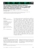
Figure 1 Genomic structure of mir-21. Location of a previously described promoter (miPPR-21) [30], our putative regulatory region (GIS) [19], a
H3K4me3 binding site as determined by previous ChIP-seq [32], and a STAT3 binding site according to Loffler et al. [34]. Both miPPR-21 and GIS regions
are highly conserved.
pri-miR-21
miPPR-21
GIS
STAT3
H3K4-me3
Choy et al. Genome Medicine 2010, 2:37
/>Page 6 of 14
Figure 2 p53 and NF-κB cooperate to induce mir-21. (a) Primary neonatal rat cardiac fibroblasts were treated with or without DFX and the NF-κB
inactivator (NFI; 1 μM quinazoline) and mir-21 was quantified using the TaqMan miRNA assay. (b) Nuclear extracts from cardiac fibroblasts with or with-
out DFX and NFI as in (a) were isolated and western blotted (WB) for p53 and RELA. Splicing factor 2 (SF2), was used to confirm equal protein loading.
{
(c) GIS-luciferase (GIS-Luc) and TK-renilla control (RL) were transfected into p53
-/-
MEF cells with or without plasmids encoding p53 or RELA, and in-
cubated with or without NFI as indicated. Firefly luciferase gene reporter activity was normalized to renilla control. (d) GIS, GIS with an AAA mutation
engineered into the putative NF-κB binding site (GISmAAA), and GIS with the NF-κB binding site deleted (GISdel) were cloned upstream of firefly lu-
ciferase. Constructs were transiently transfected together with a TK-renilla luciferase plasmid and plasmids encoding p53 and RELA into p53
-/-
MEF
cells and firefly luciferase reporter gene activity (FL) was quantified and normalized against renilla (RL). Results represent a fold-difference between the
three different GIS-constructs. (e) RelA
-/-
(-/-) MEF cells and RelA
-/-
MEF cells that were reconstituted with RELA using lentiviral overexpression (RA) were
treated with or without DFX, and mir-21 quantification was performed. (f) RelA
-/-
and reconstituted RelA
-/-
MEF cells were treated with or without DFX,
and whole cell (WCE) and nuclear (NE) extracts were western blotted for MDM2 (arrow), BAX and RELA. RhoGDI and SF2 were used to demonstrate
protein loading. miRNA quantification is shown as mean ± standard error, at least n = 3. Luciferase reporter assays are presented as mean ± standard
error for at least four independent replicates. Asterisks represent P < 0.05 (paired t-test).
0
0.5
1
1.5
2
0
5
10
15
20
pcDNA p53 NF-kB p53 + NF-kB p53 + NF-kB +
NFI
0
10
20
30
40
50
60
70
80
90
100
GIS GIS mA A A GISd e l2 2
0
5
10
15
20
25
30
123
WB: p53
WB: RELA / p65
WB: SF2
++
-
DFX
NFI
30
20
10
0
(a)
(b)
+
GCCTTAAATTGGGAGGACTCCAAGCCGGGAAGGAAAATTAAATTTTCCAA
GCCTTAAATTGGGAGGACTCCAAGCCGGGAAGGAAAATTCCCTTTTCCAA
0
10
20
30
40
50
60
70
80
90
100
FL / RL (%)
0
GIS
GISdel
20
40
60
80
100
Luciferase
GCCTTAAATT TTTTCCAA
GIS
GISmAAA
GISdel
0
5
10
15
20
p53
RELA
+
+
+
+
+
+
+
NFI
(c)
(d)
GIS-Luc / RL
GISmAAA
Transfect:
GIS
[arbitary units]
Abundance of mir-21
++
-
DFX
NFI
+
DFX
-
+
-/-
-/-
RA RA
WB: MDM2
WB: RELA
WB: BAX
WB: RhoGDI
WB: SF2
WCE
NE
0
5
10
15
20
++
(f)
(e)
DFX
Rela
-/-
Rela
-/-
(RA)
[arbitary units]
Abundance of mir-21
*
*
*
*
*
*
Choy et al. Genome Medicine 2010, 2:37
/>Page 7 of 14
Figure 3 p53 and NF-κB form a complex and occupy the putative GIS regulatory region simultaneously. (a) ChIP was performed on cardiac
fibroblasts with or without DFX using antibodies against either p53 or RELA. Results show fold enrichment of real-time qPCR for the putative regulatory
sequence (GIS). (b) ChIP using a p53 antibody was performed on cardiac fibroblasts with or without DFX and NFI. ChIP results are presented as mean
± standard error for three independent experiments performed in triplicate. (c) Using cell lysates from cardiac fibroblasts treated with DFX with or
without NFI, RELA or control IgG immunoprecipitation (IP) was performed followed by western blotting (WB) for p53 (left), and vice versa (right). Ar-
rows indicate RELA. (d) Cardiac fibroblasts were treated with or without DFX and NFI, and p53 ChIP was performed followed by 'release' of the chro-
matin, and RELA re-ChIP. Results represent fold enrichment of real-time qPCR for GIS. Re-ChIP results are presented as mean ± standard error for two
independent experiments performed in triplicate. (e) Lysates from cardiac fibroblasts treated with or without DFX were incubated with streptavidin-
coated beads on which biotinylated GIS duplexes (oligo pulldown) or scrambled sequence duplexes (scrambled) were immobilized. Proteins bound
to these duplexes were eluted and western blotted for p53, RELA and NF-κB subunit p50. Asterisks represent P < 0.05 (paired t-test).
0
70
140
210
280
350
420
490
-A +A oA
0
1
2
3
4
5
6
7
8
123
0
1
2
3
4
5
6
7
1234
DFX
IP: p53
RELA
0
100
200
300
+
+
-
-
50
150
250
350
(a)
(b)
DFX
NFI
WB: p53
++
+
+
+
+
-
-
(c)
Enrichment
[arbitary units]
Enrichment
[arbitary units]
IP:IgG
IP:RELA
WB: RELA
DFX
NFI
++
+
+
+
+
-
-
IP:IgG
IP:p53
WB: p53
WB: RELA
0
50
100
150
200
250
300
350
+
+
+
-
-
-
DFX
NFI
IP:
p53 followed by RELA
+-+- +-+-
DFX
oligo-pulldown
scrambled
Enrichment
[arbitary units]
WB: p53
WB: RELA
(d) (e)
DFX
NFI
++
+
-
-
-
IP:p53
400
300
200
100
0
WB: p50
*
*
*
*
Choy et al. Genome Medicine 2010, 2:37
/>Page 8 of 14
bated with protein lysates from cells that had been
treated with or without DFX. The GIS oligonucleotide,
but not a scrambled control, effectively pulled down p53
and RELA in DFX-treated cells (Figure 3e). Similarly, we
found that the GIS oligonucleotide also pulled down the
NF-κB subunit p50 (Figure 3e) but not p52 (data not
shown), suggesting that the p53-RELA complex included
this subunit of NF-κB.
The presence of a κB motif instead of a p53 consensus
sequence on GIS prompted us to consider if p53 was
behaving as a co-factor and if the p53-GIS interaction was
indirect and independent of the p53 DNA binding
domain. We therefore performed another oligonucle-
otide-pull-down using lysates from p53-deficient cells
(Soas2) pre-transfected with vector only, wild-type p53 or
p53 bearing a mutation in the DNA binding domain
(p53R175H). Both wild-type and mutant p53 associated
with the GIS oligonucleotide (Figure 4a). Consistent with
this, we also found that the p53-RELA interaction was
independent of the p53 DNA binding domain (Figure 4b);
moreover, mutant p53 was potentially capable of upregu-
lating mir-21 expression (Figure 4c). Taken together, our
data support the conclusion that p53 does not contribute
to the GIS binding interface but instead behaves as a co-
factor in this molecular complex and utilizes RELA for its
association with GIS to transactivate mir-21 expression.
Sites of active chromatin at regulatory sequences are
associated with the characteristic Histone-3 mark of
lysine-4 tri-methylation (H3K4me3) [31]. We therefore
performed ChIP using a specific H3K4me3 antibody and
detected a marked enrichment of GIS compared to
miPPR21 (Additional file 6). The association of the GIS
genomic location with H3K4me3 has also been mapped
by others in a genome-wide ChIP scan using human
embryonic stem cells [32].
Since levels of mir-21 are significantly elevated in
dilated human hearts and murine hearts with decompen-
sated hypertrophy [6,7,23], and Thum et al. [23] recently
validated the therapeutic value of targeting mir-21 in a
mouse model of heart failure, we undertook further anal-
ysis using human left ventricular tissues from patients
who had undergone cardiac transplantation for end-stage
dilated cardiomyopathy and age-matched normal control
left ventricular tissues from individuals involved in road
traffic accidents (Additional file 1). Despite different eti-
ologies of heart failure (such as ischemic and non-isch-
emic), end-stage cardiomyopathy is collectively
characterized by disease processes and molecular path-
ways such as apoptosis, dysregulated calcium signaling,
decompensated contractility, G-protein coupled receptor
down-regulation, maladaptive angiogenesis and fibrosis.
Hence, as predicted for our heterogeneous series of end-
stage cardiomyopathic hearts, we found significant
nuclear accumulation of RELA in both myocytes and
non-myocytes from cardiomyopathic hearts compared to
control (Figure 5a-c). Significant p53 activation was
detectable only in non-myocytes (Figure 5e-g). As a func-
tionally significant output of the piggyback mechanism,
we found that both p53 and RELA were simultaneously
resident at the GIS site, and this association was signifi-
cantly enriched in cardiomyopathic hearts compared to
normal controls (Figure 5h).
We predicted that although different mechanisms may
determine p53-RELA complex formation and its chroma-
tin association, the specificity for this complex at some
genomic locations such as mir-21 GIS may be assisted by
additional factors. In order to investigate this, we per-
formed parallel high-throughput sequencing with eight
human cardiac sequential chromatin immunoprecipitates
(four diseased and four controls; Additional file 1). We
identified 26,628 genomic locations in normal hearts and
33,578 in diseased hearts (model fold = 100) aligned to
the reference human genome (Gene Expression Omnibus
[GES21356]). Among these, 12,311 tag locations, exclud-
ing repetitive elements, were unique to disease and had
significant conservation across species (Additional files 7
and 8, and listed in Additional file 9). Of note, only 3%
(381 out of 12,311) were identical to a previous global
ChIP for RELA [22], although in the latter, ChIP was gen-
erated using a non-cardiac cell line and a stimulus unre-
lated to hypoxia. Including a location adjacent to mir-21,
1,344 out of 12,311 (10.9%) were identified to contain the
bona fide κB consensus motif. This observation suggested
that a diverse range of p53-RELA complexes may be
involved in its chromatin association and most appear to
be independent of the κB motif. Nonetheless, using CEAS
[17], we analyzed these 1,344 genomic locations and the
previous global RELA ChIP [22] in parallel. Several tran-
scription factor motifs were overrepresented and com-
mon to both our subset of locations and the global RELA
ChIP, except for STAT1, STAT3, STAT5 and STAT6
(Additional files 10, 11 and 12), with the STAT3 motif
being the most prominent. The JAK/STAT3 pathway is
particularly important for the secretory function and sur-
vival of cardiac fibroblasts [33]. Moreover, in multiple
myeloma cancer cell lines, mir-21 expression is STAT3-
mediated and two conserved STAT3 binding sites lie
upstream of mir-21 [34]. We therefore examined whether
the p53-RELA piggyback mechanism was STAT3-depen-
dent. By using structure-based virtual screening, the cell-
permeable compound S3I-201 was previously identified
to bind to the STAT3 Src homology 2 (SH2) domain, and
inhibit STAT3 dimerization, phosphorylation and DNA-
binding [35]. We used S3I-201 and found that STAT3
inhibition disrupted p53-RELA- GIS association (Figure
6a, b), and inhibited mir-21 upregulation (Figure 6c),
without altering p53 and RELA nuclear abundance (Fig-
ure 6d). Moreover, p53-RELA remained in complex,
Choy et al. Genome Medicine 2010, 2:37
/>Page 9 of 14
although STAT3 inhibition had blocked this complex
from interacting with GIS and disrupted the interaction
between STAT3 and p53-RELA complex (Figure 6e).
Using Stat3
-/-
MEF cells, we found that STAT3 deficiency
blocked DFX-induced mir-21 but this was recovered in
Stat3
-/-
MEF cells that were reconstituted with wild-type
Stat3 (Figure 6f), further demonstrating that STAT3 is
required for p53-RELA-mediated mir-21 gene expres-
sion.
Discussion
Part of the p53 response to cell death stimuli requires
activation of NF-κB [36], whereas under other circum-
stances, NF-κB and p53 may instead be mutually repres-
sive [37]. Likewise, the interaction between NF-κB/RELA
and STAT3 may either transactivate [38] or inhibit [39]
gene expression at different cis-regulatory elements; and
the unphosphorylated STAT3-NF-κB/RELA complex has
been shown to transactivate a subset of κB-dependent
genes [40]. Our study links the activity of all three factors
Figure 4 NF-κB forms a complex at the GIS regulatory region with both wild-type p53 and p53 with a DNA-binding domain mutation. (a)
p53-deficient Soas2 cells were transfected with wild-type p53 (p53WT), DNA binding domain mutant p53 (p53R175H) or vector control, and cell lysates
were incubated with GIS duplexes as in Figure 2e. Proteins bound to the GIS duplex were western blotted (WB) for p53 (left panel). The right panel
shows input from transfected Soas2 cell lysates. (b) p53-deficent Soas2 cells were transfected with wild-type p53 (p53WT), mutant p53R175H (p53RH)
or vector control. Cell lysates were co-immunoprecipitated with anti-RELA antibody or isotypic IgG control, and western blotting was performed for
p53. (c) As in Figure 2c, GIS-luciferase and TK-renilla control were transfected into p53
-/-
MEF cells with or without plasmids encoding p53 (WT and RH,
respectively) and RELA, and incubated with or without NFI as indicated. Firefly luciferase gene reporter activity was normalized to renilla control. As-
terisks represent P < 0.05 for treatment versus control, and with inhibitor versus without inhibitor. Luciferase reporter assays are presented as mean ±
standard error for at least three independent replicates.
Input
p53WT
p53R175H
vector
DFX - +
-+
-+
p53WT
p53R175H
vector
DFX - +
-+
-+
Oligo-pulldown
WB: p53
WB: RhoGDI
WB: p53
(a)
IP: RELA
IP: IgG
p53WT
p53RH
vector
p53WT
p53RH
vector
WB: p53
WB: RELA
(b)
(c)
0
5
10
15
20
12345
GIS-Luc / RL
20
15
10
5
0
-
-
-
-
p53
RELA
WT
+
+
+
+
NFI
Transfect:
RH
+
WT RH
+
**
Choy et al. Genome Medicine 2010, 2:37
/>Page 10 of 14
Figure 5 The p53-NF-κB complex is present at the GIS regulatory region in human dilated cardiomyopathic hearts. (a-c) Human left ventric-
ular tissue sections immunostained for NF-κB/RELA showed that NF-κB/RELA (in brown) was predominantly cytoplasmic in control left ventricule (a)
but nuclear in both myocytes (open arrows) and fibroblasts or non-myocytes (closed arrows) of cardiomyopathic left ventricule (b,c). (d) No primary
antibody control. (e,f) Sections were also immunostained for both a marker of oxidative DNA damage (8-oxoG, in brown) and activated p53 (phospho-
p53
ser15
(e); phospho-p53
ser20
(f); in black with closed arrows). (g) Myocytes were distinguished from non-myocytes and fibrotic tissue both by their
characteristic striations and positive staining for ankyrin (in brown). Bar represents 100 μm. (h) Left ventricular tissues from normal hearts and cardio-
myopathic hearts were used for p53-RELA re-ChIP. Results represent fold enrichment of real-time qPCR for GIS and are representative of two replicated
experiments using the same eight left ventricular samples. **P < 0.0005. Patient details for these left ventricular samples are in Additional file 1.
(e)
(a) (b)
(c)
(d)
Control Cardiomyopathy
10
20
30
40
0
Enrichment
[arbitary units]
**
(f)
(h)
(g)
Choy et al. Genome Medicine 2010, 2:37
/>Page 11 of 14
Figure 6 p53-NF-κB mediated mir-21 expression is dependent on STAT3. (a) Cardiac fibroblasts were treated with DFX with or without an inhib-
itor of STAT3 DNA binding (S3I-201), and cell lysates were incubated with streptavidin-coated beads on which biotinylated GIS duplex (oligo pull-
down) or a scrambled sequence duplex (scrambled) was immobilized. Proteins bound to these duplexes were eluted and western blotted (WB) for
STAT3, RELA and p53. (b) p53-RELA sequential ChIP was performed on cardiac fibroblasts with or without DFX and S3I-201. Results are presented as
mean ± standard error for three independent experiments performed in triplicate. (c) Cardiac fibroblasts were treated with or without DFX and S3I-
201, and mir-21 was quantified using the TaqMan miRNA assay. (d) Nuclear extracts (NE) from cardiac fibroblasts with or without DFX and S3I-201
were isolated and western blotted for STAT3, RELA and p53. (e) Using cell lysates from cardiac fibroblasts treated with DFX with or without S3I-201,
RELA or control IgG immunoprecipitation (IP) was performed and western blotted for p53, STAT3 and RELA. (f) Stat3
-/-
MEF cells and Stat3
-/-
MEF cells
that were re-constituted with wild-type Stat3 were treated with or without DFX and quantification for mir-21 was performed. All miRNA quantification
results are presented as mean ± standard error, from three independent experiments and performed in triplicate. Asterisks represent P < 0.05.
0
4
8
12
-DFX + DFX + DFX+ SI
0
0.5
1
1.5
2
2.5
1234
0
50
100
150
200
250
300
123
DFX
S3I-201
-
+
+
+
+
+
+
-
oligo-pulldown
scrambled
WB: STAT3
WB: RELA
WB: p53
NE
DFX
S3I-201
++
+
-
WB: STAT3
WB: RELA
WB: p53
(b)
(c)
(d)
(a)
0
100
200
Enrichment
[arbitary units]
+
+
+
-
-
-
DFX
p53 followed by RELA
S3I-201
300
0
10
20
30
DFX
S3I-201
+
+
+
-
-
-
[arbitary units]
Abundance of mir-21
WB: STAT3
WB: RELA
WB: p53
DFX
S3I-201
-
+
+
+
+
+
+
-
IP: IgG
IP: RELA
(e) (f)
0
5
10
15
20
25
[arbitary units]
Abundance of mir-21
DFX
-
+
-
+
Stat3
Stat3
(Stat3)
-/-
-/-
*
*
*
Choy et al. Genome Medicine 2010, 2:37
/>Page 12 of 14
and demonstrates a specific cooperative mechanism by
which p53 controls gene expression as a co-factor inde-
pendent of its DNA binding domain. The p53-GIS inter-
action may be representative of other multiple
transcription-factor loci where p53 is occupant and func-
tional regardless of the mutation it bears. This may
indeed represent a subset of genes that are transactivated
by both mutant and wild-type p53.
Profiling studies of miRNA expression following p53
activating stimuli such as doxorubicin show a robust
induction of up to ten-fold of another miRNA, mir-34
(reviewed in [12]). In those same studies, significant two-
to four-fold induction of mir-21 was also detected. The
difference in p53-dependent mir-34 and mir-21 upregula-
tion may reflect the different molecular mechanisms: in
the case of mir-34, p53 binds directly to a p53 consensus
motif in the mir-34 regulatory element; for mir-21, p53
binds as a co-factor to RELA to control mir-21 expres-
sion. Our analysis of the genome-wide map of p53-NF-κB
binding sites suggests that the molecular complex may
regulate gene expression more widely, although the exact
mechanism by which it operates in each location is not
identical.
The central role of the transcription factor p53 in the
transition from compensated cardiac hypertrophy to
dilated cardiomyopathy was recently demonstrated [11].
Myocardial stress response through NF-κB activation and
STAT3 signaling has also been described elsewhere
[32,41,42]. Our findings here combine these results to
suggest the role of a molecular complex comprising all
three factors in regulating at least the expression of mir-
21 in this disease context. The therapeutic benefit of
inhibiting mir-21 has previously been shown [23] and our
work adds to the understanding of how mir-21 is upregu-
lated in heart failure.
On a wider level, our findings add to an emerging para-
digm that revises our understanding of how transcription
factors regulate gene expression at cis-regulatory ele-
ments. Genome-wide scans now show that, in the large
majority of cases, where association between a transcrip-
tion factor and genomic location is demonstrated, a clas-
sical consensus motif for the transcription factor cannot
be identified. Instead, a transcription factor may utilize
the consensus motif of a binding partner to regulate gene
expression cooperatively. For example, we recently
showed that the estrogen receptor-binding site of the
ERBB2 gene contains a paired box 2 (PAX) consensus
motif so that an estrogen receptor-PAX complex is
recruited to repress ERBB2 gene expression following
tamoxifen treatment [2]. The mechanism we have identi-
fied here may also underlie the ability of wild-type or
mutant p53 to function as co-factors in order to regulate
other target genes whose promoters lack a p53 consensus
sequence.
Conclusions
We have demonstrated that the cooperation between
three transcription factors in a multi-protein complex
may control gene expression program in heart failure.
Understanding the way that different DNA binding pro-
teins interact with DNA regulatory elements and modu-
late gene expression will provide information for drug
therapy design for diseases such as heart failure.
Additional material
Additional file 1 Details of human cardiomyopathic and normal con-
trol left ventricular explants.
Additional file 2 (a) H9c2 cardiac cells were treated with or without
doxorubicin (an activator of p53). Small RNAs were isolated and quanti-
fied using TaqMan miRNA assays. (b) Primary neonatal rat cardiac fibro-
blasts were incubated in normoxia or <1% hypoxia for 48 h and mir-21
quantification was performed. miRNA quantification is shown as mean ±
standard error from three independent experiments performed in triplicate.
Bottom panels: western blot of nuclear fraction demonstrating p53 accu-
mulation with either doxorubicin or hypoxia stimuli. (c) Western blot dem-
onstrating significant p53 accumulation in the nucleus, but not the cytosol,
following DFX treatment (as in Figure 1b). Blots with anti-SF2 (nuclear
marker) and anti-RhoGDI (cytosol marker) demonstrate effective cellular
fractionation for the two compartments. Asterisks represent P < 0.05 for
treatment versus control.
Additional file 3 Conservation between the human, rat and mouse
GIS regulatory sequences is shown. Box represents the putative NF-κB
motif.
Additional file 4 The previously described mir-21 promoter [30]was
cloned upstream of firefly luciferase (miPPR21-Luc) and transfected
together with TK-renilla control, with or without plasmids encoding
p53 and RELA, and incubated with or without NFI as indicated. Assays
are presented as mean ± standard error for four independent replicates.
Additional file 5 p53 binds RELA through the RELA transactivation
domain. Purified recombinant GST-RELA peptides (amino terminus, DNA
binding domain; carboxyl terminus, transactivation domain; in the left
panel, the arrow indicates the GST-RELA 428-551 amino acid peptide) were
mixed with purified recombinant His-tagged full-length p53 (right lower
panel), and analyzed by immunoprecipitation (IP) and western blotting
(WB) (right upper panel).
Additional file 6 H3K4me3 and control IgG ChIP were performed on
cardiac fibroblasts in the presence of DFX. Results represent fold enrich-
ment of real-time qPCR for the previously described mir-21 promoter
(miPPR-21) and GIS. ChIP results are presented as mean ± standard error for
two independent experiments performed in triplicate.
Additional file 7 Location of p53-RELA binding sites relative to
genomic structures, as annotated using CEAS in Ji et al. [17]. Proximal
promoters, 1 kb upstream from RefSeq 5' start; immediate downstream, 1
kb from RefSeq 3' end.
Additional file 8 Average conservation plot from the analysis of the
p53-RELA binding sites using CEAS demonstrating that the 12,311 tag
locations of binding sites unique to disease were strongly conserved
across species.
Additional file 9 re-ChIP-seq p53-RELA binding sites.
Additional file 10 (a-e) Motifs that were overrepresented in the sub-
set of p53-RELA re-ChIP sites (total of 1,344 locations containing the
bona fide κB motif (a)), compared to RELA alone ChIP sites: STAT3 (b),
STAT6 (c), STAT1 (d), STAT5A (e).
Additional file 11 List of transcription factor motifs enriched in the
subset of p53-RELA binding sites (total of 1,344 sites) containing the
bona fide κB motif.
Additional file 12 List of transcription factor motifs enriched in the
previously published [22]genome-wide RELA binding sites dataset
(PET2 and PET3 clusters).
Choy et al. Genome Medicine 2010, 2:37
/>Page 13 of 14
Abbreviations
bp: base pair; CEAS: the Cis-regulatory Element Annotation System; ChIP: chro-
matin immunoprecipitation; DFX: deferroxamine; MEF: mouse embryo fibro-
blast; miRNA: microRNA; NF: nuclear factor; NFI: NFκB activation inhibitor;
qPCR: quantitative PCR; re-ChIP: sequential ChIP.
Competing interests
The authors declare that they have no competing interests.
Authors' contributions
MKC and RF carried out the cellular, genetic and immunoprecipitation experi-
ments. MKC performed ChIP on human myocardial tissue, and analyzed the
sequencing results. MM, LS and AV carried out quantitative PCR and western
blot experiments. MG collected and dissected the human myocardial tissue. AS
and NP provided essential RELA reagents, including retrovirally transfected
Rela
-/-
MEF cells. NF performed immunohistochemistry. MKC, MB, JC and RF
designed the experiments. MKC, MB and RF drafted the manuscript. All authors
read and approved the final manuscript.
Acknowledgements
We thank members of the Cardiovascular Laboratory, Cambridge for discussion
and input; Claire Fearnley and Llewelyn Roderick (Babraham Institute, Cam-
bridge, UK) for primary rat cardiac fibroblasts; Thomas Down (Gurdon Institute,
Cambridge, UK) for advice on bioinformatics and analyses leading to Addi-
tional files 9 and 10; Logan Mills for his technical assistance. This work was sup-
ported by British Heart Foundation grants FS/07/035 (RS-YF) and RG04/001
(MRB), and the NIHR Cambridge Biomedical Research Centre.
Author Details
1
Department of Medicine, University of Cambridge, Addenbrooke's Centre for
Clinical Investigation, Hills Road, Cambridge, CB2 0QQ, UK,
2
Department of
Histopathology, Papworth Hospital, Papworth Everard, Cambridge, CB23 3RE,
UK,
3
Department of Cellular and Molecular Medicine, University of Bristol,
School of Medical Sciences, University Walk, Bristol, BS8 1TD, UK and
4
Cancer
Research UK, Cambridge Research Institute, Li Ka Shing Centre, Robinson Way,
Cambridge CB2 0RE, UK
References
1. Chen X, Xu H, Yuan P, Fang F, Huss M, Vega VB, Wong E, Orlov YL, Zhang
W, Jiang J, Loh YH, Yeo HC, Yeo ZX, Narang V, Govindarajan KR, Leong B,
Shahab A, Ruan Y, Bourque G, Sung WK, Clarke ND, Wei CL, Ng HH:
Integration of external signaling pathways with the core
transcriptional network in embryonic stem cells. Cell 2008,
133:1106-1117.
2. Hurtado A, Holmes KA, Geistlinger TR, Hutcheson IR, Nicholson RI, Brown
M, Jiang J, Howat WJ, Ali S, Carroll JS: Regulation of ERBB2 by oestrogen
receptor-PAX2 determines response to tamoxifen. Nature 2008,
456:663-666.
3. Farnham PJ: Insights from genomic profiling of transcription factors.
Nat Rev Genet 2009, 10:605-616.
4. Kim VN: MicroRNA biogenesis: coordinated cropping and dicing. Nat
Rev Mol Cell Biol 2005, 6:376-385.
5. Guttman M, Amit I, Garber M, French C, Lin MF, Feldser D, Huarte M, Zuk O,
Carey BW, Cassady JP, Cabili MN, Jaenisch R, Mikkelsen TS, Jacks T,
Hacohen N, Bernstein BE, Kellis M, Regev A, Rinn JL, Lander ES: Chromatin
signature reveals over a thousand highly conserved large non-coding
RNAs in mammals. Nature 2009, 458:223-227.
6. Sayed D, Hong C, Chen IY, Lypowy J, Abdellatif M: MicroRNAs play an
essential role in the development of cardiac hypertrophy. Circ Res 2007,
100:416-424.
7. Thum T, Galuppo P, Wolf C, Fiedler J, Kneitz S, van Laake LW, Doevendans
PA, Mummery CL, Borlak J, Haverich A, Gross C, Engelhardt S, Ertl G,
Bauersachs J: MicroRNAs in the human heart: a clue to fetal gene
reprogramming in heart failure. Circulation 2007, 116:258-267.
8. van Rooij E, Sutherland LB, Liu N, Williams AH, McAnally J, Gerard RD,
Richardson JA, Olson EN: A signature pattern of stress-responsive
microRNAs that can evoke cardiac hypertrophy and heart failure. Proc
Natl Acad Sci USA 2006, 103:18255-18260.
9. van Rooij E, Liu N, Olson EN: MicroRNAs flex their muscles. Trends Genet
2008, 24:159-166.
10. Foo RS, Chan LK, Kitsis RN, Bennett MR: Ubiquitination and degradation
of the anti-apoptotic protein ARC by MDM2. J Biol Chem 2007,
282:5529-5535.
11. Sano M, Minamino T, Toko H, Miyauchi H, Orimo M, Qin Y, Akazawa H,
Tateno K, Kayama Y, Harada M, Shimizu I, Asahara T, Hamada H, Tomita S,
Molkentin JD, Zou Y, Komuro I: p53-induced inhibition of Hif-1 causes
cardiac dysfunction during pressure overload. Nature 2007,
446:444-448.
12. Hermeking H: p53 enters the microRNA world. Cancer Cell 2007,
12:414-418.
13. Foo RS, Siow RC, Brown MJ, Bennett MR: Heme oxygenase-1 gene
transfer inhibits angiotensin II-mediated rat cardiac myocyte apoptosis
but not hypertrophy. J Cell Physiol 2006, 209:1-7.
14. Adamson DL, Money-Kyrle AR, Harding SE: Functional evidence for a
cyclic-AMP related mechanism of action of the beta(2)-adrenoceptor
in human ventricular myocytes. J Mol Cell Cardiol 2000, 32:1353-1360.
15. Clarke MC, Figg N, Maguire JJ, Davenport AP, Goddard M, Littlewood TD,
Bennett MR: Apoptosis of vascular smooth muscle cells induces
features of plaque vulnerability in atherosclerosis. Nat Med 2006,
12:1075-1080.
16. Zhang Y, Liu T, Meyer CA, Eeckhoute J, Johnson DS, Bernstein BE,
Nussbaum C, Myers RM, Brown M, Li W, Liu XS: Model-based analysis of
ChIP-Seq (MACS). Genome Biol 2008, 9:R137.
17. Ji X, Li W, Song J, Wei L, Liu XS: CEAS: cis-regulatory element annotation
system. Nucleic Acids Res 2006, 34:W551-554.
18. Bailey TL, Boden M, Buske FA, Frith M, Grant CE, Clementi L, Ren J, Li WW,
Noble WS: MEME SUITE: tools for motif discovery and searching.
Nucleic Acids Res 2009, 37:W202-208.
19. Wei CL, Wu Q, Vega VB, Chiu KP, Ng P, Zhang T, Shahab A, Yong HC, Fu Y,
Weng Z, Liu J, Zhao XD, Chew JL, Lee YL, Kuznetsov VA, Sung WK, Miller
LD, Lim B, Liu ET, Yu Q, Ng HH, Ruan Y: A global map of p53
transcription-factor binding sites in the human genome. Cell 2006,
124:207-219.
20. miRBase [ />21. Cai X, Hagedorn CH, Cullen BR: Human microRNAs are processed from
capped, polyadenylated transcripts that can also function as mRNAs.
RNA 2004, 10:1957-1966.
22. Lim CA, Yao F, Wong JJ, George J, Xu H, Chiu KP, Sung WK, Lipovich L, Vega
VB, Chen J, Shahab A, Zhao XD, Hibberd M, Wei CL, Lim B, Ng HH, Ruan Y,
Chin KC: Genome-wide mapping of RELA(p65) binding identifies E2F1
as a transcriptional activator recruited by NF-kappaB upon TLR4
activation. Mol Cell 2007, 27:622-635.
23. Thum T, Gross C, Fiedler J, Fischer T, Kissler S, Bussen M, Galuppo P, Just S,
Rottbauer W, Frantz S, Castoldi M, Soutschek J, Koteliansky V, Rosenwald
A, Basson MA, Licht JD, Pena JT, Rouhanifard SH, Muckenthaler MU, Tuschl
T, Martin GR, Bauersachs J, Engelhardt S: MicroRNA-21 contributes to
myocardial disease by stimulating MAP kinase signaling in fibroblasts.
Nature 2008, 456:980-984.
24. Rius J, Guma M, Schachtrup C, Akassoglou K, Zinkernagel AS, Nizet V,
Johnson RS, Haddad GG, Karin M: NF-kappaB links innate immunity to
the hypoxic response through transcriptional regulation of HIF-1alpha.
Nature 2008, 453:807-811.
25. Cummins EP, Berra E, Comerford KM, Ginouves A, Fitzgerald KT,
Seeballuck F, Godson C, Nielsen JE, Moynagh P, Pouyssegur J, Taylor CT:
Prolyl hydroxylase-1 negatively regulates IkappaB kinase-beta, giving
insight into hypoxia-induced NFkappaB activity. Proc Natl Acad Sci USA
2006, 103:18154-18159.
26. Belaiba RS, Bonello S, Zähringer C, Schmidt S, Hess J, Kietzmann T, Görlach
A: Hypoxia up-regulates hypoxia-inducible factor-1alpha transcription
by involving phosphatidylinositol 3-kinase and nuclear factor kappaB
in pulmonary artery smooth muscle cells. Mol Biol Cell 2007,
18:4691-4697.
27. Tobe M, Isobe Y, Tomizawa H, Nagasaki T, Takahashi H, Hayashi H:
Discovery of quinazolines as a novel structural class of potent
inhibitors of NF-kappa B activation. Bioorg Med Chem 2003, 11:383-391.
28. Choi S, Kim JH, Roh EJ, Ko MJ, Jung JE, Kim HJ: Nuclear factor-kappaB
activated by capacitative Ca2+ entry enhances muscarinic receptor-
mediated soluble amyloid precursor protein (sAPPalpha) release in SH-
SY5Y cells. J Biol Chem 2006, 281:12722-12728.
Received: 4 February 2010 Revised: 12 March 2010
Accepted: 14 June 2010 Published: 14 June 2010
This article is available from: 2010 Choy et al.; licensee BioMed Cen tral Ltd. This is an open access article distributed under the terms of the Creative Commons Attribution License ( which permits unrestricted use, distribution, and reproduction in any medium, provided the original work is properly cited.Genome Medicine 2010, 2:37
Choy et al. Genome Medicine 2010, 2:37
/>Page 14 of 14
29. Clarke TB, Davis KM, Lysenko ES, Zhou AY, Yu Y, Weiser JN: Recognition of
peptidoglycan from the microbiota by Nod1 enhances systemic innate
immunity. Nat Med 2010, 16:228-231.
30. Fujita S, Ito T, Mizutani T, Minoguchi S, Yamamichi N, Sakurai K, Iba H: miR-
21 gene expression triggered by AP-1 is sustained through a double-
negative feedback mechanism. J Mol Biol 2008, 378:492-504.
31. Guenther MG, Levine SS, Boyer LA, Jaenisch R, Young RA: A chromatin
landmark and transcription initiation at most promoters in human
cells. Cell 2007, 130:77-88.
32. UCSC Genome Browser []
33. Freed DH, Borowiec AM, Angelovska T, Dixon IM: Induction of protein
synthesis in cardiac fibroblasts by cardiotrophin-1: integration of
multiple signaling pathways. Cardiovasc Res 2003, 60:365-375.
34. Löffler D, Brocke-Heidrich K, Pfeifer G, Stocsits C, Hackermüller J,
Kretzschmar AK, Burger R, Gramatzki M, Blumert C, Bauer K, Cvijic H,
Ullmann AK, Stadler PF, Horn F: Interleukin-6 dependent survival of
multiple myeloma cells involves the Stat3-mediated induction of
microRNA-21 through a highly conserved enhancer. Blood 2007,
110:1330-1333.
35. Siddiquee K, Zhang S, Guida WC, Blaskovich MA, Greedy B, Lawrence HR,
Yip ML, Jove R, McLaughlin MM, Lawrence NJ, Sebti SM, Turkson J:
Selective chemical probe inhibitor of Stat3, identified through
structure-based virtual screening, induces antitumor activity. Proc Natl
Acad Sci USA 2007, 104:7391-7396.
36. Ryan KM, Ernst MK, Rice NR, Vousden KH: Role of NF-kappaB in p53-
mediated programmed cell death. Nature 2000, 404:892-897.
37. Ravi R, Mookerjee B, van Hensbergen Y, Bedi GC, Giordano A, El-Deiry WS,
Fuchs EJ, Bedi A: p53-mediated repression of nuclear factor-kappaB
RelA via the transcriptional integrator p300. Cancer Res 1998,
58:4531-4536.
38. Hagihara K, Nishikawa T, Sugamata Y, Song J, Isobe T, Taga T, Yoshizaki K:
Essential role of STAT3 in cytokine-driven NF-kappaB-mediated serum
amyloid A gene expression. Genes Cells 2005, 10:1051-1063.
39. Yu Z, Zhang W, Kone BC: Signal transducers and activators of
transcription 3 (STAT3) inhibits transcription of the inducible nitric
oxide synthase gene by interacting with nuclear factor kappaB.
Biochem J 2002, 367:97-105.
40. Yang J, Liao X, Agarwal MK, Barnes L, Auron PE, Stark GR:
Unphosphorylated STAT3 accumulates in response to IL-6 and
activates transcription by binding to NFkappaB. Genes Dev 2007,
21:1396-1408.
41. Gupta S, Young D, Maitra RK, Gupta A, Popovic ZB, Yong SL, Mahajan A,
Wang Q, Sen S: Prevention of cardiac hypertrophy and heart failure by
silencing of NF-kappaB. J Mol Biol 2008, 375:637-649.
42. Fischer P, Hilfiker-Kleiner D: Survival pathways in hypertrophy and heart
failure: the gp130-STAT axis. Basic Res Cardiol 2007, 102:393-411.
doi: 10.1186/gm158
Cite this article as: Choy et al., High-throughput sequencing identifies
STAT3 as the DNA-associated factor for p53-NF-?B-complex-dependent gene
expression in human heart failure Genome Medicine 2010, 2:37