Craniomaxillofacial Reconstructive and Corrective Bone Surgery - part 8 ppsx
Bạn đang xem bản rút gọn của tài liệu. Xem và tải ngay bản đầy đủ của tài liệu tại đây (4.01 MB, 81 trang )
Different donor sites for autologous bone grafts have been
proposed and used including anterior and posterior iliac
crest,
112,113
rib,
113
mandible,
114–117
calvarium,
82,118
tibia,
83
pe-
riosteal flaps,
119–121
and periosteal grafts.
120,122,123
The decision
in favor of one or other of the different donor sites depends
among other things on the age of the patient at operation, that
is, the quantity of cancellous bone at the different donor sites in
different ages. In an optimal recipient site, one can obtain good
results with every graft, although autologous cancellous bone is
the most proven successful graft. With it one can fill out the de-
fect completely. It allows vessels to grow into the graft from the
recipient site and to transform the graft into the locally adapted
bone in the easiest and most rapid way. Moreover, cancellous
bone has the highest resistance against infection.
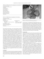
In patients older than 2 years, cancellous bone can be har-
vested from the iliac crest with the help of a trocar (Figures
48.2 and 48.3). This procedure diminishes the extension of the
secondary intervention and the pain at the donor site, and the
resulting graft is compressed. Alternatively, and especially when
large quantities are required, the iliac crest itself can be raised
as an osteoplastic flap, cancellous bone chips removed, and the
lid replaced. The key to prevention of postoperative morbidity
at this site is the avoidance of any muscle stripping in particu-
lar on the lateral aspect of the crest and the use of a long-
acting local anesthetic agent (e.g., bupivicaine) titrated over 24
hours postoperatively into the wound via an epidural cannula.
Adequate stability is always important especially in bilat-
eral alveolar clefts. During the first postoperative weeks, bone
grafting cannot abolish the mobility of the premaxilla. Indeed,
mobility of the fragments may well prevent bone union be-
tween the fragments and across the cleft(s). Some form of fix-
ation of the fragments is needed, for example, by external de-
vices such as dentally fixed splints or arch wires. Internal
fixation methods such as plates and screws can be applied
in a simultaneous osteotomy of the premaxilla or in a sec-
ondary intervention with the need for bigger grafts (Figures
48.4–48.6). Some authors describe a simultaneous palato-
osteoplasty. Their intention is to reconstruct all the layers cor-
responding to the normal anatomic situation. We have no per-
sonal experience with this procedure because we cannot see
the functional need.
The Basel Approach
In 1983, Honigmann described a method that had been
adopted in 1980.
124
This technique involved closure of the
soft palate and the lip in one stage in uni- and bilateral com-
plete clefts at the age of 6 months. The alveolar and the hard
palate cleft were closed in a second intervention at the age of
3 to 5 years with bone grafting into the alveolar cleft. The
bone graft was harvested from the iliac crest, and from 1985
onward using a trocar. In some cases the bone chips were mixed
with a granulate of tricalcium phosphate.
125
The aims of the
timing were to construct the labial and velar muscle systems
as soon as possible for optimal functional development, to re-
544 K. Honigmann and A. Sugar
F
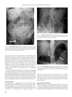
IGURE 48.2 Bone collection from the iliac crest by trocar.
FIGURE 48.3 Bone harvested from the iliac crest by trocar.
F
IGURE 48.4 A bilateral cleft lip and palate with a big bone defect;
status after Le Fort I osteotomy and fixation with 2.0-mm plates and
screws.
duce the number of interventions for primary cleft repair, and
to enable the children to enter school with a completely closed
cleft and normal colloquial speech. The failure rate in bone
grafting at that time was 11.3%. Normal colloquial speech at
school entrance was achieved in 91.6% of the children.
28
In 1991, this concept was changed with the aim of obtain-
ing a completely closed cleft at the end of the first year of
life for a better functional and psychological development of
the cleft child. Based on the aim of reducing the number of
surgical interventions and thus hospitalizations, an attempt
was made to try to close all forms of clefts in one stage at
least by the age of 6 months. Because of modern methods of
pediatric anaesthesia, there were no significant problems even
in a 4-hour operation, which was needed in complete bilat-
eral clefts. Subsequently it was found that this all-in-one pro-
cedure for unilateral cleft lip and palate patients had been pro-
posed in 1966.
126
The late results of that work were reported
at the 7th International Congress on Cleft Palate and Related
Craniofacial Anomalies in 1993 at Broadbeach, Australia.
127
The operative steps in detail are as follows.
The child’s head is placed in the ‘Rose’ position, that is,
the surgeon is seated with the child’s head on his/her knees.
The mouth is opened by a Rosenthal retractor (the widely used
Dingman retractor covers the lip and the alveolar cleft with
its extraoral frame, so it is impossible to get the view needed
for the alveolo-osteoplasty). The incision of the soft palate
edges continues with the dissection of pedicled palatal flaps
including the preparation and mobilization of the palatal ves-
sels (Figure 48.7). This provides a good view for the intra-
velar muscle dissection. With the aid of mucoperiosteal
vomerine flaps and the mobilized lateral nasal mucoperios-
teum, the nasal meatus can be formed in the complete alve-
olar and palatal cleft (Figure 48.8), and in bilateral clefts the
two nasal meati can be separated (Figure 48.9).
Suture of the mobilized and posteriorly directed soft palate
muscle stumps and pushback of the totally mobile palatal soft
48. CLP Osseous Defects and Deformities 545
FIGURE 48.5 Same patient as in Figure 48.4 grafted with cortico-
cancellous bone from the iliac crest; fixation with 2.0-mm plates and
screws.
F
IGURE 48.7 Dissection of the soft palate muscles and the pedicled
palatal flaps.
F
IGURE 48.6 Same patient as in Figure 48.6; oral cover of the graft
with a tongue flap.
F
IGURE 48.8 Formation of the nasal meatus in the unilateral alveo-
lar and palatal cleft.
tissues lengthens the soft palate into a normalized anatomic
situation (see Figure 48.1). The palatal flaps are sutured only
in the midline and then lightly pressed against the palatal bone
with the aid of a palatal dressing. Thus a dead space between
the palatal bone and soft tissues can be avoided, and with it
a hematoma and the resulting thicker scar. After reposition-
ing the child onto the table, a rib bone graft is harvested (Fig-
ure 48.10) and the alveolar cleft(s) filled with the cancellous
bone (Figure 48.11). Integrated into the final lip repair is cover
of the bone graft by mucoperiosteum advanced from the
vestibular side of the lesser maxillary segment and its sutur-
ing with the tips of the palatal flaps.
In this manner, alveolar bone grafting is a part of an all-
in-one closure of all clefts. More than 80 complete uni- and
bilateral clefts have been closed in this all-in-one procedure
(case 1: Figure 48.12 and case 2: Figure 48.13). At this time,
the rate of healing complications is 5.9% (3 partial hard palate
dehiscences, 2 bone graft losses), and the first functional re-
sults with regard to speech development and hearing disor-
ders are very encouraging.
The Swansea Approach
By contrast, Sugar’s approach to alveolar bone grafting in
Swansea (and until 1994 in Chepstow) has been unchanged
since 1985. Grafting has been carried out ideally in the mixed
dentition shortly before the eruption of the permanent maxil-
lary canine teeth, the classic secondary graft. This approach
has varied little from the method proposed by Boyne and
Sands
80
and reported by Abyholm and colleagues.
81
However,
in our patients, operating on children whose primary surgery
has been carried out by a number of surgeons, there has been
a clear need for a significant amount of orthodontics, primarily
to correct collapsed or misplaced alveolar segments, before
grafting can take place. Only cancellous bone harvested from
the anterior iliac crest has been used and with consistently
good results.
During this period, a significant number of cleft patients
presented who had, for various reasons, missed the opportu-
nity of receiving a graft into their alveolar clefts during the
mixed dentition phase. In most cases these have been man-
aged with careful orthodontic preparation with fixed bands
and tertiary alveolar grafting in exactly the same way as men-
tioned.
128
This has applied equally to those patients who have
not required orthognathic surgery, the graft not only facili-
tating closure of fistulae but also giving support to dental
restorations with or without osseointegrated implants. When-
ever grafting is carried out during orthodontic therapy, the or-
thodontist places in advance either lateral retaining arms from
molar bands or rigid arch wires to maintain arch width. This
is usually reinforced by a transpalatal bar, positioned suffi-
ciently far posteriorly and relieved from the mucosa to enable
any required palatal surgery to be performed.
In all cases the complete alveolar cleft is identified. Any
labial fistula is excised and this excision incorporated into the
mucoperiosteal flap(s) of the lesser segment(s) (see Figures
48.14a–g–48.22). These flaps critically include keratinized gin-
givae. In unilateral cases, a mucoperiosteal flap is also raised
up to one unit on the greater segment. In bilateral cases, virtu-
546 K. Honigmann and A. Sugar
F
IGURE 48.9 Separation of the two nasal meati in a bilateral cleft.
FIGURE 48.10 Rib graft resection.
FIGURE 48.11 Primary alveolar cleft bone grafting.
48. CLP Osseous Defects and Deformities 547
FIGURE 48.12 Case 1. (a) Five-month-old boy with a unilateral cleft lip and palate (CLP). (b) Intraoral aspect. (C) Two years old, after the
one-stage closure. (d) Intraoral aspect. (e) X-ray of the grafted alveolar cleft, 18 months postoperative.
a b
c
d
e
ally no dissection is permitted on the premaxilla, whose blood
supply is perilous. The closure of anterior palatal fistulae in
two layers at this stage is mandatory. The repair of posterior
palatal fistulae away from the alveolar cleft is optional, but the
opportunity to do this simultaneously is difficult to resist.
Scar tissue within the alveolar cleft is excised and the nasal
mucosa repaired. It is important that this repair is carried out
in such a way that the nasal floor lies at the same height as
the normal side. This, together with excision of the scar tis-
sue in the cleft, redefines the complete alveolar deficit into
which are then packed the cancellous bone chips. The lateral
flaps are then advanced, aided by appropriate division of pe-
riosteum, and closed with keratinized fixed gingivae over the
alveolar crest. These flaps are sutured across the crest to the
palatal oral mucosa. The posterior deficits of mucoperiosteum
over the alveolus buccally from where the flaps have been
advanced are allowed to heal by secondary epithelialization.
Antibiotics are administered intravenously during the opera-
tion. Even when large fistulae have been present we have al-
ways been able to use local flaps, although on occasion the
palatal flaps have had to be ‘islanded’ (i.e., Millard island
flaps) when advancement has been required. We have never
needed or used a Burion flap in this situation.
Case 3 (Figure 48.14)
A 10-year-old with left unilateral complete cleft of lip and
alveolus.
Treatment:
1. Raising of mucoperiosteal flaps
2. Excision of sinus and scar tissue within cleft
3. Removal of supernumerary tooth
4. Repair of nasal mucosa at level of normal nasal floor
5. Harvesting of cancellous bone from anterior iliac crest
6. Insertion of graft into alveolar defect
7. Flap advancement and closure over graft
The Role of Osseointegrated Implants
Although modern cleft surgery aims to create a dentition with-
out gaps, this aim is not always achieved. The incidence of
hypodontia in cleft patients is higher than in the noncleft pop-
ulation, and it is not always possible for this to be disguised
with the help of grafting, orthodontic treatment, and orthog-
nathic surgery alone. There are also many patients who have
not received alveolar bone grafts and also those who have lost
548 K. Honigmann and A. Sugar
FIGURE 48.13 Case 2. (a) Bilateral complete CLP
in a 6-month-old boy. (b) Same boy, aged 1 year
and 6 months, after the one-stage closure. (c) In-
traoral aspect.
a
c
b
48. CLP Osseous Defects and Deformities 549
FIGURE 48.14 Case 3. (a) X-ray of secondary alveolar defect. (b) In-
cisions for alveolar bone grafting outlined with excision of labial fis-
tula. (c) Scar tissue within the alveolar cleft. (d) Alveolar defect af-
ter excision of scar tissue and repair of the nasal mucosa. (e) Inci-
sion (continuous line) marked lateral to the left anterior iliac crest
(interrupted line) for harvesting of cancellous bone.
Continued.
a
c
e
b
d
550 K. Honigmann and A. Sugar
f
h
i
g
j
FIGURE 48.14. Case 3. Continued. (f) Alveolar defect packed with
cancellous bone chips harvested from the anterior iliac crest. (g) Flap
closure over the bone graft; note the advancement of the flap from
the lesser segment including gingivae and leaving a posterior defect
over the lateral maxilla, which is left to epithelialize by secondary
intention. (h) Diagram of procedure. (i) X-ray of the alveolus in the
grafted area in the same patient 6 months after surgery. (j) Oral view
of the same patient 6 months after surgery.
teeth early and whose conventional dental restorative treat-
ment is problematic.
The restoration of gaps in the dentition is ultimately the re-
sponsibility of the restorative dentist. Their options include
dentures and fixed bridgework supported by teeth. The avail-
ability of titanium osseointegrated implants now adds to this
repertoire the possibility of crowns or bridges supported by
implants, as well as implant-supported overdentures.
Case 4 (Figure 48.15)
A 25-year-old with left unilateral complete cleft lip and palate,
not having received an alveolar bone graft and missing the
left maxillary lateral incisor.
Treatment:
1. Alveolar bone grafting with autogenous cancellous iliac
bone as described in Figure 48.14
2. Orthodontic arch alignment
3. Insertion of Bränemark titanium fixture into grafted area
with additional small bone graft for labial defect provided
from suction filter during the drilling process and covered
with resorbable membrane (two-stage implant procedure)
4. Construction of implant-retained crown
(Restorative treatment courtesy of Will McLaughlin, Consul-
tant in Restorative Dentistry, University Dental Hospital,
Cardiff, Wales)
Case 5 (Figure 48.16)
A 16-year-old with bilateral complete cleft lip and palate as-
sessed following orthodontics and bilateral alveolar bone
grafting and with regard to two missing teeth in the left cleft.
Treatment:
1. Insertion of two Bränemark titanium fixtures (two-stage
procedure) into maxillary alveolus, previously grafted in
conjunction with orthodontics
2. Construction of implant-retained bridge
(Restorative treatment courtesy of Arshad Ali, Consultant in
Restorative Dentistry, Morriston Hospital, Swansea, Wales)
Maxillary Osteotomies
Secondary deformities in patients with repaired cleft lip and
palate present an interesting, if not difficult, surgical chal-
lenge. Careful assessment of the patient in the years follow-
ing primary repair needs to take into consideration speech,
hearing, facial growth, and dental development. The presence
of fistulae, lip scars, and poor lip function, as well as resid-
ual nasal deformity and nasal resistance, needs to be assessed
for correction. Alveolar defects and occlusion should be con-
sidered along with dental overcrowding, missing, malformed
and misplaced teeth, caries, and periodontal health. The abil-
ity and desire of the patient (and in the case of children, their
family) to comply with what can often be prolonged treat-
ment needs to be determined and taken into account.
This heterogeneity of problems requires the cooperation of
a number of different specialties, foremost of which are a sur-
geon, speech therapist/pathologist, hearing specialist, and or-
thodontist, all preferably with a special interest in cleft prob-
lems. In late adolescence, a specialist in restorative dentistry
is a valuable addition to the team. It is particularly useful to
attempt to identify at as early an age as possible those chil-
dren with significant midface hypoplasia that may require
later surgical correction. If orthognathic surgery is to be de-
layed until approximately 16 years of age when most jaw
growth is complete, early identification of those children is
helpful.
Timing
In most cases speech patterns will have developed by the age
of 4, and it should be possible to assess the need for a pharyn-
goplasty to correct velopharyngeal incompetence. Speech as-
sessment and recording, anenometry, nasendoscopy, and
video-fluoroscopy all assist in that decision. Ideally this
should be carried out before school entry.
At the age of 8 years, and with the aid of orthopantomo-
gram (OPT) and oblique occlusal and lateral cephalometric
radiographs, it is useful to start to consider the need for den-
tal extractions for orthopedic alignment of displaced and col-
lapsed arches and for grafting of alveolar defects. When fa-
cial growth appears to be essentially normal, definitive
orthodontics can then continue.
A clinical evaluation of facial form, noting the presence
or absence of midface hypoplasia, a class III malocclusion,
and dental compensation, may lead the team to the conclu-
sion that jaw osteotomies are indicated in due course. This
in turn allows the decision that orthodontics should be lim-
ited at that stage to the orthopedic alignment of segments
and perhaps the correction of minor anterior incisal dis-
crepancies. Definitive presurgical fixed-band orthodontics
can then be delayed until the approximate age of 14 years
when the patient can be prepared for orthognathic correc-
tion by osteotomies at 16. This has the merit of saving the
child from 6 to 8 years of continuous orthodontic treatment
with the inconvenience and almost inevitable lack of com-
pliance that can result.
The Role of Alveolar Bone Grafting
Primary Grafting
We have described in our previous section the purpose of con-
sidering and carrying out alveolar bone grafting as well as a
number of different approaches to it. Primary alveolar bone
48. CLP Osseous Defects and Deformities 551
a
c
e
d
b
f
48. CLP Osseous Defects and Deformities 553
g
h
i
FIGURE 48.15 Case 4. (a) X-ray of alveolar defect. (b) Diagram of
alveolar defect. (c) X-ray of grafted alveolar defect. (d) Diagram of
grafted alveolar defect. (e) Intraoral x-ray of implant in grafted alve-
olar defect. (f) Lateral cephalogram showing position of implant. (g)
Oral view with implant/abutment in situ. (h) Diagram showing im-
plant in situ. (i) Oral view showing implant retained crown in situ.
grafting is that which is carried out during the primary den-
tition or even before the eruption of the deciduous teeth. We
do not yet have available from Basel medium- or long-term
results of this approach, and much of the hostility to primary
grafting has come from the apparently poor effect on maxil-
lary growth.
129
However, others
130
have reported very en-
couraging results in this respect more recently. Rosenstein et
al.
130
have presented the long-term results in a regimen of cleft
repair that has included primary bone grafting of the alveo-
lar cleft at 4 to 6 months of age. This remains an area of con-
siderable controversy.
Secondary Grafting
Secondary alveolar bone grafting, by which we mean graft-
ing shortly before the eruption of the permanent maxillary ca-
nine teeth, has by contrast become very widely accepted. The
method described by Boyne and Sands
80
was popularized by
the reporting of large series by Abyholm and his colleagues.
81
It has undoubtedly made an important difference to the man-
agement of cleft patients. It makes the simultaneous repair of
residual fistulae easier and by producing a one-piece maxilla
facilitates a future maxillary osteotomy if needed. The pro-
554 K. Honigmann and A. Sugar
a
b
c
FIGURE 48.16 Case 5. (a) X-ray of implants in grafted alveolus. (b) Oral view of abutments. (c) Implant-retained bridge in situ.
duction is facilitated by well-aligned and continuous dental
arches, with good bone support for the maxillary permanent
canine and adjacent teeth. If there are gaps in the dental arch,
it produces a stable base for the construction of fixed bridge-
work and implant-retained crowns and bridges. The over-
whelming majority of compliant cleft children with an alve-
olar defect that has not been previously grafted will benefit
from secondary alveolar bone grafting provided that the
preparation and timing are carefully considered and the
surgery well executed.
The popularizing of this technique in Norway was based,
in the main, on children who did not have grossly collapsed
dental arches. It has been the experience of the authors that
secondary bone grafting of alveolar clefts without prior cor-
rection of misplaced segments creates significant difficulties.
The segments may become fixed in an abnormal position with
orthopedic movement no longer possible or at best very dif-
ficult (Figure 48.17).
Case 6 (Figure 48.17)
Patient with bilateral complete cleft lip and palate.
Treatment:
1. The alveolar clefts had been bone grafted before orthope-
dic expansion and alignment of segments.
2. The premaxilla was thus fixed in its position significantly
displaced inferiorly and to the right as were the lateral seg-
ments in their contracted position.
3. Later orthodontics was thus made very difficult. In some
cases the problem can only be resolved with the help of
multipiece osteotomies (see case 8, Figure 48.19).
48. CLP Osseous Defects and Deformities 555
a
b c
F
IGURE 48.17 Case 6. (a–c) Result of grafting of bilateral alveolar clefts before orthopedic alignment of the segments.
Careful assessment with an orthodontist experienced in the
management of clefts is therefore essential to determine the
presurgical needs, which should include alignment of any mis-
placed segments. After this, the orthodontist will design an
appliance that will both retain the parts which have been
moved and not impede surgery. Because the latter may well
involve the repair of residual palatal oronasal fistulae, the ap-
pliance in situ during surgery must not cover any part of the
palate to which access is required.
Tertiary Grafting
Patients who present after the eruption of the permanent canine
teeth and at the end of the mixed dentition phase of develop-
ment sometimes have not received any form of alveolar bone
graft. Others have poor results from earlier grafting attempts
and have inadequate bone for orthodontic movement of teeth,
for support for prostheses, or for carrying out a maxillary os-
teotomy in one piece. In these cases, and notwithstanding the
allegedly poor results that have been claimed for such late graft-
ing by some authors (relative to secondary grafting), it has been
our reported experience that excellent results can still be ob-
tained.
128
We therefore always consider, in conjunction with
our multidisciplinary team, tertiary grafting in such cases.
Investigation
Facial Appearance
The principal tool in the diagnosis of residual facial deformity
is clinical evaluation by an experienced surgeon. It is useful to
document those parts of the upper, middle, and lower face that
show anteroposterior, vertical, and transverse deficiencies or ex-
cesses. Dysmorphology and abnormality should be noted in all
areas and in particular of the nasal bones, septum, tip, columella,
and alar bases, as well as of the philtrum and upper lip.
Measurement of some aspects of the face in both frontal
and profile views and comparison with norms is of value. The
exposure at rest and when smiling of the upper incisor teeth,
as well as measurement of the clinical crown height, are just
a few examples. These enable the surgeon to determine the
vertical movements needed of the anterior maxilla to create
an ideal relationship with the upper lip, but consideration
needs to be given to the need for lip revision in this respect
and any of the resultant effects on lip–tooth relationship.
The interalar distance needs to be known if only to avoid
making it worse after maxillary advancement; sometimes si-
multaneous revision of this distance needs to be built into the
treatment plan. The intercanthal distance and nasofrontal an-
gle may also increase in Le Fort II or Le Fort III osteotomies
and should be recorded. The relationship between the maxil-
lary and mandibular dental centers and the facial midline and
chin needs to be known so that attempts at creating symme-
try may be made. The presence of missing teeth in the cleft
patient may make this particularly difficult.
Many forms of cephalometric measurement are available,
some of which are particularly designed for analysis of the
patient with a jaw deformity. While these can be useful, al-
lowance does need to be made for the different values that
are observed in cleft patients. A particularly relevant exam-
ple is the cranial base to which the position of the maxilla
and mandible is usually related. When the cranial base angle
is abnormal (that is, it is outside the normal range of values),
the angles of SNA and of SNB also vary widely, and this
needs to be taken into consideration.
Occlusion
Dental study casts are essential in the overall analysis. In this
way, the precise needs of presurgical orthodontics can be de-
termined and results monitored.
Speech
It is always desirable that the cleft patient should be managed
in coordination with a speech therapist/pathologist with ex-
perience of and interest in cleft patients. Children should be
assessed at regular intervals during their development. The
axiom that treatment should aim at producing an individual
who “looks well and speaks well” remains valid today.
In relation to midface osteotomies, it is well recognized that
these have the significant potential for improving the articu-
latory aspects of speech by correcting malocclusion and skele-
tal disproportion. However they also carry the unwanted risk
of producing, or making worse, velopharyngeal incompetence
(VPI). Consequently all cleft patients should have a thorough
speech assessment immediately before undergoing midface
advancement. This should involve a standard form of assess-
ment with speech recording and anenometry. Nasendoscopy
and videofluoroscopy may be valuable but can usually be re-
served for those cases with problems postoperatively. The ex-
perienced speech therapist/pathologist, especially working in
the same team and with the same surgeon, should be able to
identify those patients most at risk of developing VPI.
Deformities/Diagnosis
Maxillary hypoplasia in cleft patients has a clear relationship
to both the original deformity and the consequences of early
surgical repair. We now describe the principal forms.
Unilateral Complete Cleft Lip and Palate (UCLP)
In this cleft defect, when midfacial hypoplasia is present it is
manifested predominantly by an anteroposterior deficiency of
the maxilla with lack of support to the nasal tip. There is of-
ten a vertical deficiency producing a lack of exposure of the
upper incisor teeth at rest, influenced by any distortion of the
upper lip. There will usually be an alveolar defect on the side
of the cleft unless it has been grafted previously. Even with-
out a previous periosteoplasty,
119
bone bridging across the
alveolar defect is sometimes seen. Transverse collapse of the
alveolar segments may also occur, perhaps the most common
556 K. Honigmann and A. Sugar
being displacement inward (palatally) and upward (cranially)
of the lesser segment.
Several studies have shown that the mandible often lacks
some forward growth in the repaired UCLP patient. In rela-
tion to surgery, it is questionable whether this usually requires
correction. There is, however, often a lack of chin prominence
but an excess of chin height. These contribute to an unesthetic
and often drooping or ptotic appearance of the lower lip and
warrant intervention.
Although the principal secondary nasal deformities are pre-
dominantly cartilage and soft tissue, the lack of support to the
nasal tip may be severe. The dorsum of the nose is usually
described as being essentially normal, but cases are seen
where it is retropositioned and asymmetry is not uncommon.
Labial or palatal fistulae may be present, communicating with
the nasal cavity. The septum is usually deviated to the non-
cleft side and is often quite wide. In the authors’ experience,
septa more than 1 cm wide can occur with complete block-
age of the nasal airway. The inferior turbinate on the cleft
side is usually hypertrophied.
Bilateral Complete Cleft Lip and Palate (BCLP)
Although class III malocclusions are seen in BCLP patients,
very much depending on the method of primary repair, the
principal finding is prominence of the premaxilla (and pro-
labium), especially vertically. In ungrafted cases, the pre-
maxilla is usually mobile, poorly inclined (retroclined), and
to one side or the other. The patient will often have, or with
the aid of orthodontics be capable of having, a class I incisor
relationship.
Class II-based deformities with mandibular retrognathia or
retrogenia are seen in BCLP patients (Figure 48.18), and
sometimes this is the only skeletal defect that requires cor-
rection. Occasionally bimaxillary advancement is indicated.
Case 7 (Figure 48.18)
Patient with bilateral complete cleft lip and palate and an-
teroposterior deficiency of the mandible.
Treatment:
1. Fixed-band orthodontics commenced in both arches to re-
move dental compensation, align teeth, and produce com-
patible arches on the basis of three-point contact following
orthognathic surgery.
2. Before the movement of teeth adjacent to the alveolar
clefts, these clefts were bone grafted in the way that we
have described.
3. Following completion of the presurgical phase of ortho-
dontics, mandibular advancement was carried out using bi-
lateral sagittal split osteotomies of the mandibular rami,
fixation being by four 2.7-mm titanium position screws
(two on each side) inserted transbuccally.
4. Orthodontics was then completed.
(Orthodontics courtesy of Jeremy Knox, Dept. of Child Den-
tal Health, University Dental Hospital, Cardiff, Wales)
It is common for teeth in the premaxilla of bilateral cleft pa-
tients to be poorly formed and prone to caries or crumbling;
such teeth are not a good support for orthodontic devices. Nev-
ertheless, malposition of the premaxilla and lateral segments can
usually be corrected by the orthodontist before cleft bone graft-
ing. Jones and Sugar
128
have reported one case in whom this
was carried out with an orthodontic device when the patient had
no teeth on the premaxilla. There are, however, instances in
which repositioning of the premaxilla can be difficult or im-
possible. In such occasional cases, surgical repositioning of the
premaxilla before grafting should be considered (see case 9, Fig-
ure 48.20). The nose in the bilateral cleft patient may be broad
at the alar bases and often also at the bridge with a short col-
umella. Anteroposterior deficiency of the dorsum is rare.
Cleft Palate (CP)
The patient with a repaired isolated cleft of the palate may also
exhibit anteroposterior and sometimes vertical deficiency of the
maxilla. It has been argued that many deformities of this kind
in these and complete cleft lip and palate patients are not nec-
essarily cleft related. Undoubtedly instances of class III skele-
tally based malocclusion of familial rather than cleft origin do
occur, but the relative rarity of class II deformities in cleft pa-
tients is food for thought. The patient in case 8 (Figure 48.19a)
has a repaired cleft palate with maxillary hypoplasia. Figure
48.19(c) shows her “identical” twin sister who has no cleft.
Case 8 (Figure 48.19)
Patient in Figure 48.19(a) has a repaired cleft of the secondary
palate with anteroposterior and vertical deficiency of the max-
illa. Figure 48.19(c) shows her identical twin sister who had no
cleft, the photographs being taken on the same day as those of
her sister. The principal difference noticeable between the sis-
ters is the maxillary hypoplasia exhibited by the sister with a
repaired cleft.
Surgery:
1. One-piece Le Fort I maxillary advancement and downward
movement
2. Fixation using four L-shaped titanium 2-mm miniplates
3. Augmentation of the anterior maxillary bone steps only
with corticocancellous blocks harvested from the medial
aspect of the anterior iliac crest
Indications for Orthognathic Surgery
The principal indications for carrying out orthognathic surgery
in patients with repaired cleft lip and/or palate are as follows.
1. To improve facial aesthetics and in particular the appear-
ance of the midface, including the upper lip and nose
48. CLP Osseous Defects and Deformities 557
558 K. Honigmann and A. Sugar
a
e
c
b
d
48. CLP Osseous Defects and Deformities 559
f
h
g
i
FIGURE 48.18 Case 7. Anteroposterior mandibular deficiency in a pa-
tient with bilateral cleft lip and palate (BCLP). (a–d) After ortho-
dontic preparation but before surgery. (e) Diagram of surgical pro-
cedure (sagittal split advancement). (f–i) Following surgery and
completion of orthodontics.
560 K. Honigmann and A. Sugar
a
c
e
g
b
d
f
F
IGURE 48.19 Case 8. (a,b) Patient with repaired cleft palate before orthognathic surgery. (c,d) Identical twin sister of patient in a,b who
had no cleft. (e,f) Patient in a,b after Le Fort I maxillary advancement and correction of vertical deficiency. (g) Diagram of procedure.
2. To permit the full correction of skeletally based malocclu-
sions
3. To improve the nasal airways by reducing nasal resistance
4. To improve speech, especially the articulatory aspects
Orthodontic Requirements
To achieve these aims optimally, orthodontic management is
required to accomplish these aims:
1. Correct major displacement of segments by orthopedic
movements
2. Permit the ideal choice of timing for alveolar bone grafting
3. Correct crowding and adopt a rational approach to tooth
position where teeth are missing (hypodontia)
4. Remove dental compensation, especially abnormal incli-
nations of upper and lower incisors
5. Produce well-coordinated dental arches that will be com-
patible after surgery
6. Fine-tune tooth positioning and occlusion following
surgery
Planning, Soft Tissue Effects, and Predictions
Planning in orthognathic surgery
131
is the process by which
the assessment, investigation, and resulting diagnosis are
translated into a coherent treatment plan. It should be based
predominantly on a clinical determination of treatment ob-
jectives. In the typical case with moderate to severe antero-
posterior and vertical deficiency of the maxilla, and provided
that the alveolar segments were aligned before bone grafting,
it will probably involve the advancement of the maxilla at the
Le Fort I level. Although every patient needs to be assessed
individually, there is a tendency in some quarters to avoid
large maxillary advancements by “splitting the difference”
and moving the mandible back simultaneously. There are un-
doubtedly cases of true mandibular prognathism in which this
is called for, but it is still necessary to carry out full correc-
tion of a retropositioned maxilla. Advancements of more than
2 cm may be necessary.
Model surgery is an absolute requirement in all cases.
Models should be set up on a semiadjustable anatomic ar-
ticulator after face bow recording. Reference lines are drawn
and various distances in three planes are recorded. The de-
sired movements are then carried out and the measurements
retaken and recorded. These movements need to relate to the
clinical treatment objectives, and it is valuable to test the
achievement of those objectives against a predictive com-
puter program. Once the movements have been finalized,
acrylic occlusal wafers should be constructed, one in the
case of a single-jaw osteotomy and two (including an inter-
mediate position) in the case of bimaxillary procedures.
These at least will remove some of the guesswork from the
operating room, although vertical determinations will still
need to be made.
Most computer packages for orthognathic surgery planning
are based on surgery on a digitized lateral cephalometric ra-
diograph. They are not infallible but can be a remarkably valu-
able indication of what will happen. We have analyzed two
of the most commonly used such packages in the United States
and U.K. specifically for internally fixed Le Fort I osteotomies
including clefts.
132,133
Soft tissue changes in cleft patients
have a tendency to differ from those in noncleft patients, prob-
ably because of the lack of elasticity of the enveloping tis-
sues. It is hoped that in the future such programs will be able
to take this into consideration and thus give more accurate
predictions. It is questionable whether more sophisticated
(and expensive) techniques of three-dimensional prediction
are of much value in the average case. However, video cap-
ture techniques with color print predictions of the result of
surgical movements allow the patient to see a reasonable sim-
ulation of what surgery can achieve. They may also be help-
ful to the surgeon.
Treatment planning should take into consideration the
views of the speech therapist or speech pathologist on the
likelihood of the development or worsening of velopha-
ryngeal incompetence. When very large advancements are
considered, this may dictate a modification of surgical tech-
nique.
Surgical Procedures
Premaxillary Osteotomy
Osteotomies of the maxilla of cleft patients have to be tai-
lored to the different anatomy, to the blood supply of the dif-
ferent parts of the maxilla, and to the nature and effects of
previous surgery. This is especially the case when the part to
be moved is the premaxilla. In the bilateral cleft patient, the
bone of the premaxilla is attached very narrowly to the nasal
septum. Its blood supply is derived principally from the labial
mucoperiosteum. These need to be taken into consideration
when designing the surgical approach and osteotomy tech-
nique if the premaxilla is not to become a free graft.
Premaxillary osteotomies will be needed only rarely because
orthodontic methods are quite good at guiding this bone into
the correct position. When needed, it will usually be because
the bone would not move in this way. The bony attachment of
the premaxilla may be approached from the palatal side or lat-
erally (Figure 48.20), in both cases from within the cleft. It is
also possible to use a midline labial vertical mucoperiosteal in-
cision. Following osteotomy of the narrow attachment, the bone
may then be moved digitally and fixed in its new position with
the guidance of an occlusal wafer.
Fixation is best achieved with a strong arch wire within
preexisting fixed orthodontic bands. It is unlikely, however,
that this premaxilla will then become stable without the arch
wire. It will eventually be stabilized by bilateral alveolar bone
grafting, and the authors consider that this is visually best car-
ried out as a separate procedure a few months later.
48. CLP Osseous Defects and Deformities 561
562 K. Honigmann and A. Sugar
Case 9 (Figure 48.20)
A 10-year-old girl with bilateral complete cleft lip and palate.
The premaxilla is misplaced and would not move with or-
thodontic appliances.
Treatment:
1. Model surgery to reposition the premaxilla and fabricate
an occlusal wafer
2. Noted that this was only possible with the surgical removal
of part of the premaxilla including a developing supernu-
merary (or abnormal lateral incisor) tooth germ
3. Securing of orthodontic fixed bands and fabrication of a
strong arch wire that would support the premaxilla in its
new position
4. Surgery in which the premaxilla was approached through
a small lateral incision, permitting the removal of both the
required amount of the premaxilla and division with a small
osteotome of its bony attachment
5. Digital movement of the premaxilla into its new position,
temporary fixation into the preformed occlusal wafer, and
stabilization with a strong arch wire. The wafer was then
removed
6. Three months later, bilateral alveolar bone grafting was car-
ried out with simultaneous repair of the palatal fistula
7. Continued orthodontics
(Orthodontics courtesy of Prof. Malcolm Jones, Consultant
Orthodontist and Head of Department of Child Dental Health,
University Dental Hospital, Cardiff, Wales)
Le Fort I Osteotomy
The Le Fort I osteotomy is the most valuable procedure in
cleft adolescents with maxillary hypoplasia. We consider that
the most important aims must be full mobilization and good
fixation.
Nasal airway obstruction, a severely retropositioned maxilla,
and previous pharyngoplasty may all conspire to make nasal
endotracheal intubation in these patients difficult. However, it
is most unusual for the nares to prevent passage of an endo-
tracheal tube at least on one side. Forewarning of the problem
of the tube hitting the posterior pharyngeal wall enables the
anesthetist to carefully redirect it inferiorly. This can sometimes
be helped by a finger placed in the mouth above and behind
the soft palate, where the tube can be palpated and brought for-
ward and downward. Pharyngoplasties, especially superiorly or
inferiorly based pharyngeal flaps, may limit access for intuba-
tion. The presence of such flaps should be noted preoperatively;
most can be bypassed without damage but the patient should
be warned of the risk of the pharyngoplasty being damaged or
in extreme cases of it having to be divided and repaired. For-
tunately, dynamic pharyngoplasties have become more popu-
lar and they present much less restriction to intubation.
In the past, multipiece and segmental procedures were ef-
fectively forced on surgeons with what was then the stage of
development of orthodontic support and before the common
use of alveolar bone grafting. The work of Tideman et al. is
particularly recognized in this context,
134
with his innovative
use of substantial closure of the alveolar cleft by advance-
ment of the lesser or lateral segments. Posnick
135
has also de-
veloped a closely related approach based on orthodontics and
multipiece osteotomies in the ungrafted cleft patient. Case 10
(Figure 48.21) demonstrates an adaptation of these techniques
in a previously bone-grafted bilateral cleft patient where
presurgical orthodontic preparation could not be completed to
permit a one-piece osteotomy.
Case 10 (Figure 48.21)
An 18-year-old with bilateral complete cleft lip and palate.
The occlusion was mildly class III with the premaxillary teeth
proclined. Further orthodontic preparation was not possible
because of the very short roots on the upper central incisors.
Successful bilateral alveolar bone grafting had been carried
out elsewhere.
Treatment:
1. Following presurgical orthodontics, sectional arch wires
were placed
2. Le Fort I osteotomy carried out from a lateral approach at-
tempted to preserve the maximum mucoperiosteal attach-
ment to the premaxilla both palatally and labially
3. Ostectomies carried out in the previously grafted clefts bi-
laterally
4. Positioning of the three bone segments of the maxilla into
a preformed occlusal wafer and wiring of a prefabricated
arch wire across all segments fixed to the orthodontic
brackets. The proclined premaxilla was retroclined, and the
lateral segments advanced to close off the gaps in the den-
tal arch coinciding with the alveolar clefts
5. Internal bone fixation with titanium 2-mm L-shaped mini-
plates was followed by removal of the wafer and inter-
maxillary fixation (IMF)
6. Grafting of the anterior bone steps with corticocancellous
blocks harvested from the medial aspect of the anterior il-
iac crest, and of the interdental bone cuts with cancellous
bone chips
(Orthodontics courtesy of David Howells, Consultant Ortho-
dontist, Morriston Hospital, Swansea, Wales)
With these particular methods, special care is required for
blood supply, and tunneling incisions are usually advisable
anteriorly. Difficulty may be encountered because of the pres-
ence of scar tissue from the primary palate repair and poor
access to break it down; this can be a particular problem for
large advancements. Loss of part of the maxilla is rarely re-
ported but is not unknown when carrying out maxillary os-
teotomies in cleft patients, and it is arguable that segmental
procedures increase the risk. Although demonstrating good
results, it has been shown
136
that grafting the cleft at the time
48. CLP Osseous Defects and Deformities 563
a
b
d
c
e
FIGURE 48.20 Case 9. (a) A 10-year-old girl with BCLP and a
malpositioned premaxilla. (b) Model of maxillary arch. (c)
Model surgery to reposition the premaxilla. (d) OPT before pre-
maxillary surgery. (e) Lateral cephalogram before premaxillary
surgery.
Continued.
f
h
i
g
j
FIGURE 48.20 Case 9. Continued. (f) Surgical approach to the pre-
maxilla marked. (g) Repositioned premaxilla after osteotomy. (h)
OPT of repositioned premaxilla with bilateral alveolar bone grafts.
(i) Lateral cephalogram taken at same time as h. (j) Patient follow-
ing this treatment.
48. CLP Osseous Defects and Deformities 565
a
c
b
d
FIGURE 48.21 Case 10. Multipiece maxillary Le Fort I osteotomy in a bilateral
cleft lip and palate (CLP) patient using a modified Tideman/Posnick technique.
This patient had been grafted previously elsewhere, but the proclination of the
premaxilla and condition of the roots of the upper incisor teeth prevented com-
plete orthodontic decompensation and correction of the position of the pre-
maxilla (a,b). Consequently, osteotomies were carried out through the grafted
alveolus on each side. The premaxilla was then retroclined, the lateral segments
advanced to close off the alveolar clefts, and the whole maxilla advanced. Fix-
ation was aided by a temporary acrylic wafer and intermaxillary fixation (IMF),
both during surgery only, and was maintained with an arch bar wired to the or-
thodontic brackets and four 2-mm titanium L-shaped miniplates (c,d).
of osteotomy is not quite as effective as grafting as a sepa-
rate procedure. Our own experience in South Wales (before
1985 when the present approach was adopted) was of a much
greater difference in the results with much more successful
graft take in the alveolar cleft when this was performed as a
procedure separate from maxillary osteotomy.
Three developments have permitted us to change our ap-
proach:
1. Improved primary surgery, leaving the maxilla in a better
developed condition with less hypoplasia and less arch col-
lapse.
2. Improved dental health and sophisticated orthodontics so
that cleft patients can now expect to have a complete den-
tition (with the exception of those teeth that have not de-
veloped) with oral hygiene and tooth condition such that
they can be offered fixed-band orthodontics.
3. Alveolar bone grafting which, in a good multidisciplinary
team, can be timed to fit in with bone and tooth develop-
ment and with other procedures, and will usually produce
a continuous maxillary dental arch.
It is the view of the authors, therefore, that segmental os-
teotomies in the cleft maxilla can usually be avoided. Most
maxillae will present in one piece following successful sec-
ondary (or primary) alveolar grafting. In those rare cases when
the grafting has been less than totally successful, it can be re-
peated and other patients who have not received a primary or
secondary graft at all can be prepared for tertiary grafting in
the way that we have described.
128
Having taken this ap-
proach, it is not very logical or sensible to follow with sec-
tioning of the maxilla into multiple pieces. Consequently, we
try to carry out all cleft osteotomies with a one-piece maxilla
using a downfracture approach. To date only one case (a
BCLP case bone grafted in another unit) has shown signs of
the maxilla failing to remain in one piece, and the minor
cracks that occurred in the grafted area did not compromise
the result, the segments being held in a strong arch wire.
The incision is placed anteriorly (Figure 48.22), being mod-
ified from the standard Le Fort I approach. It commences high
in the cheek, just above and anterior to the openings of the
parotid ducts. It is then continued down across the inside of
the upper lip. This permits a broader posterolateral pedicle to
supplement the palatal supply and still gives good access to
the pterygoid area. This incision is used for grafted unilateral
and bilateral cases alike, and since it was adopted 11 years
ago not a single instance of compromised blood supply has
been encountered.
The osteotomy is carried out using saws and fine os-
teotomes and with separation of tuberosities from pterygoid
plates with a chisel. The cuts are placed high to facilitate in-
ternal miniplate fixation. Mobilization is carried out digitally
and with disimpaction forceps and mobilizers. The nasal mu-
cosa is preserved on both sides but in places may have to be
cut to separate it from the oral (principally palatal) mucosa.
There is often very little space for disimpaction forceps in the
palate, especially with rubber protection for the blades. There
is also a small risk of damaging previous palate repairs. We
therefore always use a purpose-constructed metal palatal cov-
erage plate (Figure 48.23) first designed in our unit by Ross
and Bocca that permits use of the forceps without rubber cov-
ers and protects the palate effectively during mobilization. We
always break down digitally the palatal scar tissue holding
the maxilla back, and we do this from above through the open-
ing created by the downfracture.
With the maxilla displaced downward, it is then possible
to assess the internal nasal structures. A broad septum may
be reduced, and inferior (partial) turbinectomy carried out if
indicated. A preformed acrylic wafer is attached to the teeth
by orthodontic powerchain and intermaxillary fixation (IMF)
placed with more powerchain.
137
The maxilla is fixed using
566 K. Honigmann and A. Sugar
FIGURE 48.22 Modified incision for one-piece Le Fort I osteotomies
in all grafted cleft patients.
FIGURE 48.23 Palatal protection plate for mobilizing cleft maxillae
with disimpaction forcep (Designed by Ross and Bocca).
L-shaped 2-mm miniplates, long L-shaped plates being par-
ticularly valuable for large advancements (Case 11, Figure
48.24).
Case 11 (Figure 48.24)
A 17-year old with bilateral complete cleft lip and palate and
treated hypertelorbitism.
Treatment:
1. Orthopedic expansion and alignment of segments
2. Bilateral alveolar bone grafting and palatal fistula repair
3. Presurgical orthodontic preparation
4. One-piece Le Fort I osteotomy as described in the text, in-
ternally fixed with long L-shaped titanium 2-mm mini-
plates and bone grafted. The advancement in this case was
22 mm and the downward movement anteriorly was 10
mm
5. Completion of orthodontics. The stability of the result is
demonstrated in the lateral cephalometry in Figure 48.24(h)
2 years after surgery
(Orthodontics courtesy of Prof. Malcom Jones, Consultant
Orthodontist and Head of Dept. of Child Dental Health, Uni-
versity Dental Hospital, Cardiff, Wales, and David Bach-
meyer, Sydney, New South Wales, Australia)
We always bone graft these cleft osteotomies, using au-
togenous corticocancellous blocks harvested from the me-
dial aspect of the anterior iliac crest. The grafts are placed
anterolaterally and occasionally are fixed with screws.
Grafts are never placed into the region behind the maxilla,
where they are in any event unstable. The wounds are closed
primarily and without tension with no attempt to use the
so-called V to Y single or multiple advancements. We con-
sider that these closures, designed to produce vertical lip
lengthening, actually produce increased anteroposterior lip
projection and a tight wound and lip. In our hands, IMF is
always removed at the end of the operation and before ex-
tubation. We have never encountered instability in these
cases, even for the largest maxillary advancements (more
than 2.5 cm in some cases), and have never had to resort
to later IMF.
Case 12 (Figure 48.25)
A 21-year-old with repaired complete unilateral cleft lip and
palate, anteroposterior and vertical midface deficiency and
retrogenia, and secondary alveolar bone graft having been in-
serted previously.
Treatment:
1. Presurgical orthodontics
2. One-piece Le Fort I maxillary osteotomy as described
above with miniplate fixation
3. Advancement genioplasty (horizontal sliding osteotomy)
with 2-mm miniplate fixation
4. Completion of orthodontics
(Orthodontics courtesy of Russell Samuels, Consultant Or-
thodontist, Glenfield Hospital, Leicester, England)
Case 13 (Figure 48.26)
A 16-year-old with right unilateral complete cleft lip and
palate. Secondary alveolar bone graft inserted after arch ex-
pansion at the age of 9 years with simultaneous closure of a
large palatal fistula. Presented with significant anteroposte-
rior and vertical maxillary deficiency.
Treatment:
1. Presurgical orthodontic preparation
2. Le Fort I maxillary advancement (1.5 cm) and downward
movement (5 mm) as described above, fixation being with
2-mm titanium L-shaped miniplates and anterior maxillary
grafting with corticocancellous blocks harvested from the
medial aspect of the anterior iliac crest
3. Completion of orthodontics
(Orthodontics courtesy of Simon James, Consultant Ortho-
dontist, Withybush General Hospital, Haverfordwest, Wales)
This approach has of course made these procedures much
more popular with our anesthetic colleagues with safer post-
operative airway management. Expensive intensive or high
dependency care management in the immediate postoperative
period can almost always be avoided. It has also made the
postoperative period much more comfortable for the patient,
who has often had to endure many operations.
The downfracture approach has been criticized in some
quarters because of the risk of making velopharyngeal func-
tion worse. Using the technique described here, this has not
been our experience. Velopharyngeal incompetence only
seems to be present postoperatively in patients in whom it
was present before surgery. It has been reported that the de-
velopment of VPI can be avoided if a palatal approach to the
osteotomy is adopted, the intention being to leave the palatal
musculature and soft palate behind when the maxilla is ad-
vanced.
138–141
This certainly has merit but unfortunately also
has some disadvantages. Intraoperatively, there is reduced ac-
cess anteriorly to the nose and for fixation, and among the
postoperative complications there is a high incidence of resid-
ual oronasal fistulae that require further surgery. Average
skeletal relapse in the position of the maxilla anteroposteri-
orly has been reported as high as 29% in one series.
140
The literature and experience indicate that Le Fort I os-
teotomies in cleft patients can be associated with particularly
high incidences of relapse in the opposite direction to the move-
ments carried out. It is our clear impression that this is no longer
the case with our approach.
142
This is discussed further later.
48. CLP Osseous Defects and Deformities 567
a
c
e
b
d
f