General Principles for Approaches to the Facial Skeleton - part 5 pot
Bạn đang xem bản rút gọn của tài liệu. Xem và tải ngay bản đầy đủ của tài liệu tại đây (621.12 KB, 18 trang )
74
Step 3. Incision
Cross hatches or dye markings across the proposed site of incision assist in properly aligning the
scalp during closure. The first is made in the midline and subsequent marks are made laterally at
approximately equal distances from the midline (Fig. 6-10). Crosshatches made with a scalpel tip
should be deep enough (until bleeding) so that their location is visible at the end of the surgical
procedure.
The initial portion of the incision is made with a no. 10 blade or special diathermy knife,
extending from one superior temporal line to the other. For routine coronal exposure, the incision is
made through skin, subcutaneous tissue, and galea (see Fig. 6-10), revealing the subgaleal plane of
loose areolar connective tissue overlying the pericranium. The flap margin may be rapidly and
easily lifted and dissected above the pericranium. Limiting the initial incision through the
temporalis fascia into the temporalis musculature, which bleeds freely.
The skin incision below the superior temporal line should be to the depth of the glistening
superficial layer of temporalis fascia. This depth is into the subgaleal plane, continuous with the
dissection above the superior temporal line. An easy method to ensure that the incision is made to
the proper depth is to bluntly dissect in the subgaleal plane from above, toward the zygomatic arch,
with curved scissors and incising to that depth (Fig . 6-11).
Preauricular extension of the incision is within a preauricular skin fold to the level of the
lobule. The dissection severs the preauricular muscle and follows the cartilaginous external auditory
canal, similar to the dissection described in Chapter 12.
75
Figure 6- 10 Draping of the patient and the initial incision. The drapes are secured with staples and/or
sutures just posterior to the location of the planned incision. Cross-hatches are scored into the scalp at
several locations for realignment of the flap during closure. The initial incision extends from one superior
temporal line to the other, to the depth of the pericranium (see inset). The dissection will be in the
subgaleal plane, which is loose connective tissue and cleaves readily.
76
Figure 6- 2 11 One technique for incising the scalp in the temporal region. Scissor dissection of the scalp
in the subgaleal plane can proceed inferiorly from the previous incision made above the superior temporal
line. While the scissors are spread, a scalpel incises to them, preventing the surgeon from incising the
temporalis fascia and the muscle, which bleeds freely.
77
Step 4. Elevation of the Coronal Flap and Exposure of the Zygomatic Arch
After elevation of the anterior and posterior wound margins for 1 to 2 cm, hemostatic clips (Raney
clips) are applied or bleeding vessels are isolated and cauterized. Indiscriminate cauterization of the
edge of the incised scalp can result in areas of alopecia and should be avoided. A technique to
expedite clip removal before closure involves positioning an unfolded gauze sponge the cut edge of
the scalp before clip application. The gauze can be pulled off the scalp before closure, removing the
accompanying row of clips. In some instances, bleeding encountered during the procedures is from
small emissary veins exiting through the pericranium or exposed skull. Cauterization, bone wax, or
both are useful for these vessels.
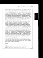
Figure 6- 12 Two methods of dissecting the flap in the subgaleal plane. Left, finger
dissection readily cleaves the areolar tissue in the subgaleal plane. Several centimeters
above the orbital rims, however, the pericranium is more tightly bound to the frontalis
muscle and the periosteum may strip from the bone when using this technique in this
location. Right, dissection with a scalpel. The flap is lifted gently with retractors and/or
hooks to maintain gentle tension. The back (dull) edge of the scalpel rests on the
pericranium and is swept back and forth, allowing the point of the scalpel to incise the
subgaleal tissue. This technique is especially useful in flaps elevated for a second or third
time, where adhesion in the subgaleal layer are more common and must be sharply incised.
78
The flap may be elevated atop the pericranium with finger dissection, with blunt periosteal
elevators, or by back-cutting with scalpel (Fig. 6-12). As dissection proceeds anteriorly tension
develops because the flap is still attached laterally over the temporalis muscles. Dissecting that
portion of the flap below the superior temporal line from the temporalis fascia relieves this tension
and allows the flap to retract farther anteriorly. Along the lateral aspect of the skull, the glistering
white temporalis fascia becomes visible where it blends with the pericranium at the superior
temporal line. The plane of dissection is just superficial to this thick fascial sheet.
Dissection of the flap continues anteriorly in the subgaleal fascial plane to a point 3 to 4 cm
superior to the supraorbital rims. A finger is used to palpate and locate the superior temporal lines,
and a horizontal incision is made through pericranium from one superior temporal line to the other
(Fig. 6-13). The surgeon should not extend the incision beyond the superior temporal line or the
temporalis muscle will be cut and begin to bleed. A subperiosteal dissection then continues to the
supraorbital rims.
Figure 6-13 Incision of periosteum across the forehead from one superior temporal line to
the other. The tension throu
g
h
p
eriosteum should be 3 to 4 cm su
p
erior to the orbital rims.
79
The lateral portion of the flaps is dissected inferiorly atop the temporalis fascia. Once the
lateral portion of the flap has been elevated to within 3 to 4 cm of the body of the zygoma and
zygomatic arch, these structures usually can be palpated through the covering fascia. Near the ear,
the flap is dissected inferiorly to the root of the zygomatic arch. The
superficial layer of temporalis
fascia
is incised at the root of the zygomatic arch, just in front of the ear, and continues anteriorly
and superiorly at a 45
o
angle, joining the cross-forehead incision previous made through
pericranium at the superior temporal line. Incision of the superficial layer of temporalis fascia
reveals fat and areolar tissue (Fig. 6-14). Further dissection inferiorly within this layer provides a
safe route of access to the zygomatic arch, because the temporal branch of the facial nerve is always
lateral to the superficial layer of temporalis fascia (Fig. 6-15). Metzenbaun scissors are used to
bluntly dissect just under the superficial layer of temporalis fascia, within the space containing the
superficial temporal fat pad (see Fig 6-15). Once the superior surface of the zygomatic arch and
posterior border of the body of the zygoma are palpable or visible, an incision through periosteum is
made along their superior surface. The incision progresses superiorly along the posterior border of
the body of the zygoma and orbital rim, ultimately meeting the cross-forehead horizontal incision
through pericranium. Subperiosteal elevation exposes the lateral surfaces of the zygomatic arch,
body of the zygoma, and a lateral orbital rim (Fig. 6-16). To allow the flap to fold anteriorly, it may
be necessary to continue the preauricular component inferiorly and to dissect the flap from the TMJ
capsule.
Figure 6 -14 Anatomic dissection showing incision through the superficial layer of temporalis fascia
(forceps) several centimeters above the zygomatic arch. Note the underlying fat between this layer of
fascia and the deep layer of temporalis fascia. The tempoparietal fascia with the temporal branch of the
facial nerve is folded inferiorly (below).
80
Figure 6-15 Incision made through the superficial layer of the temporalis fascia. Incision begins at the
root of the zygomatic arch (above the temporomandibular joint) upward and forward to join the incision
made across the forehead in periosteum. One method to approach the posterior portion of the lateral
orbital rim and superior surface of the zygomatic arch is also demonstrated. Dissection with incisors is
continued deep to the superficial layer of temporalis fascia (see inset), within the superficial temporal fat
pad, until bone is encountered. Sharp incision is then made through the periosteum on the superior surface
of the z
yg
omatic arch and the
p
osterior surface of the z
yg
oma.
81
Figure 6-16 Anatomic dissection showing the zygomatic arch (ZA) and body (ZB). The
superficial layer of the temporalis fascia and periosteum is retracted inferiorly and anteriorly.
Note the masseter muscle (MM) attachment to the inferior portion of the zygomatic arch.
82
Step 5. Subperiosteal Exposure of the Periorbital Areas
To allow functional access to the superior orbits and/or nasal region, it is necessary to release the
supraorbital neurovascular bundle from its notch or foramen. This maneuver involves dissecting in
the subperiosteal plane completely around the bundle, including inside the orbit. If no bone is noted
inferior to the bundle, the bundle can be gently removed from the bony bridge along the
supraorbital rim to release the bundle (Fig. 6-17).
Figure 6-17 Technique of removing bone inferior to the supraorbital foramen (when
present) so the neurovascular bundle can be released. Relaxing incisions in the sagital
plane through the elevated periosteum over the bridge of the nose are also shown. Use of
this technique greatly facilitates dissection more inferiorly along the nasal dorsum.
83
Further retraction of the flap inferiorly may be accomplished by subperiosteal dissection
into the orbits. The orbital contents attached to the lateral orbital tubercle are stripped, allowing
dissection deep into the lateral orbit. Release of the periosteum around the inferior rim of the orbit
allows exposure of the entire orbital floor and infraorbital region. Access to the infraorbital area is
easiest after overlying tissue of the zygomatic arch and body are released to relax the overlying
envelope.
Dissection of the periosteum from the superior and medial orbital walls releases the flap and
allows retraction down to the level of the junction of the nasal bones and upper lateral cartilages.
This technique is facilitated by carefully incising the periosteum of the nasofrontal region (see Fig.
6-17). Dissection can proceed along the dorsum to the nasal tip, if necessary (Fig. 6-18).
Figure 6-18 Dissection inferiorly to the top of the nose with a
p
eriosteal elevator.
84
The medial canthal tendons should not be inadvertently stripped from their attachments to
the posterior and anterior lacrimal crest. They are identified as dense fibrous attachments in the
nasolacrimal fossa (Fig. 6-19). The entire medial orbital wall may be exposed without stripping the
canthal tendons. As subperiosteal dissection proceeds posteriorly along the medial orbital wall, the
surgeon should be on the lookout for the anterior (and posterior) ethmoidal artery. A simple method
to identify and cauterize the artery is to strip the periosteum along the roof of the orbit and inferior
to where the artery pierces the medial orbital wall. With a periosteal elevator on each side of the
foramen, retraction allows the periosteum attached to the foramen to "tent" outward (Fig. 6-20).
Bipolar cauterization of the artery may be performed, followed by transection. Dissection can then
proceed posteriorly by subperiosteal elevation.
Figure 6-19 Anatomic dissection showing the posterior limb of the medial canthal tendon
(
MCT
)
of the ri
g
ht orbit.
85
Figure 6-20 Dissection of the medial orbital wall. Periosteal elevators are placed above an
d
b
elow the anterior ethmoidal neurovascular bundle, allowing bipolar cauterization an
d
sectioning.
86
After the dissections just described, the upper and middle facial regions are completely
exposed (Fig. 6-21). The entire orbit can be dissected from the orbital rims to the apex; the only
remaining structure is the medial canthal tendon, unless it was intentionally or inadvertently
stripped.
Figure 6-21 Amount of exposure obtained with complete dissection of the upper and middle facial bones
using the coronal approach. Note maintenance of attachment of the medial canthal tendon. The
infraorbital areas are also exposed if retraction is performed from the side of the orbit.
87
Step 6. Exposure of the Temporal Fossa
Access into the temporal fossa is possible by stripping the anterior edge of the temporalis muscle
from the temporal surfaces of the zygomatic, temporal, and frontal bones. The entire temporalis
muscle can be stripped subperiosteally from the temporal fossa if necessary, but care is needed to
preserve the blood supply to the temporalis muscle.
Step 7. Exposure of the Temporomandibular Joint and/or Mandibular Condyle/Ramus
Access to the TMJ region may be accomplished by dissection below the zygomatic arch, as
described in Chapter 12. Exposure of the lateral surface of the mandibular subcondylar region and
ramus may commence lateral to the capsule of TMJ. An incision through the periosteum just
inferior to the insertion of the TMJ capsule at the condylar neck will expose the neck of the
condyle.
Wider access below the zygomatic arch can be enhanced with two maneuvers. In the first
approach, the masseter muscle is cut or released from its origin along the zygomatic arch and body,
and then stripped from the lateral surface of the mandibular ramus to expose the ramus of the
mandible (Fig. 6-22). The temporalis muscle at the depth of this dissection may be noted at it inserts
into the coronoid process. Another approach is to perform an osteotomy of the zygomatic arch,
leaving it pedicled to the masseter muscle, and to dissect between the masseter and temporalis
muscles, stripping the masseter from the lateral surface of the mandibular ramus. One anatomic
consideration is valid with either of these wide exposure methods. The vascular and neural supply
to the masseter muscle courses from the medial side of the mandible through the sigmoid notch into
the masseter muscle. Therefore, stripping the masseter from above may severely affect its function.
Figure 6-22 Anatomic dissection showing exposure of the superior portion of the mandibular ramus
through the coronal approach. In this dissection, the masseter muscle (MM) was stripped from its origi
n
along the undersurface of the zygomatic arch (ZA). The facial nerve is retracted inferiorly an
d
anteriorly. Note the temporomandibular joint (TMJ) capsule, which has not been entered. The
temporalis muscle (TM) is still attached to the coronoid process (CP) and the medial surface of the
mandible.
88
Step 8. Harvesting Cranial Bone Grafts
One of the many advantages of the coronal approach is that cranial bone graft harvesting is
facilitated. An incision through the periosteum allows exposure for harvesting a bone graft (Fig. 6-
23). Closure of the periosteum proceeds scalp closure. Alternatively, subperiosteal dissection
posteriorly from the point of the original coronal incision also exposes the cranium for harvesting
bone grafts.
Figure 6-23 Bone graft harvest using the coronal approach.
89
Step 9. Closure
Closed suction drainage may be employed using a flat drain exiting the hair bearing region of the
scalp posterior to the incision. Proper closure of the detached tissues is critical to produce optimal
esthetic results. After wide exposure of the malar and infraorbital regions, suture resuspension of
the soft tissue is necessary. Slowly resorbing 3-0 sutures are passed through the deep surface of the
periosteum of the malar region and then suspended to the temporalis fascia or another stable
structure. One or two well-placed sutures are effective to prevent "drooping" of the soft tissues. A
lateral canthopexy is also necessary if the attachments to the lateral orbital tubercle were stripped
from bone. Toothed forceps are used to identify the superficial portion of the lateral canthal tendon
within the deep surface of the coronal flap. One slowly resorbing or permanent 3-0 suture is placed
through the lateral canthus from the deep surface of the coronal flap. Location of the proper vertical
position of the canthal tendon can be determined by drawing the suture upward or downward while
observing the configuration of the palpebral fissure. Ideally, lateral canthopexy of the deep portion
of the lateral canthal tendon is performed by drilling a large hole through the lateral orbital rim just
below the frontozygomatic suture. The suture and tendon are pulled into this hole. In many
instances, however, canthopexy may be accomplished by passing the suture through the anterior
portion of the lateral canthal tendon, around the front of the lateral orbital rim, and securing it to a
bone screw, a hole in the bone, or the temporalis fascia.
Whenever the temporalis muscle is stripped from the temporal surface of the orbit, it should
also be suspended to prevent a hollow appearance in the temporal region. An easy method involves
drilling holes through the posterior edge of the orbital rim and suturing the anterior edge of the
temporalis muscle with slowly resorbing 3-0 sutures.
Closure of the periosteum around the lateral orbital rim is performed with 4-0 resorbable
sutures. Ideally, the periosteum over the zygomatic arch should be closed, but this effort can be
difficult owing to the small amount of periosteum available. Suturing the periosteum may also
injure the temporal branch of the facial nerve, which is just superficial to the periosteum. Instead,
"oversuspension" of the superficial layer of the temporalis fascia is performed. The inferior edge of
the superficial layer of the temporalis fascia, which was incised during the approach, is sutured
approximately 1 cm superior to the superior edge of the incised fascia (Fig. 6-24). Running
horizontal 3-0, slowly resorbing sutures are used for this purpose. Thus, the tissue lateral to the
zygomatic arch are suspended tightly in a location that is more superior than it would have been had
the incised superficial temporalis fascia simply been sutured.
It is not necessary to close the horizontal periosteal incision across the forehead. The
periosteum in this area is thin and does not hold sutures. Closure of the coronal incision will bring
the periosteal tissue into acceptable approximation.
The scalp incision is closed in two layers using 2-0 slowly resorbing sutures through the
galea/subcutaneous tissues and 2-0 resorbable or permanent skin sutures (smaller sutures are used in
children), or staples. As noted previously, use of a suction drain (usually 7 mm flat) is optional. The
skin sutures/staples are removed in 7 to 10 days.
The preauricular component of the coronal approach should be closed in layers as for any
other preauricular approach.
Pressure dressing are optional, but if used, they should not be tight. Periorbital edema
increases greatly with tight pressure dressings on the scalp after coronal approaches.
90
Figure 6-24 Suturing the superficial layer of the temporalis fascia. Note that the inferior edge of fascia is
sutured is a more su
p
erior location that the cut su
p
erior ed
g
e.
91
ALTERNATIVE INCISIONS
The coronal incision has been modified repeatedly by surgeons. The principal difference in these
surgical techniques involves the position of the skin incision. A major modification has been
placement of the incision behind the ear (Fig. 6-25) (5,6). The advantage of this positioning is
further camouflage of the scar. Any inferior extension of the coronal incision can be hidden within
the postauricular fold or along the hairline.
Figure 6-25 Postauricular placement of the coronal incision. The incision can be extended into the
p
ostauricular sulcus or within the hairline.