General Principles for Approaches to the Facial Skeleton - part 6 pot
Bạn đang xem bản rút gọn của tài liệu. Xem và tải ngay bản đầy đủ của tài liệu tại đây (686.63 KB, 18 trang )
92
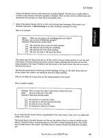
Even with well-placed incisions, the scar that forms may produce a separation of the hair
that can become visible when the hair is wet, such as during swimming. A modification of the
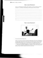
incision has been the use of a zig-zag incision instead of a straight incision within the hairline (Fig.
6-26) (7). The zig-zag incision helps break up the scar and make it less noticeable, even when the
hair is worn short. The major disadvantage of this incision is the increased time needed for closure.
Fi
g
ure 6-26 Zi
g
-za
g
incision to make the scar less obvious.
93
S E C T I O N
IV
TRANSORAL
APPROACHES TO THE
FACIAL SKELETON
The midfacial and mandibular skeleton can be readily exposed through incisions placed
inside the oral cavity. The approaches are rapid and safe and the exposure is excellent. The
greatest advantage of such approaches is the hidden scar. This section includes descriptions
of maxillary and mandibular vestibular approaches to the facial skeleton.
97
7
Maxillary Vestibular
Approach
The Maxillary vestibular approach is one of the most useful when performing any of a wide variety
of procedures in the midface. It allows relatively safe access to the entire facial surface of the
midfacial skeleton, from the zygomatic arch to the infraorbital rim to the frontal process of the
maxilla. The greatest advantage of the approach is the hidden intraoral scar that result. The
approach is also relatively rapid and simple, and complications are few. Damage to the branches of
the facial nerve is nonexistent as long as one stays within the subperiosteal plane, and damage to the
infraorbital nerve is unusual with proper technique.
SURGICAL ANATOMY
Infraorbital Nerve
The only neurovascular structure of any significance that must be negotiated with procedures in the
midfacial region is the infraorbital neurovascular bundle. The infraorbital nerve is the largest
cutaneous branch of the maxillary division of the trigeminal nerve. The artery and vein that
accompany the infraorbital nerve are surgically insignificant. The nerve exits the infraorbital
foramen, 7 to 10 mm inferior to the infraorbital rim just medial to the zygomaticomaxillary suture,
or approximately at the medial and middle thirds of the orbit. The infraorbital nerve divides after
exiting the infraorbital foramen into terminal branches that spread fanwise from into the lower
eyelid; the nasal branches supply the skin on the lateral surface of the lower half of the nose. Three
of the four superior labial branches enter the lip between its muscles and the mucous membrane.
These nerves supply not only the mucous membrane of the upper lip, but also its skin, which they
reach by perforating the orbicularis oris muscle. Damage to this nerve results in loss of sensation to
these areas, and possibly dysesthesia.
Nasolabial Musculature
The attachments of facial muscles of the nasolabial region may be disrupted during the maxillary
vestibular approach. Therefore, these muscles should be properly repositioned during closure to
prevent disturbing esthetic changes. The important muscles are the nasalis group, the levator labii
superioris alaeque nasi, the levator labii superioris, The levator anguli oris, and the orbicularis oris
(Fig. 7-1).
98
The nasali group has transverse nasal and alar parts. It originates along the midline of the
nasal dorsum and spreads laterally over the external aspect of the upper lateral cartilages where it
intermingles with fibers of the levator labii superioris alaeque nasi and the levator labii superioris.
Part of the transverse nasali inserts into the skin at the nasolabial groove, where it intermingles with
fibers from the levator labii superioris alaeque nasi and oblique fibers of the orbicularis oris,
forming a lateral nasal modiolus. Another portion of the transverse nasalis inserts onto the incisal
crest and anterior nasal spine and is deeply in contact with the depressor septi muscle. The alar
portion is ultimately reflected inward, forming the anterior floor of the nose.
Several muscle groups elevate the upper lip. The levator labii superioris alaeque nasi arises
from the frontal process of the maxilla alongside the nose and passes obliquely in two segments.
One segment inserts onto the lateral crus of the alar cartilage and skin of the nasalis muscle,
depressor septi, and oblique bands of the orbicularis oris. The levator labii superioris arises from the
infraorbital margin of the maxilla beneath the orbicularis oculi. It extends downward and medially,
superficial to and intermingling with the orbicularis oris, beneath the skin of the ipsilateral lower
philtral columns and the upper lip. The levator anguli oris muscle lies deep to the levator labii
superioris and the zygomaticus muscle. It arises from the canine fossa of the maxilla and courses
downward and medially to the commissure, where it intermingles with the fibers of the orbicularis
oris muscle.
The orbicularis oris muscle consists of three distinct strata. Horizontal fibers extend from
one commissure to the other, passing beneath the philtrum. Oblique bands extend from the
commissures to the anteroinferior aspect of the nasal septal cartilage, anterior nasal spine, and floor
of the nose. The incisal bands extend from the commissures deeply to insert onto the incisive fossa
of the maxilla. All of these muscles and their investing fascia jointly contribute significantly to the
position and configuration of the lateral nasal and labial regions.
Figure 7- 1 Important facial musculature when performing the maxillary vestibular
h
99
The maxillary vestibular incision and the subperiosteal dissection attendant to this approach
cut some of the muscular origins and strip the origins and insertions of most muscles from the bone
(see Fig. 7-1), causing superolateral retraction of the tissue by the action of the zygomaticus
muscles and the natural tendency for muscles to reattach in a shortened position. Lateral
displacement of the nasal modiolus causes widening of the alar base with flaring of the alae from
unopposed action of the dilator naris. This displacement causes deepening of the alar groove and
splaying of the alar bases, nostrils, and nasal tip (Fig. 7-2). Loss of soft tissue fullness in the
nasolabial region results in changes similar to those seen in the aging face: thinning and retraction
of the upper lip, reduced vermilion exposure, and a more obtuse nasolabial angle. Downturning of
the corners of the mouth may occur when the levators of the upper lip are detached from their
origin, because the depressor of the mouth are then unopposed.
Figure 7- 2 Effects of the maxillary vestibular approach if simple closure is performed :
the nasal ti
p
loses
p
ro
j
ection
,
the alar bases widen
,
and the u
pp
er li
p
rolls inward.
100
Buccal Fat Pad
The buccal fat pad consists of a main body and four extensions: bucal, pterigoid,
pterygomandibular, and temporal. The body is centrally positioned. The buccal extension lies
superficially within the cheek, and the pterigoid, pterygomandibular, and temporal extension are
situated more deeply.
The main body of the fat pad is located above the parotid duct and extends along the upper
portion of the anterior border of the masseter. It then courses medially to rest on the periosteum of
the posterior maxilla (Fig. 7-3). In this region, the body of the fat pad overlies the uppermost fibers
of the buccinator muscle and travels forward along the vestibule overlying the maxillary second
molar. Posteriorly, it wraps around the maxilla and travels through the pterygomaxillary fissure,
where it is in intimate contact with branches of the internal maxillary artery and maxillary division
of the trigeminal nerve.
Figure 7- 3 Axial
section through the
maxilla at the level of
the tooth root apices
showing the
relationship of the
buccal fat pad (BFP)
to the lateral maxilla.
Note that the fat pad
extends anteriorly to
approximately the first
molar. Also, posterior
to the origin of the
buccinator muscle on
the maxilla, the buccal
fat pad is just lateral to
the periosteum.
101
TECHNIQUE
The facial surface of the midface can be exposed using the maxillary vestibular approach. The
length of incision and amount of subperiosteal dissection depend on the area of interest and the
extent of the surgical intervention. If the area of interest involves only one half of the midface, for
instance with a unilateral zygomaticomaxillary fracture, the incision can be made on one side only,
leaving the other side intact.
Step 1. Injection of a Vasoconstrictor
The oral mucosa, submucosa, and facial muscles are lushly vascularized. Submucosal injection of a
vasoconstrictor can dramatically reduce the amount of hemorrhage during incision and dissection.
Step 2. Incision
The incision is usually placed approximately 3 to 5 mm superior to the mucogingival junction.
Leaving unattached mucosa on the alveolus facilitates closure. This tissue has many elastic fibers
and contracts following incision, although during closure, the tissue can be grasped and holds
sutures well. The surgeon should not make the incision more superior in the anterior region because
entrance into the piriform aperture, with puncture of the nasal mucosa, may result. Some individuals
have extremely low piriform apertures, which makes this possibility a reality. Palpation of the
inferior extent of the piriform aperture and/or anterior nasal spine ensures incision placement
inferior to these structures. In the edentulous maxilla, where atrophy of the alveolar bone brings the
alveolar crest and floor of the nose in close apposition, incision along the alveolar crest is an
excellent choice.
The incision extends as far posteriorly as necessary to provide exposure, usually of the first
molar tooth, and traverses mucosa, submucosa, facial muscles, and periosteum (Fig. 7-4). The
mucosa retracts during incision, exposing underlying tissues.
Figure 7- 4 Incision through the mucosa, submucosa, facial musculature, and periosteum.
102
Step 3. Subperiosteal Dissection of Anterior Maxilla and Zygoma
Periosteal elevators are used to elevate the tissues in the subperiosteal plane (Fig. 7-5). Dissection
should be orderly, first elevating tissues superiorly, then along the piriform aperture, then
posteriorly behind the zygomaticomaxillary buttress. While the tissues are elevated superiorly in the
subperiosteal plane, small perforating vessels are encountered and are easily distinguishable from
the infraorbital neurovascular bundle. The bundle is encountered and the periosteum is dissected
completely around the foramen. Dissection proceeds superiorly to the infraorbital rim.
Subperiosteal dissection along the piriform aperture strips the attachments of the nasolabial
musculature, allowing upward and lateral retraction of the muscles.
Subperiosteal dissection proceeds posteriorly to the pterygomaxillary fissure. Perforation of
the periosteum at or behind the zygomaticomaxillary buttress produces herniation of the buccal fat
pad into the surgical field, a nuisance during surgery. A helpful suggestion is to keep the tip of the
periosteal elevator always in an intimate contact with bone when proceeding posteriorly around the
zygomaticomaxillary buttress. The only anatomic hazards are the infraorbital neurovascular bundle
above and posterior superior alveolar vessels along the posterior maxilla, which infrequently cause
bleeding.
Fi
g
ure 7- 5 Sub
p
eriosteal dissection of the anterior maxilla.
103
The entire anterior face of the zygoma can be easily exposed, but reaching the zygomatic
arch necessitates detachment of some of the masseter muscle attachments. Sharp dissection is
needed to free these tenacious fibers. Dissection below the piriform aperture up the anterior nasal
spine should be performed carefully to maintain the integrity of the nasal mucosa. When violated, it
bleeds profusely.
Step 4. Submucosal Dissection of Nasal Cavity
If it is necessary to strip the nasal mucosa from the lateral wall, floor, or septum of the nose, this
maneuver is done carefully with periosteal or Freer elevators. A forked right-angle retractor is
placed over the anterior nasal spine and subperiosteal dissection superiorly allows the retractor to
retract the septum and nasal mucosa above the level of the anterior nasal spine. A scalpel is used to
make a horizontal incision on top of the anterior nasal spine, freeing the cartilaginous septum from
the top of the spine and thus the attachment of the nasal mucosa from the anterior nasal spine. The
rim of the piriform aperture is thin and sharp, and the nasal mucosa is adherent. Periosteal elevators
are used to strip the mucosa from the entire circumference of the piriform rim.
Figure 7- 6 Submucosal dissection of the nasal cavity. Note the tip of the periosteal elevators inside
the
p
iriform a
p
erture.
104
Dissection into the nasal cavity is easiest to perform along the lateral wall and floor. The
anteroinferior margin of the piriform rim is usually located above the nasal floor. Thus, after freeing
the nasal mucosa from the piriform rim, the elevators should be inserted inferiorly before advancing
posteriorly (Fig. 7-6). Dissection of the lateral wall of the nose is performed by gently inserting a
periosteal elevator between the nasal mucosa and the lateral wall of the nasal cavity. It is not
advanced deeply until the entire circumference of the lower one half of the piriform has been
dissected. The previously taut nasal mucosa can then relax somewhat so that the elevator can be
advanced more deeply along the lateral wall. The elevator is advanced in a sweeping motion to free
the entire lateral wall and floor of its mucosa to the level of the inferior turbinate. The posterior
edge of the nasal floor is approximately 45 mm posterior to the piriform aperture and can be felt
when the elevator steps off the posterior edge.
Once the lateral wall and floor of the nose are stripped of mucosa, the elevator is placed at
the junction of the floor of the nose and the nasal septum. A tenacious attachment of the mucosa to
the septal crest of the maxilla must be carefully elevated to prevent perforation. A simple maneuver
for stripping the septal mucosa from this approach is to place a Freer elevator along the junction of
the septum and the floor of the nose and twist it so that the edge against the septum is twisted
superiorly, freeing the mucosa on the septum.
Step 5. Closure
Restitution of the nasolabial muscles is performed as three uniform steps during closure of the
maxillary vestibular incision. The first step involves identification and resetting of the alar bases,
the second involves eversion of the tubercle and vermilion, and the last involves closure of the
mucosa.
To help control the width of the alar base, an alar cinch suture is placed before suturing the
lip. Suture placement is accomplished in one of two ways. With one technique, small toothed
forceps placed through the vestibular incision grasp the insertion of the transverse nasalis muscle.
Pulling the forceps medially allows one to see the change that occurs in the alar base. A slowly
resorbing suture is passed through this tissue, taking care to engage adequate tissue to resist the pull
of the suture, but not so much that a subcutaneous dimple occurs when the suture is pulled medially.
The suture is then passed through the opposite side and temporarily tightened to examine the effect
of the medial pull of the alar bases on the nose (Fig. 7-7). Another method is to evert the tissue into
the incision area by pressing the thumb or finger into the alar facial groove (Fig. 7-8). A suture can
then be passed through the incision, into the tissues, the depth of placement being guided by
palpation of the thumb or finger. Whichever method is used to pass the suture through the nasalis
muscle, the appearance must be symmetric and the desired curvature and definition of the alar base
should be achieved after provisional tying. Tying the suture is delayed until a second suture is
passed. The second suture is placed at a higher level or more laterally on the alar base, depending
on the desired rotation of the ala. Generally, two sutures are adequate.
105
Figure 7- 7 Effect of the alar cinch technique of the width of the alar base. Note the difference after tying
the suture.
106
Figure 7- 8 Tip of the finger (or thumb) everts the lip and nasal base while suture is passed.
107
A V-Y advancement closure of the maxillary vestibular incision is recommended where the
incision has been placed across the base of the nose and subperiosteal dissection of the tissues along
the piriform aperture has occurred. In closing the maxillary vestibular incision, a skin hook engages
the labial mucosa incision in the midline and pills it away from the maxilla (Fig. 7-9). Three or four
interrupted resorbable sutures are used to gather lip tissue in the midline. The mucosa and labia
musculature are engaged by the needle on either side of the incision and then sutured. In most cases,
1 cm of tissue is closed in this manner, creating a pout in the midline of the lip. When this step is
performed properly, the lip bulges anteriorly in the midline and the exposed vermillion is full.
Within 7 to 10 days, this fullness gradually settles and a more normal appearance returns.
Figure 7- 9 V-Y closure of a lip incision. A skin hook is placed in the midline and tissue is gathered for
a
pp
roximatel
y
1 cm with suture.
108
After closing the vertical limb of the V-Y advancement, a single suture is placed across the
incision in the midline to ensure symmetric closure of the horizontal posterior incisions. When
closing the horizontal incision, one should begin in the posterior and work anteriorly with a running
resorbable suture (3-0 chromic catgut) through mucosa, submucosa, musculature, and periosteum.
The superior aspect of the incision is gradually advanced toward the midline by passing the needle
anteriorly in the lower margin of the incision as compared to the upper margin. This maneuver, in
addition to the V-Y closure, helps lengthen the relaxed musculature so that it reattaches in its proper
position (Fig. 7-10). From the canine-to-canine area, the suture is passed close to the edges of the
incision to prevent gathering of the mucosa, which rolls the lip inward and reduces the amount of
exposed vermilion.
Figure 7- 10 The remainder of the incision is closed so that the superior edge is pulled anteriorly.
8
Mandibular Vestibular
Approach
he mandibular vestibular approach if useful for a wide variety of procedures. It allows
relatively safe access to the entire facial surface of the mandibular skeleton, from the condyle
to the symphysis. An advantage of this approach is the ability of constantly access the dental
occlusion during surgery. The greatest benefit to the patient is the hidden intraoral scar. The
approach also relatively rapid and simple, although access is limited in some regions, such as the
lower border of the mandible at the angle and parts of the ramus. Complications are few but include
mental nerve damage and lip malposition, both of which are minimized with proper technique.
SURGICAL ANATOMY
Mental Nerve
The only neurovascular structure of any significance that must be negotiated with procedures in the
mandibular body/symphysis region is the mental neurovascular bundle. The artery and vein that
accompany the mental nerve are insignificant from a surgical standpoint. The mental nerve is a
terminal branch of the inferior alveolar nerve (mandibular nerve), and provides sensory innervation
to the skin and mucosa of the lower lip, the skin in the region of the chin, and the facial gingiva of
the anterior teeth.
The mental nerve exists the mental foramen, located midway between the alveolar and basal
borders of the mandible and usually below or slightly anterior to the second bicuspid tooth (Fig. 8-
1). The mental nerve divides under the depressor anguli oris muscle into three main branches; one
T
FIGURE 8- 1 Anatomic
dissection of mental
nerve branches.
110
descends to the skin of the chin and the other two ascend to the skin and mucous membrane of the
lower lip and gingiva. The branching pattern is variable, however, and several finer branches may
be noted. As the branches enter the lower lip, they become superficial and can usually be seen just
beneath the mucosa of the lower lip when it is everted.
Facial Vessels
The facial artery and vein are usually not encountered during the mandibular vestibular approach
unless dissection through the periosteum occurs in the region of the mandibular anterogonial notch.
The facial artery arises from the external carotid artery in the carotid triangle of the neck. At
or close to its origin, it is crossed by the posterior belly of the digastric muscle, the stylohyoid
muscles, and the hypoglossal nerve. In the submandibular triangle, the facial artery ascends deep to
the submandibular gland, grooving its deep and superior aspect, and passes superficially to reach
the inferior border of the mandible. As it crosses the mandible at the anterior border of the masseter
muscle, the artery is covered on its superficial surface by skin and platysma muscle, and its
pulsation can be felt at this location.
The facial vein is the drainage vein of the angular and ultimately the labial vessels. It is
usually located more posterior and superficial to the artery. Of surgical significance, however, is the
fact that the facial artery and vein are close to the mandible in the region of the inferior border. The
only structure separating them from the bone is the periosteum (Fig. 8-2).
Mentalis Muscle
FIGURE 8- 2 Anatomic dissection of the mandibular body showing relationship of facial vessels to
b
one. The onl
y
tissue between them is the
p
eriosteum.
111
The only muscle of facial expression that is important from a surgical standpoint when using the
mandibular vestibular approach is the mentalis muscle. All of the other muscles of facial expression
are stripped from the mandible by subperiosteal dissection and are readily reattached with soft
tissue closure. The mentalis muscle is unique, however, in that it is the only elevator of the lower lip
and chin. If this muscle is not properly repositioned during closure, the chin will “droop” and the
lower lip will take on a lifeless, sagging appearance, exposing more lower teeth.
The mentalis muscle are paired, small, conical muscles arising from the mandible,
beginning at the midroot level of the lower incisor teeth and continuing inferiorly to a point below
the apices. They are separated from one another by a firm septum and adipose tissue.
At the inferior portion of its origin, the mentalis muscle attaches lateral to the pogonial
trigone. The fibers of this muscle pass from their origin inferiorly, inserting into the skin of the chin
at the soft tissue chin prominence (Fig. 8-3). The most superior fibers are the shortest and pass
almost horizontally into the skin of the upper chin. The lower fibers are the longest and pass
obliquely or vertically to the skin at the lower part of the chin. The origin of the mentalis muscle
determines the depth of the labial sulcus in the anterior portion of the mouth. The mentalis muscle is
innervated by the marginal mandibular branch of the facial nerve.
FIGURE 8- 3 Anatomic dissection showing cross section of soft tissues of the chin. Note the direction
of the mentalis muscle fibers.