General Principles for Approaches to the Facial Skeleton - part 10 pps
Bạn đang xem bản rút gọn của tài liệu. Xem và tải ngay bản đầy đủ của tài liệu tại đây (1.17 MB, 18 trang )
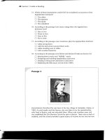
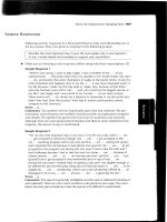
Figure 12 5 Coronal section of the temporomandibular joint (TMJ) region. SMAS = superficial
musculoaponeurotic system; TF = temporalis fascia (note that it splits inferior to this point into superficial
and deep layers); TPF = temporoparietal fascia; VII = temporal branch of the facial nerve.
TECHNIQUE
Several approaches to the TMJ have been proposed and are used clinically. The standard and
most basic is the Preauricular approach. Other approaches differ in term of placement of the
skin incision as well as access to the joint. The dissection down to the TMJ, however, is similar
in all approaches. In this discussion, the standard Preauricular approach is described first. Later,
variants are briefly presented.
Step 1. Preparation of the Surgical Site
Preparation and draping should expose the entire ear and lateral canthus of the eye. Shaving the
Preauricular hair is optional. A sterile plastic drape can be used to keep the hair out of the
surgical field. Cotton soaked in mineral oil or antibiotic ointment may be placed into the
external auditory canal.
168
Step 2. Marking the Incision
The incision is outlined at the junction of the facial skin with helix of the ear. A natural skin
fold along the entire length of the junction of the incision can be used. If none is present,
posterior digital pressure on the Preauricular skin usually creates a skin fold that can be marked.
The incision extends superiorly to the top of the helix, and may include an anterior (hockey-
stick) extension.
Step 3. Infiltration of Vasoconstrictor
The Preauricular area is quite vascular. A vasoconstrictor can be injected subcutaneously in the
area of the incision to decrease incisional bleeding. If a local anesthetic is also being injected,
however, it should not be injected deeply because it may be necessary to use a nerve stimulator
on exposed facial nerve branches.
Step 4. Skin Incision
The incision is made through skin and subcutaneous connective tissues (including
temporoparietal fascia) to the depth of the temporalis fascia (superficial layer) (Fig. 12-6). Any
bleeding skin vessels are cauterized before deeper dissection proceeds.
Step 5. Dissection to the TMJ Capsule
Blunt dissection with periosteal elevators undermines the superior portion of the incision (that
above the zygomatic arch) so that a flap can be retracted anteriorly for approximately 1 to 1,5
cm (Fig. 12-7). This flap is dissected anteriorly at the level of the superficial (outer) layer of
temporalis fascia. This layer is usually hypovascular. The superficial temporal vessels and
auriculotemporal nerve may be retracted anteriorly in the flap. Failure to develop the flap close
to the cartilaginous external auditory canal increases the risk of damage to these structures.
Below the zygomatic arch, dissection proceeds bluntly adjacent to the external auditory
cartilage. Scissor dissection proceeds along the external auditory cartilage in an avascular plane
between it and the glenoid lobe of the parotid gland (see Fig. 12-7). The external auditory
cartilage runs anteromedially and the dissection is parallel to the cartilage. The depth of the
dissection at this point should be similar to that above the zygomatic arch.
169
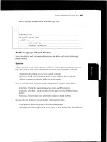
Figure 12 6 Initial incision made in the preauricular skin fold.
170
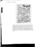
Figure 12 7 Dissection above the zygomatic arch to the level of the superficial layer of the temporalis
fascia. Dissection below the zygomatic arch along the external auditory meatus to the same depth.
171
Attention again turns to the portion of the incision above the zygomatic arch. With the
flap retracted anteriorly, an incision is made through the superficial (outer) layer of temporalis
fascia beginning from the root of the zygomatic arch just in front of the tragus anteroposteriorly
toward the upper corner of the retracted flap (Fig. 12-8). The fat globules contained between the
superficial and deep layers of temporalis fascia are then exposed. At the root of the zygoma, the
Figure 12 8 Oblique incision through the superficial layer of the temporalis fascia. Fat is visible deep to
the fascia.
172
incision can be through both the superficial layer of temporalis fascia and periosteum of the
zygomatic arch. The sharp end of a periosteal elevator is inserted in the fascial incision, deep to
the superficial layer of temporalis fascia, and swept back and forth to dissect this tissue from the
underlying areolar and adipose tissues (Fig. 12-9). The undermining proceeds inferiorly toward
Figure 12 9 A periosteal elevator inserted beneath the superficial layer of the temporalis muscle is used
to strip periosteum off the lateral portion of the zygomatic arch, and continues the dissection below the
arch just superficial to the capsule of the temporomandibular joint
173
the zygomatic arch, where the sharp end of the periosteal elevator cleaves the attachment of the
periosteum at the junction of the lateral and superior surfaces of the zygomatic arch, freeing the
periosteum from its lateral surface. The periosteal elevator can then be used to continue bluntly
dissecting inferiorly with the black-and-forth motion, taking care not to dissect medially into the
TMJ capsule (Fig. 12-10). Blunt dissection with scissors can also be used to dissect inferiorly to
the zygomatic arch. Once the dissection is approximately 1 cm below the arch, the intervening
tissue is sharply released posteriorly along the plane of the initial incision (Fig. 12-11).
The entire flap is then retracted anteriorly, and blunt dissection at this depth proceeds
anteriorly until the articular eminence is exposed. The entire TMJ capsule should then be
revealed. Because of subperiosteal dissection along the lateral surface of the zygomatic arch, the
temporal branches of the facial nerve are located within the substance of the retracted flap (see
Fig. 12-10). To help determine the location of the articular space, the mandible can be
manipulated open and closed.
Figure 12 10 Coronal section showing the layer of dissection.
VII = relative position at temporal branch during dissection.
174
Figure 12 11 Vertical incision made through intervening tissues just in front of the external auditory
meatus to the depth of the periosteal elevator.
175
Figure 12 12 After retraction of tissues superficial to the temporomandibular joint (TMJ) capsule,
scissors are used to enter the capsule. Initial point of entry is just below the zygomatic arch, continuing
parallel to the contour of the TMJ fossa.
176
Step 6. Exposing the Interarticular Spaces
With retraction of the developed flap, the joint spaces can be entered. With the condyle
distracted inferiorly, pointed scissors enter the upper joint space anteriorly along the posterior
slope of the eminence (Fig. 12-12). The opening is extended anteroposteriorly by cutting along
the lateral aspect of the eminence and fossa. The incision is continued inferiorly along the
posterior portion of the capsule until the capsule blends with the posterior attachment of the
disk. Lateral retraction of the capsule allows entrance into the superior joint space.
The inferior joint space is opened by making an incision in the disk along its lateral
attachment to the condyle within the lateral recess of the upper joint space (Fig. 12-13). The
incision may be extended posteriorly into the attachment tissues. The inferior joint space is then
entered.
Step 7. Closure
The joint spaces are irrigated thoroughly and any hemorrhage is controlled before closure. The
inferior joint space is closed with permanent or slowly resorbing suture by suturing the disk
back to its lateral condylar attachment (Fig. 12-14). The superior joint space is closed by
suturing the incised edge with the remaining capsular attachments on the temporal component
of the TMJ (Fig. 12-15). If no such attachments were left attached to bone, the capsule can be
resuspended over the zygomatic arch to the temporalis fascia.
177
Figure 12 13 Incision through the lateral attachment of the temporomandibular joint disk, entering the
inferior joint space.
178
Figure 12 14 Closure of the inferior joint space using running suture between lateral disk attachments
and the joint capsule.
179
Figure 12 15 Closure of the superior joint space using running suture between remnants of the
temporomandibular joint (TMJ) capsule on the zygomatic arch and the TMJ capsule below.
180
Subcutaneous tissues are closed with resorbable sutures. No sutures deeper than
subcutaneous tissues are required. The skin is then closed. A running subcuticular suture makes
removal simple and allows a delay in removal if necessary (Fig. 12-16). A pressure dressing is
usually applied, taking care to bolster posterior to the ear.
Figure 12 16 Closure of the preauricular skin incision with running subcuticular suture.
181
ALTERNATE APPROACHES
Other approaches to the TMJ have been described and used clinically. The extended temporal
and coronal incision can proceed inferiorly in the same fashion as for a Preauricular incision to
expose the TMJ. The “extended” preauricular approach incision is similar to the preauricular
approach, but an anterosuperior extension(hockey-stick) is made in the hair-bearing temporal
skin (Fig. 12-17). Some surgeons choose to bring the preauricular incision behind the tragus
(endaural incision) to hide a portion of it (Fig. 12-18). This choice may be especially useful in
individuals, often young patients, who do not have a well-demarcated preauricular skin fold. A
retroauricular skin incision further hides the incision and helps to protect the auriculotemporal
nerve. This approach requires an arc-shaped incision behind the ear (Fig. 12-19). The external
auditory canal must be transected at a wide portion to prevent stenosis, and the ear is reflected
anteriorly to gain access to the joint. The same deeper dissection is effective for all of the
approaches just described.
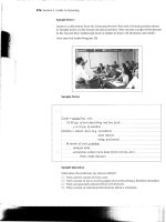
Figure 12 17. Preauricular incision with an oblique anterosuperior extension ("hockey stick").
182
Figure 12 18 A and B. Preauricular incision with a retrotragal portion, hiding scar within
the scar.
183
184
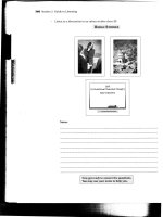
Figure 12 19 Retroauricular approach to the temporomandibular joint (TMJ). A, initial
curvilinear incision in the retroauricular crease. B, Transection of the external auditory
meatus. C, Retraction of the external ear anteriorly, exposing the TMJ capsule.
REFERENCE
1. Al-Kayat A, Bramley P; A modified pre-auricular approach to the temporomandibular joint
and malar arch, Br J Oral Maxillofac Surg 17:91,1979.
185