Fundamentals of Clinical Ophthalmology - part 6 pps
Bạn đang xem bản rút gọn của tài liệu. Xem và tải ngay bản đầy đủ của tài liệu tại đây (340.63 KB, 20 trang )
shown that patients may require larger
volume. More recently a conoid shape with a
flat front surface has been suggested.
In 1885 Mules first suggested the insertion
of a glass ball into the scleral cup.
Subsequently inert materials such as silicone
and methyl methacrylate have been
developed. More recently, a natural
component of coral reefs known as porous
hydroxyapatite has proved to be an ideal
implant material. This allows fibro-vascular
ingrowth, the implant becoming fully
integrated rather than forming a sequestrated
foreign body, as was the case with inert ball
implants. Synthetic and cheaper forms of
hydroxyapatite and other integrateable
materials such as Medpor (porous
polyethylene) are also now available.
Orbital implants are generally wrapped to
allow ease of placement and to allow
attachment of the extra-ocular muscles. Inert
implants are best inserted posterior to Tenon’s
capsule whilst hydroxyapatite should be
inserted within Tenon’s capsule.
The aim of orbital implantation is to
increase orbital volume and promote
prosthesis mobility. It is essential that the
orbital implant is stable and does not extrude.
Until recently attempts to improve mobility in
the form of partially exposed peg-type
implants have led to a high extrusion rate.
Hydroxyapatite, as it becomes fully integrated
with fibro-vascular ingrowth, allows direct
coupling of the orbital implant and prosthesis.
Following implantation and integration of the
Hydroxyapatite a drill hole is placed, into
which a peg can be inserted.This can be made
to fit a depression in the artificial eye which
can further improve prosthesis mobility
although it is not always necessary.
Complications of orbital implants
Extrusion of implant
• Early (in the first six weeks)
– Inadequate suturing of Tenon’s
capsule and conjunctiva
– Infection
– Too large an orbital implant
•Late
– Chronic infection
– Pressure necrosis
– Poorly fitted prosthesis
– Inappropriate orbital implant.
Early extrusion may be controlled with
resuturing of Tenon’s capsule and conjunctiva.
Chronic extrusion requires patching the
extruded area with sclera or fascia lata. In the
presence of infection removal of the orbital
implant may be necessary.
Migration of the implant
Here the implant migrates outside the
muscle cone leading to decentration of the
artificial eye. This requires removal and
secondary implantation.
Dermis fat grafts
In certain circumstances, such as following
implant extrusion, it may be inappropriate to
reinsert a foreign body into the orbit. A useful
autogenous graft to replace orbital volume,
and if necessary to increase socket lining, is
de-epithelialised dermofat. Dermofat grafts
PLASTIC and ORBITAL SURGERY
92
Box 9.2 Classification of materials
used in orbital implants
Orbital implant materials:
• Synthetic – silicone, Medpor
• Naturally occurring – Hydroxyapatite
• Autogenous – dermofat graft
Wrapping materials:
• Synthetic – Gortex, Vicryl mesh
• Homologous – fascia lata, dura, sclera
• Autogenous – temporalis fascia, fascia
lata
do not fare well in extensively traumatised
sockets nor in severely contracted sockets with
poor vascularity.
De-epithelialised dermofat is harvested
from a donor site, generally the upper outer
quadrant of the buttocks. Here, even in thin
individuals, a moderate degree of fat exists
and the donor site is easily hidden.
A horizontal ellipse is marked of
appropriate size allowing a circle of 2·5cm
diameter of dermis with attached fat of 3–4cm
in depth to be harvested. The size of the graft
should be tailored to the amount of orbital
replacement required, allowing for an
expected shrinkage of at least 25%. One per
cent lignocaine with adrenaline is injected
superficially into the dermis to allow a split
skin graft to be taken. Once the epithelium has
been removed in this way the ellipse of dermis,
with attached fat to a depth up to twice the
diameter of the dermis, is removed. 3·0 catgut
is used to close the fat and 4·0 black silk or
nylon to close the skin. A pressure dressing
should be applied and the patient should be
advised not to soak the wound in a bath until
it is fully healed.
The socket is prepared as for the insertion
of other orbital implants. All measures to
encourage vascularity of the graft are taken.
These include opening Tenon’s capsule to
encourage ingrowth of blood vessels,
attachment of four rectus muscles to the graft
and suturing the conjunctiva and Tenon’s
capsule to the surface of the graft. If muscles
cannot be identified the subconjunctival
fibrous tissue should be opened and sutured
to the graft as this will contain the muscle
insertions. Particular care should be paid to
haemostasis and minimal handling of the graft
to maximise graft survival.
Complications
Donor site
• Wound dehiscence – avoid physical activity
and soaking of the skin edges. Sutures can
be left in for up to 3 weeks and removed in
stages.
• Wound infection – this is minimised by the
routine use of post operative systemic
antibiotics.
By harvesting dermofat from the upper
outer buttocks post operative discomfort and
unsightly scarring are minimised.
Socket
• Early
– Graft failure, partial.
Here, central necrosis and ulceration
occurs as vascularisation of the centre
of the graft is delayed. This area
frequently heals with time or if
necessary the central avascular ulcer
can be excised and the edges sutured
directly.
– Graft failure, total.
Here, shrinkage and pallor of the graft
occurs within the first few weeks
following the operation. If
appropriate, repeated surgery may be
necessary.
– Infection – minimised by routine post
operative systemic antibiotics.
•Late
– Residual epithelium – if skin and
conjunctiva co-exist this can be
associated with a creamy discharge
from the socket which may
require removal of the residual skin
epithelium.
– Hair growth – hair may appear on the
surface of the graft. This often
disappears within a period of months, if
not the hair can be removed by
electrolysis.
– Granuloma formation – post operative
granulomas may need to be removed
surgically.
93
SOCKET SURGERY
The volume deficient socket
(post-enucleation socket
syndrome)
Main features
• Enophthalmus
• Ptosis
• Deep upper lid sulcus
• Lax lower lid.
With the loss of the globe and post
operative fat atrophy enophthalmos of the
prosthesis occurs. Attempts to improve this by
fitting a larger artificial eye lead to lower lid
laxity and downward displacement of the
lower lid with the loss of the inferior fornix
and associated deepening of the upper lid
sulcus. The prosthesis no longer provides an
adequate fulcrum for the levator muscle so
ptosis results. In some cases retraction of the
upper lid rather than ptosis is seen as a feature
of a volume deficient socket. This is due to
retraction of the levator complex with
posterior rotation of the orbital contents. This
further deepens the superior sulcus and there
is associated forward redistribution of the
orbital fat and upward displacement of the
inferior rectus, all resulting in a backwards tilt
of the prosthesis.
Management of post-enucleation
socket syndrome
Each of the features of the post enucleation
syndrome should be assessed:
• Enophthalmos – evident clinically but may
be quantified using exophthalmometry
measurements.
• Ptosis – assessment of the degree of ptosis
and amount of levator function is necessary.
The margin reflex distance and skin crease
should be recorded. The tarsoconjunctival
surface should also be examined.
• Deep upper lid sulcus – evident as hollowing
above the upper lid.
• Lower lid laxity – the degree of lower lid
laxity and the strength of the medial
canthal tendon should be assessed. The
inferior fornix depth should be reviewed as
lid laxity may be associated with a shallow
inferior fornix.
To correct the features of the volume
deficient socket its components must be
managed in an appropriate order. Volume
replacement is the primary requirement
followed by the surgical correction of the lax
lower lid and shallowing of the inferior fornix.
Finally, once all other features have been
resolved, any residual ptosis can be addressed
following the principles described in ptosis
surgery elsewhere.
By supplementing orbital volume and
correcting enophthalmos, a lighter well-
positioned prosthesis will provide a better
fulcrum for levator. The prosthesis becomes
more stable and cosmetically acceptable.
• Replacement of orbital volume with an orbital
implant. Where an inadequate orbital
implant exists this should be replaced with
a larger implant. The details of this
procedure are covered in the section on
enucleation (page 93). In the presence of a
previously extruded orbital implant,
autogenous material such as dermofat
should be employed, as described earlier.
• Replacement of orbital volume with sub-
periosteal implant. Using a subciliary
blepharoplasty approach a skin and muscle
flap is raised to expose the inferior orbital
rim.The periosteum is incised and elevated
to reveal the orbital floor. A flat topped,
wedge shaped block of silicone or Medpor
is inserted deep into the periosteum this
acts to elevate the orbital contents,
displacing them superiorly and anteriorly.
The periosteum is closed with 4/0 Vicryl
and the skin and muscle flap sutured using
6/0 black silk.
• Horizontal lid laxity. A full thickness lid
resection or lateral tarsal strip should
PLASTIC and ORBITAL SURGERY
94
be undertaken. These procedures are
described in Chapter 3.
• Lower lid fascial sling. If the medial canthal
tendon is lax, lateral canthal tightening will
result in the lateral displacement of the
inferior punctum. This can be avoided
using a fascialata sling between the medial
and lateral canthal tendons. Such a sling
will support a heavy prosthesis if necessary.
Fascia lata is harvested as for brow
suspension. Stored fascia lata can be used
as an alternative material.
Three incisions are made in the lower lid.
A vertical medial incision over the medial
canthal tendon, a central subciliary incision
and a lateral horizontal incision which overlies
the lateral orbital rim and exposes the lateral
canthal tendon. A 3mm wide strip of facia,
cut parallel to the line of the collagen fibres, is
used. It is looped over the medial canthal
tendon and sutured to itself. Using a Wright’s
fascial needle, introduced from the central
subciliary incision, the free end of fascia is
drawn laterally deep to orbicularis and pulled
out through the central lid incision.
The fascia should pass deep into the
orbicularis but superficial to the tarsal
plate. The Wright’s needle is reinserted
from the lateral canthal incision and the
fascia drawn further laterally.
Finally the free lateral end of the fascia
is passed through the upper limb of the
lateral canthal tendon and sutured to the
orbital periosteum. Alternatively burr holes
can be made in the lateral wall and the
fascia anchored in this way.
• Shallowing of the inferior fornix. This may
occur if the fornix is not well maintained in
the early post operative period or forward
migration of the orbital implant occurs.
Symblepharon may develop with abnormal
adhesion between the bulbar and palpebral
conjunctiva. A heavy prosthesis that rests
on the lower lid, stretching it, may lead to
further shallowing of the inferior fornix. It
can be treated by
• Removal of the cause. For example,
reposition intra-orbital implant.
• Reconstitution of the inferior fornix.
Commonly some element of cicatrisation
occurs but if the conjunctiva is adequate the
inferior fornix can be reformed using fornix
deepening sutures attached to the orbital
rim. If cicatrisation exists the conjunctiva of
the inferior fornix is opened and dissection
continued down to the orbital rim. Any scar
tissue should be excised. A buccal mucous
membrane graft is inserted deep within
the inferior fornix and sutured to the
conjunctival edges. A silicone rod or gutter
is held in the inferior fornix and 4/0 nylon
sutures attached to the gutter are passed
through the inferior periosteum to emerge
through the skin well below the lid margin.
These sutures are tied on the skin surface
over bolsters. The sutures are left in place
for three weeks. Fornix deepening can be
coupled with lid shortening procedures.
• Ptosis. Once adequate volume replacement
has been achieved a better fitting artificial
eye re-establishes the normal fulcrum for
levator complex and ptosis improves. Any
residual ptosis may be due to damage of
the levator complex at the time of injury or
surgery and correction is dependent upon
the degree of levator function. With a good
levator function a levator resection should
be performed, if the levator function is
poor a brow suspension procedure is a
more appropriate operation. It is preferable
to avoid any operation which will interfere
with the tarso-conjunctiva of the upper lid
such as Fasanella Servat as this tends to
shallow the upper fornix.
Contracted socket
Congenital small socket
The most extreme form of contracted
socket occurs in children born without an eye
(anophthalmos) or with a very small eye
95
SOCKET SURGERY
(microphthalmos). The management is to fit
expanders into the socket at as young an age
as possible to stretch the tissue and try to
stimulate conjunctival, lid, and bony orbital
growth. Various expanders can be tried from
the conventional fitting of a series of larger
shapes to the use of hydrophilic shapes or
silicone balloons which can be progressively
inflated.These can be placed either within the
conjunctival sac or in the orbit itself, which
may produce better bone expansion. When
no further expansion of the tissues can
be achieved with conservative measures,
consideration must be given to enlarging the
soft tissues with mucous membrane grafts and
possible skin flaps and enlarging the bony
orbit with bone grafts.
Localised contracture
A band of contracted mucous membrane
may be elongated using a Z-plasty technique.
Severe contracture
If there is severe shortage of socket lining a
graft must be used to supplement the
deficient conjunctiva. When the socket is
moist, buccal mucous membrane is the
preferred material. In a dry socket split skin
may be employed but the results are often
disappointing. If skin is used to line a moist
socket it tends to desquamate and may lead
to irritation and discharge. If the socket is
volume deficient and mildly contracted a
dermofat graft can be used to correct both
these defects.
In severely contracted sockets or
postexenteration sockets a spectacle borne
prosthesis may be more acceptable than
attempted major surgical reconstruction.
Discharging sockets
Socket discharge is a problem frequently
encountered in patients with prostheses.
Causes
Prosthesis
• Poor fit. Dead space occurring behind the
prosthesis allowing pooling of secretions
• Mechanical irritation – Scratched or
cracked prosthesis
• Hypersensitive reaction to the prosthetic
material (methylmethacrylate) or to protein
deposited on the surface of the prosthesis
• Poor prosthesis hygiene.
Orbital implant
• Extrusion of the implant. Partially extruded
implant producing irritation and increased
secretions
PLASTIC and ORBITAL SURGERY
96
Box 9.3 Causes of contracted socket
Congenital
• Anophthalmos
• Microphthalmos
Acquired
• Radiotherapy
• Alkaline or chemical burns
• Fractured orbit
• Chronic infection especially if
associated with extrusion of the
implant
• Failure to wear prosthesis
• Excessive loss of conjunctiva during
enucleation
Acquired contracted socket
Mild contracture
This may present with an upper or lower lid
entropion which can be corrected with
entropion surgery.
• Conjunctival inclusion cysts produced by
implantation of conjunctiva or epithelial
downgrowth at the site of implant
extrusion
• Granuloma formation.
Lids
• Poor closure. Shortage of skin and/or
conjunctiva; implant too large
• Infected focus. Blepharitis or meibomianitis.
Socket lining
Attempts at surgical correction using a
mixture of skin and mucous membrane can
lead to chronically discharging socket.
Lacrimal system
• Defective tear production. Resulting in dry
socket with crusting of secretions on the
surface of the prosthesis
• Defective tear drainage. Because of poorly
positioned puncta or nasolacrimal
blockage
• Infected focus. Such as dacryocystitis
producing retrograde spread of infection.
All patients wearing prostheses should be
advised to handle them as little as possible
In acute infection antibiotic drops should be
prescribed. In the case of chronic discharge
both steroid and antibiotic drops may be
effective after the socket has been swabbed
and the scraping sent for microbiology and
cytology. Regular polishing of the prosthesis
and a viscus lubricant, usually polyvinyl
alcohol, may help to clear the prosthesis of
dried secretions. If the prosthesis is heavily
“caked” patients should be advised to wash
the prosthesis in a mild household detergent.
If the implant is extruding this should be
addressed and conjunctival inclusion cysts or
granulomata excised. Lid and socket surgery
should be performed to provide adequate
closure over the prosthesis. In mild cases of
socket contracture entropion correction is
often sufficient but if the socket is grossly
contracted, a mucous membrane graft may be
necessary. Lid surgery, which repositions the
puncta improving epiphora, may be necessary
but if nasolacrimal or canalicular blockage
exists lacrimal drainage surgery may be
required.
Further reading
Collin JRO. Socket surgery. A manual of systemic eye lid surgery.
London: Churchill Livingstone, 1989.
Dutton JJ. Coralline Hydroxyapatite as an ocular implant.
Ophthalmology 1991; 98:370–7.
Jones CA, Collin JROC. A classification and review of the
causes of discharging sockets. Trans Ophthal Soc UK 1983;
103:351–3.
Jordan DR, Allen L, Ells A et al. The use of Vicryl mesh to
implant hydroxyapatite implants. Ophthal Plast Reconstr
Surg 1995; 11:95–9.
Jordan DR, Gilberg SM, Mawn L, Grahovac SZ. The
synthetic Hydroxyapatite implant: a report on 65 patients.
Ophthal Plast Reconstr Surgery 1998; 14:250–5.
Kaltreider SA, Jacobs LJ, Hughes MO. Predicting the ideal
implant size before enucleation. Ophthal Plast Reconstr
Surg 1999; 15:37–43.
Karesch JW, Dresner SC. High density porous polyethylene
(Medpor) as a successful anophthalmic socket implant.
Ophthalmology 1994; 101:1688–96.
Levine MR, Pou CR, Lash RH. Evisceration: Is sympathetic
ophthalmla a concern in the new millennium. Ophthal
Plast Reconstr Surg 1999; 15:4–8.
McNab AA. Orbital Exenteration.Manual of orbital & lachrymal
surgery (2nd Ed.). Oxford: Butterworth Heinemann, 1998.
Nunery WR, Chen WP. Enucleation and evisceration. In:
Bosniak S, ed. Principles and practice of ophthalmic plastic
and reconstructive surgery. London: WB Saunders, 1995.
Perry AC. Advances in enucleation. Ophthal Plast Reconstr
Surg 1991; 7:173–82.
Shaefer DP. Evaluation and management of the
anophthalmic socket and socket reconstruction. Smith’s
Ophthalmic Plastic and Reconstructive Surgery (2nd
Ed.). London: Mosby, 1997.
Smit TJ, Koornneef L, Zonneveld FW, Groet E, Oho AJ.
Primary and secondary implants in the anophthalmic
orbit: pre-operative and postoperative computer
tomographic appearance. Ophthalmology 1991; 98:106–10.
Smith B, Petrelli R. Dermis fat graft as a movable implant
within the muscle cone. Am J Ophthalmol 1978; 85:62–6.
Soll DB. The anophthalmic socket. Ophthalmology 1982; 89:
407–23.
Thaller VT. Enucleated volume measurement. Ophthalmic
Plast Reconstr Surg 1997; 13:18–20.
Tyers AG, Collin JRO. Orbital implants and post-
enucleation socket syndrome. Trans Ophthalmol Soc UK
1982: 102:90–2.
97
SOCKET SURGERY
98
Although many conditions can affect the
orbit, the symptoms of orbital disease are
relatively limited (Box 10.1) and most diseases
are of structural, inflammatory, infectious,
vascular, neoplastic or degenerative origin. A
thorough history and systematic examination
usually provides the astute clinician with a
concise differential diagnosis and will guide
appropriate further investigation; in
particular, the temporal sequence and speed
of events is very important in suggesting the
likely disease. A general medical history, a
history of trauma or prior malignancy, and a
family history of systemic diseases (for
example, thyroid or other autoimmune
diseases) are also very important.
10 Investigation of lacrimal and orbital
disease
Timothy J Sullivan
Assessment of orbital disease
History taking for orbital disease
Pain
Patients should be questioned closely on the
nature, intensity, location, radiation and
duration of pain: those with thyroid orbitopathy
may, for example, have either deep orbital pain,
due to increased intraorbital pressure, or ocular
surface pain related to exposure keratopathy.
Deep-seated, relentless ache may be found in
neoplasia, sclerosing inflammation or with
some specific inflammatory diseases, such as
Wegener’s granulomatosis.
Factors that relieve or exacerbate the pain
should be sought, the pain of orbital myositis
typically being worse with eye movements
away from the field of action of affected
muscles. Pain worse during straining or with
the head dependent suggests the filling and
congestion of a distensible venous anomaly or
pain of sinus origin.
Proptosis and globe displacement
Whilst some patients may be aware of
displacement of the globe, in some only
relatives or friends will have noted these
symptoms. Old photographs may be helpful in
establishing the duration of displacement.
Posteriorly located lesions cause axial
proptosis, while anterior lesions tend to displace
the globe away from the mass (Figure 10.1a and
Box 10.1 Main presenting symptoms
of orbital disease
• Pain • Visual loss
• Proptosis • Diplopia
• Globe • Sensory
displacement disturbance
• Mass • Epiphora
• Periorbital • Exposure
(including lid) symptoms
changes
10.1b). Enophthalmos may be seen with post-
traumatic enlargement of the orbital cavity,
orbital venous anomalies, scirrhous tumours
(typically breast or bronchial carcinoma) or
with hemifacial atrophy (Figure 10.1c).
carotico-cavernous fistulae, or rarely with
tumours having a significant arterial supply.
CSF pulsation occurs with the sphenoid wing
hypoplasia of neurofibromatosis or after
surgical removal of the orbital roof.
Visual loss
Sudden loss of vision is often due to a
vascular cause and associated nausea and
vomiting suggests orbital haemorrhage.
Although periorbital or subconjunctival
ecchymosis may be evident at presentation,
often it does not track forward from the orbit
(and become visible) for several days. Vaso-
obliterative conditions, such as orbital
mucormycosis or Wegener’s granulomatosis,
may also be associated with multiple cranial
nerve deficits.
Optic nerve compression generally causes a
progressive loss of function, which the patient
will notice as failing colour perception and a
“drab”, “washed-out” and “grey” quality to
their vision. Slow-growing retrobulbar masses
may compress the globe and affect vision by
inducing hypermetropia (or premature
presbyopia) or by causing choroidal folds.
Gaze evoked amaurosis – with visual failure
on certain ductions – may occur with large
and slowly growing retrobulbar masses that
stretch the optic nerve.
Diplopia
Double vision arises from neurological
deficit, muscle disease or due to distortion of
orbital tissues. True binocular diplopia may be
intermittent or constant, the images may be
displaced horizontally, vertically or obliquely,
and the diplopia may be worse in different
positions of gaze. Thyroid orbitopathy and
trauma are the commonest orbital cause of
diplopia, although disease at the apex may
cause multiple cranial nerve palsies. Anteriorly
located tumours tend to displace the globe
rather than cause diplopia.
99
INVESTIGATION of LACRIMAL and ORBITAL DISEASE
Figure 10.1 Various forms of ocular displacement
due to orbital disease: (a) axial proptosis associated
with intraconal haemorrhage; (b) hypoglobus due
to cholesterol granuloma of the frontal bone;
(c) enophthalmos due to hemi-facial atrophy.
(a)
(b)
(c)
Variability of globe position is important
and proptosis increasing with the Valsalva
manoeuvre suggests a distensible venous
anomaly. Pulsation may be due to transmission
of vascular or cerebro-spinal fluid (CSF)
pressure waves. Arterial vascular pulsation is
normal in young children, but otherwise occurs
with orbital arterio-venous malformations,
PLASTIC and ORBITAL SURGERY
100
Sensory disturbance
Although periorbital sensory changes, either
paraesthesia or hypaesthesia, are uncommon,
they provide a valuable guide to location of
orbital disease. Sensory loss may occur with
orbital inflammation or with malignant
infiltration, particularly perineural spread
from orbital or periorbital tumours. Specific
enquiry should be made for these symptoms,
as most patients will not volunteer them.
Exposure symptoms and epiphora
Where proptosis is associated with
lagophthalmos, or an incomplete blink cycle,
the patient will often have ocular “grittiness”,
redness and episodic watering; such symptoms
being common, and often very troublesome, in
patients with thyroid eye disease.
Examination for orbital disease
To avoid missing important orbital signs,
the examination should follow a set sequence:
visual functions, ocular displacement, ocular
balance and ductions, periorbital functions,
intraocular signs and signs of systemic disease.
Visual functions
The best-corrected visual acuity and colour
perception should be obtained prior to pupillary
examination. Ishihara colour plates, although
designed for the assessment of hereditary colour
anomalies, provide a widely available test for
subtle defects of optic nerve function and
the speed of testing and number of errors
should be recorded. Likewise, the subjective
degree of desaturation of a red target, compared
with the normal eye, may be assessed. The
pupillary reactions, including an approximate
quantitative assessment of a relative afferent
pupillary defect, should be tested last.
Evidence of mass
Displacement of the globe in each of the
three dimensions should be measured and, if
there is a manifest ocular deviation, it is
important to assess the position whilst in
primary position (if possible), covering the
eye not being assessed. Evidence of variation,
either with arterial pulsation or with the
Valsalva manoeuvre, should be sought and
the presence of a palpable thrill or bruit
recorded.
The resistance of the globe to retropulsion
is hard to assess, but may be markedly
increased where intraorbital pressure is raised
in thyroid orbitopathy.
The size, shape, texture and fixation of an
anterior orbital mass provide guidance to the
likely site of origin and possible diagnosis.
Tenderness suggests an acute inflammation,
such as that seen with dacryoadenitis. Dermoid
cysts in the supero-temporal quadrant, when
mobile, are typical (Figure 10.2a); when fixed,
they may simply have periosteal attachment,
or they may extend through a defect in the
lateral orbital wall. Fixed lesions in the
supero-medial quadrant are usually frontal
mucocoeles in adults, but dermoid cysts in
children (Figure 10.2b) or – very rarely – an
anterior encephalocoele. Soft masses causing
swelling of the eyelids should be regarded as
infiltrative tumours or inflammation, until
otherwise proved, and a “salmon patch”
subconjunctival lesion is characteristic of
lymphoma (Figure 10.3).
Ocular balance and ductions
Binocular patients should be examined for
latent or manifest ocular deviations and the
approximate extent of uniocular ductions in
the four cardinal positions estimated.
A forced duction (traction) test under
topical anaesthesia will assist differentiation
of neurological from mechanical causes of
restricted eye movements. Likewise, retraction
of the globe during an active duction suggests
fibrosis of the ipsilateral antagonist muscle,
this being a common sign with chronic orbital
myositis.
101
INVESTIGATION of LACRIMAL and ORBITAL DISEASE
Periorbital and eyelid signs
Swelling is the commonest eyelid sign of
orbital disease, but lid retraction, lag or
incomplete closure are also very common and
hallmarks of thyroid orbitopathy (Figure 10.4).
An S-shaped contour of the upper lid may be
associated with a number of conditions:
plexiform neurofibroma of the upper eyelid,
Figure 10.3 Conjunctival “salmon patch” lesion of
lymphoma.
Figure 10.2 Periocular dermoids: (a) typical lesion in
the supero-temporal quadrant; (b) the superomedial
dermoid has a differential diagnosis of anterior
encephalocoele.
(a)
(b)
Figure 10.4 Signs typical of dysthyroid orbitopathy:
(a) bilateral proptosis and upper lid retraction; (b) lid
lag, best demonstrated by asking the patient to follow
a slowly descending target; (c) lagophthalmos on
gentle eyelid closure; (d) festoons due to marked
periorbital oedema.
(b)
(a)
(c)
(d)
if present, confirms the diagnosis of peripheral
neurofibromatosis; dacryoadenitis, either acute
or chronic, may be associated with inflammatory
signs; tumours or infiltration of the lacrimal
gland.Anterior venous anomalies give a blue hue
to eyelid skin and xanthomatous lesions may
present as a yellow plaque.
Corkscrew episcleral vessels suggest a low-
flow dural shunt (Figure 10.5a) or, in the
presence of more extreme vessels and
chemosis, a small carotico-cavernous fistula
and these are often associated with a raised
and widely-swinging intraocular pressure.
Markedly dilated, tortuous vessels with a
palpable thrill or audible bruit suggest a high-
flow carotico-cavernous fistula or arterio-
venous malformation (Figure 10.5b). Raised
pressure in the retinal venous circulation leads
to loss of the spontaneous pulsation of the
central retinal vein, and the presence or
absence of pulsation should be noted in both
fundi.
Periocular sensory loss should be assessed,
as it provides a good guide to location of the
orbital disease, and loss of corneal sensation
must be noted.
Examination of the nose and mouth is
important: palatal varices may indicate orbital
varices as a cause of spontaneous orbital
haemorrhage, or the presence of a nasal mass
or palatal necrosis may indicate a sino-orbital
tumour or infection (such as mucormycosis).
Signs of intraocular or systemic disease
Slit lamp bio-microscope examination of the
ocular surface and the anterior and posterior
segments should be performed: conjunctival
chemosis may be seen in inflammatory
conditions, including thyroid related
ophthalmopathy, and superior limbic kerato-
conjunctivitis is typically related to thyroid
orbitopathy. The pathognomonic Lisch nodules
of neurofibromatosis are readily apparent in
the postpubertal patient (Figure 10.6).
Anterior or posterior segment inflammation
may accompany the orbital inflammatory
syndromes as a secondary phenomenon.
PLASTIC and ORBITAL SURGERY
102
Figure 10.5 (a) Dilated episcleral veins in a patient
with a low-flow dural shunt; (b) the grossly abnormal
vasculature, with conjunctival chemosis, in a patient
with a high-flow orbital arterio-venous malformation.
(a)
(b)
Figure 10.6 Typical Lisch nodules of neurofibro-
matosis Type I.
With compression of the globe due to tight
inferior recti in thyroid orbitopathy, the
measured intra-ocular pressure is often
elevated during fixation in primary gaze; a
true measure of the underlying pressure is
given by placing the chin forward, in front of
the rest, and having the patient look in slight
down-gaze. A widely-swinging pulsation of the
mires during applanation tonometry suggests
an arterio-venous communication affecting
the orbital circulation, or transmitted dural
pulsation – as with dysplasia of the sphenoid
in neurofibromatosis.
Choroidal striae result from globe
indentation by an orbital mass, from optic
nerve meningiomas or can be idiopathic; the
folds occur almost exclusively at the macula
and are not related to the position of the
orbital mass. Atrophy or swelling of the optic
disc may be due to many causes and optico-
ciliary shunt vessels develop with longstanding
optic nerve compression as, for example, with
optic nerve meningioma.
The regional lymph nodes should be
examined for enlargement or tenderness, and
the presence of widespread lymphadenopathy
sought. Lymphadenopathy, particularly where
due to haematological malignancy, may be
associated with splenomegaly. In a patient
with an orbital mass, clubbing of the finger-
nails may indicate underlying bronchogenic
carcinoma and the changes of thyroid
acropachy or pretibial myxoedema would
suggest thyroid orbitopathy.
Ancillary tests in orbital disease
Visual field assessment provides additional
information, together with a permanent
record, of optic nerve function and may be
either static or kinetic tests.
Fields of monocular ductions are somewhat
variable, but large changes with time may be
of value in monitoring the severity and
treatment of thyroid orbitopathy involving
the extraocular muscles. Likewise, serial
measurement of the field of binocular single
vision (BSV) and Hess chart is a useful and
permanent record of binocular motility and
balance in various orbital conditions, such as
thyroid ophthalmopathy, orbital fractures and
orbital myositis.
The clinical and imaging features of most
orbital conditions will guide the clinician
toward the correct diagnosis and for some
there may be appropriate systemic blood tests
(Table 10.1). Diagnosis of specific forms of
inflammatory orbital disease remains elusive.
103
INVESTIGATION of LACRIMAL and ORBITAL DISEASE
Table 10.1 Systemic investigations in orbital disease.
Orbital disease Tests for causative systemic diseases
Thyroid Ultra-sensitive TSH
orbitopathy Free T3, Free T4
TSH receptor antibodies
Anti-peroxidase antibodies
Anti-thyroglobulin antibodies
Orbital cellulitis Full blood count
Blood cultures
(Cultures of abscess contents)
Orbital Full blood count
inflammatory Erythrocyte sedimentation rate
disease C-reactive protein
Angiotensin converting enzyme
Syphilis serology
Sputum acid fast bacilli, Mantoux
Viral serology (EBV, coxsackie)
Bartonella henselae (Cat Scratch
disease)
cANCA, pANCA
Antiproteinase-3
Vascular endothelial growth factor
Anti nuclear antibody
Anti double stranded DNA
Extractable nuclear antigens
Rheumatoid factor
Ro and La antibodies (Sjogrens)
Orbital Full blood count (film)
haemorrhage Activated partial thromboplastin time
Prothrombin time
Thrombin time
Fibrinogen
Factor VIII
Ristocetin
Platelet desegregation time
Bleeding time
Metastasis Carcinoembryonic antigen
Prostate specific antigen
Vanillylmandelic acid
Homovanillic acid
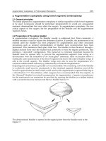
Orbital ultrasonography
With orbital diseases, ultrasonography is
principally of value in examination of the
intraocular structure (where ultrasonographic
resolution is greater than CT or MRI) and for
the examination of vascular size and flow-rates,
using colour-coded Doppler ultrasonography. It
is, therefore, particularly useful for the detection
of small intraocular tumours, intraocular
tumours in the presence of opaque media,
scleritis and inflammation in the posterior
Tenon’s space, arterio-venous malformations
and low- or high-flow vascular shunts.
With orbital vascular anomalies, there may be
not only enlargement of the superior ophthalmic
vein (compared with the normal side), but also
an arterial wave-form to the flow, together with
reversal of direction of flow within the vein.
Computed tomography
As the orbit and surrounding sinuses have
tissues with naturally high radiographic
contrast, thin-slice computed tomography is
the most effective and economical tool for the
initial imaging of the orbit (Figure 10.7).
For a suspected orbital mass, a single run of
axial scans with intravenous contrast (unless
contraindicated), together with coronal
reformats images, is generally sufficient to give
a probable diagnosis and allow planning of
surgery or medical therapy; if needed, direct
coronal imaging may give greater detail of the
relationship of the mass to the optic nerve or
rectus muscles. For planning the repair of
blowout fractures, or the diagnosis and
treatment of thyroid orbitopathy, a single run
of direct coronal scans (without contrast) is
often sufficient. Parasagittal reformats along
the plane of the vertical recti and optic nerve
may also be of help in orbital floor trauma and
in evaluating the relationship of lesions to the
optic nerve (Figure 10.8).
For orbital pathology arising in bone, or
where secondary bone invasion is suspected,
then it is important to obtain images with
both soft tissue and bone window settings.
Spiral CT allows greatly reduced imaging
time and three-dimensional studies may be
of help in planning major cranio-facial
reconstruction (Figure 10.9).
PLASTIC and ORBITAL SURGERY
104
(a)
(b)
Figure 10.7 A patient with dysthyroid orbitopathy,
showing gross proptosis and enlargement of extraocular
muscles, as imaged by (a) axial and (b) coronal soft
tissue CT scans through the mid-orbits.
Figure 10.8 Parasagittal reformatted CT, imaged
along the line of the vertical recti, showing the inferior
rectus muscle to be free from the site of a repaired
orbital floor fracture.
Magnetic resonance imaging
Magnetic resonance imaging is derived from
the signal emission when hydrogen nuclei
realign to a very strong magnetic field after the
cessation of an exciting radio frequency pulse,
the interval being termed the “relaxation time”.
Various relaxation times may be assessed and
images derived from the measured signals at
these different relaxation times: T1-weighted
images tend to show anatomical detail of the
orbit, whereas T2-weighted images – where the
high signal of tissue oedema is readily evident –
generally demonstrate pathological processes.
Orbital fat has a high signal on T1-
weighting, this often hindering the
discernment of orbital pathology, but the
contrast can be markedly improved by use of
fat-suppression software programmes to
manipulate the images. Gadolinium-DTPA
provides an intravenous contrast, highlighting
vascular lesions or tissues with leaking vessels
(Figure 10.10) but with T1-weighted images,
renders pathology less discernable unless used
in conjunction with fat-suppression.
MRI should not be used routinely for the
investigation of orbital disease, but provides
additional information to CT in certain
circumstances. It is of particular value in
determining the nature of optic nerve lesions
in the region of the optic canal and chiasm; in
demonstrating the position of the optic nerve
within large orbital tumours, where not shown
on CT; in the imaging of radiolucent foreign
bodies that are not ferro-magnetic. Although
the presence of muscular oedema on STIR
105
INVESTIGATION of LACRIMAL and ORBITAL DISEASE
Figure 10.9 Three-dimensional CT reformat for a
patient with severe clefting of the facial soft tissues
and bone.
Figure 10.10 MRI of a patient with recent
intraconal orbital haemorrhage: (a) T1- and (b) T2-
weighted images, and (c) fat-suppressed T1-weighted
image with Gadolinium-DTPA, showing normal
uptake of contrast in the extraocular muscles on the
unaffected right side. A fluid level may be seen within
the lesion of the left orbit.
(a)
(b)
(c)
images is suggestive of active inflammatory
oedema in patients with thyroid orbitopathy,
MRI used for this purpose is expensive and does
not add usefully to a thorough clinical
examination.
Although there are exceptions, most orbital
tumours have a fairly low T1 signal, a
medium-to-high T2 signal, and show variable
Gadolinium-DTPA enhancement. Non-
specific orbital inflammation tends to have a
medium T1 signal, with a relatively low signal
on T2. Lesions containing melanin, or the
breakdown products of blood, and those
with lipid, fat or mucus will give high signal
on T1-weighted images; examples include
orbital haemorrhage, orbital melanomas,
cholesterol granulomas, dermoid cysts and
sinus mucocoeles.
Angiography
Magnetic resonance angiography offers
information on the vascular dynamics of most
orbital lesions, although intra-arterial contrast
angiography (selective internal and external
carotid arteriography) remains important in
the exclusion of small aneurysms, dural low
flow arterio-venous fistulae and the
investigation of pulsatile proptosis not
explained by other imaging modalities.
Another important indication is in the surgical
planning of high flow tumours, such as
haemangiopericytoma, and consideration of
therapeutic embolisation of the arterial supply
may be considered at the time of angiography.
Positron emission tomography (PET)
and single photon emission CT (SPECT)
These modalities have yet to find a major
place in orbital assessment, but may become
important in the coming decade. Current uses
include staging patients with non-small cell
carcinoma of the lung, malignant melanoma,
Hodgkin’s disease, non-Hodgkin’s lymphoma,
colorectal carcinoma and head and neck
carcinoma.
PET scanning using fluorine-labelled
deoxyglucose radiotracer has proved as
reliable as conventional scanning for the
identification of primary or metastatic tumour
and is also superior to clinical examination
or other imaging modalities for detecting
nodal metastases; unfortunately the imaging
technique presently lacks anatomic detail. A
major current role, particularly in patients
with lymphoma, is in the differentiation of
tumour from fibrous tissue after radiotherapy.
Octreotide scintigraphy
Following the discovery of somatostatin
receptors on the activated lymphocytes
associated with thyroid orbitopathy, radio-
labelled octreotide (a somatostatin analogue)
has been used as a semi-objective tool in the
evaluation of the disease activity in this
condition. The test is extremely expensive and
its use limited to a few research centres.
Tissue diagnosis
Tissue diagnosis remains essential for the
appropriate management of almost every orbital
disease. Although fine needle aspiration biopsy
is useful for the confirmation of certain tumours
in a patient with known systemic malignancy, it
requires an experienced cytologist to interpret
results. Even with CT or ultrasonographic
guidance, fine-needle aspiration of post-
equatorial lesions is hazardous and the amount
of tissue often insufficient for the histological
studies required. Most experienced orbital
surgeons favour an open biopsy approach, in
order to correctly identify pathological tissue,
secure haemostasis, and obtain enough tissue
for pathological studies.
Assessment of lacrimal drainage
disease
The patient presenting with a watering eye
may be producing too many tears, may have
trouble delivering the tears to the drainage
PLASTIC and ORBITAL SURGERY
106
apparatus, or may have defective tear drainage.
Hypersecretion is usually due to ocular surface
irritation, trichiasis, or blepharitis. The
lacrimal pump relies on intact motor nerve
supply from the facial nerve, good tone in the
orbicularis oculi muscle and taut lids to deliver
tears to the lacrimal drainage apparatus. The
drainage apparatus can be intrinsically affected
at the level of the puncti, canaliculi, lacrimal
sac and the nasolacrimal duct, and can also be
adversely affected by nasal pathology. A
thorough history and meticulous examination
will usually aid the elucidation of this
polyfactorial symptom.
History for patients with lacrimal
disease
Apart from helping to elucidate the cause
of the epiphora, the history (Box 10.2) allows
assessment of the degree of functional
disturbance to the patient. In some, epiphora
is simply a mild nuisance, whereas in others it
significantly interferes with their quality of
life, often with a profound effect on reading
and driving. In most cases tears spill over at
the medial canthus, whereas lateral spillover
usually occurs with lower lid laxity (Figure
10.11). The nature of the discharge, whether
water, mucus, pus or blood-stained tears, is a
useful guide to the likely type of block; blood-
stained tears, although most commonly due
to severe Actinomyces canaliculitis, may
indicate a tumour of the lacrimal drainage
system. Bilateral epiphora associated with
itching, foreign body sensation, pain
or photophobia is indicative of reflex
hypersecretion. Obstructive epiphora is often
unilateral and usually worse outdoors in cold,
windy conditions. A history of cicatrising
conditions such as trachoma, herpes simplex,
Stevens-Johnson syndrome, systemic
chemotherapy with 5-fluorouracil, ocular
chemical burns or chronic ocular medication,
and pemphigoid should raise suspicion of
canalicular disease.
Examination of patients with
lacrimal disease
Careful examination of the eyelids and ocular
surface should exclude causes of hypersecretion
such as marginal blepharitis (Figure 10.12),
107
INVESTIGATION of LACRIMAL and ORBITAL DISEASE
Box 10.2 Main aspects of history
from the lacrimal patient
• Duration of symptoms
• Unilateral or bilateral
• Severity
• Constant or intermittent
• Precipitating factors (for example, cold
or windy weather)
• Spillover of tears at medial or lateral
canthus
• Associated symptoms (for example,
discharge, blurred vision, skin
excoriation)
• Past history: cicatricial skin or ocular
diseases, herpetic disease, eyelid
trauma, dacryocystitis
• Chronic nasal disease, nasal injury and
surgery to the nose or sinuses
• Drug history: including topical
medications, anticoagulants,
antiplatelet drugs
• Fitness for surgery
Figure 10.11 Lateral canthal spillover of dye in a
patient with lower lid laxity as the main cause for
epiphora.
trichiasis, dry eyes, pingueculum and corneal
pathology.
The normal punctum is directed into the
tear lake and, although frank lower lid
ectropion is easily recognisable, mild punctal
ectropion may be missed and is often
associated with secondary punctal stenosis. A
pouting punctum with a plug of stringy pus
that is almost impossible to express is
suggestive of Actinomyces canaliculitis (Figure
10.13). Eyelid laxity, even in the absence of lid
or punctal malposition, can result in
troublesome epiphora due to lacrimal pump
failure and “gravitation” of the tear-line on the
sagging lower lid margin. Facial weakness
should be noted and the presence of aberrant
muscular movements suggests aberrant
reinnervation and the possibility of “crocodile
tears” as a cause of the patient’s symptoms.
The presence of a lacrimal sac mucocoele or a
mass may only become evident after palpation
of the lacrimal sac fossa; a readily expressible
mucocoele suggests a patent canalicular
system with nasolacrimal duct obstruction and
requires no further investigation.
Each tear film should be stained with a
partial drop of 2% fluorescein and the height
of the tear meniscus and stability (break-up
time) of the tear film assessed. Corneal
staining suggests the possibility of episodic
reflex hypersecretion due to unstable tear film
or reduced background tear secretion. The
rate of dye disappearance from the conjunctival
sac, particularly useful in children, gives a
good indication of lacrimal drainage especially
when both sides are compared (Figure 10.14).
Lacrimal syringing is invariably performed
as part of the assessment of the adult patient
with epiphora. Good technique is essential not
only to obtain maximum information, but also
to avoid canalicular damage and subsequent
fibrosis; it is possible that many canalicular
obstructions are iatrogenic. After instilling a
topical anaesthetic, the punctum may be
dilated without rupturing the surrounding
ring of connective tissue or annulus. Lateral
traction is applied to the eyelid to straighten
the canaliculus and a fine lacrimal cannula on
a 2ml saline-filled syringe is used to gently
probe the appropriate canaliculus (Figure
10.15). In cases of canalicular obstruction a
PLASTIC and ORBITAL SURGERY
108
Figure 10.12 Epiphora caused by severe blepharo-
keratitis in a patient with acne rosacea.
Figure 10.13 Stringy, non-expressible pus at the
punctum of a canaliculus affected by Actinomyces.
Figure 10.14 Asymmetrical tear lines and dye
disappearance in a child with nasolacrimal duct
stenosis.
“soft stop” is reached. Reflux of clear fluid
through the same punctum in individual
canalicular obstruction or through the
opposite punctum in common canalicular
obstruction: with individual canalicular
obstruction, the point of obstruction may be
assessed by grasping the cannula at the
punctum with fine forceps before withdrawing
it from the canaliculus. In the absence of
canalicular disease a “hard stop” is felt as the
cannula reaches the medial wall of the
lacrimal sac and, in such cases, the irrigation
fluid that reaches the nose if the nasolacrimal
duct is patent or only partially obstructed;
reflux of fluorescein-stained fluid, with or
without mucus, from opposite punctum and
failure of fluid to reach the nose indicates total
nasolacrimal duct obstruction.
Intranasal examination (with a headlight
and speculum or, ideally, an endoscope) may
be performed, looking for the presence of
fluorescein in the inferior meatus, polyps,
allergic rhinitis, septal deviation, turbinate
impaction (rare), or other intranasal diseases
(Figure 10.16). Preoperative nasal endoscopy
is essential in the assessment of patients for
endonasal lacrimal procedures.
Ancillary tests for lacrimal
assessment
Dacryocystography
Dacryocystography provides very good
anatomical detail of the outflow system –
revealing occlusion, stenosis or dilatation of
the outflow tract and also, in some cases,
diverticulae, stones, or tumour (Figure 10.17) –
but does not give a true measure of the
physiological function. However, where the
system is patent during injection of contrast,
the failure of spontaneous clearance of oil-
based contrast media after the patient resumes
109
INVESTIGATION of LACRIMAL and ORBITAL DISEASE
(a)
(b)
(c)
Figure 10.15 Analysis of lacrimal probing and
syringing: (a) “hard stop” with a patent canalicular
system; (b) medial “soft stop” with obstruction of
common canaliculus; (c) lateral “soft stop” due to
lower canalicular obstruction.
Figure 10.16 Intranasal tumour causing epiphora.
the upright posture is suggestive of a reduced
physiological clearance (so-called “functional
block”).
Dacryocystography is indicated in planning
endonasal lacrimal surgery, or with surgery for
congenital lacrimal anomalies, after trauma,
after cranio-facial repair, with revisional
lacrimal surgery, or where a tumour or
sequestrum within the system is suspected. A
dilated canalicular system with filling defects
may be evident in Actinomyces canaliculitis
(Figure 10.18). There is no indication for
dacryocystography where clinical signs indicate
an uncomplicated lacrimal sac mucocoele.
Lacrimal drainage scintigraphy
This study uses a gamma camera to follow
the passage of a drop of radio-labelled fluid
PLASTIC and ORBITAL SURGERY
110
Figure 10.17 Dacryocystography showing: (a) an anatomically normal left system but a dilated right lacrimal
sac with filling defect due to a stone; (b) an anatomically normal right system (although contrast reflux suggests
distal stenosis within the outflow tract) and a tiny, non-functional left surgical anastomosis after endonasal
dacryocystorhinostomy, (c) a small right and large left mucocoele; (d) a functional right dacryocystorhinostomy
with direct drainage of contrast to the nasal space.
(a)
(b)
(c)
(d)
Figure 10.18 A typically dilated canaliculus in a
patient with Actinomyces canaliculitis.
(usually Technetium 99) from the conjunctival
sac to the nasal passages, and provides a
measure of physiological tear clearance where
there is a patent system on clinical
examination or dacryocystography (Figure
10.19). In this situation, scintigraphy will
generally reveal whether there is a failure of
gathering of tears into the drainage system
(often due to lid anomalies), or a failure of
clearance of tears that are otherwise rapidly
entering the system from the tear lake.
Computed tomography
Computed tomography is indicated when a
lacrimal sac tumour is suspected and may be
helpful in planning surgery for trauma cases,
particularly when plating systems have been
used. Craniofacial disorders and sclerosing
bony dysplasias may have unusual bone
anatomy shown on CT, and these changes
may influence the approach to surgery and the
prognosis.
111
INVESTIGATION of LACRIMAL and ORBITAL DISEASE
Figure 10.19 Lacrimal scintigraphy showing a
normal right drainage pattern but a marked delay in
the exit of tracer from the left lacrimal sac.