Fundamentals of Clinical Ophthalmology - part 7 ppsx
Bạn đang xem bản rút gọn của tài liệu. Xem và tải ngay bản đầy đủ của tài liệu tại đây (357.64 KB, 20 trang )
112
Dysthyroid eye disease is the commonest cause
of proptosis in adults and the disease typically
presents as Graves’ disease in the third and
fourth decade, with a four- to seven-fold
predominance in females. Bilateral orbital
inflammation is often accompanied by eyelid
retraction and restriction of ocular movements.
Asymmetrical involvement and extensive
fibrosis are less frequent presentations and the
diagnosis of dysthyroid eye disease should
always be suspected in the presence of any
inflamed orbit or with proptosis.
Sight is threatened by corneal exposure due
to incomplete eyelid closure over a proptotic
globe, uncontrolled ocular hypertension or
optic nerve compression. One-fifth of patients
with untreated compressive optic neuropathy
develop irreversible visual impairment (to
6/36 or less).
Treatment of dysthyroid eye disease aims to
conserve or restore normal visual function,
relieve ocular pain and achieve an acceptable
appearance.
Pathogenesis
In Graves’ hyperthyroidism it is likely that
thyroid damage leads to activation of
autoimmune thyroid disease by activation of
anti-receptor antibodies to the thyrotrophin
receptor (TSH receptor). Eye disease is
clinically evident in 40% of patients with
Graves’ disease but, in contrast, in only 3% of
patients with Hashimoto’s thyroiditis and very
11 Dysthyroid eye disease
Carol Lane
rarely with primary hypothyroidism. The high
correlation between Graves’ disease and
orbital disease suggests a shared antigen,
such as TSH receptor, thyroglobulin or
thyroid peroxidase. Circulating activated T
lymphocytes infiltrate the orbital tissues,
where they release cytokines which, in turn,
stimulate proliferation of fibroblasts and
deposition of glycosaminoglycans (GAGs).
Intense lymphocytic infiltration, fibroblast
proliferation and perimysial oedema result in
expansion of orbital contents and proptosis.
Subsequent fibrosis of involved perimysial
connective tissue results in varying degrees of
muscular contracture.
Hales and Rundle described the natural
history of dysthyroid eye disease, with the
disease typically peaking after six months
and active inflammation resolving within 18
months. Of 67 patients followed for an average
of 15 years, those with gross eye disease
persisting for more than six months after control
of thyroid status had a worse outcome. The
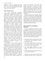
disease tends to be more severe in males and in
smokers, and the elongated myopic globe is at
greater risk of exposure keratopathy, whereas a
tight orbit without proptosis is at greater risk of
compressive optic neuropathy (Figure 11.1).
Features of dysthyroid eye
disease
Although Werner’s early “NOSPECS”
classification of dysthyroid eye disease
underlines the concept of a gradation of
severity of the condition, it has largely been
superseded by classifications based upon the
degree of inflammation – such as that of
Mourits or that of others (Table 11.1). A
simple “activity score” may be assigned by
awarding one point for each of retrobulbar
pain, pain on eye movement, eyelid erythema,
eyelid oedema, conjunctival injection,
conjunctival chemosis, caruncular swelling,
deteriorating vision, diplopia and worsening
appearance; an activity score of 3 or more (out
of 10) indicates active disease.
An objective deterioration in visual acuity,
reduced colour perception, an acquired visual
field defect, impaired visual-evoked potentials
or corneal ulceration are signs of serious
sight-threatening disease for which urgent
intervention is essential.
Treatment of the thyrotoxicosis of Graves’
disease tends to improve eye signs, although
hypothyroidism after suppression of
the hyperthyroid state may exacerbate
ophthalmopathy and this should be avoided
by regular blood tests during control of the
thyroid gland. Recent evidence suggests that
radio-iodine treatment for thyrotoxicosis may
adversely affect ophthalmopathy and systemic
steroids during therapy may prevent
exacerbation of the eye disease.
Although most patients with the clinical
features of dysthyroid eye disease have
abnormal thyroid function, some will be
euthyroid and the clinical diagnosis may be
supported only by raised levels of serum
thyroid auto-antibodies.
Orbital imaging in dysthyroid eye disease
(most readily with CT scan) tends to show
enlargement of several extraocular muscles,
the inferior and medial recti being affected
most frequently, the superior and lateral recti
less often and involvement of the oblique
muscles being relatively rare. Other features
include changes in the orbital fat and, with
longstanding disease, changes in the thin
113
DYSTHYROID EYE DISEASE
Figure 11.1 A 39-year-old woman with dysthyroid
compressive optic neuropathy: (a) before and (b) after
orbital decompression.
(a)
(b)
Table 11.1 Assessment of common clinical features of
dysthyroid eye disease (after Thyroid 1992; 2:235–6).
Feature Clinical assessment
Eyelid Maximal fissure width
Upper lid to limbus distance and lower
lid to limbus distance
Cornea Exposure keratopathy assessed by Rose
Bengal or fluorescein staining (indicates
presence of absence of staining)
Extraocular Binocular single vision in central
muscles 30° field (indicate presence or absence,
with or without prisms) and one or
more of the following measurement
techniques: Maddox rod test; alternate
cover test; Hess chart or Lancaster
red–green test.
Optional: intraocular pressures, CT
scan or MRI scan
Proptosis Exophthalmometry
(CT or MRI scan may also be used for
measurement)
Optic nerve Visual acuity, fields and colour vision
activity score Sum one point for each of the
following: spontaneous retrobulbar
pain; pain with eye movement; eyelid
erythema; eyelid oedema; conjunctival
injection; conjunctival chemosis;
caruncular swelling
Patient Satisfaction with the following
self-assessment (indicate change of each with therapy,
using a scale such as “greatly improved,
improved, unchanged, worse, much
worse”): appearance; subjective visual
function; ocular discomfort; diplopia
PLASTIC and ORBITAL SURGERY
114
orbital walls (Box 11.1). Enlargement of the
posterior part of the medial rectus is most
likely to crowd the orbital apex and cause
optic neuropathy (Figure 11.2a) and direct
coronal CT scans are valuable for showing
“crowding” of the optic nerve at the orbital
apex, with loss of the fat spaces, in
compressive optic neuropathy (Figure 11.2b).
MRI scans, particularly STIR (short-tau
inversion recovery) sequences, may provide an
indication of the water content of extraocular
muscles – this being a reflection of the degree
of inflammatory myositis – but the relatively
costly investigation adds little to clinical
examination. Likewise, B-mode ultrasono-
graphy may be used to assess the size of the
anterior part of the extraocular muscles, but
provides poor images of the posterior orbital
structure.
Treatment of dysthyroid eye
disease
Most patients with thyroid eye disease will
have relatively few symptoms and signs, and
many will require only topical lubricants
during the active phase of the disease and no
long-term therapy. Patients without proptosis
when the disease is inactive, but with
persistent lid retraction or incomplete lid
closure, may need eyelid surgery to protect the
cornea (Chapter 7). Likewise, squint surgery
Box 11.1 Typical features of
dysthyroid eye disease on CT or MR
imaging
• Enlarged extraocular muscles;
tendinous insertion often spared
• Orbital fat normal, diffusely increased
opacity or increased in quantity
• Occasional slight bowing of the medial
orbital wall (lamina papyracea); the
“Coca-Cola bottle” sign
• Frequent inferior rectus enlargement
on axial scan, the mass of which may
simulate an orbital tumour
• Crowding of the optic nerve, at the
orbital apex, by enlarged extraocular
muscles
• Lacrimal gland rarely enlarged, but
often prolapsed forwards
• Fat prolapse from the orbit into the
cranium at the superior orbital fissure
• Absence of orbital masses, vascular
anomaly or sinus involvement
Figure 11.2 (a) Axial and (b) coronal CT for a
patient with compressive optic neuropathy, shown in
Figure 11.1. All extraocular muscles are enlarged and
there is loss of the fat planes around the optic nerve at
the orbital apex.
(a)
(b)
115
DYSTHYROID EYE DISEASE
may be needed when the eye disease has been
shown to be stable and inactive for some
months.
Management of more severe and significant
thyroid eye disease should be first directed
towards suppression of orbital inflammation
and later the restoration of orbital function.
Suppression of orbital
inflammation in dysthyroid eye
disease
Patients with significant signs or symptoms
of active orbital inflammation, or with optic
neuropathy or significant exposure keratopathy,
should receive systemic therapy to reduce the
degree of orbital inflammatory congestion.
Those with an activity score of 3 or more are
likely to benefit, as are those with a muscle
oedema shown on STIR-sequence MRI.
Systemic steroids at high dosage (either
intravenous methyl prednisolone or oral
prednisolone) should be administered and the
patient checked for improvement after a few
days. The patient should be monitored for
hyperglycaemia and hypertension during
treatment and the prescription of a gastric
proton-pump inhibitor or Histamine-2
receptor antagonist considered; patients on
long-term steroids, especially the elderly,
should be given calcium supplementation to
counteract steroid-induced osteoporosis. If
systemic steroids produce an improvement in
the inflammatory orbitopathy, the dosage
should be slowly reduced towards about 20mg
daily if possible and the patient referred for
low-dose, (2000–2400 cGy) lens-sparing
radiotherapy to the posterior tissues of the
orbit; some authors consider radiotherapy
contraindicated in diabetics, as it may hasten
the development of retinopathy.
Steroids probably suppress dysthyroid
orbitopathy by inhibition of the production of
cytokines by activated T cells and macrophages
and fibroblasts within the orbit. It has been
reported that treatment with steroids and
radiotherapy is more effective than treatment
of orbitopathy with steroids alone. As there
may be an increase in orbital inflammation
and oedema whilst undergoing orbital
radiotherapy, it is prudent to continue a
moderate steroid dosage (for example,
prednisolone 20mg daily) during this
treatment.
Surgical rehabilitation of the
patient with dysthyroid eye disease
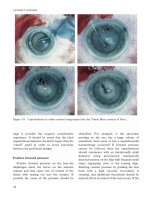
Severe conjunctival chemosis is self-
perpetuating due to the “throttling” effect of
the lower eyelid on the prolapsed conjunctiva
and will, in some patients, prevent eyelid
closure (Figure 11.3a). After subconjunctival
injection of local anaesthetic with adrenaline,
drainage of subconjunctival fluid and
placement of Frost sutures in the upper and
lower eyelids will typically allow closure of the
eyelids under an occlusive dressing, with
topical application of a steroidal ointment
(Figure 11.3b). This typically produces a
dramatic improvement within 12 hours
(Figure 11.3c), allows the cornea to rehydrate
and gives time for systemic antiinflammatory
therapy to act.
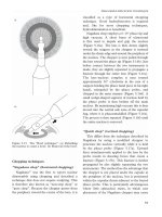
Orbital decompression is necessary if visual
function deteriorates despite the use of high-
dose systemic steroids (Figure 11.1b). As
compression of the optic nerve occurs mainly
at the orbital apex, decompression for visual
failure must include removal of the posterior
part of the medial wall (Figure 11.4); in a few
patients the most posterior part of the medial
wall being the lateral wall of the sphenoid
sinus. Pre-operative CT is required to confirm
the diagnosis (especially with unilateral
disease), to exclude underlying sinus disease
and to detect any cranio-facial anomalies,
such as a midline encephalocoele.
Although Olivari has described reduction of
proptosis by meticulous excision of orbital fat
from the intraconal and extraconal spaces,
most orbital decompressions involve removal
PLASTIC and ORBITAL SURGERY
116
Figure 11.3 (a) Severe conjunctival chemosis
preventing any movement of the right eyelid and
causing dehydration of the right cornea. After
drainage of the chemosis under local anaesthesia and
placement of multiple eyelid traction sutures (b), the
eyelid was padded closed for 12 hours with a dramatic
improvement in the clinical state (c).
(a)
(b)
(c)
of a combination of the medial wall, floor and
lateral wall of the orbit. Removal of the medial
wall is necessary for relief of optic neuropathy
(Figure 11.5), removal of the floor adds the
Figure 11.4 Patient referred with persistent
compressive optic neuropathy on the left side, due to
the failure to remove the posterior half of the left
medial orbital wall.The right side had successful relief
of optic neuropathy after a complete ethmoidectomy
reaching the orbital apex.
Figure 11.5 The medial orbital wall, showing the
lamina papyracea with the foramina for the anterior
and posterior ethmoidal arteries in relation to the
optic canal.
(a)
(b)
most to reduction in proptosis and removal of
the lateral wall allows reduction of lacrimal
gland prolapse and reduces the deleterious
effect of medial wall decompression on ocular
muscle balance. Decompression requires
adequate hypotensive general anaesthesia and
a reverse Trendelenburg positioning of the
patient to reduce bleeding during this
complex surgery.
Other surgery for dysthyroid eye disease
Upper eyelid retraction in thyroid eye
disease occurs due to a combination of
primary factors (adrenergic stimulation,
inflammation and fibrosis) and secondary
retraction due to inferior rectus fibrosis – with
secondary overaction of the superior
rectus/levator complex. If secondary upper
eyelid retraction is present, the restriction of
ocular motility should be addressed first, with
inferior rectus recession. Primary upper eyelid
retraction is treated by one of the several
techniques for graded levator tenotomy
(Chapter 7), but with all methods it is
particularly important to completely divide
the lateral horn of the levator aponeurosis and
to maintain a levator action on the medial part
of the upper eyelid.
Lower lid displacement, with excessive
scleral show below the lower limbus, is due to
proptosis and is almost always corrected by
adequate orbital decompression – which
should probably be considered in any patient
with exophthalmos of 24 mm or more. True
lower lid retraction, due to an overaction of the
retractor fascia in the lower lid, probably
occurs only after inferior rectus recession.
Lower lid retraction may require surgery to
elevate the eyelid using an implant of sclera,
hard palate mucosa or ear cartilage.
Lateral tarsorrhaphy invariably stretches
with time and, with appropriate surgery to
address the other position of the globe and
upper eyelid, there is almost no indication
for this rather disfiguring procedure in the
patient with dysthyroid eye disease. Likewise,
skin-reduction blepharoplasty should be used
with caution, as removal of anterior lamella in
these patients may risk exacerbation of
exposure keratopathy.
Methods for bone-removing orbital
decompression
Orbital decompression can be achieved
through several approaches: transnasal or
transantral endoscopic decompression leaves
no external incision, but can provide only a
limited decompression (of the medial wall and
medial part of the floor); likewise, the post-
caruncular transconjunctival incision also
provides access for medial wall decompression,
but can present some surgical difficulty due
to the presence of unrestrained orbital fat in
the operative field. The lateral canthotomy
approach provides the most aesthetic approach
for decompression of up to three walls, which
may be required where exophthalmos is greater
than 25 mm (Figure 11.6).
Although the use of a bicoronal flap for
orbital decompression has been widely reported
in the past, there is no advantage to the use of
this large-incision approach. Likewise, the
Lynch incision of the external ethmoidectomy
approach often leaves an unsightly scar and
gives only limited access – to the medial wall
and medial part of the orbital floor.
Lynch external ethmoidectomy approach
A gently curving incision is placed from the
medial end of the brow, past the attachment of
the medial canthal tendon, towards the orbital
floor (Figure 11.7). After securing haemostasis
within the orbicularis muscle, the periosteum
is opened in front of the anterior lacrimal crest
and the lacrimal sac and medial orbital
periosteum raised from the bone. The anterior
ethmoidal artery may be exposed, cauterised
and divided, although this should not be
necessary as the artery provides a key
landmark to the level of the cribriform plate –
the upper limit of decompression.
117
DYSTHYROID EYE DISEASE
The lamina papyracea is infractured
medially and the ethmoidectomy completed,
keeping posterior to the posterior lacrimal
crest and below the level of the anterior
ethmoidal artery; bone excision is continued
inferiorly until the medial part of the orbital
floor is removed. The periosteum is incised
widely, to allow free prolapse of orbital fat into
the areas of bone removal, and the anterior
periosteal incision and superficial tissues
closed in layers.
Extended lateral canthotomy approach
The lateral canthotomy approach
(Figure 11.7), with extension of the incision
along the lower conjunctival fornix (the “lower
PLASTIC and ORBITAL SURGERY
118
Figure 11.6 Ten millimetre reduction in left
proptosis after three-wall orbital decompression
performed through an extended lateral canthotomy
incision (a). Preoperative views (b,c) and five weeks
after surgery (d,e).
(a)
(b)
(c)
(d)
(e)
lid swinging flap”), provides excellent access
to the orbital floor, although decompression
of the medial wall requires greater dexterity
than with the Lynch approach as access is
more restricted. A 1–2 cm extension of the
canthotomy into the lateral part of the upper
eyelid skin crease (Figure 11.8a) eases access
to, and decompression of, the lateral wall.
A horizontal canthotomy of 1.5cm is made
and the orbicularis oculi cauterised and
divided infero-laterally to the orbital rim;
division of this muscle must be continued
until there is a clear release of the lateral
tethering of the lower eyelid. The conjunctiva
is divided at a point 1mm below the lower
border of the lower tarsus and the conjunctival
edge attached to the upper eyelid with a
4/0 nylon suture – this acting to protect the
cornea and to keep the lower orbital septum
tight during subsequent preparation of a
pre-septal skin-muscle flap (Figure 11.8b).
The periosteum is opened at the rim, raised
widely across the orbital floor and medially up
to the level of the ethmoido-frontal suture.
The medial part of the orbital floor is
fractured with a surgical clip, as much as
necessary of the floor and medial wall
removed (Figure 11.8c) and the periosteum
excised or incised widely over the area of bone
removal; it is prudent to preserve the infero-
medial bone strut between the maxilla and
ethmoid, as this maintains aeration of the
maxillary sinus.
If the lateral wall is to be removed, the outer
quarter of the upper eyelid skin-crease is
divided to reach the level of the superficial
temporalis fascia and the periosteum divided
8 mm outside the rim. The periosteum is
raised over the rim and into the orbit, the
lateral wall removed in part (Figure 11.8d) or
whole, and the periosteum below the orbital
lobe of the lacrimal gland incised or excised.
The periosteal incision may be continued
upwards just anterior to the orbital lobe and,
when clear of the gland, directed posteriorly
along the orbital roof to allow the orbital lobe
to fall posteriorly into the defect in the lateral
wall (Figure 11.8e) – this repositioning of the
gland, together with the marked reduction in
proptosis, restoring the depth of the upper
eyelid sulcus.
The lateral periosteal strip, to which the intact
upper limb of the lateral canthal tendon is still
attached, is fixed around the residual bone of
the rim and, after placement of a vacuum drain
in the intraconal space and sub-temporalis
space, the lower fornix incision and canthotomy
closed in layers with absorbable sutures.
After instillation of an antibiotic ointment
into the conjunctival sac, a 4/0 lower lid traction
(“Frost”) suture is placed on traction to the
forehead and a firm eye dressing applied for
12–18 hours.The vacuum drain and patient are
monitored for abnormal haemorrhage.
Subciliary blepharoplasty approach
This approach is similar to the extended
lateral canthotomy, except that the preseptal,
post-orbicularis plane is reached through a
subciliary incision. The technique and view is
otherwise identical for the two procedures.
Bicoronal scalp-flap approach
The scalp is shaved 2–3 cm behind the
hairline, the operative field prepared and both
eyelids closed with tarsorrhaphy sutures. A
scalp incision, down to the periosteum, is
placed parallel to the hair line (Figure 11.7),
compressive haemostatic clips placed, and the
flap raised down to the brow ridge. The deep
layer of the temporalis fascia is followed down
to the level of the zygomatic arch, thereby
avoiding branches of the facial nerve.
The pericranium is incised 2 cm above the
orbital rim and the periosteal flap raised
inferiorly, using (if necessary) an osteotome to
119
DYSTHYROID EYE DISEASE
Bicoronal flap
approach
c
Lynch external
ethmoidectomy
approach
a
Lateral
canthotomy
approach
b
Figure 11.7 Incisions for orbital decompression
through (a) Lynch external ethmoidectomy approach;
(b) lateral canthotomy approach; (c) the bicoronal
flap approach.
release the supraorbital neurovascular bundle
from its canal. Using malleable retractors in a
hand-over-hand technique, the periosteum is
raised across the roof and lateral wall of the
orbit and, likewise, the temporalis muscle is
raised from its fossa. The thinnest part of the
lateral wall is shown by transillumination and
an osteotome used to breach the wall at this
point, the orbital contents being protected at
all times; the breach is extended with rongeurs
until adequate lateral wall removal has been
accomplished. A series of incisions, to the
depth of Richter’s muscle, are made in the
periosteum above the orbital rim, this
increasing flap mobility and reducing thyroid
“frown”. The medial wall and accessible parts
of the orbital floor are removed, the
periosteum incised to maximise prolapse of
orbital fat and the temporalis fascia closed
with 3/0 non-absorbable sutures. Vacuum
drains are placed across the subgaleal space
from each temporalis fossa, the periosteum
closed with a 4/0 absorbable suture and the
PLASTIC and ORBITAL SURGERY
120
Figure 11.8 Extended lateral canthotomy approach
to orbital decompression: (a) skin incisions for three
wall decompression; (b) lower conjunctiva closed over
the cornea, with preparation of a lower eyelid
swinging flap which provides excellent access to the
orbital floor; (c) infraorbital nerve visible after
removal of the bone of the orbital floor; (d) the lateral
wall has been removed behind an undisturbed orbital
rim; (e) the lacrimal gland settles backwards behind
the orbital rim, restoring the depth of the upper eyelid
sulcus.
(a)
(b)
(c)
(d)
(e)
scalp incision closed with surgical staples. A
firm scalp dressing is applied and the vacuum
drains maintained until dry.
Post operative management and
complications
The patient should be nursed half-seated on
bed-rest, to minimise post operative swelling,
and (where accessible) the pupils checked for a
few hours after surgery. If the patient complains
of severe or increasing pain, the affected side
should be examined for signs of rising
intraorbital pressure due to haemorrhage and
appropriate measures taken if it is impairing
optic nerve function.
Post operative antibiotics and anti-
inflammatory drugs (such as prednisolone
80mg daily, tailing over about ten days)
should be administered and the patient
instructed to avoid nose-blowing and
strenuous exercise over this period. Forced
ocular ductions are to be encouraged, as this
probably encourages clearance of post
operative oedema and recovery of normal
ocular balance and movements.
With administration of post operative
systemic antibiotics, early infection is rare but
sinusitis, particularly maxillary, can be a
recurrent late complication of decompression
and may require middle meatal antrostomy or
other corrective procedure. Under- or over-
correction of the proptosis may occur
(Figure 11.9a) and, very rarely, the latter
(enophthalmos) may be accompanied by
hypoglobus.
Loss of vision is extremely rare, but the
patient must be made aware of this remote
possibility prior to surgery.
Diplopia is almost universal after surgery,
will settle within a few weeks in most cases,
but presents the greatest practical problems
with the activities of daily living. If diplopia is
troublesome, it is worthwhile occluding one
eye in the early post operative period for those
activities (such as descending stairs) where a
second image is distracting or dangerous.
Where there is no risk with diplopia – as, for
example, with watching television – the
patient should be encouraged to try and fuse
the two images.
Other complications, such as persistent
infraorbital neuropraxia, nasolacrimal duct
obstruction with epiphora, or secondary lower
lid entropion, are uncommon. Likewise,
major intraoperative or post operative
(Figure 11.9b) haemorrhage, or loss of
cerebro-spinal fluid is relatively rare.
121
DYSTHYROID EYE DISEASE
Figure 11.9 Complications of orbital decompression:
(a) late enophthalmos due to maxillary atelectasis; (b)
limited orbital haemorrhage after third-time revisional
orbital decompression.
(a)
(b)
Further reading
Bartalena L, Marcocci C, Bogazzi F et al. Relation between
therapy for hyperthyroidism and the course of Graves’
ophthalmopathy. New Engl J Med 1998; 338:73–8.
Burch HB, Wartofsky L. Graves’ ophthalmopathy: current
concepts regarding pathogenesis and management.
Endocrinology Rev 1993; 14:747–93.
Char DH. Thyroid eye disease (3rd ed.) Boston: Butterworth-
Heinemann, 1997.
Claridge KG, Ghabrial R, Davis G et al. Combined
radiotherapy and medical immunosuppression in the
management of thyroid eye disease. Eye 1997; 11:717–22.
Consensus of an ad hoc committee. Classification of eye
changes of Graves’ disease. Thyroid 1992; 2:235–6.
Fatourechi V, Garrity JA, Bartley GB, Bergstralh EJ,
DeSanto LW, Gorman CA. Graves’ ophthalmopathy.
Results of transantral orbital decompression performed
primarily for cosmetic indications. Ophthalmology 1994;
101:938–42.
Garrity JA, Fatourechi V, Bergstralh EJ et al. Results of
transantral orbital decompression in 428 patients with
severe Graves’ ophthalmopathy. Am J Ophthalmol 1993;
116:533–47.
Hales JB, Rundle FF. Ocular changes in Graves’ disease: a
long term follow-up study. QJM 1960; 29:113–9.
Kalmann R, Mourits MP, van der Pol JP, Koornneef L.
Coronal approach for rehabilitative orbital decompression
in Graves’ ophthalmopathy. Br J Ophthalmol 1997;
81:41–5.
Lyons CJ, Rootman J. Orbital decompression for disfiguring
exophthalmos in thyroid orbitopathy. Ophthalmology 1994;
101:223–30.
McCord CD Jr. Orbital decompression for Graves’ disease;
exposure through lateral canthal and inferior fornix
incision. Ophthalmology 1981; 88:533–41.
Metson R, Dallow RL, Shore JW. Endoscopic orbital
decompression. Laryngoscope 1994; 104:950–7.
Mourits MP, Koornneef L, Wiersinga WM, Prummel MF,
Berghout A, van der Gaag R. Clinical criteria for the
assessment of disease activity in Graves’ ophthalmopathy;
a novel approach. Br J Ophthalmol 1989; 73:639–44.
Mourits MP, Koornneef L, Wiersinga WM, Prummel MF,
Berghout A, van der Gaag R. Orbital decompression for
Graves’ ophthalmopathy by inferomedial, by inferomedial
plus lateral, and by coronal approach. Ophthalmology
1990; 97:636–41.
Naniaris N, Hurwitz JJ, Chen JC, Wortzman G. Correlation
between computed tomography and magnetic resonance
imaging in Graves’ orbitopathy. Can J Ophthalmol 1994;
29:9–19.
Olivari, N. Transpalpebral decompression of endocrine
ophthalmopathy (Graves’ disease) by removal of
intraorbital fat: experience with 147 operations over 5
years. Plast Reconstr Surg 1979; 87:627–41.
Perros P, Crombie AL, Kendall-Taylor P. Natural history of
thyroid associated ophthalmopathy. Clin Endocrinol Oxf
1995; 42:45–54.
Prummel MF, Wiersinga WM. Medical management of
Graves’ ophthalmopathy. Thyroid 1995; 5:231–4.
Prummel MF, Wiersinga WM, Mourits MP et al. Effect of
abnormal thyroid function on severity of Graves’
ophthalmopathy. Arch Intern Med 1990; 150:1098–101.
Shine B, Fells P, Edwards OM, Weetman OP. Association
between Graves’ ophthalmopathy and smoking. Lancet
1990; 335:1261–3.
Tagami T, Tanaka K, Sugawa H, et al. High-dose intravenous
steroid pulse therapy in thyroid-associated opthalmopathy.
Endocr J 1996; 43:689–99.
Trokel S, Kazim M, Moore S. Orbital fat removal.
Decompression for Graves’ orbitopathy. Ophthalmology
1993; 100:674–82.
Werner SC. Classification of changes in Graves’ disease.
J Clin Endocrinol Metab 1969; 29:982–9.
Wulc A, Popp JC, Bartlett SP. Lateral wall advancement in
orbital decompression. Ophthalmology 1990; 97:1358–69.
PLASTIC and ORBITAL SURGERY
122
123
Benign orbital diseases, for many of which the
clinical history and examination findings are
diagnostic, cover a wide spectrum. The
incidence varies with age but, although
obviously dependent upon referral patterns, in
childhood the most common benign lesions
are dermoid and epidermoid cysts (6–37%),
capillary haemangiomas (8–13%) and trauma
(7%), whereas adult series are typically
dominated by thyroid orbitopathy (Chapter
11), orbital trauma (Chapter 14) and orbital
infections.
Benign cystic anomalies of the
orbit
Orbital cysts generally arise from
epithelium sequestered within the orbit during
embryological development, by implantation
after trauma or due to expansion of epithelial-
lined sinus lesions into the orbit.
Dermoid and epidermoid cysts
These lesions arise from surface epithelium
implanted at sites of embryological folding
and, if situated anteriorly within the orbit, are
commonly noted soon after birth. Due to the
accumulation of epithelial debris and
sebaceous oil in the lumen of the cyst, the
cysts slowly enlarge and leakage of the
contents into the surrounding tissues may
cause marked inflammation – with deeper
dermoid cysts tending to present in this
12 Benign orbital disease
Christopher J McLean
fashion. A dermoid cyst contains dermal
structures (hairs, sebaceous glands), whereas
more rarely there is only an epithelial
(epidermoid cyst) or a conjunctival lining
(conjunctival dermoid).
Implantation cysts may arise and develop in
a similar fashion to congenital dermoids, but
do not respect the characteristic anatomic
sites of the latter and will generally present in
patients with a past history of periocular
trauma.
The commonest dermoid cysts are firm and
smooth, mobile preseptal masses overlying the
supero-temporal quadrant of the orbit and,
less commonly, the supero-nasal quadrant.
Many cysts have a variable periosteal
attachment near the underlying fronto-
zygomatic or fronto-ethmoidal sutures, but
occasionally the dermoid will pass into or
through defects in the neighbouring bone. In
some cases the dermoid is incompletely
separated from the skin surface and presents
as a chronically inflamed and discharging
sinus (Figure 12.1).
A clinically characteristic lesion presenting
in childhood does not require radiological
investigation if anteriorly situated and mobile.
Likewise, fixed anterior masses do not
necessitate imaging, provided the orbital
surgeon is adequately experienced to follow
the lesion to its limits – if necessary within
the orbital depths. Deep orbital dermoids,
often presenting as orbital inflammation or
proptosis, require thin-slice CT with bone
windows to show associated clefts or canals in
the bone (Figure 12.2); MRI is a poor
investigation for these orbital abnormalities.
CT will often show a smooth, “scalloped”
erosion of the neighbouring bone as a result of
pressure from the mass, although this is a non-
specific sign suggesting a longstanding benign
orbital lesion.
All cysts develop inflammatory changes and
should be removed, generally at preschool age
before childhood trauma encourages rupture.
They are excised through an incision hidden
in the upper lid skin-crease or brow line and it
is important to divide tissues right down to
the cyst (there being a tendency to dissect a
plane too far from the surface of the cyst) and
then follow the plane by blunt dissection; in
some cases it is necessary to remove some of
the underlying periosteum or follow the lesion
into or through the orbital walls.
Deep orbital dermoids often involve the
greater wing of the sphenoid and may require
lateral orbitotomy or complicated anterior
orbitotomy for their removal.
Rupture of the cyst during surgery may lead
to a marked post operative inflammation and
any spilt contents should be removed.
Incomplete excision of the epithelial lining
will lead to recurrent inflammation with
formation of a discharging cutaneous fistula
to the operative site.
Dermolipoma
Although dermolipomas are not cystic, they
are conveniently classified with dermoid cysts
as they arise from cutaneous epithelium
sequestered within the conjunctival recesses –
typically laterally and occasionally associated
with a minor clefting of the outer canthus or
with Goldenhar’s syndrome. The abnormal
epithelium, which often bears hairs and
sebaceous glands that cause chronic
conjunctivitis, is associated with localised
prenatal formation of cutaneous-type fat,
which is evident as an underlying bright
yellow mass. The differential diagnosis is
subconjunctival fat prolapse, which tends to
present in obese adults and is not associated
with an abnormal conjunctival surface.
Dermolipomas require removal if they are
causing chronic conjunctivitis or if easily
visible at the palpebral aperture. Excision
should be performed under the operating
microscope as this aids preservation of all
except the abnormal epithelium and reduces
the risk of damage to the lacrimal gland
PLASTIC and ORBITAL SURGERY
124
Figure 12.1 Dermoid cyst in communication with
the skin, presenting as a discharging fistula.
Figure 12.2 CT scan showing orbital and
temporalis fossa components of a dermoid cyst, with
associated bone changes.
ductules. To avoid adherence between the
lateral rectus and the orbital rim, only fat
anterior to the rim should be removed and
this abnormal cutaneous fat has a subtle plane
of dissection free from the normal orbital fat.
Incautious excision of dermolipomas is a
source of medico-legal cases, as there is a
significant risk of damage to the lacrimal gland
ductules, restriction of eye movements and
ptosis if the lesions are not managed properly.
Paranasal sinus mucocoeles
Mucocoeles, most commonly in the
anterior ethmoid and frontal sinuses, are
slowly-enlarging, mucus-filled, cystic lesions
that arise from paranasal sinus mucosa and
gradually encroach into the orbit; occasionally
the contents of a mucocoele become infected,
which may lead to orbital cellulitis. Most
mucocoeles will present with a gradual
displacement of the globe and proptosis
(Figure 12.3), although those of the maxillary
sinus may lead to collapse of the orbital floor
and secondary enophthalmos.
The CT appearance of a mucocoele is a
cystic cavity smoothly expanding the bone of
a paranasal sinus and necessary with patchy
thinning or loss of bone. T1- and T2-weighted
MR images can show a wide variation in
signal intensities due to continuing changes in
the contents of the mucocoele.
Severe acute sinusitis or orbital cellulitis
requires admission for intravenous antibiotic
therapy and drainage of the orbital abscess if
threatening vision. Once the infection has
been shown to be under control, the
mucocoele and other sinus disease should
receive definitive treatment under the care of
an otorhinolaryngologist. In general,
treatment involves removal of the mucocoele
lining and re-establishment of a new drainage
pathway for the affected sinus.
Orbital cellulitis secondary to infected
mucocoeles may lead to an orbital abscess or
the formation of a transcutaneous fistula,
typically in the medial aspect of the upper
eyelid. Late presentation of mucocoeles within
the sphenoid or posterior ethmoid sinuses can
lead to compressive optic neuropathy and
irreversible visual loss.
Cranial and orbital anomalies
Microphthalmos with cyst
Microphthalmos with cyst arises from
incomplete closure of the fissure in the optic
vesicle, with formation of a cyst below a
microphthalmic globe. The cysts can vary
greatly in size and may slowly enlarge.
Small cysts can be left and may be in
communication with the vitreous cavity. Large
cysts are cosmetically unacceptable, cause
excessive orbital bony expansion, and
generally need to be removed together with
the microphthalmic globe; a ball implant can
be placed at the same operation and, in all
cases, a suitable fornix conformer must be
placed (Chapter 9).
Excision of small or moderately sized orbital
cysts may retard orbital growth and lead to
problems with prosthetics fitting later in life.
Ball implantation can, on occasion, be difficult
due to abnormal extraocular musculature.
Cephalocoeles
Congenital clefts of the skull, with
herniation of intracranial contents, leads to
125
BENIGN ORBITAL DISEASE
Figure 12.3 Outward displacement of the left globe
due to ethmoidal mucocoele.
cephalocoeles: the herniating contents can be
meninges (meningocoeles), brain tissues
(encephalocoeles), or both tissues (meningo-
encephalocoeles). When involving the orbit,
they often present in childhood as fullness
above the medial canthus, this swelling
increasing with straining or bending. Some
patients with orbital encephalocoeles will have
neurofibromatosis and the association of
colobomatous optic disc with basal
encephalocoele is known as “morning glory
syndrome’’.
Direct coronal CT scanning is best for
identifying the skull base deformity that
always accompanies orbital meningocoeles
and encephalocoeles.
Orbital cephalocoeles are removed as part
of the major reconstruction in affected
children, who often have multiple cranio-
facial anomalies, and defects within the
sphenoid bone are hard to correct compared
to those of the frontal bone.
Benign vascular anomalies
of the orbit
Many vascular anomalies, such as varices,
lymphangiomas and cavernous haemangiomas,
are probably present from birth but may only
become manifest in early adulthood.
Capillary haemangiomas
Capillary haemangiomas occur in 1–2% of
infants and are more common in females and
children of low birth weight; most appear soon
after birth, can enlarge dramatically and then
undergo a spontaneous involution – with 75%
resolving within five years. Involvement is
usually unilateral and the intradermal eyelid
lesions are bright red and dimpled (so called,
“strawberry naevus”; Figure 12.4), whereas
the deeper orbital lesions have a blue
colouration and spongy texture; both may
increase slightly in size with crying or
straining.
The rapid growth of a deep orbital capillary
haemangioma may mimic the highly
malignant rhabdomyosarcoma and it is
important to be aware of this differential
diagnosis; Doppler ultrasonography will,
however, show high reflectivity and vessels
with very high flow-rate (over 50cm/s) within
the capillary haemangioma. CT scan is rarely
necessary, but typically shows an irregular,
poorly defined lesion with marked contrast
enhancement.
Affected children should be monitored for
impairment of visual development, being
refracted when at an age suitable for spectacle
correction of anisometropia or marked
astigmatism. If the child is tending to develop an
amblyopic eye, then appropriate corrective
measures should be taken to maintain vision and
consideration be given to treating the lesion.
Many capillary haemangiomas will regress
rapidly, or their growth be slowed, by injecting
them with corticosteroids under general
anaesthesia; a useful regime being 40mg
depomedrone in the lesion and 4mg soluble
dexamethasone around the lesion, this being
repeated at six-weekly intervals for two further
sessions. Before injecting, the plunger must be
drawn back and, if blood is present, the needle
should be resited to avoid intravascular injection.
Systemic interferon has been used to treat
steroid-resistant, life-threatening capillary
haemangiomas, but the systemic side effects
render it inapplicable to orbital lesions.
Because of the risk of major haemorrhage,
surgery is not recommended for most
capillary haemangiomas.
PLASTIC and ORBITAL SURGERY
126
Figure 12.4 Orbital capillary haemangioma in an
infant.
When the haemangioma has regressed, it
may be necessary to remove redundant
atrophic eyelid skin or correct ptosis resulting
from disinsertion of the levator muscle
aponeurosis.
The complications of intralesional steroid
injections are necrosis of the skin overlying the
capillary haemangioma, and atrophy of
subcutaneous fat or dermis. Rarely growth
retardation and blindness have been reported
with this therapy.
Cavernous haemangiomas
Cavernous haemangioma is the most
common benign orbital tumour of adults and
may be a developmental hamartoma that
presents late in life, typically in the fourth or
fifth decades, with gradually increasing
painless proptosis. It is usually solitary and lies
in the retrobulbar space, thereby causing axial
proptosis, induced presbyopia, choroidal folds
and optic disc congestion. There is often a
global reduction in the extremes of eye
movement.
CT scanning reveals a well defined, round
intraconal lesion that commonly displaces the
optic nerve medially and, due to a very slow
blood flow, shows a very slow and patchy
contrast enhancement (Figure 12.5). Some
haemangiomas are wedged in the orbital apex
and these tend to present early due to optic
neuropathy. On MRI scanning, cavernous
haemangiomas are hypointense to fat on T1-
weighted images and isointense to vitreous
and hyperintense to fat and vitreous on T2-
weighted images.
Patients with asymptomatic tumours,
discovered by chance on imaging for other
reasons, can be monitored for orbital signs
and many presumed haemangiomas show
minimal change over many years. Indications
for removal include optic neuropathy,
proptosis and diplopia.
Lateral orbitotomy with intact excision of
the tumour is usually required, as many
haemangiomas are large and intraconal. The
tumour typically is like a purple plum and
contains large blood-filled cystic spaces.
Method for lateral orbitotomy
An upper eyelid skin-crease incision is
extended laterally to about 1cm below the
lateral canthus (Figure 12.6a) and the tissues
opened to the supero-lateral orbital rim. The
periosteum is incised 6mm outside the rim,
from the lateral one-third of the upper rim to
the level of the zygomatic arch, the origin of the
temporalis muscle separated from the bone over
its antero-superior 2cm, and the periosteal
incision extended backwards over the zygomatic
arch (Figure 12.6b).The periosteum is elevated
over the rim of the orbit and separated from the
inner aspect of the lateral wall, with particular
care being taken to cauterise and divide any
bridging vessels.Two parallel saw cuts are made,
in the coronal plane, at the upper and lower
ends of the lateral osteotomy, drill holes placed
either side of the cuts and the inner aspect of the
lateral wall fragment weakened 1cm behind the
rim, using a burr; the fragment is then broken
away and trimmed, to be swung outwards on
the temporalis muscle (Figure 12.6c), and the
periosteum opened to provide access for the
intraorbital procedure.
After achieving intraorbital haemostasis, a
vacuum drain is placed within the intraconal
127
BENIGN ORBITAL DISEASE
Figure 12.5 CT scan appearance of well-defined
intraconal cavernous haemangioma; the differential
diagnosis being the rarer orbital neurilemmoma.
space and passed out through the skin overlying
the temporalis fossa. The bone is swung
medially into the correct position and fixed into
place with a 4/0 absorbable suture passed
through the drill holes (Figure 12.6d). The
deep subcutaneous tissues over the outer
canthus and further laterally are closed with a
4/0 or 5/0 absorbable suture and the skin
incision closed with a running 6/0 nylon suture.
The patient should be nursed upright after
surgery and it is important that any severe or
increasing pain is reported. Where pain is
severe or increasing, the vision in the affected
eye and the state of the orbit should be
checked; a very tense orbit with markedly
decreased vision, a relative afferent pupillary
defect and loss of eye movements, suggests
accumulation of orbital haemorrhage and this
may lead to irreversible visual loss. If this
emergency appears to be developing, the drain
should be moved slightly to see if drainage of
fluid from the orbit occurs; if this does not
succeed, the operative site should be reopened
at the “bedside”, without delay, and any
accumulation of blood allowed to drain.
The vacuum drain is removed when active
fluid drainage has ceased (usually 12–18 hours
after surgery) and post operative systemic
anti-inflammatory medications at high dosage
are useful, particularly where there has been
manipulation in the region of the superior
orbital fissure or optic nerve. The patient
should refrain from vigorous exercise for 10
days after surgery, normal ocular ductions
PLASTIC and ORBITAL SURGERY
128
(a)
(b)
(c)
(d)
Figure 12.6 Lateral orbitotomy: (a) the largely hidden skin incision; (b) the periosteum being raised over the
lateral rim; (c) the lateral wall hinged outwards on temporalis muscle; (d) the bone fixed in position with a 4/0
absorbable suture.
encouraged and the skin suture removed at
one week.
Complications
Excision of cavernous haemangiomas is
curative, although the induced hyperopia and
choroidal folds do not resolve in all cases.
Complications with removal of cavernous
haemangiomas are more related to the lateral
orbitotomy and the need to displace tissues to
reach the tumour. It is common to get a
transient weakness of ocular ductions,
particularly abduction, and this typically
improves over several weeks. Motor
neuropraxias, which may recover over many
months, are also fairly common with surgery
near the orbital apex and superior orbital
fissure; post operative mydriasis, probably due
to denervation at the ciliary ganglion, is
relatively common and may be permanent.
Blindness due to optic nerve compression or
ischaemia is a distinct risk with any surgery
involving the posterior half of the orbit.
Orbital varices and
lymphangiomas
Orbital varices and lymphangiomas are a
spectrum of congenital low-pressure vascular
malformations with venous-type channels that
typically are unilateral and may involve
ipsilateral parts of the face and brain, as well
as the orbit.
In the absence of lymphatics from the
human orbit, the term “lymphangioma”
appears to be a misnomer. It serves, however,
to emphasise an important clinical distinction
between the two groups of low-pressure
vascular anomalies that occur in various
admixtures within different patients. Varices
are largely blood-filled and generally in free
communication with the normal low-pressure
vascular system of the orbit, whereas
“lymphangiomas” are largely isolated from
the venous system and have a much greater
component of inflammatory infiltration.
Whilst an ophthalmologist should supervise
the day-to-day management of visual
development of children with these
malformations, the surgical management is an
ophthalmic specialist field, being both difficult
and liable to complication.
Lymphangiomas
These typically present between the ages of
6 and 10 years, as haemorrhage within cystic
spaces deep in the orbit (so-called “chocolate
cysts”) or as superficial lesions with multi-
loculated cysts of the conjunctiva or lid
margin; proptosis and displacement of the
globe occur as deep components enlarge.
An increase in the size of lymphangiomas
during respiratory infections, possibly due to
lymphoid hypertrophy or vascular congestion,
is frequently noted with these malformations.
Orbital CT scan will demonstrate irregular,
multi-loculated cystic opacities within the
normal (but displaced) structures of an
expanded orbit. Ultrasonography often shows
acoustically empty cystic spaces.
An attempt should be made to optimise
visual development in the eye affected by the
orbital malformation, with treatment of
anisometropia or astigmatism, and occlusion
of the unaffected eye where necessary.
Complete surgical excision of orbital
lymphangiomas is, effectively, impossible and
would be liable to damage the interspersed
normal orbital structures. Surgery is,
therefore, reserved for debulking the lesion
anterior to the equator of the globe to improve
cosmesis – for example, where there is
prolapse of abnormal tissues through the
palpebral aperture. Otherwise deep
components may be drained or resected
where there is gross displacement of the globe
or compressive optic neuropathy.
Despite all efforts to maintain visual
development, large lymphangiomas almost
inevitably result in some degree of amblyopia.
Compressive optic neuropathy may result
from large intraorbital haemorrhages and
129
BENIGN ORBITAL DISEASE
there is a risk to all orbital structures during
surgery to drain or excise these lesions.
Orbital varices
Although very rarely secondary to orbital
arterio-venous communication, almost all
varices are primary, congenital, low-pressure
malformations that typically present in the
second or third decade. Many patients will first
notice intermittent proptosis, occasionally
painful, on bending or straining and this may
be simulated, during examination, by the
Valsalva manoeuvre; in some cases the varices
are non-distensible and may present with a
sudden onset of painful proptosis due to
haemorrhage within the varix.
Orbital enlargement on CT scan is common
with varices and the serpiginous opacities of
the malformation (Figure 12.7) may show
phleboliths, small flecks of calcification within
intravascular thrombi. Management of orbital
varices is similar to that for the allied
lymphangiomas, with maintenance of visual
development and limited surgical intervention
for anterior lesions, or large malformations
causing visual problems or a major
interruption of life-style; surgical resection
carries, however, a significant risk of major
haemorrhage and blindness.
Orbital arterio-venous
communications
High-pressure arterio-venous communi-
cations within the orbit are characterised by
pulsatile proptosis and chemosis, a global
reduction in eye movements and dilation of
episcleral veins with raised intraocular
pressure. The high pressure and flow within
the orbital veins may result from an arterio-
venous shunt within the orbit or in the
anterior part of the intracranial circulation.
Intraorbital arterio-venous
malformations
Branches of both the internal and
external carotid arteries commonly supply
orbital arterio-venous malformations, either
spontaneous or post-traumatic. CT scan
typically shows unilateral proptosis with mild
enlargement of all extraocular muscles, a
diffuse increase in opacity of the orbital fat and
widespread engorgement of tortuous orbital
vessels. Orbital Doppler ultrasonography will
show widespread engorgement of vessels and
arterial waveforms within veins – particularly
the superior ophthalmic vein where there may
be reversal of the (normally posteriorly-
directed) flow.
Super-selective angiography of branches of
the internal and external carotid arteries is
required, with embolisation of the vessels
supplying the abnormal communication.
Resection of remaining abnormal vessels may
be undertaken, although surgery for these
lesions tends to be difficult and the results
somewhat unsatisfactory.
Dural shunts
Dural shunts commonly present with a
chronic “red eye” (Figure 12.8) and are due to
a spontaneous fistula between a minor dural
vessel and the cavernous venous sinus. CT
scan shows orbital changes similar to those of
an intraorbital arterio-venous communication
PLASTIC and ORBITAL SURGERY
130
Figure 12.7 CT scan of orbital varices in an
enlarged orbit.
but, in addition, there may be engorgement of
the ipsilateral cavernous sinus and possibly
also some subtle changes in the contralateral
cavernous sinus and orbit. Doppler
ultrasonography is, again, valuable in the
diagnosis of these lesions.
Most low-flow dural arterio-venous shunts
will resolve spontaneously over many months
and treatment (with arteriography and possible
embolisation) is required only where a
high-flow fistula is causing visual failure,
unacceptable proptosis, or persistent severe
proptosis.
Carotico-cavernous fistula
These high-pressure, high-flow communi-
cations generally present with acute proptosis,
eyelid swelling, chemosis with engorged
episcleral vessels, raised intraocular pressure,
retinal haemorrhages and ocular ischaemia; in
some cases palsies of the third and sixth
cranial nerves may be present. They arise
spontaneously in atheromatous individuals,
with rupture of the intracavernous internal
carotid artery into the venous sinus, or occur
after severe head injury (Figure 12.9).
Radiological imaging shows a more extreme
version of the changes seen with low-flow
dural shunts and arteriography is required in
most cases. Balloon occlusion of the fistula is
effective in 90% of cases and has a low
morbidity.
Benign lacrimal gland disease
The lacrimal gland is liable to inflammation,
cysts and benign tumours, but these
conditions can present in a similar fashion to
malignancy and this complicates the clinical
management of these patients. Inappropriate
management of benign conditions can lead to
serious consequences – as with, for example,
malignant recurrence after biopsy of a benign
pleomorphic adenoma.
Dacryocoele (Dacryops)
Dacryocoele is a retention cyst of a gland
ductule and often presents in young adults,
with a variable swelling in the supero-
temporal conjunctival fornix and bursts of
apparent lacrimation. The clinical diagnosis is
absolute and imaging is not required.
Microsurgical opening of the cyst, to allow
free drainage of the affected ductule, is
indicated only where the cyst is large and
persistent. Surgery should be with an operating
microscope and the greatest of care taken to
avoid damage to the normal lacrimal gland
ductules – for fear of a post operative dry eye.
131
BENIGN ORBITAL DISEASE
Figure 12.8 Dilated episcleral veins due to a low-
flow dural arterio-venous shunt.
Figure 12.9 Gross chemosis, proptosis and vascular
dilation due to high-flow post-traumatic carotico-
cavernous fistula.