Fundamentals of Clinical Ophthalmology - part 10 ppt
Bạn đang xem bản rút gọn của tài liệu. Xem và tải ngay bản đầy đủ của tài liệu tại đây (281.11 KB, 20 trang )
in the post operative period. The silicone
intubation is typically removed after 6–12
weeks and the function of the anastomosis
assessed at about six months.
Complications specific to endonasal surgery
may include canalicular damage as a result of
the greater instrumentation, collateral laser
damage to the mucosa of the nose or lacrimal
sac, or the formation of granulation tissue at
the rhinostomy or scarring during the healing
phase. If the rhinostomy fails due to fibrosis,
the anastomosis may be revised either with
further endonasal surgery or by external DCR
(Chapter 15).
Various success rates have been reported
(Tables 16.1 and 16.2), but the perioperative
use of a topical anti-metabolite, such as
Mitomycin C, appears to reduce the failure
rate by decreasing the fibrosis associated with
secondary intention healing.
Further reading
Boush GA, Lempke BN, Dortzbach RK. Results of endo-
nasal laser-assisted dacryocystorhinostomy. Ophthalmology
1994; 101:955–9.
PLASTIC and ORBITAL SURGERY
172
B
MT
ST
16.7b
16.7a
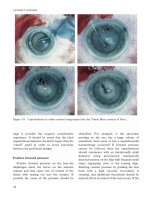
Figure 16.8 Holmium-YAG laser is being used to
ablate nasal mucosa, just anterior to the area of
transillumination, during left endonasal laser DCR.
Figure 16.3 a–16.7b Right endonasal surgical DCR:
16.3a The light beam is visible in the middle meatus on the lateral nasal wall; 16.3b view of light-pipe transillumination
with 30° Hopkins endoscope, Sϭseptum, LRϭlacrimal ridge, MTϭmiddle turbinate, Eϭendoscope.
16.4a A freer elevator is placed close to the lacrimal ridge, in preparation for raising the mucosal flap (visible
blood is from local anaesthesia); 16.4b the mucoperiosteal flap is raised with a freer elevator (F).
16.5a Blakesley forceps are used to grasp and excise nasal mucosa; 16.5b the lacrimal bone is removed with
Blakeseley forceps (B).
16.6a The lacrimal sac mucosa is opened with an angled keratome; 16.6b an angled keratome (K) is used to open
the lacrimal sac.
16.7a Silicon intubation is passed and knotted in the nasal space; 16.7b the intubation is retrieved from the nose
using curved artery forceps, STϭSilicone tube, Bϭbodkin.
Caldwell GW. Two new operations for obstruction of the
nasal duct with preservation of the canaliculi, and an
incidental description of a new lacrymal probe. Am J
Ophthalmol 1993; 10:189–95.
Camera JG, Bennzon AU, Henson RD. The safety and
efficacy of mitomycin C in endonasal endoscopic laser-
assisted dacryocystorhinostomy. Ophthal Plast Reconstr
Surg 2000; 16:114–18.
Gonnering RS, Lyon DB, Fisher JC. Endoscopic laser-assisted
lacrimal surgery. Am J Ophthalmol 1991; 111:152–7.
Hartikainen J, Grenman R, Puukka P, Seppa H. Prospective
randomised comparison of external dacryocystorhinostomy
and endonasal laser dacryocystorhinostomy. Ophthalmology
1998; 105:1106–13.
Jokinen K, Karja J. Endonasal dacryocystorhinostomy. Arch
Otolaryngol 1974; 100:41–4.
Massaro BM, Gonnering RS, Harris GJ. Endonasal laser
dacryocystorhinostomy. A new approach to nasolacrimal
duct obstruction. Arch Ophthalmol 1990; 108:1172–6.
McDonough M, Meiring JH. Endoscopic transnasal
dacryocystorhinostomy. J Laryngol Otol 1989; 103:585–7.
Metson R. The endoscopic approach for revision
dacryocystorhinostomy. Laryngoscope 1990; 100:1344–7.
Orcutt JC, Hillel A, Weymuller EA. Endoscopic repair of
failed dacryocystorhinostomy. Ophthal Plast Recontr Surg
1990; 6:197–202.
Rouviere P, Vaille G, Garcia C, Teppa H, Freche C, Lerault
P. La dacryocysto-rhinostomie par voie endo-nasale. Ann
Otolaryngol Chir Cervicofac 1981; 98:49–53.
Sadiq SA, Hugkulstone CE, Jones NSS, Downes RN.
Endoscopic holmium:YAG laser dacryocystorhinostomy.
Eye 1996; 10:43–6.
Sprekelsen MB, Barberan MT. Endoscopic dacryo-
cystorhinostomy: surgical technique and results.
Laryngoscope 1996; 106:187–9.
Steadman MG. Transnasal dacryocystorhinostomy.
Otolaryngol Clin North Am 1985; 18:107–11.
Szubin L, Papageorge A, Sacks E. Endonasal laser assisted
dacryocystorhinostomy. Am J Rhinol 1999; 13:371–4.
Weidenbecher M, Hoseman W, Buhr W. Endoscopic
endonasal dacryocystorhinostomy: results in 56 patients.
Ann Otol Rhino Laryngol 1994; 103:363–7.
West JM. A window resection of the nasal duct in cases of
stenosis. Trans Am Ophthalmol Soc 1909-11; 12:654–8.
West JM.The intranasal lacrimal sac operation. Its advantages
and its results. Arch Ophthalmol 1926; 56:351–6.
Whittet HB, Shun-Shin GA, Awdry P. Functional
endoscopic transnasal dacryocystorhinostomy. Eye 1993;
7:545–9.
Woog JJ, Metson R, Puliafito CA. Holmium:YAG endonasal
laser dacryocystorhinostomy. Am J Ophthalmol 1993;
116:1–10.
Yung MW, Hardman-Lea S. Endoscopic inferior
dacryocystorhinostomy. Clin Otolaryngol 1998, 23:152–7.
Zilelioglu G, Ugurbas SH, Anadolu Y, Akiner M, Akturk T.
Adjunctive use of Mitomycin C on endoscopic lacrimal
surgery. Br J Ophthalmol 1998; 82:63–6.
173
LASER-ASSISTED and ENDONASAL LACRIMAL SURGERY
174
Lacrimal canalicular obstruction presents a
difficult area for assessment and treatment
and the management of traumatic telecanthus
and canthal dystopia also falls within this
setting. The management of acute lacrimal
trauma, including canalicular lacerations, is
covered in Chapter 2.
Assessment
The assessment of the lacrimal system is
similar to that for more simple lacrimal
disorders (Chapter 15) but, in addition, a
more extensive assessment of the eye, eyelids,
medial canthus and lacrimal system is
essential to establish a plan of management. In
addition, the nasal structure and cavity should
also be carefully examined.
Lacrimal canalicular obstructions may
rarely be idiopathic, but are generally the
result of infection (primary Herpes simplex
and zoster, or Actinomyces canaliculitis),
trauma (direct, iatrogenic or irradiation),
cicatrising mucous membrane diseases
(pemphigoid, chronic ocular medication, or
topical drug reactions such as Stevens-
Johnson syndrome), or involvement with
tumours (papillomas or secondary to skin
tumours). With these causes in mind,
associated abnormalities should be sought
during the ocular examination; for example, in
the presence of a progressive disease such as
ocular pemphigoid, it may be undesirable to
place a canalicular bypass tube for fear of
17 Specialist lacrimal surgery and
trauma
Alan A McNab
exacerbating inner canthal scarring or
worsening an underlying dry eye syndrome.
The shape and position of the medial
canthus should be assessed and, if abnormal,
the lateral or vertical displacement should be
measured relative to the midline and compared
with the other side (if normal); the normal
adult intercanthal distance is about 30mm, or
15mm from the midline to each canthus. The
shape of the canthus may be relevant both for
cosmesis and, where required, for the
likelihood of being able to successfully place a
lacrimal canalicular bypass tube.
Clinical assessment of the lacrimal system is
directed towards establishing at what level
obstruction lies (Chapter 10).With canalicular
obstructions the length of patent canaliculus,
both upper and lower, should be measured; the
critical length in planning surgery is 8mm.
Where there is at least this amount of one
remaining canaliculus, it is generally feasible to
perform a canaliculo-dacryocystorhinostomy
(canaliculo-DCR). If there is less than 8mm, a
Lester Jones canalicular bypass tube may be
required unless the obstruction lies in the
proximal canaliculus; in the latter instance the
distal remnants of the canaliculi may be
normal and may be opened into the tear lake
by retrograde probing from within the lacrimal
sac and canaliculostomy, or by direct cut-down
along the eyelid margin and intubation of the
openings. Although such procedures may be
performed without dacryocystorhinostomy, it
is more logical to perform DCR at the time of
175
SPECIALIST LACRIMAL SURGERY and TRAUMA
primary canalicular surgery. DCR not only
increases canalicular conductance by having
bypassed the physiological resistance of the
nasolacrimal duct, but adequate primary
rhinostomy allows the relatively straight-
forward closed placement of a canalicular
bypass tube should the primary canalicular
surgery fail to control symptoms.
Where there is blockage of each individual
canaliculus, the length of patent canaliculus can
be estimated clinically and dacryocystography is
not possible. With common canalicular
obstruction, where syringing leads to reflux of
dye-free fluid from the opposite punctum
(Chapter 10), a dacryocystogram is helpful in
establishing the extent of common canalicular
disease. Lateral obstruction, with complete
obliteration of the common canaliculus,
requires canaliculo-DCR whereas medial
obstruction, due to adherence and fibrosis of
the mucosal valve over the common canalicular
opening, may be dealt with by excision of the
membrane at the time of DCR and intubation.
There is no need for CT of the facial
skeleton when considering lacrimal surgery
after previous mid-facial trauma, provided
that the presence of a nasal space alongside
the site of future rhinostomy is established by
clinical inspection or nasal endoscopy. Where
there has been major facial trauma, however,
CT of this region may be useful in case other
procedures – such as septoplasty, sinus
surgery or intercanthal wiring – are to be
combined with the lacrimal reconstruction.
Surgical options
Canaliculo-dacryocystorhinostomy
(CDCR)
Canaliculo-dacryocystorhinostomy is
indicated where there is bicanalicular block
with canalicular obstruction situated a
minimum of 8mm from at least one of the
puncta and for lateral common canalicular
block, in which several millimetres of common
canaliculus have been obliterated by scar
tissue. The principle of the procedure is to
excise the block of scar tissue and unite the
medial end of one or both canaliculi to the
nose, using the lacrimal sac mucosa as a
bridging flap; the operation, although
technically feasible, carries a much lower
success rate than the standard external DCR
or surgery for a more medial common
canalicular obstruction (Chapter 15) and
closed placement of a Lester Jones canalicular
bypass tube may be required later if the
operation fails.
A standard DCR incision is made but,
before mobilising the lacrimal sac and
periosteum, probes are placed in the blocked
canaliculi, and the overlying medial canthal
tendon divided and dissected laterally using
blunt or sharp dissection (Figure 17.1a). This
dissection is continued laterally until the tips
of the canalicular probes are revealed in the
underlying tissues, the ends of the canaliculi
are transected at their most medial point and
fine silicone tubing inserted and pulled
laterally, to aid in retraction. If only one
canaliculus is patent, either a monocanalicular
stent can be used, or the other end of a
bicanalicular intubation can be returned to
the nasal space through a “blind” passage (via
the punctal annulus, if present), ensuring
entry into the nasal space well away from the
one remaining functional canaliculus.
A large rhinostomy is created and nasal
mucosal flaps fashioned (Figure 17.1b); with
canaliculo-DCR, however, a very large
anterior nasal flap is required and relatively
small posterior flap. The lacrimal sac is
opened, not in the mid-part of its medial
wall – as with ordinary DCR (Chapter 15) –
but much more anteriorly at the junction of
the medial wall and anterior border; this
allows the lacrimal sac to be “unfurled”
posteriorly to create a large bridging flap
between the back wall of the canaliculi and the
small posterior nasal flap. The canalicular
mucosa is united to the small anterior edge of
PLASTIC and ORBITAL SURGERY
176
the lacrimal sac mucosa with two or three 8/0
absorbable sutures and the posterior edge of
the sac sutured to the nasal mucosa using 6/0
absorbable sutures.The canalicular intubation
is knotted as with a standard DCR, passed
into the nasal space and the anterior mucosal
anastomosis between the large nasal flap and
the anterior edge of the canalicular remnants
completed with multiple 8/0 absorbable
sutures (Figure 17.1c); it is important to avoid
snagging the intubation with the cutting edge
of the needles whilst performing the anterior
mucosal union. Finally the DCR wound is
closed in a standard fashion and the
intubation left in place for several months. If
watering continues at 9–12 months after
canaliculo-DCR, closed placement of a Lester
Jones canalicular bypass tube is required.
Complications
Although canaliculo-DCR has the same
spectrum of complications as simple DCR
(Chapter 15), the commonest specific
complication is failure of tear drainage due to re-
obstruction of the fine surgical anastomosis.
Trephination and silicone intubation may be
tried where the obstruction is a small membrane,
but in most cases the closed placement of a
Lester Jones bypass tube will required.
Dacryocystorhinostomy with
retrograde canaliculostomy
Indications
This procedure is designed to open onto
the lid margin, in the region of the medial
tear lake, canaliculi that are blocked within
their first 6–7mm but are patent in the distal
part; it being, of course, only possible to
establish this at the time of surgery and so the
patient must be warned that a glass
canalicular bypass tube might be required if
there is insufficient canaliculus to allow a
retrograde canaliculostomy.
Figure 17.1 Representation of canaliculo-
dacryocystorhinostomy:
(a) The lacrimal sac in its fossa with an obstruction of
the distal canaliculus and the area of bone removal for
the rhinostomy (hatched).
(b) The sites of incision marked: “a” denotes an
incision across the most medial portion of the patent
canaliculus, “b” the incision in the anterior aspect of
the lacrimal sac and “c” the incision in the nasal
mucosa to make a large anterior mucosal flap and a
small posterior flap.
(c) The anastomosis performed with the anterior nasal
mucosal flap sutured to the canaliculus and the
lacrimal sac opened out and sutured as a “bridging
flap” between the nasal mucosa and the posterior
edge of the lacrimal canaliculi.
a
b
c
(a)
(b)
(c)
177
SPECIALIST LACRIMAL SURGERY and TRAUMA
A standard external DCR is performed to
the point of opening the lacrimal sac and
suturing of the posterior mucosal flaps
(Chapter 15). The common canalicular
opening is located in the usual fashion and a
“0” gauge lacrimal probe, bent perpendicularly
on itself at about 8–10mm from the end, is
then passed from the sac, into the common
canaliculus (Figure 17.2a) and as far laterally
as possible along each canaliculus. The probe
is pushed up against the lid margin and a cut
down made onto the end of the probe,
opening the canaliculus onto the lid margin
(Figure17.2b) and the same manoeuvre
repeated for the other canaliculus, if possible.
The “false” puncta are intubated and the
DCR completed in a standard fashion; if only
one canaliculus is present, the other end of the
intubation is returned to the nasal space
through a “blind” passage. A monocanalicular
stent placed in the pseudo-punctum is
unlikely to remain in position in the absence
of the normal punctal annulus.
The intubation can be removed when the
epithelium of the canaliculus and the
conjunctiva have united and there is little need
to leave them more than 3–4 weeks, or the
tubes will tend to “cheese wire” through the
tissues and cause a medial cross-union
between the eyelids. If the pseudo-puncta fail
to control symptoms, closed placement of a
canalicular bypass tube is required.
Dacryocystorhinostomy and Jones
canalicular bypass tube
The canalicular bypass tube is designed to
establish tear drainage, from the medial tear
lake into the nose, by way of a false conduit;
the most used device being the Pyrex glass
(Lester Jones) canalicular bypass tube.
Placement of a bypass tube is indicated where
the extent of canalicular obliteration is such as
to preclude either canaliculo-DCR or DCR
with retrograde canaliculostomy, or where
watering continues in the face of a functioning
standard DCR – as, for example, in patients
with facial nerve palsy.
As with DCR and retrograde canaliculo-
stomy, a standard external DCR is performed
to the stage of suturing the posterior mucosal
flaps although, if no lacrimal sac is present or
flaps cannot be formed, the posterior nasal
mucosa should be sutured to the soft tissues of
the lacrimal sac fossa. Although a large
rhinostomy is important for all lacrimal
surgery (Chapter 15), it is particularly
important when placing a canalicular bypass
tube, or the tube tends to become displaced
due to its bearing on the bone at the lower
edge of the rhinostomy. The nasal cavity
should also be examined and, if necessary, the
anterior part of the middle turbinate should be
(a)
(b)
Figure 17.2 Retrograde canaliculostomy during
dacryocystorhinostomy: (a) the angled “0” gauge
probe is being directed towards the internal opening
of the common canaliculus in the left lacrimal sac;
(b) a cut-down on the lid margin is directed onto
the most lateral point reached by the probe placed
retrogradely into the canaliculus.
PLASTIC and ORBITAL SURGERY
178
de-boned or trimmed to make extra room for
the medial end of the tube.
If retrograde exploration of the common
canaliculus fails to reveal any useful tissues for
retrograde canaliculostomy, a carunculectomy
may be performed with care being taken to
avoid damage to the plica semilunaris. A sharp
guide wire is inserted from the medial canthus
into the nose (Figure 17.3).The point of entry
is critical to the functional success of the tube
and should be at the level of the undisplaced
lower eyelid margin, at the site of
carunculectomy. An alternative site is the
lateral 2–3mm of the canaliculus which, if
present, can be laid open to accommodate the
lateral flanged end of the bypass tube. The
wire marks the future track of the tube and is
directed about 15–25° downhill in the coronal
plane, the medial end passing into the nose in
the vicinity of the lacrimal sac fossa.
A small trephine (1·5–2mm diameter) is
passed over the guide wire to remove a narrow
core of tissue and, whilst the trephine is in the
tissues, the sharp wire may be replaced with a
blunt one. A glass canalicular bypass tube is
slipped over the guide wire and pushed firmly
through the tissues so that the distal end is at
least 2mm clear within the nasal space; the
end of a thumb-nail should be used to drive
the tube into the tissues, as instruments tend
to shatter the glass flange of the tube. The
neck of the tube is encircled with three turns
of a 6/0 nylon suture, passed through the
medial end of the lower lid and tied over the
bolster – this suture lifting the tube slightly
laterally to allow epithelial healing around
(and not over) the lateral end of the bypass
tube. In most cases a 12mm tube with a
3·5mm flange is suitable for caruncular
placement, whereas a somewhat longer
(16mm) tube may be needed where the neck
of the bypass tube is placed within the lateral
canalicular remnant.
The anterior mucosal anastomosis and
surface closure is completed as with standard
external DCR. The encircling suture is
removed at 7–10 days after surgery, at the time
of suture removal from the skin incision.
Canalicular bypass tubes are subject to a
number of common complications and
require regular monitoring and maintenance,
otherwise they become caked or blocked with
mucus debris from the tear film and this
results in recurrent conjunctivitis. Patients
should be encouraged to sniff water from the
tear lake, through the tube and into the nose,
on a daily basis and they should also be taught
to place one of their fingers over the tube
when violently sneezing or nose blowing.
Figure 17.3 A system for insertion of a Lester Jones
bypass tube using a guide wire, trephine and
“dipstick”. (Reproduced with permission from Morlet
GC. A modern approach to lacrimal surgery. Aust NZ
J Ophthalmol 1988; 16:202.)
179
SPECIALIST LACRIMAL SURGERY and TRAUMA
Lateral migration of the tube occurs
most often and the tube will sometimes
be completely dislodged, when closed
replacement should be undertaken. Repeated
episodes of tube extrusion generally occur due
to residual bone in the area of the rhinostomy
and, in such cases, open revision of the
rhinostomy should be undertaken. More rarely
the tube will sink medially into the tissues and
may require a cut-down to retrieve it.
Malposition of the ocular end of the tube
may result in failure of tear drainage where the
tube is too anterior or, more usually, the tube
is too posterior and becomes embedded in
conjunctiva or against the globe, causing
episcleritis. In more extreme cases, the
irritation will cause formation of a pyogenic
granuloma – this being particularly
troublesome where the tube has become filthy
through neglect. In such cases of malposition
with secondary inflammation, the tube should
be removed and replaced in a better position
at a later date when the inflammatory changes
have settled.
Build-up of tear-film debris on the surface
of a bypass tube tends to lead to obstruction
and repeated ocular infections. If the
obstruction cannot be cleared with the tube in
place, the device should be removed and
cleaned, or else replaced.
Closed placement of a canalicular
bypass tube
Indications
Closed secondary placement of a glass
canalicular bypass tube is indicated when a
previously inserted bypass tube has become
dislodged, after failed canaliculo-DCR or
retrograde canaliculostomy, or where, in the
absence of reflex lacrimation, a functioning
DCR fails to control watering.
The procedure is similar to a primary (open)
bypass tube, except that the DCR has already
been performed and the rhinostomy does not
have to be opened; in other words, the tube is
inserted in a closed fashion. For optimum
positioning of the distal end of the bypass tube,
nasal examination is required and is best
achieved with endoscopy, although a good
headlight and nasal speculum are often
adequate. A 3mm diameter rigid sucker is
required to clean the nasal space during
surgery. The procedure is best performed
under general anaesthesia as vasoconstriction
from the endonasal local anaesthesia
encourages, due to an artificially shrunken
middle turbinate and septal mucosa, a
misjudgement of the position of the nasal end
of the tube.
First-time placement of a closed Jones’
tube is similar to the open canalicular bypass
tube, using and positioning the guide wire
and trephine in the same way; the intranasal
position of the introducer should be checked
endoscopically and, if necessary, the anterior
part of the middle turbinate removed to make
room for the tube. If there has recently been
a satisfactorily functioning tube, the double-
ended (“bullhorn”) dilator that accompanies
the commercial sets of tubes may be used to
dilate the previous track and the tube forced
into place along an “0” gauge probe
introduced into the dilated track. After
placement of any bypass tube, the position of
both the ocular and the nasal ends of the
unsupported tube should be checked and it is
particularly important to verify that the nasal
end lies free within the nasal cavity and not
up against the septum, lateral wall or
turbinate. A newly-placed tube needs to be
secured in the same way as a primary tube, but
a replacement tube does not need fixation.
Medial canthoplasty during
lacrimal surgery
Where injury to the lacrimal drainage
system has been accompanied by significant
midfacial trauma, there may be traumatic
telecanthus or canthal dystopia and
repositioning of the canthus may be required
PLASTIC and ORBITAL SURGERY
180
as part of the lacrimal surgical repair.
Malpositions of the canthus secondary to
trauma are, however, notoriously difficult to
correct and, despite the best efforts, the
canthus tends to drift back towards its
previous position.
Traumatic telecanthus may be due to a
widening of the fractured midfacial skeleton
and the first step required may be to remove
excess bone at the inner canthi, this then
presenting difficulty with the medial fixation of
the inner canthi in the absence of a firm bony
anchor. In this setting a transnasal wire is
helpful (Chapter 6) or a small T-plate or anchor-
screw – to which to fix the canthal tendon – may
be inserted into the remaining fragments of the
nasal bones. To facilitate a medial repositioning
of the lids, it is important to widely mobilise the
medial attachments of the eyelids.
If canthal repositioning is being performed
at the time of open lacrimal surgery, advantage
can be made of exposure of the posterior
lacrimal crest and this fascia used as an anchor
point for elevating the medial end of the
lower eyelid. Although some results will be
encouraging (Figure 17.4), on occasion
this site of postero-superior fixation lacks
rigidity.
The superficial tissues may need to be
redistributed at the time of surgery. In
traumatic canthal dystopia, the canthus is
generally shifted downwards and a triangular
pedicle flap of skin, based medially on the side
of the nasion, may be transposed from the
upper eyelid to the lower; this thereby helping
to raise the inner canthus by correcting any
vertical shortage of tissues below the medial
canthus (Figure 17.5).
Figure 17.4 Medial canthoplasty performed at the
time of open lacrimal surgery: (a) pre- and (b) post
surgery. Although there is an improvement in the
canthal position and elevation after postero-superior
fixation of the lower eyelid, this is limited by scarring
at the site of the previous injury.
(a)
(b)
A
(a)
A
(b)
Figure 17.5 Redistribution of the soft tissues as part
of a medial canthoplasty for inferior displacement of
the medial canthus. The apex ‘A’ of the flap of skin
and muscle is transposed from the upper eyelid (a)
into the lower (b) after fixing the canthal structures
deeply to bone or periosteum.
181
SPECIALIST LACRIMAL SURGERY and TRAUMA
Further reading
Bartley GB, Gustafson RO. Complications of malpositioned
Jones tubes. Am J Ophthalmol 1990; 109:66–9.
Call NB, Welham RAN. Epiphora after irradiation of medial
eyelid tumors. Am J Ophthalmol 1981; 92:842–5.
Chapman KL, Bartley GB, Garrity JA, Gonnering RS.
Lacrimal bypass surgery in patients with sarcoidosis. Am J
Ophthalmol 1999; 127:443–6.
Coster DJ, Welham RAN. Herpetic canalicular obstruction.
Br J Ophthalmol 1979; 63:259–62.
Henderson ON. A modified trephining technique for
insertion of Lester Jones tube. Arch Ophthalmol 1985;
103:1582–5.
Hicks C, Pitts J, Rose GE. Lacrimal surgery in patients with
congenital cranial or facial anomalies. Eye 1994; 8:583–91.
Jones BR. The surgical cure of obstruction of the common
canaliculus. Trans Ophthalmol Soc UK 1960; 80:343–56.
Kwan ASL, Rose GE. Lacrimal drainage surgery in Wegener’s
granulomatosis. Br J Ophthalmol 2000; 84:329–31.
McLean CJ, Rose G.E. Post-herpetic lacrimal obstruction.
Ophthalmology 2000; 107:496–9.
McNab AA. Lacrimal canalicular obstruction associated
with topical ocular medication. Aust NZ J Ophthalmol
1998; 26:219–24.
McNab AA. Diagnosis and investigation of lacrimal disease.
In: McNab AA, ed, Manual of orbital and lacrimal surgery
(2nd ed). Oxford: Butterworth Heinemann, 1988: 91–8.
Morlet GC. A modern approach to lacrimal surgery. Aust
NZ J Ophthalmol 1988; 16:199–204.
Rose GE, Welham RAN. Jones’ lacrimal canalicular bypass
tubes: twenty-five years’ experience. Eye 1991; 5:13–19.
Sanke RF, Welham RAN. Lacrimal canalicular obstruction
and chicken pox. Br J Ophthalmol 1982; 66:71–4.
Steinsapir KD, Glatt HJ, Putterman AM. A 16-year study of
conjunctival dacryocystorhinostomy. Am J Ophthalmol
1990; 109:387–93.
Wearne MJ, Beigi B, Davis G, Rose GE. Retrograde
intubation dacryocystorhinostomy for proximal and mid-
canalicular obstruction. Ophthalmology 1999; 106:2325–8.
182
A-sutures 13, 13
accentuation measurement in ptosis 33
achrocordon 45
acne rosacea 108
actinomyces canaliculitis 107, 108, 108, 110, 110
adenocarcinoma 147
adenoid cystic carcinoma 142, 147
adenoma
pleiomyorphic 132, 132
sebaceous 45
adnexal benign tumours 45
adrenaline addition to anaesthesia 4
advanced trauma life support (ALTS) system 7
airgun pellet foreign body 157
amaurosis, gaze-evoked 99
amblyopia 126
from lymphangioma 129
prevention 11
in ptosis 34
anaesthesia
for cosmetic surgery 70–80
for dacyrocystorhinostomy 161–2
for endonasal dacyrocystorhinostomy 169, 169
for eye surgery 5
anatomy of eye 1–6
angiography
in intraorbital arterio-venous malformations 130
in orbital disease 106
anisometropia 126
annulus of Zinn 4, 4
anophthalmos 95–6
anterior lamella 1
repair 11
repositioning with everting sutures 29
anterior orbitotomy 133–4
anterior venous anomalies 102
antibiotic
and central ectropion 16
following dacyrocystorhinostomy 166
for orbital cellulitis 135
prophylaxis in bite wounds 9
following surgery for dysthyroid eye disease 121
anti-inflammatories following surgery for dysthyroid
eye disease 121
anti-mitotic agents 12
antitetanus prophylaxis 7
aponeurosis 2
complications 40
surgery 39–40, 40
tuck 40, 40
aponeurotic ptosis 37
arterio-venous communications, orbital 130–1
arterio-venous malformation 102, 102
ultrasonography 104
aspirin, withdrawal prior to surgery 21
astigmatism 126
B-cell lymphoma 144
basal cell carcinoma 45–7, 46, 146–7
common 46, 47
complex 47
management principles 51
radiation 53, 54
Bell’s palsy
assessment in ptosis 34
prognosis 67
symptoms 67
benign tumours 45, 123–39
misdiagnosis 52
simulating malignancy 48
types 45
Bick procedure, modified 16–17, 17
bicoronal scalp-flap for orbital decompression 117,
118, 119, 119–21
biopsies
incisional 133
in orbital disease 106
of tumours 45
bite wounds 9
blepharitis
causing trichiasis 30
marginal 107–8, 108
Index
Page numbers in bold refer to figures; those in italic refer to tables or boxed material
INDEX
183
blepharo-conjunctivitis and sebaceous gland
carcinoma 49
blepharo-keratitis causing epiphora 108
blepharophimosis syndrome 35
blepharoplasty 82–5
complications 85
laser use 87
lower lid 17
skin-reduction, in dysthyroid eye disease 117
subciliary in dysthyroid eye disease 119
blepharoptosis see ptosis
blepharospasm 27
blindness
following blepharoplasty 85
following lateral orbitotomy 129
from surgical optical trauma 159
blood supply to eye 5
“blowout” fracture 4, 150–5, 151
imaging 104
blue naevus 45
bone invasion of orbit, imaging 104
bone-removing orbital decompression in dysthyroid
eye disease 117–21
bony orbit 4, 4
botulinum toxin for spastic entropion 27
brain abscess and intraorbital foreign bodies 157
Breslow melanoma classification 50
bronchogenic carcinoma 103
brow see eyebrow
browpexy 80–1, 80
browplasty 81
bruising after ectropion surgery 21–2
“bullhorn” dilator 179
Burkitt’s lymphoma 144
Burrow’s triangle 59
canalicular bypass tube 174, 177–9, 178
closed placement 179
canalicular configuration 3
laceration repair 11–12
canaliculo-dacycystorhinostomy 174, 175–6,
176
canaliculostomy, retrograde, dacycystorhinostomy
with 176–7, 177
canthal corner, anterodisplacement 22
canthal tendons 2, 15
laxity see ectropion
medial anterior 19, 19
medial posterior 19–20, 20
medial resection 20, 20
plication 18
canthloysis
for lower lid reconstruction 57, 57
for upper lid reconstruction 62, 62
canthoplasty, medial 69–70, 69
during lacrimal surgery 179–80
canthotomy
lateral, orbital decompression 117, 118, 118–19,
119, 120
for lower lid reconstruction 57, 57
for upper lid reconstruction 62
canthus assessment 174
capillary haemangioma 45, 126–7, 126
differential diagnosis 140
carbon dioxide lasers 85–6
skin resurfacing 86–7, 87
carcinoma
metastasising to orbit 148, 148
see also malignant tumours
carotico-cavernous fistula 102, 131, 131
carotid arteries 5
carunculectomy 178
cavernous haemangioma 127–9, 127
complications 129
cellulitis
orbital 134–5, 134
in rhabdomyosarcoma 140, 140
cephalocoeles 125–6
cerebrospinal fluid leak following
dacyrocystorhinostomy 166
chalazion and sebaceous gland carcinoma 48
cheek rotation flap 60–1, 61
chemosis 102, 102, 131
in dysthyroid eye disease 115, 116
chemotherapy for idiopathic orbital inflammation
136
children
amblyopia prevention 11
malignant orbital disease 140–2
chloroma see leukaemia, acute myeloid
“chocolate cysts” 129
cholesterol granuloma imaging 106
choroidal folds 99
choroidal striae 103
cicatrical entropion 22, 27
avoiding 5
diagnosis 13
treatment 13
Clark melanoma classification 50
cocaine test for Horner’s syndrome 38
Coherent CO
2
laser 85
colour perception tests in orbital disease 100
computed tomography (CT) imaging
cavernous haemangioma 126, 126
dacryoadenitis 133
dermoid cysts 124
dysthyroid eye disease 113–14, 114, 114
foreign bodies 156, 156
idiopathic orbital inflammation 136
lacrimal gland carcinoma 142, 143
lacrimal sac 110
lacrimal surgery 175
lymphangioma 129
optic nerve glioma 137
optic nerve meningioma 138
orbital 104
orbital floor fracture 151, 151
orbital lymphoma 144–5, 145
INDEX
184
computed tomography (CT) imaging – Continued
orbital varices 130
sphenoid wing meningioma 138
conformer 91
congenital clefts of skull 125–6
congenital ectropion 15–16
congenital myogenic ptosis 35–7
congenital naevus 45
congenital ocular fibrosis syndrome and ptosis 36
conjunctiva
bulbal graft 65
chemosis 102, 102
in dysthyroid eye disease 115, 116
exposure problems 16
inclusion cysts 97
prolapse and ptosis surgery 40–1
tight 73
conjunctivodacryocystorhinostomy 12
corkscrew episcleral vessels 102
cornea
abrasion in Fasanella–Servat procedure 43
exposure 16, 73
features in dysthyroid eye disease 113
limbus 1
protection 7–8, 74–7
sensation assessment in ptosis 34
cosmetic defects in facial palsy 67–8
management 68
cosmetic surgery 78–88
patient evaluation 78–80
cranial anomalies 125–6
Crawford method for eyebrow suspension 41, 41
“crocodile tear” syndrome 67, 108
cryotherapy
for malignant eyelid tumours 54
complications 54
for trichiasis 31
CT see computed tomography
cutaneous horn 45
Cutler–Beard reconsutrction 63–4, 64
cyst
excision for ectropion 21
malignant 46
microphthalmos with 125
orbital benign 123–5
dacryoadenitis 100, 102, 132–4, 133
differential diagnosis 142
dacryocoele 131–2
dacryocystitis 97
dacryocystography 109–10, 110
dacryocystorhinostomy 12, 110
anaesthesia 161–2
endonasal 168–73
indications 168–9
postoperative management and complications
170–2
results 168
technique 169–70
complications 166–7, 166
indications 161
and Jones canalicular bypass tube 177–9
post-operative management 165–6
with retrograde canaliculostomy 176–7, 177
surgical technique 162–5
vasoconstriction and haemostasis 162
dacryops see dacryocoele
debridement, minimal 8
dermatofibroma 45
dermis
benign tumours 45
fat (dermofat) grafts 14, 92–3
complications 93
dermoid cysts 100, 101
benign 123–4, 124
imaging 106
dermolipoma, benign 124–5
Dexon sutures 8
diplopia
following blepharoplasty 85
following surgery in dysthyroid eye disease 121
in orbital floor fracture 151
in orbital disease 99
dirt removal 7–8
distichiasis 31
Down’s syndrome, ectropion 15
down-gaze restriction in orbital floor fracture 151,
152
drainage see lacrimal drainage
dry eye following surgery for lacrimal gland
carcinoma 143
duction test 100
dural shunts 130–1, 131
low-flow 102, 102
dysthyroid eye disease 100, 102, 112–22
features 112–14, 113
pathogenesis 112
postoperative management and complications 121
treatment 114–21
see also thyroid orbitopathy
ear cartilage harvesting 30
ecchymosis in orbital disease 99
ectropion 15–23
acquired 16
caused by entropion surgery 22
central 16–17
cicatricial 21
avoiding 5
diagnosis 13
treatment 13
classification 15
complications 21–3
congenital 15–16
entropion following surgery for 22–3
following blepharoplasty 85
involutional 16
lateral 17–18
INDEX
185
management 68
mechanical 20
medial 18–20
paralytic 21
and ptosis surgery 41
punctal 108
recurrent 22
electrolysis for trichiasis 31
encephalocoeles 126
endonasal lacrimal surgery 168–73, 171–2
enophthalmos 99, 150, 151
following enucleation 94
following surgery in dysthyroid eye disease 121
entropion 24–31
acquired 25, 26
caused by ectropion surgery 22
cicatricial 22, 27, 29
classification 25
congenital 25
involutional 25, 27
spastic 27
surgery
ectropion following 22
for lower lid 27–9
for upper lid 29–30
enucleation 89–91, 91
indications 12, 89
from injury 157, 158
volume deficiency following 94–5
eosinophilic granuloma 142
epiblepharon 25
epidermis, benign tumours 45
epidermoid cysts, benign 123–4
epiphora
assessment 107–9
in ectropion 18
in orbital disease 100
episcleral vessels, corkscrew 102
episcleritis 134
Erbium YAG laser 85–6, 87–8
estheioneuroblastoma 147
ethibond 9
ethmoid
fracture 151
mucocoele 125, 125
ethmoidoectomy incision, Lynch external 117–18, 119
everting technique
for ectropion 27–8
for entropion 8
see also ectropion; entropion
evisceration 89, 90
Ewing’s sarcoma, metastatic 144
excretory system of eye 3–4
exenteration 91, 146, 147
exophthalmos 150
expanders for congenital small socket 96
exposure symptoms in orbital disease 100
extraconal space 4, 5
extraocular muscles
damage 157
features in dysthyroid eye disease 113
function measurement in ptosis 33–4
eye ointment for central ectropion 16
eye socket see socket
eyebrow
configuration analysis 78
lift, direct 68, 72–3, 72
ptosis 79,80
suspension in 41–2
surgery, cosmetic 80–82
coronal lift 81–2
direct lift 81
endoscopic forehead and brow lift 82
internal fixation 80–1, 80
mid forehead list 81
temporal lift 81
eyelash
distorted 30
dysplastic 30
follicles 25
ptosis 41
see also trichiasis
eyelid
anatomy 1–3, 1, 24–5, 24–5
assessment for cosmetic surgery 78–9
asymmetry following blepharoplasty 85
basal cell carcinoma 45–7, 46
common patterns 46, 47
complex 47
management principles 51
benign tumours 45
closure, causes of inadequacy 73–4
strength measurement in ptosis 34
ectropion see ectropion
entropion see entropion
fatiguability measurement in ptosis 34
features in dysthyroid eye disease 113
full-thickness loss 65
guard 7–8
inversion see entropion
Kaposi’s sarcoma 50, 50
management principles
lacerations 10–11
lag in thyroid orbitopathy 101
laxity 16
causing epiphora 108
malignant tumours 146–7
cryotherapy 54
features 44
management principles 50–3
Mohs’ micrographic surgery 51, 52, 54–5
radiation 52, 53–4
margin 1
melanoma 49–50
management principles 52–3
metastatic tumours 50
management principles 53
movements, aberrant/synkinetic, in ptosis 34
INDEX
186
eyelid – Continued
oriental 25
peaked, in Fasanella–Servat procedure 42
position measurement in ptosis on downgaze 35
problems causing discharge following surgery 97
rare tumours 50
reconstruction 55–65
lower 56–62
upper 62–5
retractors 1,2
tight 73
reattachment to tarsal inferior border 20–1
sebaceous gland carcinoma 48–9, 48
management principles 52
signs in orbital disease 101–2
skin preservation in exenteration 91
squamous cell carcinoma 47–8, 48
management principles 51–2
stability 24–5
surgery, cosmetic 82–5
see also under ectropion; entropion
tightening, decisions 15
tissue loss 157, 158
trauma and reconstruction 7–14
tissue preservation 8
see also lower eyelid and upper eyelid
facial asymmetry analysis 78
facial palsy see seventh nerve palsy
facial soft tissue and bone clefting 104, 105
facial trauma, tissue preservation 8
Fasanella–Servat procedure 38, 42–3, 42
fascia lata for eyebrow suspension 41
fascial sling, lower lid, following enucleation 95
fat
excision problems 85
pads, eyelid 2
fatiguability of eyelid measurement in ptosis 34
festoons in thyroid orbitopathy 101
fibroblast activity and scarring 12
fibrosarcoma, orbital 144, 144
fingernail clubbing 103
Fitzpatrick’s classification for skin colouring 79
“flying man” incision 35
follicular keratosis, inverted 45
forceps 9
foreign bodies
intraorbital 156–7
removal 7–8
fornix, interior, shallowing following enucleation 95
Fox method for eyebrow suspension 41, 41
frontalis muscle action measurement in ptosis 34, 35
full-thickness composite graft 64
gaze-evoked amaurosis 99
glands of Krause 3
glands of Moll 25
glands of Wolfring 3
glands of Zeiss 25
glioma, optic nerve 137, 137
globe
displacement in orbital disease 98–9, 99, 100
“frozen” 47
removal see enucleation
rupture 150
gold weight implantation in seventh nerve palsy 68,
71–2, 72
Goldenhar’s syndrome 124
graft see skin graft
granuloma
cholesterol, imaging 106
eosinophilic 142
formation from dermofat graft 93
pyogenic 45
Graves’ hyperthyroidism 112
“grey line” of lid margin 1
haemangioma
capillary 45, 126–7, 126
cavernous 127–9, 127
complications 129
haemangiopericytoma 144
imaging 106
orbital 148–9
haematoma
subperiosteal 159
following dacyrocystorhinostomy 165, 166
following ectropion surgery 22
following orbital floor repair 153–4
orbital 159
on weapon removal 10
haemostasis for dacyrocystorhinostomy 162
haemostats 9
hair follicle, benign tumours 45
hair growth on dermofat graft 93
hard palate
harvesting 30
spacer 76, 77
Hashimoto’s thyroiditis 112
head posture, compensatory, measurement in
ptosis 34
Hering’s law 33
histiocytoma, malignant fibrous 144
histiocytosis, Langerhans cell 141–2
history taking in orbital disease 98–103
Horner’s syndrome
miosis 33
ptosis 37–8
Hughes’ flap 58–60, 58–9
hydroxyamphetamine test for Horner’s syndrome 38
hydroxyapatite for orbital implant 92
hypermetropia 99
hyperthyroidism see Graves’ hyperthyroidism
hypoglobus following surgery in dysthyroid
eye disease 121
idiopathic orbital inflammation 135–7, 136
implant see orbital implant
INDEX
187
incision
for canaliculo-dacyrocystorhinostomy 176
for dacyrocystorhinostomy 163, 167
for eye surgery 5, 6
for lower lid surgery 1, 5
see also specific types, e.g. Mustarde’s “flying man”
incision
incisional biopsy 133
infection
following dacyrocystorhinostomy 166, 167
following orbital floor repair 154, 154
inflammatory disease, benign orbital 134–7
infraorbital nerve hypoaesthesia following orbital
floor repair 154
intranasal examination 109
intranasal tumour 109, 109
intraocular disease 102–3
intraocular tumours 148
intraocular pressure, raised, in orbital disease 102, 103
intraoperative care 5
intraorbital arterio-venous malformations 130
intraorbital foreign bodies 156–7
Ishihara colour plates 100
jaw-wink in ptosis 34, 38
Jones procedure 18, 28
canalicular bypass tube 174, 177–9, 178
closed placement 179
Kaposi’s sarcoma 50, 50
management principles
Kearns Sayre syndrome and ptosis 36
keratectomy and evisceration 89
keratitis
following orbital roof fractures 155
from dacryoadenitis 134
keratitis sicca in pleiomyorphic adenoma 132
keratoacanthoma 45
Kuhnt–Symanovsky type procedure 17
lacerations to eyelid 10–11
lacrimal canal obstruction 174
lacrimal drainage disease 106–11
anatomy 3–4, 3
ancillary tests 109–11
damage following orbital floor repair 154
and endonasal anastomosis 168
examination 107–9
history taking 107, 107
problems causing discharge 97
scintigraphy 110–11, 111
surgery and trauma 174–81
assessment 174–5
surgical options 175–81
external 161–7
laser-assisted and endonasal 168–73, 171–2
syringing 108–9, 109
lacrimal gland 3, 5
carcinoma 142–3
disease, benign 131–4
tumours 102
lacrimal instruments 9
lacrimal pump 4, 4
lacrimal sac 3
mucocoele 108
stone 110
tumour imaging 110
lagophthalmos
following blepharoplasty 85
following orbital roof fractures 155
following upper eyelid reconstruction 62
and proptosis 100
in ptosis 34, 40
in thyroid orbitopathy 101
lamellae, eyelid 1, 24, 24
repair 11
repositioning with everting sutures 29
lamina papyracea breakdown 146
Langerhanas cell histiocytosis 141–2
lasers
lacrimal surgery 168–73, 172
oculoplasty surgery 85–8
incisional surgery 87
skin resurfacing 86–8
lash see eyelash
lateral canthal tendon 2
lateral orbitotomy 127–9, 128
lateral tarsal strip for ectropion 17–18, 18
lateral tarsorrhaphy 68–9, 69
Lazy-T procedure 18, 19, 19
Le Fort fractures 155
leiomyosarcoma 144
lentigo senilis 45
lentigo simplex 45
Lester Jones see Jones
leukaemia, acute myeloid 141, 141
levator muscle
dysgenesis in ptosis 35
function measurement in ptosis preservation in
upper lid reconstructin 62
laceration repair 10
resection 39, 39
complications 40–1
levator palpebrae superioris 2
lines of minimum skin tension 5, 6
liposarcoma 144
Lisch nodules 102, 102
lower eyelid
blepharoplasty 83–5
anterior approach 83–4, 84
external direct 84–5
transconjunctival 84
cicatricial retraction following orbital floor repair
154
displacement in dysthyroid eye disease 117
fascial sling following enucleation 95
laxity 79
causing epiphora 107, 108
INDEX
188
lower eyelid – Continued
following enucleation 94
lesion excisions 1, 5
reconstruction 56–62
margin rotation and inversion 64–5
retractors 3, 24
recession 76–7
swinging flap 118
lymphadenopathy and orbital disease 103
lymphangiomas 129–30
lymphatic drainage from eye 5
lymphoid hyperplasia 144
lymphoid tumours 50
management principles 53
lymphoma in adults, orbital 144–6, 145
Lynch external ethmoidoectomy incision 117–18,
119
magnetic resonance imaging
cavernous haemangioma 126
dysthyroid eye disease 114, 114
foreign bodies 156
optic nerve glioma 137
orbital 105–6
malignant orbital disease 140–9
malignant tumour
clinical signs of malignancy 44
features 44
management and repair following excision 44–66
management principles 50–3
types 44
Marcus Gunn jaw-winking ptosis 38
margin–reflex distance (MRD) 32–3, 32
masquerade syndrome 48
mattress sutures 8
maxillary fractures 155
maxillary nerve 5
mechanical ptosis 38
medial canthal tendon 2
laceration repair 11
medial canthoplasty 69–70, 69
during lacrimal surgery 179–80, 180
medial wall
fracture 150–5
removal in dysthyroid eye disease 115–16, 116
medial wedge resection 70–1, 70
meibomian gland
eyelash growing from 31
tumours 146–7
melanoma 49–50
fungating 147, 147
management principles 51–2
nodular malignant 50, 50
orbital imaging 106
uveal malignant 148
meningioma
optic nerve 103, 137, 138
sphenoid wing 137–8, 138
meningocoeles 126
meningo-encephalocoeles 126
Merkel cell tumour 50, 51
MERRF see myoclonic epilepsy and ragged red fibre
syndrome
mesenchymal tumours 143–4
metastatic orbital disease 148, 148
microphthalmos 96
with cyst 125
mid-face fracture 155–6
miosis measurement in ptosis 33
mitochondrial myopathy and ptosis 36
Mohs’ micrographic surgery for malignant eyelid
tumours 51, 52, 54–5
“morning glory syndrome” 126
MRI see magnetic resonance imaging
mucocoele
frontal 100
imaging 106
lacrimal sac 108
paranasal sinus 124, 125
mucormycosis 102
in orbital disease 99
mucosal flaps in dacyrocystorhinostomy 164–5,
164–5
mucous membrane graft
for contracted socket 96
harvesting 30
with distal margin rotation 29–30
with tarsal dissection 29
Müller’s muscle 2
surgery in seventh nerve plasy 68, 71, 71
muscular dystrophy and ptosis 36
Mustarde’s “flying man” incision 35
cheek rotation flap 60–1, 61
myasthenia gravis and ptosis 36
mycosis fungoides 50
mydriasis in ptosis 33
myoclonic epilepsy and ragged red fibre syndrome
and ptosis 36
myogenic ptosis 35–7
myositis, orbital 135, 136
pain with 98
myotonic dystrophy and ptosis 36
naevocellular naevus 45
naevus 45
malignant 46
nasal mass 102
nasojugal fold 1
nasolacrimal duct 3–4
obstruction surgery 161–7
stenosis 108
needle holders 9
nerve supply to eye 5
neural lesions, benign 137–8
neurilemmomas 137
malignant 148
neuroblastoma 140, 141, 141
neurofibroma 45
INDEX
189
plexiform 137
neurofibromatosis
Lisch nodules 102, 102
peripheral 102, 103
neurogenic ptosis 37–8
neurological examination 150
neuropathy, optic 134
neuropraxia following lateral orbitotomy 129
non-Hodgkin’s B-cell lymphoma 144
NOSPECS classification of dysthyroid eye disease
112–13
nylon sutures 8
ocreotide scintigraphy in orbital disease 106
ocular balance and ductions 100
ocular duction following lateral orbitotomy 129
ocular fibrosis syndrome, congenital, and ptosis 36
ocular measurement in ptosis 35
oculopharyngeal dystrophy and ptosis 36
oculoplastic instruments 9
ophthalmia, sympathetic, prevention 12
ophthalmic nerve 5
optic canal decompression 158–9
optic nerve
activity score in dysthyroid eye disease 113
compression 99
injury 157–8, 158
lesion imaging 105
meningioma 103
neuropathy following orbital roof fractures 154
tumours, benign 137, 138
orbicularis fibres, lacerations near 10
orbicularis muscle 2, 24, 25
function and corneal exposure 73
spasm 15
orbita
anatomy 4–5
anomalies 125–6
arterio-venous communications 130–1
benign vascular anomalies 126–31
blowout fractures imaging 104
cellulitis 134–5, 134
from mucocoele 125
in rhabdomyosarcoma 140, 140
decompression, bone-removing, in dysthyroid eye
disease 117–21
in dysthyroid eye disease 115
disease 98–106
ancillary tests 103–6
benign 123–39
benign cysts 123–5
examination 100–3
history taking 98–100
malignant 140–9
metastatic 148, 148
presenting symptoms 98
exenteration 146, 147
fat
causing MRI problems 105
prolapse following dacyrocystorhinostomy 166
haemorrhage 159
imaging 105, 106
imaging in dysthyroid eye disease 113–14
inflammation, treatment in dysthyroid eye disease
115
inflammatory disease, benign 134–7
idiopathic 135–7, 136
inflammatory syndrome 102
lobe tumours 132
myositis 135, 136
roof fracture 155–6, 155
septum 2
soft tissues injury 157–9
trauma 150–60
surgical 155, 159, 159
tumour, imaging 104, 106
rare primary malignant, in adults 148–9
secondary 146–8
signs 103
varices 129, 130, 130
orbital floor
fracture 150–5
assessment 151–2
complications 153–5
management 152
repair 152–3
trauma imaging 104, 104
orbital implant 90–1, 91–2
volume calculation 91–2, 91
materials 92, 92
complications 92, 94–5
migration 92
replacement 94
extrusion 96
in orbital floor fracture 152–3, 153, 154, 154
causing discharge 96–7
see also prosthesis
orbitotomy
anterior 133–4
lateral 127–9, 128
orthoptics assessment in orbital floor fracture 151
osseous lesions, benign 137–8
osteosarcoma, orbital 143–4
pagetoid pattern of sebaceous gland carcinoma 49, 52
pain in orbital disease 98
palatal varices 102
palpebral aperture 32
palpebral lobe tumours 132
papillary reaction tests in orbital disease 100
paranasal sinus
mucocoeles 125, 125
tumours 147–8
patient
evaluation for cosmetic surgery 78–80
preparation for eye surgery 5
self-assessment in dysthyroid eye disease 113
survey and resuscitation in trauma 7
INDEX
190
peau d’orange effect in dermis–fat grafting 14
pentagon excision for central ectropion 16–17, 17
periorbital oedema in thyroid orbitopathy 101
periorbital signs in orbital disease 101–2
periosteal flap for lower eyelid reconstruction 60
PET see positron emission tomography
phenylephrine test in ptosis 35
pigmentary benign lesions 45
pilomatrixoma 45
pleiomyorphic adenoma 132, 132
plication for ectropion 18, 19, 19
pneumatocoele following orbital floor repair 154, 154
positron emission tomography in orbital disease 106
post-caruncular transconjunctival orbital
decompression 117
post-enucleation syndrome 94–5
posterior lamella 1
advance following lid split 29, 30
lengthening 29–30
repair 11
pre-aponeurotic fat pad 2
presbyopia, premature 99
proptosis 73, 131, 150
in dysthyroid eye disease 113, 115–17
following surgical trauma to orbit 159
and lagophthalmos 100
in orbital disease 98–9
in thyroid orbitopathy 101
under/over correction in dysthyroid eye disease
121
prosthesis causing discharge 96–7
see also orbital implant
pseudocapsule breach 132
pseudoepitheliomatous hyperplasia 54
pseudoptosis 38
ptosis 32–43
aponeurotic 33, 37
assessment 32–5
brow 79,80
classification 35–8
following blepharoplasty 85
following enucleation 94, 95
mechanical 38
myogenic 25–7
neurogenic 37–8
props 43
repair 11
S-shaped 132–3, 133
surgical correction 39–43
pulsation 99
punctal ectropion 18–19, 108
pupil size measurement in ptosis 33
Quickert’s procedure 28
radiography for weapon demonstration 9
radio-iodine adverse effects 113
radiotherapy 53–4
complications 53, 54
for dysthyroid eye disease 115
for idiopathic orbital inflammation 137
for orbital lymphoma 145
for orbital metastases 148
for sebaceous gland carcinoma 52
Ramsay–Hunt syndrome 67
rectus muscle attachment to orbital implant 91
“red eye”
from dural shunts 130
management 68
painful 67
refraction measurement in ptosis 34
retention cyst 131
retinoblastoma, extraocular extension 148
retractors see eyelid retractors
retrobulbar masses 99
rhabdomyosarcoma 140–1, 140
differential diagnosis 126
rhinostomy 164, 164
saline irrigation 8
“salmon patch” lesion 100, 101, 144, 145
sarcoma, orbital 143–4
scalp-flap, bicoronal, for orbital decompression 117,
118, 119, 119–21
scarring
following dacyrocystorhinostomy 162, 163, 167
management 12–14
minimising formation 5
Schwannomas 137
scintigraphy, lacrimal drainage 110–11, 111
scissors 9
scleral defect 158
scleral spacer donation 76
scleritis 134
sebaceous gland
benign tumours 45
carcinoma 48–9, 48
management principles 51–2
seborrhoeic keratosis 45
secretory system of eye 3
semicircular flap
for lower eyelid reconstruction 57, 58
for upper lid reconstruction 62–3, 63
sensory disturbance in orbital disease 100
seventh nerve
aberrant regeneration 38
palsy 15, 16, 21, 67–73
assessment 67–8
management 68
prognosis 67
surgery 68–73
silk sutures 8
single photon emission CT in orbital disease 106
sino-orbital cavity following exenteration 146
sino-orbital tumour 102
sinus
mucocoeles imaging 106
paranasal, mucocoeles 125, 125
INDEX
191
sinusitis following surgery for dysthyroid eye disease
121
skin
analysis for cosmetic surgery 79
crease measurement in ptosis 33, 33
eyelid, anatomy 1–2
graft to eyelids 1, 11, 13
for entropion 27
full-thickness composite 64–5
see also eyelid reconstruction
necrosis following dacyrocystorhinostomy 167,
167
resurfacing, laser 86–8, 87
tag 45
tight, causes 73
see also dermis
skull, congenital clefts 125–6
Smith’s Lazy-T procedure 19
socket
complications following dermofat graft 93
congenital small 95–6
contracted 95–6
acquired 96
causes 96
discharging 96–7
lining problems causing 97
preparation for dermofat graft 93
surgery 89–97
volume-deficient, following enucleation
94–5
soft tissue
diagnosis in orbital disease 106
injury, orbital 157–9
loss in eyelid repair 11
preservation 8
redistribution in medial canthoplasty 180, 180
repair, principles 7–8
spacer for eyelid retractor recession 74, 76–7
SPCET see single photon emission CT
sphenoid wing meningioma 137–8, 138
spiradenoma, eccrine 45
squamous carcinoma 47–8, 48, 146, 147
management principles 51–2
stab wounds 9–10
Steristrips 8
steroid therapy
adverse effects 113
in dysthyroid eye disease 115
following optic nerve injury 158
for idiopathic orbital inflammation 137
“strawberry naevus” 126, 126
subciliary blepharoplasty in dysthyroid eye disease
119
subconjunctival lesion, “salmon patch” 100, 101
subperiosteal haematoma 159
subperiosteal implant following enucleation 94
subperiosteal space 4, 5
superior limbus 1
superior transverse ligament 3
surgical rehabilitation in dysthyroid eye disease
115–17
surgical spaces of orbit 4–5
sutures
adjustable, in ptosis surgery 40, 40
everting 25, 27–8, 28,29
for browpexy 80
for browplasty 81
for canalicular repair 12
for dacyrocystorhinostomy 163, 163,165,167
and Jones canalicular bypass tube 178
for eyelid laceration repair 10
inverting 20–1, 21
for lower eyelid reconstruction 56–61
for orbital exenteration 146
removal 8
types 8–9
for upper eyelid reconstruction 62–5
for wound closure 8
see also specific types
sweat gland benign tumours 45
“Swiss cheese” pattern biopsy 143
symblepharon following enucleation 95
synkinesis 68
syringoma 45
systemic disease 102–3, 103
T cell lymphoma 144
T lymphocytes in Graves’ disease 112
tarsal dissection with or without mucous membrane
grafting 29
tarsal eversion, total 20–1
tarsal margin split 29
tarsal plates 3
tarso-conjunctival diamond excision 18, 19
tarso-conjunctival free graft 60
tarso-conjunctival flap
pedicle 58–60, 58–9
sliding 63
tarsorrhaphy, lateral 68–9, 69
in dysthyroid eye disease 117
tarsotomy 71, 72
tarsus 15
tattoo of tissues, prevention 7
tear film
assessment in ptosis 34
staining 108
tears
overflow in ectropion 17, 18
production/drainage, defective 97, 106–11
telecanthus, traumatic 180
temporalis transfer surgery 68
Tensilon test in myasthenia gravis 36
Tenzel flap 57, 58
Tenzel’s lateral canthal sling modification 17–18
third nerve palsy and ptosis 37
thyroid orbitopathy 99, 100, 101, 101, 103
imaging 104, 104, 106
see also dysthyroid eye disease