Positioning Techniques in Surgical Applications - part 9 ppsx
Bạn đang xem bản rút gọn của tài liệu. Xem và tải ngay bản đầy đủ của tài liệu tại đây (1.67 MB, 33 trang )
18
251
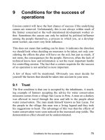
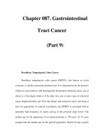
. Fig. 18.28. Legs positioned on divided CRP leg plates with individu-
al adjustment
. Fig. 18.29. Fluoroscopy possible through 360°
. Fig. 18.30. Legs positioned on single-section CRP module 1150.45
. Fig. 18.31. Legs positioned on CRP operating table 1150.16
. Fig. 18.32. Minimally invasive operation to the tibia with image
intensifier in anteroposterior position
. Fig. 18.33. Lateral fluoroscopy of the tibia with left leg lowered
18.4 · Lower leg
Chapter 18 · Lower extremities252
18
18.5 Foot
18.5.1 Supine position
. Figs. 18.34–18.39.
Indications
Ventral, anterolateral, medial and posteromedial access to the ankle joint,
access to the inner malleolus and outer malleolus, lateral and medial access
to the talocalcaneonavicular, front, medial and plantar access to the middle
foot and to the toes for fractures, posttraumatic, congenital and acquired
misalignment, arthrosis, synovitis, osteochondral lesions, soft tissue lesions
and tumours.
Preparations
4 Arm positioning devices
4 Wedge cushion, body support
4 Shaving in the area of the incision and preoperative skin cleansing
4 Apply a tourniquet in position
Positioning
4 Standard operating table position 1, position 2 or universal operating
table
4 Anaesthetic preparation and induction in supine position with 2 adapted
arm positioning devices
4 Normal positioning of the operating table in the theatre
4 When positioning the patient, take appropriate measures to prevent
decubitus at areas which are subjected to pressure
4 Position both arms on the arm positioning device in abduction position
4 Apply the neutral electrode and connect to the HF surgery device
4 Connect the compressed air supply to the tourniquet
4 Arrange absorbent drapes or self-adhesive covers for preoperative skin
disinfection
4 Position the operating lights
4 Patient warming system
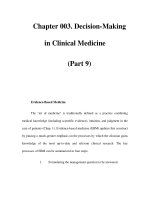
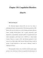
. Fig. 18.34. Legs positioned on divided CRP
leg plates with individual adjustment
18
253
. Fig. 18.36. Divided leg plates with individual adjustment
. Fig. 18.35. Legs positioned on divided leg plates with use of the
image intensifier
. Fig. 18.37. Optimum scanning possibility with lateral ray path due
to lowered leg plate
. Fig. 18.38. Body support with wedge cushion padding under the
pelvis on the other side
. Fig. 18.39. Preoperative skin disinfection and additional moisture
protection during the operation
18.5 · Foot
Chapter 18 · Lower extremities254
18
18.5.2 Lateral position
. Figs. 18.40–18.43.
Indications
Access to the fibula and Achilles tendon, lateral access to the calcaneus and
talocalcaneonavicular, medial and posteromedial access to the calcaneus and
talocalcaneonavicular for fractures, posttraumatic, congenital and acquired
misalignment, arthrosis, synovitis, osteochondral lesions, tumours and soft
tissue lesions.
Preparations
4 Arm positioning devices
4 Gel ring, gel cushion, Goepel leg holder, lateral supports, radial adjusting
clamps, padded cushions (normal and flat) and wedge cushions or tunnel
cushions, body belts
4 Shaving in the area of the incision and preoperative skin cleansing
4 Apply a tourniquet in position
Positioning
4 Standard operating table position 1, position 2 or universal operating
table
4 Anaesthetic preparation and induction in supine position with 2 adapted
arm positioning devices
4 Normal positioning of the operating table in the theatre
4 When positioning the patient, take appropriate measures to prevent
decubitus at areas which are subjected to pressure
4 Fit the radial adjusting clamp to the side rail of the head plate, position the
Goepel leg holder and place a gel mat on the operating table
4 Spread out the arm on the side not being operated
4 Move the patient onto the healthy side
4 Move the lower arm forwards so that the weight of the upper body does
not lie directly on the shoulder
4 Fit the radial adjusting clamps to the side rails of the back plate and posi-
tion the body supports on the level of the coccyx and symphysis
4
1st possibility: position the legs with the padded cushions (normal and
flat) and possibly wedge cushions
4 Fix the lower leg and the positioning aids with the body belts
4
2nd possibility: position the legs with the tunnel cushion
4 Apply the neutral electrode and connect to the HF surgery device
4 Connect the compressed air supply to the tourniquet
4 Arrange absorbent drapes or self-adhesive covers for preoperative skin
disinfection
4 Position the operating lights
4 Patient warming system
18
255
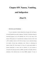
. Fig. 18.40. Legs positioned on single-
section CRP module 1150.45 with padded
cushion
. Fig. 18.43. Stable positioning of the foot
and optimum access for the surgeon with use
of the image intensifier from the opposite
side in both levels
. Fig. 18.42. Lateral positioning on CRP operating table 1150.16 with CRP accessory adapter
and lateral positioning cushion for anatomic positioning of the lower arm
. Fig. 18.41. Legs positioned on divided CRP
leg plates with tunnel cushion
18.5 · Foot
Chapter 18 · Lower extremities256
18
18.5.3
Prone position
. Figs. 18.44, 18.45.
Indications
Posterolateral access to the talocalcaneonavicular and concealed osteosyn-
thesis in the calcaneal part of the foot.
Preparations
4 Arm positioning devices
4 Shaving in the area of the incision and preoperative skin cleansing
4 Apply a tourniquet in position
Positioning
4
table
4 Anaesthetic preparation and induction in supine position with 2 adapted
arm positioning devices
4 Transfer to the prepared operating table in the induction room
4 Position both tables next to each other, with the prepared table lowered
4 Place the patient in prone position on the padded cushion of the prepared
operating table and take him into the theatre
4
4 When positioning the patient, take appropriate measures to prevent
decubitus at areas which are subjected to pressure
4 Position the head on the special head positioning cushion
4 Position both arms on the arm positioning devices
4
4 Connect the compressed air supply to the tourniquet
4 Arrange self-adhesive covers for preoperative skin disinfection
4 Position the operating lights
4 Patient warming system
Standard operating table position 1, position 2 or universal operating
Normal positioning of the operating table in the theatre
Apply the neutral electrode and connect to the HF surgery device
18
257
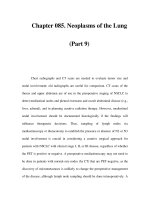
. Fig. 18.44. Prone position on universal
operating table 1150.30 with arms positioned
in maximum 90° abduction
. Fig. 18.45. Use of the image intensifier in
anteroposterior position. C-arm with colour-
ed handles for better communication bet-
ween surgeon and operator
18.5 · Foot
19
19 Positioning on the extension table
19.1 Extension table proximal femur – 260
19.1.1 Supine position – 260
19.2 Extension table thigh – 262
19.2.1 Supine position – 262
19.3 Extension table lower leg – 264
19.3.1 Supine position – 264
Chapter 19 · Positioning on the extension table260
19
19.1 Extension table proximal femur
19.1.1 Supine position
. Figs. 19.1–19.5.
Indications
of extension and fluoroscopy in two levels, and displacement osteotomy of the
proximal femur.
Preparations
4 Arm positioning devices
4
4 Shaving in the area of the incision and preoperative skin cleansing
4
Positioning
4 Universal operating table for traumatology and orthopaedic procedures
(extension table)
4 Anaesthetic preparation and induction in supine position with 2 adapted
arm positioning devices
4 If necessary, diagonal positioning of the operating table in the theatre
4 When positioning the patient, take appropriate measures to prevent
decubitus at areas which are subjected to pressure
4
4
4 Insert the countertraction post on the side being treated
4 Insert the long telescopic bar in the extension bar on the side not being
operated
4 Insert the short telescopic bar in the extension bar on the side being ope-
rated
4 Fit the foot plate adapter
4 Fit the screw tension device
4 Fit the rotating and tilting clamp to the screw tension device
4 Place a double wedge cushion on the operating table
4 Fit the arm positioning device to the side rails of the lower back plate on
the side not being operated
4 Fit the anaesthesia screen with a radial adjusting clamp to the side rail of
the upper back plate on the left-hand side
4 Fit the anaesthesia screen extensions and possibly suspend 2 arm straps
4 Position the foot plates before transferring the patient
4 Transfer the patient from the induction table to the prepared operating
table in supine position
4 Fit the positioned foot plates to the screw tension device and foot plate
adapter, constantly pulling the legs at the rotating and tilting clamp
4 Position the arms
4 Reposition the fracture using the image intensifier and position the legs
4 Check all screwed and clamped connections
4 Apply the neutral electrode and connect to the HF surgery device
4 Arrange self-adhesive covers for preoperative skin disinfection
4 Position the operating lights
4 Patient warming system
Risks
4 Secondary dislocation
4
Osteosynthesis of the proximal femur entailing reposition with the possibility
Extension table accessories
G-arm, alternatively 1 or 2 C-arms
Longitudinal adjustment of the operating table towards the feet (1150.20)
Swivel the extension bars in a V-shape towards the feet
Nerve injuries (n. pudendus)
19
261
. Fig. 19.1. Operating table 1150.20 with foot plates fitted for
both legs
. Fig. 19.2. Operating table 1140.20 with foot plates fitted for both
legs and use of the G-arm
. Fig. 19.3. Operating table 1140.20 with foot plates fitted for both
legs and use of the G-arm for DHS operation (DHS, dynamic hip screw)
. Fig. 19.5. Foot plate fitted to the rotating and tilting clamp with
padded, fixed foot
. Fig. 19.4. Preoperative skin disinfection
19.1 · Extension table proximal femur
Chapter 19 · Positioning on the extension table262
19
19.2 Extension table thigh
19.2.1 Supine position
. Figs. 19.6–19.8.
Indications
Preparations
4 Two arm positioning devices
4
4 Shaving in the area of the incision and preoperative skin cleansing
Positioning
4 Universal operating table for traumatology and orthopaedic procedures
(extension table)
4 Anaesthetic preparation and induction in supine position with 2 adapted
arm positioning devices
4 If necessary, diagonal positioning of the operating table in the theatre
4 When positioning the patient, take appropriate measures to prevent
decubitus at areas which are subjected to pressure
4
4
4 Insert the countertraction post on the side being treated
4 Insert the short telescopic bar in the extension bar on the side being
operated
4 Fit the screw tension device
4 Fit the rotating and tilting clamp to the screw tension device
4 Place a double wedge cushion on the operating table
4 Fit the arm positioning device to the rails of the lower back plate on the
side not being operated
4 Fit the side rail extension to the side rail of the seat plate
4 Fit the Goepel leg holder to the side rail extension with a radial adjusting
clamp
4 Fit the anaesthesia screen with a radial adjusting clamp to the side rail of
the upper back plate on the left-hand side
4 Fit the anaesthesia screen extensions and possibly suspend 2 arm straps
4
4 Transfer the patient from the induction table to the prepared operating
table in supine position
4
rotation, constantly pulling the leg
4 Position the left leg in the Goepel leg holder
Position the arms
4 Reposition the fracture using the image intensifier
4 To stabilise the patient, possibly support the thorax from the side
4 Check all screwed and clamped connections
4 Apply the neutral electrode and connect to the HF surgery device
4 Arrange self-adhesive covers for preoperative skin disinfection
4 Position the operating lights
4 Patient warming system
Risks
4 Iatrogenic damage caused by inserting the Steinmann nail
4 Pressure injuries
4 Compartment syndrome from overdistraction
Medullary nailing, intramedullary reaming.
Extension table accessories
Longitudinal adjustment of the operating table towards the feet (1150.20)
Swivel the extension bars in a V-shape towards the feet on the side being
operated
Fit the Kirschner wire bow before transferring the patient
Fit the positioned Kirschner wire bow to the traction stirrup clamp with
19
263
. Fig. 19.6. Operating table 1150.20 with
fitted foot plate, healthy leg is in abduction
on a Goepel leg holder
19.2 · Extension table thigh
. Fig. 19.7. Operating table 1150.20 with
fitted Kirschner wire bow and thorax support
. Fig. 19.8. Operating table 1150.20 with
fitted foot plate, healthy leg is positioned
downwards on a special support
Chapter 19 · Positioning on the extension table264
19
19.3 Extension table lower leg
19.3.1 Supine position
. Figs. 19.9–19.11.
Indications
Preparations
4 Two arm positioning devices
4 Shaving in the area of the incision and preoperative skin cleansing
Positioning
4 Universal operating table for traumatology and orthopaedic procedures
(extension table)
4 Anaesthetic preparation and induction in supine position with 2 adapted
arm positioning devices
4 If necessary, diagonal positioning of the operating table in the theatre
4 When positioning the patient, take appropriate measures to prevent
decubitus at areas which are subjected to pressure
4 Longitudinal adjustment of the operating table towards the fe
4 Fit the tibia device to the seat plate on the side being operated
4 Unlock the unit at the lower radial joint and lower towards the feet to
improve scanning the knee in anteroposterior ray path after positioning
4 Lock the safety lever again
4 Insert the short telescopic bar in the extension bar on the side being
operated
4 Fit the screw tension device
4 Fit the rotation tilt clamp to the screw tension device
4 Place a double wedge cushion on the operating table
4 Fit the arm positioning device to the side rails of the lower back plate on
the side not being operated
4 Fit the side rails extension to the side rail of the seat plate
4 Fit the Goepel leg holder to the side rail extension with a radial adjusting
clamp
4 Fit the anaesthesia screen with a radial adjusting clamp to the side rail of
the upper back plate on the side not being operated
4 Fit the anaesthesia screen extensions and possibly suspend 2 arm straps
4 Fit the tension hoop before transferring the patient
4 Transfer the patient from the induction table to the prepared operating
table in supine position
4
rotation, constantly pulling the leg
4 Position the healthy leg in the Goepel leg holder
4 Position the arms
4 Reposition the fracture using the image intensifier
4 Check all screwed and clamped connections
4 Apply the neutral electrode and connect to the HF surgery device
4 Arrange self-adhesive covers for preoperative skin disinfection
4 Position the operating lights
4 Patient warming system
Risks
4 Iatrogenic damage caused by inserting the Steinmann nail
4 Pressure injuries
4 Compartment syndrome from overdistraction
Medullary nailing, intramedullary reaming.
et (1150.20)
Fit the positioned Kirschner wire bow to the traction stirrup clamp with
19
265
. Fig. 19.9. Operating table 1150.20 with
tibia device and fitted Kirschner wire bow,
healthy leg is spread out on a Goepel leg
holder
. Fig. 19.11. Image intensifier in lateral
position
. Fig. 19.10. Tibia device is lowered to
optimise anteroposterior fluoroscopy
19.3 · Extension table lower leg
20
20 Arthroscopic procedures
20.1 Shoulder – 268
20.1.1 Beach-chair position – 268
20.1.2 Lateral position – 270
20.2 Hips – 272
20.2.1 Supine position on the extension table – 272
20.3 Knee – 274
20.3.1 Supine position – 274
20.4 Foot/ankle – 276
20.4.1 Supine position – 276
Chapter 20 · Arthroscopic procedures268
20
20.1 Shoulder
20.1.1 Beach-chair position
. Figs. 20.1–20.5.
Indications
drome, rotator cuff rupture, tendinosis calcarea, shoulder instability, arthrosis/
osteolysis of the acromioclavicular joint and synovitis.
Preparations
4 Arm positioning devices
4 Horseshoe-shaped headrest
4 Remove the Gilchrist bandage
4 Shaving in the area of the incision and preoperative skin cleansing
Positioning
4 Beach-chair operating table position 2 or universal operating table with
shoulder plate
4 Anaesthetic preparation and induction in supine position with 2 adapted
arm positioning devices
4 Crosswise positioning of the operating table in the theatre
4 When positioning the patient, take appropriate measures to prevent
decubitus at areas which are subjected to pressure
4 The patient’s shoulders end at the upper edge of the operating table
4 Bring the operating table gradually to the half-sitting (beach-chair)
position
4 Raise the back plate and alternately lower the head of the complete oper-
ating table until the final position is reached
4 Change the Bowden cable over and lower the legs to the horizontal (system
4 Position the head on a horseshoe-shaped headrest and fix with transparent
plaster right across the forehead or use a head holder for shoulder opera-
tion (U-shaped helmet)
4 Position the infusion arm on an arm positioning device
4 Cover the arm on the side being operated while leaving it free to move and
position it at the body with arm protection or place it on the arm posi-
tioning device
4 Apply the neutral electrode and connect to the HF surgery device
4 Fit the thorax support to the rail of the shoulder plate
4 Arrange self-adhesive covers for preoperative skin disinfection
4 Position the operating lights
4 Patient warming system
Risks
4
Diagnostic and therapeutic arthroscopy procedures for impingement syn-
1120)
Nerve injuries (brachial plexus)
20
269
. Fig. 20.5. Thorax support
. Fig. 20.1. Beach-chair positioning on special shoulder plate with
helmet for safe positioning of the head
. Fig. 20.2. The thorax support offers additional safety
. Fig. 20.3. The universal operating table with special shoulder plate
is adapted to the body
. Fig. 20.4. A segment is removed to leave free access to the rear
shoulder
20.1 · Shoulder
Chapter 20 · Arthroscopic procedures270
20
20.1.2 Lateral position
. Figs. 20.6, 20.7.
Indications
drome, rotator cuff rupture, tendinosis calcarea, shoulder instability, arthrosis/
osteolysis of the acromioclavicular joint and synovitis.
Preparations
4 Arm positioning devices
4 Gel ring, gel cushion, Goepel leg holder, side supports, radial adjusting
clamps, padded cushions (normal and flat) and wedge cushions, tunnel
cushions, body belts
4 Gallows for arm extension
4 Shaving in the area of the incision and preoperative skin cleansing
Positioning
4
table
4 Anaesthetic preparation and induction in supine position with 2 adapted
arm positioning devices
4 Crosswise positioning of the operating table in the theatre
4 When positioning the patient, take appropriate measures to prevent
decubitus at areas which are subjected to pressure
4
1st possibility: fit the radial adjusting clamp to the side rail of the head
plate and position the Goepel leg holder
4
2nd possibility: fit gallows for suspending the arm being operated with
counterweight (extension) to the foot end of the operating table
4 Spread out the arm on the side not being operated
4
4
not lie directly on the shoulder
4 Fit the body supports to the side rails and brace on the level of the sacrum
and symphysis
4
1st possibility: position the legs with the padded cushions (normal and
flat) and possibly wedge cushions
4 Fix the lower leg and the positioning aids with the body belts
4
2nd possibility: position the legs with the tunnel cushion
4 Apply the neutral electrode and connect to the HF surgery device
4 Arrange absorbent drapes or self-adhesive covers for preoperative skin
disinfection
4 Position the operating lights without switching on
4 Patient warming system
Diagnostic and therapeutic arthroscopy procedures for impingement syn-
Standard operating table position 1, position 2 or universal operating
Move the patient onto the healthy side
Move the lower arm forwards so that the weight of the upper body does
20
271
. Fig. 20.6. Lateral position with arm exten-
sion
. Fig. 20.7. Arm extension (gallows) can be
swivelled and adjusted in height
20.1 · Shoulder
Chapter 20 · Arthroscopic procedures272
20
20.2 Hips
20.2.1 Supine position on the extension table
. Figs. 20.8, 20.9.
Indications
synovitis, fractures, labrum lesions and arthrosis.
Preparations
4 Arm positioning devices
4
4 Shaving in the area of the incision and preoperative skin cleansing
4
Positioning
4 Universal operating table for traumatology and orthopaedic procedures
(extension table)
4 Anaesthetic preparation and induction in supine position with 2 adapted
arm positioning devices
4 If necessary, diagonal positioning of the operating table in the theatre
4 When positioning the patient, take appropriate measures to prevent
decubitus at areas which are subjected to pressure
4
4
4 Insert the countertraction bar on the side being treated
4 Insert the long telescopic bar in the extension bar on the side not being
operated
4 Insert the short telescopic bar in the extension bar on the side being
operated
4 Fit the foot plate adapter
4 Fit the screw tension device
4 Fit the rotation tilt clamp to the screw tension device
4 Position the foot plates
4 Fit the positioned foot plates to the screw tension device and foot plate
adapter, constantly pulling the legs at the rotation and tilting clamp
4 Remove the (extension table) leg plates
4 Position the arms
4 Position the legs and set up the image intensifier
4 Check all screwed and clamped connections
4 Apply the neutral electrode and connect to the HF surgery device
4 Arrange self-adhesive covers for preoperative skin disinfection
4 Position the operating lights without switching on
4 Patient warming system
Risks
4
Diagnostic and therapeutic arthroscopic procedures for floating cartilage,
Extension table accessories
G-arm, alternatively 1 or 2 C-arms
Longitudinal adjustment of the operating table towards the feet (1150.20)
Swivel the extension bars in a V-shape towards the feet
Nerve injuries (pudendal nerve)
20
273
. Fig. 20.8. Supine position on extension
table with G-arm
. Fig. 20.9. Positioning the monitors at the
foot end; foot holder closure is located on the
inside for better padding of the feet on the
outside
20.2 · Hips
Chapter 20 · Arthroscopic procedures274
20
20.3
Knee
20.3.1
Supine position
. Figs. 20.10–20.15.
Indications
meniscus lesions, cartilage damage, arthrosis, floating cartilage, synovitis,
fractures and osteochondrosis dissecans.
Preparations
4 Arm positioning devices
4 Tourniquet, cotton wool padding, elastic bandages, perforated rubber
sheet knee holder, special positioning cushion
4 Shaving in the area of the incision and preoperative skin cleansing
4 Apply a tourniquet in position
Positioning
4
table
4 Anaesthetic induction in the supine position, padding under the shoul-
ders
4
4 When positioning the patient, take appropriate measures to prevent
decubitus at areas which are subjected to pressure
4
position
4 Fit the knee holder with radial adjusting clamp to the small rail of the leg
plate on the operation side
4 Fix the thigh in the knee holder
4 Position the leg not being operated on the special positioning cushion
4 Apply the neutral electrode and connect to the HF surgery device
4 Connect the compressed air supply to the tourniquet
4 Preoperative skin disinfection as far as the perforated rubber sheet
4 Position the operating lights without switching on
4 Patient warming system
Risks
4
4 Injury to the femoral nerve under hyperextension of the other hip, there-
fore positioning cushion (black) under the other thigh
Diagnostic and therapeutic arthroscopic procedures for ligament injuries,
Standard operating table position 1, position 2 or universal operating
Normal positioning of the operating table in the theatre
Position both arms on the arm positioning devices in 45° abduction
(MHH), with abduction of the leg and swing down the leg plate
Dislocation in the leg holder
20
275
. Fig. 20.10. Supine position on universal operating table with 4-sec-
tion leg plates
. Fig. 20.11. Lower segment of the leg plate folded down and thigh
fixed in a knee holder
. Fig. 20.12. Thigh padded to prevent pressure from the knee holder,
at the same time this acts as tourniquet
. Fig. 20.13. Healthy leg lowered and abducted, thigh padded/raised
with special positioning cushion (MHH) to prevent hyperextension in
the hip joint
. Fig. 20.14. Preoperative skin disinfection up to the perforated
rubber sheet
. Fig. 20.15. Special covering for arthroscopy of the knee
20.3 · Knee