Practical Plastic Surgery - part 10 ppsx
Bạn đang xem bản rút gọn của tài liệu. Xem và tải ngay bản đầy đủ của tài liệu tại đây (2.2 MB, 66 trang )
101
603
Reflex Sympathetic Dystrophy
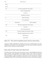
Intravenous alpha-Adrenergic Blockade
Bier block administration of intravenous phentolamine, an alpha-adrenergic re-
ceptor antagonist, should produce sympathetic blockade. If the signs and symptoms
are sympathetically-mediated as in the case of RSD, they should diminish in re-
sponse to this infusion. It is important to administer saline in a blinded fashion to
eliminate the placebo effect.
Classification
• Lankford classified RSD into five categories:
• Minor causalgia: A mild form of RSD seen after injury to a sensory nerve in
the forearm, hand or fingers.
• Major causalgia: The more severe form of causalgia in which pain and dys-
function are prominent. It occurs as a result of injury to mixed motor and
sensory nerve.
• Minor traumatic dystrophy: Mild RSD with an inciting trauma, but no
known nerve injury.
• Major traumatic dystrophy: The form most commonly thought of when
the term RSD is used. Seen after trauma or fracture of the upper extremity,
without specific nerve involvement.
• Shoulder and hand syndrome: RSD due to remote injury such as an MI or
cervical spine injury. Symptoms begin in the shoulder and spread to the hand,
sparing the elbow.
• SMPS can be classified into Type I and II:
• Type I: What is thought of when the term RSD is used. Pain follows and
inciting event and is out of proportion to the exam. The other findings typi-
cally associated with RSD are usually present.
• Type II: This type of SMPS describes causalgia, similar to the definition
given in the Lankford classification.
Staging
• RSD can also be thought of in terms of its stage: early, established or late.
• Early RSD: Defined as the first three months of symptoms. Pain is often
burning and can be caused be even light touch. Discoloration, hyperhydrosis,
and increased temperature are often present.
• Established RSD: Defined as the period between three and twelve months of
symptoms. Pain is still the dominant feature. Skin dryness, joint stiffness,
contractures and osteoporosis are common. The temperature of the hand gradu-
ally goes from warm seen in early RSD to cold, as compared to the other side.
• Late RSD: Defined as the final stage of RSD, twelve months or longer after
onset of symptoms. The pain may become less severe during this stage, how-
ever flare-ups can occur. Stiffness and joint contracture are the most promi-
nent features of late RSD. The skin can become thickened and nodular, and
severe osteoporosis is not uncommon.
Treatment
The overriding goal of treatment for RSD is elimination of persistent sources of
pain. Simple measures such as relieving pressure points or elevation of the extremity
can be very helpful. Local and regional nerve blocks help neutralize sensory nerves
as well as providing a chemical sympathectomy.
101
604
Practical Plastic Surgery
The stellate ganglion block is the most effective regional nerve block. It has
been demonstrated to provide some degree of relief; however results are variable.
Numerous studies have been published with good results ranging from zero to 100%.
However, little long-term data is available, and few studies are randomized. A satis-
factory block is indicated by warming of the upper extremity and a Horner’s sign
(unilateral pupillary constriction, ptosis, anhydrosis and facial flushing). Conven-
tional stellate blocks are done with lidocaine or bupivicaine. Good results have been
obtained with narcotic blocks (e.g., fentanyl) in refractory cases. Usually repeated
biweekly blocks are required. For patients unable to tolerate weekly treatments, a
continuous stellate block for 3 to7 days has been used successfully.
Although not widely used in the U.S., sympathetic inhibition can also be achieved
using an intravenous regional block with anti-adrenergic agents such as bretylium,
guanethidine or reserpine. These agents are infused intravenously into an extremity
using the Bier block technique to isolate the upper extremity. Other drugs such as
steroids and NSAIDs have been used as well. Good long-term pain relief has been
demonstrated with this technique.
A variety of oral medications have been used to treat RSD. Several drug regi-
mens, such as a short course of oral corticosteroids, nightly amitryptyline, and select
calcium channel blockers have met with good success. Oral phenoxybenzamine and
other anti-adrenergic drugs have been used with mixed results. Calcitionin and pheny-
toin have been used to relieve symptoms of RSD; however their use has met with
mixed results.
Physical therapy should consist of active range of motion of all joints from the
shoulder to the DIP joints. Hand therapy should not be done while the patient is
actively in pain. It can be performed immediately following sympathetic blocks when
substantial pain relief has been achieved. Progressive stress loading without joint
motion is also recommended. It involves the use of active traction and compression
exercises. Static splints can be used to keep the hand in the intrinsic plus position.
Adjunctive treatments can be helpful in dealing with RSD that does not re-
spond to traditional sympathetic blocks and hand therapy. Biofeedback, psycho-
therapy, smoking cessation, and transcutaneous electrical nerve stimulation have all
been attempted.
Surgical sympathectomy should be reserved for severe, prolonged cases, and
those that are refractory to other treatment modalities. The procedure consists of
transection of the upper thoracic sympathetic chain via an extrapleural, axillary ap-
proach. The T2 and T3 sympathetic nerves must be completely transected. Success
rates up to 90% have been reported. More recently, sympathectomies have been
performed under video-assisted thoracoscopic surgery (VATS).
Long-Term Outcomes
Very few studies have addressed the sequelae of patients successfully treated for
RSD. Overall, long-term results have been disappointing. At one year
post-treatment, roughly half of patients have cold intolerance or pain with cold
weather. Trophic changes persist in about a third of patients. Joint swelling and
stiffness, as well as decreased grip strength are also common complaints. In sum-
mary, RSD and SMPS are still poorly understood. The diagnosis of these condi-
tions can be challenging, and their treatment even more so. Active and future
research will undoubtedly shed greater light on these syndromes and offer prom-
ise for those who suffer from them.
101
605
Reflex Sympathetic Dystrophy
Pearls and Pitfalls
1. It is important for the treating physician to realize that almost any injury can be
the inciting cause for RSD. The earlier the inciting injury is recognized, the
more likely treatment is to be successful.
2. Pain free movement is probably the best therapeutic modality against RSD. Nerve
blocks and oral analgesics combined with physical therapy is a treatment goal.
3. The extremity surgeon treating this condition is a coach or motivator for the
patient, more so for this disease process than almost any other. The patient
needs frequent counseling about the disease process and the expected length of
treatment.
Suggested Reading
1. Dzwierzynski WW, Sanger JR. Reflex sympathetic dystrophy. Hand Clin 1994; 10:29.
2. Lankford LL. Reflex sympathetic dystrophy. In: Hunter JM et al, eds. Rehabilitation
of the Hand-Surgery and Therapy. 3rd ed. St. Louis: CV Mosby, 1990.
3. Nath RK, Mackinnon SE, Stelnicki E. Reflex sympathetic dystrophy. The controversy
continues. Clin Plast Surg 1996; 23:435.
4. Zyluk A. The sequelae of reflex sympathetic dystrophy. J Hand Surg (Br) 2001; 26:151.
Appendix I
Part A: Important Flaps and Their Harvest
Zol B. Kryger and Mark Sisco
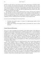
Groin Flap
The groin flap is a fasciocutaneous Type A flap based on the superficial cir-
cumflex iliac system. The skin is innervated from the T12 lateral cutaneous nerve.
This flap is used primarily as a rotational flap for coverage in the abdominal wall
and perineum; it can also be used as a free flap for distant coverage.
The skin of the lateral groin is elevated along an axis parallel and 3 cm inferior to
the inguinal ligament (Fig. AI.1). The skin flap can measure up to 25 x 10 cm. The
pedicle originates from the femoral artery roughly in the femoral canal. The main
vein drains into the saphenous vein just distal to the fossa ovalis.
The skin is incised down to the fascia. The flap is elevated distal to proximal in
the plane superficial to tensor fascia lata, which serves as the distal extent of the flap.
It is dissected free from the anterior superior iliac spine (ASIS), inguinal ligament
and external oblique fascia. The deep fascia that envelops the sartorius is included in
the flap. The pedicle is dissected to the medial edge of the sartorius muscle—the
proximal limit of the flap.
If the skin is elevated as an island flap, the pedicle should be identified in the
femoral triangle through a transverse incision, before the skin island incision is made.
The flap is elevated from distal to proximal as described above, until the dissection
meets the pedicle.
Practical Plastic Surgery, edited by Zol B. Kryger and Mark Sisco. ©2007 Landes Bioscience.
Figure AI.1. The groin flap and HS
blood supply.
AI
608
Practical Plastic Surgery
Table AI.1. Fasciocutaneous and perforator flaps
Flap Type* Size (cm) Blood Supply (D=dominant, M=minor) Common Uses
Forehead A 22 x 7 D: Superficial temporal a. Middle and inferior face, oral cavity,
M: Supratrochlear a. nose, frontal and maxillary sinus
M: Supraorbital a
Nasolabial C 2 x 5 D: Angular a. Nose, lips, floor of mouth
M: Alar branches of superior labial a.
Temporoparietal fascia A 12 x 9 Superficial temporal a. Scalp, upper and middle face, ear,
free flap
Deltopectoral C 10 x 20 D: Perforators 1-3 of IMA Middle and inferior face, neck,
M: Perforators 4-5 of IMA oral cavity, esophagus
Gluteal thigh A 12 x 30 D: Superior gluteal a. Sacrum, ischium, perineum
D: Inferior gluteal a.
M: Branches of lateral circumflex femoral a.
M: 1st perforator of profunda femoris
Scapula B 20 x 7 D: Circumflex scapular a. Neck, axilla, shoulder, back,
Radial forearm B 10 x 40 D: Radial a. free flap (mandible)
M: Musculocutaneous branches of radial Forearm, elbow, wrist, hand,
recurrent a. and inferior cubital a. free flap
Groin A 25 x 10 D: Superficial circumflex iliac Upper extremity, abdominal wall,
perineum, free flap
Lateral thigh B 7 x 20 D: 1st-3rd perforators of profunda femoris Ischium, greater trochanter, free flap
Medial plantar B 12 x 6 D: Medial plantar a. Plantar foot, ankle, free flap
M: Musculocutaneous perforators from (contralateral foot)
medial plantar a.
Anterolateral thigh (ALT) B, C 12 x 20 D: Septocutaneous branches of descending Trunk, abdomen, groin, thigh,
branch of lateral circumflex a. free flap
M: Musculocutaneous branches of
descending branch of lateral circumflex a.
Deep Inferior epigastric D: Perforator from deep inferior Free flap (breast)
perforator (DIEP) epigastric a.
Thoracodorsal perforator (TAP) D: Perforator from thoracodorsal a. Free flap (breast, extremity)
Superior gluteal perforator (SGAP) D: Perforator from superior gluteal a. Sacrum, ischium, lower back, free flap
*Mathes-Nahai classification of fasciocutaneous flaps. Type A, direct cutaneous pedicle. Type B, septocutaneous pedicle. Type C, musculocutaneous pedicle
AI
609
Appendix I
Table AI.2. Muscle and musculocutaneous flaps
Flap Type* Size (cm) Blood Supply (D = dominant, M = minor) Common Uses
Temporalis III 10 x 20 D: Anterior deep temporal a. Facial reanimation, orbit, palate, mandible
D: Posterior deep temporal a.
M: Branches of middle temporal a.
Pectoralis major V 15 x 25 D: Pectoral branch of thoracoacromial a. Face, chest, neck, shoulder, axilla, sternum,
M: Pectoral branch of lateral thoracic a. upper extremity
S: Internal mammary (1-6) and intercostal (5-7) perforators
Gluteus maximus III 24 x 24 D: Superior gluteal a. Sacrum, ischium, trochanter, vagina, free flap
D: Inferior gluteal a.
M: Branches of lateral circumflex femoral
M: 1st perforator of profunda femoral
Latissimus dorsi V 25 x 35 D: Thoracodorsal Scalp, neck, trunk, breast, abdomen, upper extremity,
S: Posterior intercostal perforators and lumbar perforators free flap
Rectus III 25 x 6 D: Superior epigastric a. Breast, trunk, abdomen, groin, perineum,
abdominis D: Deep inferior epigastric a. pelvic floor, free flap
M: Subcostal and intercostal a.
Serratus III 15 x 20 D: Lateral thoracic a. Head, thorax, axilla, posterior trunk, intrathoracic,
D: Branch of thoracodorsal a. free flap
Gracilis II 6 x 24 D: Ascending branch of medial circumflex femoral a. Perineum, groin, penis, vagina, free flap
M: 1-2 branches of SFA
Hamstring II 15 x 45 D: 1st-3rd perforators of profunda femoris Ischium
M: Branch of inferior gluteal a.
M: Superior lateral genicular a.
Tensor fascia lata I 5 x 15 D: Ascending branch of lateral circumflex femoral a. Abdomen, groin, perineum, trochanter, ischium, free flap
Vastus lateralis I 10 x 25 D: Descending branch of lateral circumflex femoral a. Ischium, trochanter, groin, perineum, knee, abdomen
M: Transverse branch of lateral circumflex femoral a.
M: Posterior branch from profunda femoris
M: Superficial branch of lateral superior genicular a.
Gastrocnemius I 20 x 8 D: Medial sural a. Knee, lower thigh, upper leg
D: Lateral sural a.
M: Anastomotic vessels from sural a.
Soleus II 8 x 28 D: Muscular branches of popliteal Middle and lower leg
D: First 2 branches of posterior tibial a.
D: First 2 branches of peroneal a.
M: Segmental branches of posterior tibial a.
Fibula V 3 x 40 D: Nutrient branches of peroneal a. Tibia, free flap (mandible)
M: Periosteal and muscular branches of peroneal a.
*Mathes-Nahai classification of muscle and musculocutaneous flaps. Type I, one pedicle. Type II, dominant and minor pedicles. Type III, two dominant pedicles. Type IV,
segmental pedicles. Type V, one dominant pedicle and secondary segmental pedicles.
AI
610
Practical Plastic Surgery
Table AI.3. Visceral flaps
Blood Supply
Flap Type* Size (cm) (D = dominant, M = minor) Common Uses
Omentum III 40 x 60 D: Right gastroepiploic a. Head and neck, trunk, intrathoracic,
D: Left gastroepiploic a. abdomen, groin, perineum, free flap
Jejunum I 7-25 D: Jejunal a. (from SMA) Free flap (esophagus)
*See classification in Table AI.2.
AI
611
Appendix I
Rectus Abdominis Flap
The rectus abdominis flap can be harvested as either a muscle or musculocuta-
neous, Type III flap. The two dominant pedicles are the superior and deep infe-
rior epigastric arteries. Minor pedicles include the intercostals and subcostal arteries,
with the T8 subcostal artery usually being the largest. The muscle and overlying skin
are innervated by segmental motor and cutaneous intercostal (7-12) nerves, re-
spectively. It is an extremely useful flap used in breast, perineal and vaginal recon-
struction, and as coverage in the thorax, abdomen, posterior trunk and groin. For
these purposes, it is primarily used as a rotational flap or island pedicle flap. It is
also an extremely versatile free flap based on the deep inferior epigastric vessels.
Contraindications for use of the rectus abdominus flap include:
• Unilateral subcostal incision (Kocher incision) for an ipsilateral flap based on the
superior pedicle
• Bilateral subcostal incisons (Chevron incision) for any flap based on the superior
pedicle
• Low transverse incison (Pfannenstiel incision) for any muscle flap based on the
inferior pedicle (exception: a deep inferior epigastric perforator flap)
• Any portion of the skin island that is lateral to a prior skin incision should not
be used
• Prior use of the internal mammary artery is a relative contraindication for a
superiorly based flap
• History of major external iliac vascular surgery is a relative contraindication for
an inferiorly based flap, unless angiography confirms otherwise.
For harvesting a muscle flap, either a longitudinal paramedian or low transverse
skin incision is used. For the musculocutaneous flap, the skin island can be marked
in multiple horizontal or vertical patterns. A transverse (horizontal) skin island can
be up to 21 x 8 to 21 x 14 cm in size. This skin can be divided into zones: zone 1 is
over the ipsilateral rectus; zone 2 is over the contralateral rectus; zone 3 is lateral to
the ipsilateral rectus; zone 4 (least reliable skin) is lateral to the contralateral rectus.
After the skin paddle is marked, the inferior border is incised down to the ante-
rior rectus sheath. The skin and subcutaneous fat are elevated from lateral to medial
off the fascia. The dissection is slowed several centimeters lateral to the midline
where the musculocutaneous perforators are encountered. The superior border of
the skin island should be incised only after confirming that the donor skin will close
without excessive tension. Alternatively, the superior incision can be made first,
followed by the inferior incision once it is clear that the abdominal skin will come
together without undue tension.
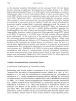
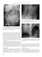
The anterior rectus sheath is opened sharply in a longitudinal direction exposing
the rectus muscle (Fig. AI.2). The muscle is dissected free from its sheath, with care
taken not to violate the posterior sheath. For the inferiorly-based flap, the muscle is
divided at or near the costal margin. The superior epigastric artery and vein are
divided at the medial border of the muscle. For the superiorly-based flap, the muscle
is divided at the level of the pubis symphysis. The deep inferior epigastric artery and
vein can be dissected for several centimeters prior to division. This can serve as an
alternative pedicle for microvascular anastomosis if the superior pedicle is insuffi-
cient. Care must always be taken to avoid injuring the musculocutaneous perfora-
tors feeding the skin paddle.
AI
612
Practical Plastic Surgery
If only a muscle flap is required, the abdominal skin and subcutaneous fat are
elevated off the anterior abdominal wall. The rectus sheath can then be opened as
described above without concern for the musculocutaneous perforators.
Once the rectus muscle is harvested, the donor site is closed. The anterior rectus
sheath can be closed primarily using a running or interrupted permanent suture. If
the fascial edges are frayed or primary closure will create under undue tension, a
synthetic mesh can be used to replace the missing segment. If necessary, small tears
in the fascial edges during primary fascial closure can be reinforced with an overly-
ing piece of mesh.
Fibula Composite Flap
The fibula free flap can be harvested as either an osseous or osseofasciocutaneous
(composite), Type V flap. In addition, cuffs of muscle are usually incorporated in
order to protect the blood supply. The dominant pedicle is the nutrient branch of
the peroneal artery. The minor pedicles are the periosteal and muscular branches
of the peroneal artery. The sensory nerve supply is from the superficial peroneal
nerve. This flap is used primarily as a free flap for mandibular reconstruction or for
reconstruction of the ipsilateral tibia and femur.
If an osseous flap is needed, a longitudinal incision is made along the posterior
border of the fibula from the head of the fibula to the lateral malleolus. If an
osseofasciocutaneous flap is used, the skin territory should be marked as a vertical
ellipse over the middle third of the fibula. The skin island can span from 6 cm below
the fibular head to 8 cm above the distal fibula, and it can measure up to 5 x 15 cm.
The width of the skin island can be extended; however closure will require a skin graft.
For harvesting the osseous flap, the lateral compartment is opened, and the pero-
neus longus and brevis are detached from the fibula leaving a small cuff of muscle
attached. The common and superficial peroneal nerves are identified and preserved.
Figure AI.2. The rectus
abdominus musculocu-
taneous flap and its dual
blood supply.
AI
613
Appendix I
The proximal and distal osteotomies are performed (4 cm inferior to fibular head
and 6 cm superior to lateral maleolus). As long as at least 6 cm of distal fibula is left
intact, ankle stability will be preserved.
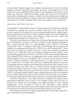
A cross section of the leg showing the compartments in relationship to the fibula
are shown in Figure AI.3. In the anterior approach, the anterior compartment muscles
are released followed by division of the interosseous membrane and entry into the
superficial and deep posterior compartment musculature. The peroneal artery and
vein are divided upon entry into the deep posterior compartment, followed by tibi-
alis posterior and flexor hallucis longus. In the posterior approach, the posterior
compartments are released first followed by entry into the anterior compartment
and release of its musculature. Cuffs of muscle should be included with the flap.
The dominant pedicle usually enters the middle third of the fibula on the medial
side via the nutrient foramen 14-19 cm (average of 17 cm) below the styloid pro-
cess. The peroneal artery and vein can be dissected proximally as needed to achieve
adequate length. Shortening of the bone should be done in the subperiosteal plane
in order to avoid injuring the blood supply.
For harvesting the osseofasciocutaneous flap, the skin island is incised
circumferentially down through the deep fascia. Anteriorly the skin island is el-
evated subfascially off the anterior and lateral compartment musculature. The pos-
terior skin island is elevated subfascially off the lateral gastrocnemius and soleus
muscles. The remainder of the dissection proceeds as described above. Special care is
taken to preserve the septocutaneous and musculocutaneous perforators. A cuff of
flexor hallucis longus and soleus should be preserved.
Figure AI.3. A cross section of the leg showing the compartments of the leg and
the anterior and posterior approaches to the deep posterior compartment.
AI
614
Practical Plastic Surgery
Pectoralis Major Flap
The pectoralis major flap (Fig. AI.4) can be harvested as either a muscle or
musculocutaneous, Type V flap. The dominant pedicle is the pectoral branch of
the thoracoacromial artery. The minor pedicle is the pectoral branch of the lat-
eral thoracic artery. Minor segmental pedicles include the first through sixth inter-
nal mammary perforators, and the fifth through seventh intercostal perforators.
The muscle is innervated by the lateral (superior) and medial (inferior) pectoral
nerves. The sensory innervation is from the intercostal (2-7) nerves. It is a versatile
pedicle flap for coverage of defects of the face, chest, neck, shoulder, axilla, sternum
and upper extremity flap. It can also be used in the intrathoracic cavity. By dividing
either its origin or insertion, it can serve as a rotational flap or island pedicle flap
for many sites. The pectoralis major muscle flap can also serve as a functional flap
for the upper extremity (elbow flexion). Rarely, it is used as a free flap for coverage
in the head and neck or perineum.
The flap can be elevated with the entire skin paddle covering the muscle, or any
part of it. The borders of the flap are the clavicle, the anterior axillary line, the sixth
intercostal space and the parasternal line. The skin territory can measure up to 20 x
28 cm. For head and neck reconstruction, a smaller skin paddle is used: an
inframammary skin island in women and a parasternal paddle in men. Most small
skin defects can be closed primarily. Larger skin paddles will leave a donor site re-
quiring skin grafting or secondary flap closure.
Harvesting the pectoralis major muscle is relatively simple. If only muscle is
required, a horizontal incision is made below the clavicle, vertically along the axil-
lary line or midsternal. The skin and subcutaneous fat are dissected free of the muscle.
As an island flap based on the thoracoacromial pedicle, the muscle fibers are divided
from their origin and from the clavicle, and the muscle is dissected from medial to
Figure AI.4. The pectoralis major muscle flap and its blood supply.
AI
615
Appendix I
lateral. The muscle is divided just lateral to the pedicle, which enters the muscle
from the deep side around the junction of the middle and lateral thirds of the clavicle.
As a turnover flap (reverse flap), the muscle is divided laterally at the level of the
lateral border of pectoralis minor, preserving the lateral one-third of the muscle. The
muscle is dissected from lateral to medial, preserving the vascular and nerve supply
to the remaining portion. The dissection continues to within 2-3 cm from the ster-
nal border until the internal mammary perforators are visualized.
For head and neck reconstruction, the horizontal, infraclavicular skin incision is
used. A skin paddle is often needed as described above. The skin island is incised
and the muscle divided distally. Mobilization occurs in a superior direction towards
the clavicle. If the entire muscle is not needed, a wide central strip of muscle is often
sufficient to vascularize the skin pedicle. The musculocutaneous flap can be pulled
through the clavicular incision.
If functional muscle transfer for the upper extremity is required, an anterior
axillary line incision is used. The portion of muscle required is outlined. The muscle
is dissected free from the subcutaneous tissue, and the origin at the ribs and sternum
is divided. The muscle is mobilized from medial to lateral towards the humerus.
Care is taken to preserve the blood supply and motor nerves. The muscle can be
tunneled through the axilla onto the arm. Elbow flexion can be achieved by suturing
the pectoralis to the biceps tendon.
Latissimus Dorsi Flap
The latissimus dorsi flap (Fig. AI.5) can be harvested as either a muscle or mus-
culocutaneous, Type V flap. The dominant pedicle is the thoracodorsal artery.
Minor segmental pedicles include a medial and lateral row of posterior intercostals
and lumbar perforators. The muscle is innervated by the thoracodorsal nerve
which travels with the vascular pedicle. The sensory innervation is from the inter-
costal nerves. It is a versatile pedicle flap for coverage of defects of the neck, trunk,
breast, abdomen and upper extremity. The latissimus muscle flap can also serve as a
functional flap for the upper extremity (elbow extension or flexion). It is also used
as a free flap for coverage in the scalp, lower and upper extremity-especially when a
thin flap is required.
The skin island, when required, is marked. Options include oblique skin is-
lands—with the superior end towards the midline or the axilla, superior posterior
skin island, superior transverse, inferior transverse, lateral or vertical orientation.
The dominant pedicle is marked in the posterior axilla entering the lateral deep
surface of the muscle about 15 cm below the humeral insertion. The skin is incised
around the island beveling away from the skin island. The muscle fibers of the latis-
simus are identified: superiorly, with the scapula and trapezius muscle. The fibers
are divided, separating the muscle from the scapula. The attachments to the verte-
bral column are divided. The lumbrosacral fascia is divided to the level of the poste-
rior axillary line. The minor pedicles are divided as they emerge from the posterior
intercostals and lumbar vessels. The direction of dissection progresses towards the
axilla where the pedicle is located. The dissection should allow sufficient flap mobil-
ity without compromising the pedicle. If needed, the insertion to the humerus can
be divided to gain mobility.
If a muscle flap alone is required, the location of the skin incision is variable.
Once the skin is incised and the superficial side of the muscle is exposed, the skin
flaps are elevated off the muscle exposing its entire dimensions. The muscle is freed
AI
616
Practical Plastic Surgery
from its origin at the midline and superiorly as described above. Dissection proceeds
towards the axilla and the pedicle. If the muscle is to be used as a free flap, adequate
pedicle length can easily be achieved. Once the pedicle is clearly identified, the
crossing branches to the serratus are divided. The circumflex scapular artery and
branches to teres major are also divided. The insertion can be divided once the
pedicle is completely dissected.
Serratus Flap
The serratus flap (Fig. AI.6) can be harvested as either a muscle or musculo-
cutaneous, Type III flap. The dominant pedicles are the lateral thoracic artery
and branches of the thoracodorsal artery. The muscle is innervated by the long
thoracic nerve. The sensory innervation is from the intercostal (2-4) nerves. It is
a versatile pedicle flap for coverage of defects of the head, thorax, axilla and pos-
terior trunk. It can also be used in the intrathoracic cavity. By dividing either its
origin or insertion, it can serve as a rotational flap or island pedicle flap. It is
used as a free flap for coverage in the head and neck or limbs. It can also be used
as a functional flap for facial reanimation. It can be harvested with the latissimus
muscle as a combined flap. It can also be elevated as an osseomusculocutaneous
flap by harvesting a portion of a rib along with the flap since the ribs are vascular-
ized through the attachments of the serratus to the periosteum.
Figure AI.5. The latissi-
mus dorsi muscle flap
and its blood supply.
AI
617
Appendix I
The serratus flap is often harvested without a skin paddle. The skin is incised
diagonally across the axilla. The muscle slips are identified. The upper three slips are
vascularized by the lateral thoracic artery. The remaining lower slips receive their
blood supply from the thoracodorsal branches. Preservation of the upper slips will
decrease the chances of scapular winging. Therefore, only the lower three or four slips
should be harvested based on the branches from the thoracodorsal artery. The slips
are divided from the ribs anteriorly and dissected posteriorly towards the scapula.
The muscle is divided and elevated. The long thoracic nerve runs on the superficial
surface of the muscle. This nerve should be preserved during the dissection. The
thoracodorsal pedicle can be lengthened by dividing the branches to the latissimus.
If an osseomusculocutaneous flap is needed, the serratus is harvested with a por-
tion of the 5th or 6th rib. The muscle slips to the desired rib are preserved, and the
rib is dissected in the extrapleural plane.
Omental Flap
The omental flap (Fig. AI.7) is a Type III visceral flap. It has two dominant
pedicles: the right or left gastroepiploic arteries. It can be used as a pedicle flap for
coverage in the head and neck, trunk, intrathoracic region, abdomen, groin and
perineum. It is useful as a free flap for head and neck reconstruction or as coverage in
the extremities.
The greater omentum lies between the greater curvature of the stomach and the
transverse colon. After exposure of the peritoneal cavity, the omentum is released
from its attachments to the colon along the antimesenteric border. The vascular branches
from the gastroepiploic arch to the greater curvature are divided. The desired pedicle
is chosen, and the other pedicle is ligated. When the right pedicle is chosen, the
omentum is mobilized to within 3 cm of the pylorus. If it is to be harvested as a free
Figure AI.6. The serratus flap and its blood supply.
AI
618
Practical Plastic Surgery
flap, the gastroepiploic artery and vein can be dissected further for increased pedicle
length. If the left pedicle is chosen, the right pedicle is divided and the omental dissec-
tion continues to within 7 cm of the gastrosplenic ligament.
Gracilis Flap
The gracilis flap (Fig. AI.8) can be harvested as either a muscle or musculocu-
taneous, Type II flap. The dominant pedicle is the ascending branch of the
medial circumflex femoral artery. The minor pedicles are the first and second
branches of the superficial femoral artery. The muscle is innervated by the an-
terior branch of the obturator nerve which enters it on its deep surface, superior
to the vascular pedicle. The sensory innervation is from the intercostal nerves. It
is a versatile pedicle flap for coverage of defects of the abdomen, pelvis, perineum,
groin, penis and vagina. The gracilis muscle flap can also serve as a functional flap
for facial reanimation. It is also used as a free flap for coverage in the head and
neck and extremities.
For muscle flap elevation, a linear incision is made 2-3 cm posterior to a line
connecting the pubis and the medial condyle. The gracilis is posterior to the adduc-
tor longus. The musculotendinous insertion of the gracilis lies posterior to the sarto-
rius and saphenous vein. The tendon is isolated with a penrose drain and then divided.
As the muscle is mobilized proximally, the minor pedicles will be encountered as
they enter the medial muscle belly. If the dominant pedicle is chosen, these minor
pedicles are divided. The dominant pedicle can be exposed by medial retraction of
the adductor longus. It passes over adductor magnus as it enters the gracilis on its
deep surface about 10 cm inferior to the pubic tubercle. If additional pedicle length
is required, as is the case for a free flap, the pedicle can be dissected proximally after
Figure AI.7. The omental flap and its blood supply.
AI
619
Appendix I
division of the branches to the adductor magnus and longus muscles. The anterior
branch of the obturator nerve should be identified as it enters the muscle superior to
the point of entry of the pedicle.
The skin island, if required, should be a vertical or horizontal ellipse overlying the
proximal or mid portions of the muscle. Once the distal gracilis muscle is exposed, the
relationship of the skin island to the underlying muscle should be confirmed prior to
incising its entire border. If it has been drawn too distally, it should be redrawn in a
more proximal position. The skin island should be incised from distal to proximal
down to the level of the fascia. The deep surface of the skin island can be sutured to
the muscle in order to avoid traction injury to the musculocutaneous perforators. The
gracilis musculocutaneous flap does not have a robust and reliable skin paddle. Surgi-
cal delay should be considered if a large skin paddle is required.
Radial Forearm Flap
The radial forearm flap can be harvested as an osseofasciocutaneous or
fasciocutaneous Type B flap. Its dominant pedicle is the radial artery and minor
pedicles are musculocutaneous branches of the radial recurrent artery and the
inferior cubital artery. The skin paddle can measure up to 10 x 40 cm, requiring skin
grafting of the donor site in most cases. Primary donor site closure can be achieved if
a very small skin paddle is used. This flap can be used locally in the arm, forearm or
hand for coverage. It is a versatile free flap, often used in head and neck reconstruction.
The harvesting of this flap and the special considerations involved in its use are
discussed in Part B of this appendix.
Figure AI.8. The gracilis muscle flap and its blood supply.
AI
620
Practical Plastic Surgery
Gluteus Flap
This flap can be harvested as a musculocutaneous, muscle or fasciocutaneous
flap (see below). It is a Type III flap with two dominant pedicles, the superior and
inferior gluteal arteries (Fig. AI.9). Minor pedicles not commonly used as sole
blood supply for the flap include the first perforator of the profunda femoral artery
and branches of the lateral circumflex femoral artery. The skin paddle can measure
up to 24x24 cm. This flap is used locally for coverage of pressure sores of the sacrum
and ischium, as well as in the trochanteric region if other flaps (e.g., TFL flap) are
not available. Bilateral flaps can be advanced medially to close a large midline sacral
defect. It can also be used for reconstruction of pelvic and vaginal defects.
The gluteal fasciocutaneous perforator flap is a Type A flap that can be used locally
for coverage of spinal defects and pressure sores. It has also been uses successfully as a free
flap, most notably for breast reconstruction. Most commonly the flap is based on the
superior gluteal artery perforator, hence the common name for this flap is the SGAP.
For fasciocutaneous flap harvest, the skin paddle is marked: either a transposition
or V-Y advancement pattern is used, keeping in mind the ability to close the donor site
directly. The skin is incised through the superficial fascia, down to the gluteal muscles.
The flap is elevated along with the muscle fascia. Care is taken to locate the perfora-
tors. The proximal perforators to be saved are skeletonized to allow greater flap mobil-
ity. Those that hinder flap mobility are ligated and divided. When used locally, the flap
is transposed or advanced into the defect and sewn into place with two layers. For the
free SGAP, the perforators are traced back to the superior gluteal artery, and the pedicle
is dissected proximally to gain sufficient length.
For musculocutaneous flap harvest, either a superior or inferior skin paddle can be
utilized. The superior skin paddle is based on the superior gluteal artery, and the infe-
rior paddles on the inferior gluteal artery (The entire muscle and buttock skin can be
Figure AI.9. The gluteus
muscle flap and its blood
supply.
AI
621
Appendix I
based on the inferior artery). The muscle flap can be advanced or rotated. The skin
and subcutaneous tissue are divided. For rotational flaps, the muscle insertion (greater
trochanter and IT band) is also divided. The inferior and lateral borders of the muscle
are divided. The muscle is detached from its origin. For ambulatory patients, the infe-
rior portion of the muscle with its insertion and origin should be preserved. The pedicle
and the sciatic nerve are located using the piriformis muscle as a landmark (the sciatic
nerve emerges from beneath this muscle). The flap is inset into the defect, and the
muscle fascia is sewn to the contralateral gluteus maximus fascia. The subcutaneous
tissue is closed in a second layer followed by skin. The donor site is closed directly over
a suction drain, and additional drains placed under and over the flap. If direct closure
is not possible, skin grafting the donor site is an option.
Anterolateral Thigh (ALT) Flap
The ALT flap is a Type B or C fasciocutaneous flap is an extremely versatile free
flap used most commonly for head and neck reconstruction. It can also be raised as a
purely cutaneous free flap without the underlying fascia. It is based on either
septocutaneous or musculocutaneous branches (much more common) of the de-
scending branch of the lateral circumflex artery. A skin paddle up to 12 x 20 cm in
size can be harvested; however any flap wider than 10 cm is difficult to close primarily.
The skin island is marked by drawing a line down the lateral thigh: from the ASIS
to the superolateral corner of the patella. The midpoint of this line represents the site
at which the greatest concentration of perforators can be found. A 6 cm diameter
circle (centered at the midpoint of the line) will capture the main perforators and
should be included within the flap. The medial border incision is made first. The
dissection can be either subfascial or suprafascial. The subfascial dissection is safer and
will create a fasciocutaneous flap; however the underlying muscles will bulge out and
make the closure more difficult. In addition, there is greater risk of injury to the sen-
sory and motor nerves that travel within and just below the fascia. The suprafascial
dissection will yield a purely cutaneous ALT flap, sparing the nerves and muscular
fascia. This dissection is challenging due to the smaller size of the perforators above the
fascia. The perforators are traced back to the lateral circumflex artery pedicle. In only
about 10-15 % of cases is there an adequate septocutaneous vessel. In the majority of
cases, the perforators are musculocutaneous and must be followed to the pedicle through
the vastus lateralis and rectus femoris muscles.
Once sufficient pedicle length has been obtained, the lateral skin incision can be
made. The advantage of not incising the entire skin border early on is most notable
in head and neck reconstruction. The harvest of the flap can be performed simulta-
neously with the resection and recipient site preparation. Prior to making the final
skin incision, the size of the required skin island will be known based on the size of
the defect and any size adjustments can be made. After the lateral aspect of the flap
is raised and joined with the medial dissection, the pedicle is ligated and the flap
transferred. A skin island less than 10 cm in width can usually be closed primarily
over suction drainage. If this is not possible, the majority of the donor site is closed,
and a skin graft is used to cover the remaining central portion.
Suggested Reading
1. Mathes SJ, Nahai F. Reconstructive Surgery: Principles, Anatomy, and Technique. New
York: Churchill Livingstone, 1997.
2. Strausch B, Vasconez LO, Findley-Hall EJ. Grabb’s Encyclopedia of Flaps, 2nd edi-
tion. Boston: Little Brown, 1998.
AI
622
Practical Plastic Surgery
Part B: Radial Forearm Free Flap
Peter Kim and John Y.S. Kim
Indications
The radial forearm free fasciocutaneous flap has become one of the mainstays
of head and neck reconstruction. The radial forearm flap provides thin, pliable
tissue from a reliable donor site based on a long vascular pedicle. It is described for
coverage of soft tissue defects in the head and neck, the posterior trunk, and upper
and lower extremities. Additionally, the radial forearm free flap has been used for
reconstruction of defects of the esophagus and penis.
Numerous modifications have broadened the application of this flap. A por-
tion of the radial cortex (no greater than one-third the circumference of the ra-
dius) can be harvested along with the skin paddle in reconstructing composite
defects such as those seen in marginal mandibulectomies and palatal resections.
The tendon of the palmaris longus can be included in the flap to be used as a sling
in lip reconstruction. The lateral and/or medial antebrachial sensory nerve can be
included to create a neurosensory flap, as is used in neophallus reconstruction.
Preoperative Considerations
Allen Test
This is the first screening test that should be performed on all potential pa-
tients. The surgeon uses his thumb and fingers to compress the radial and ulnar
arteries at the wrist. The patient exsanguinates the hand by making a fist several
times, and then opens the hand so that the fingers are in a relaxed and gently
extended position. The examiner then releases pressure from over the ulnar artery.
Capillary refill time in the hand is noted. A normal Allen test is refill in less than
5 seconds, and greater than 5 seconds indicates an abnormal Allen test. About
85-90% of patients will have a normal Allen test. If the test is normal, surgery can
proceed without further testing.
If the Allen test is abnormal, bilateral duplex ultrasonography or pulse vol-
ume recordings should be performed of hands and fingers, with and without
radial artery compression. Over 90% of these patients will have a normal
noninvasive exam and can proceed to radial forearm flap harvest safely. In most
cases, one of the two hands will demonstrate preserved flow pattern with radial
artery compression, and consequently, safe harvesting of the flap. In the rare case
that both hands demonstrate abnormal arterial flow to the hands, use of this flap
is contraindicated.
The donor site scar must be addressed during preoperative counseling. The
scar can be particularly unsightly in obese patients due to the high “step-off”
between the muscle bed and the surrounding skin. Other flaps may need to be
considered if the patient is particularly concerned about the appearance of the
donor site scar. Another relative contraindication is the use an osseocutaneous
flap in postmenopausal women. Osteoporosis places these patients at increased
risk of developing a postoperative fracture.
AI
623
Appendix I
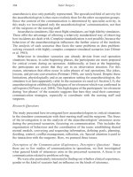
Flap Elevation
Harvest of the flap can usually be performed rapidly in a bloodless field using
a tourniquet. The radial forearm flap is a Type II fasciocutaneous flap. Its domi-
nant pedicle is the radial artery. Deep venous drainage is via the venae comitantes.
Superficial venous drainage is through the cephalic vein which is routinely in-
cluded with the flap. One large (up to 10 x 40 cm) or multiple smaller skin islands
can be harvested anywhere in the volar forearm from the antecubital fossa to the
wrist along the axis of the radial artery (Fig. AI.10).
As it bifurcates off the brachial artery, the radial artery courses between the
brachioradialis and the pronator teres muscle bellies. As it progresses distally, it
travels between the tendons of the brachioradialis and the flexor carpi radialis.
Accordingly, the distal incision is made first, identifying the flexor carpi radialis
and brachioradialis tendons, as well as the underlying artery. The cephalic vein
lies radial to the brachioradialis tendon. It can be ligated and divided at this stage.
The proximal incision is made, and the brachioradialis and the flexor carpi
radialis are identified. The radial artery can be found deep to the brachioradialis
muscle and superficial to the pronator teres. An incision is carried out along the
ulnar border of the skin island. The flap is then elevated from this ulnar border,
working toward its arterial axis. The flap is elevated off the flexor digitorum
superficialis, palmaris longus and the flexor carpi radialis. Particular care is taken
to preserve the peritenon of the tendons. Just radial to the flexor carpi radialis lays
the intermuscular septum carrying the fasciocutaneous perforators.
Lastly, the radial skin incision is made, and the deep fascia is dissected off the
brachioradialis muscle ulnarward. The perforators are identified at the ulnar bor-
der of the brachioradialis along the intermuscular septum. The brachioradialis is
retracted radially to reveal the radial artery. The radial artery is cross-clamped
distally and, if adequate arterial filling of the hand is demonstrated, the radial
artery is ligated and transected. The flap is elevated with dissection of the radial
artery and its venae comitantes. The cephalic vein can be dissected further proxi-
mally to provide an additional long venous pedicle. The radial forearm flap is then
ready for transfer to the intended defect.
Figure AI.10. The course of the radial artery. The skin island can be designed
anywhere along its axis (dashed area).
AI
624
Practical Plastic Surgery
Postoperative Considerations
Donor Site Closure
Part of the donor site can be closed in a primary fashion. The remainder of the
defect is covered with a split-thickness skin graft. The donor site is dressed according
to preference and the hand is splinted for 5-7 days to ensure graft take.
Complications
Complications are not very frequent at the donor site, and harvest of this flap is
generally well-tolerated. The following complications can occur:
• Infection
• Partial or complete skin graft loss
• Cold intolerance
• Hand claudication and other neurosensory changes
• Poor cosmetic appearance of the arm
• Radius fractures (osteocutaneous flap)
Pearls and Pitfalls
• Peritenon covering the flexor carpi radialis, brachioradialis, FDP and FDS ten-
dons should be preserved to minimize skin graft loss and tendon desiccation.
• If the hand is found to be ischemic during cross-clamping of the radial artery, a
venous interposition graft may rarely be needed to maintain adequate perfusion.
• Care should be taken to preserve the sensory branch of the radial nerve during
dissection of the radial artery.
• It is important to include the intermuscular septum to preserve the septocutaneous
perforators. The more distal the skin island, the longer the vascular pedicle.
Suggested Reading
1. Abu-Omar Y, Mussa S, Anastasiadis K et al. Duplex ultrasonography predicts safety of
radial artery harvest in the presence of an abnormal Allen test. Ann Thorac Surg 2004;
77:116.
2. Bardsley AF, Soutar DS, Elliot D et al. Reducing morbidity in the radial forearm flap
donor site. Plast Reconstr Surg 1990; 86(2):287.
3. Evans HB. The radial forearm flap. In: Buncke HJ, ed. Microsurgery: Tranplantation,
Replantation: An Atlas Text. 4th ed. Philadelphia: Lea and Febiger, 1991: Chapter 14.
4. Evans GRD et al. The radial forearm free flap for head and neck reconstruction: A
review. Am J Surg 1994; 168:446.
5. Villaret DB, Futran NA. The indications and outcomes in the use of osteocutaneous
radial forearm free flap. Head Neck 2003; 25(6):475.
6. Yang G, Chen B, Gad Y. Forearm free skin flap transplantation. Nat Med J China
1981; 61:139.
Appendix II
Practical Plastic Surgery, edited by Zol B. Kryger and Mark Sisco. ©2007 Landes Bioscience.
Surgical Instruments
Zol B. Kryger and Mark Sisco
Introduction
The purpose of this Appendix is to provide illustrations of the basic surgical
instruments used in plastic surgery. It is aimed towards the medical students, junior
residents, and non-plastic surgeons who are largely unfamiliar with the names of the
instruments and the many subtle differences. These images were reprinted with gen-
erous permission from Teleflex Medical.
Figure AII.1. Allis tissue forceps (A), Mixter forceps (B), Kelly forceps (C), Hartman
mosquito forceps (D).
A
B
C
D
AII
626
Practical Plastic Surgery
Figure AII.2. Jeweler forceps (straight, angled, curved).
A
B
C
Figure AII.3. Micro tying forceps (A), Castroviejo micro forceps (B), Bishop Harmon
forceps (C).
ABC
AII
627
Appendix II
Figure AII.4. Debakey atraumatic forceps (A), Tissue forceps (B), Russian forceps (C).
ABC
Figure AII.5. Adson tissue forceps or “Adson with teeth” (A), Adson dressing for-
ceps or “smooth Adson forceps” (B), Adson Brown forceps (C).
ABC