Recurrent Hernia Prevention and Treatment - part 4 docx
Bạn đang xem bản rút gọn của tài liệu. Xem và tải ngay bản đầy đủ của tài liệu tại đây (547.82 KB, 41 trang )
121
V
Finding the Best Abdominal Closure – An Evidence-Based Overview of the Literature
33. Gislason H, Gronbech JE, Soreide O. Burst abdomen and
incisional hernia after major gastrointestinal operations
– comparison of three closure techniques. Eur J Surg 1995;
161:349–354
34. Trimbos JB, van Rooji J. Amount of suture material needed for
continuous or interrupted wound closure: An experimental
study. Eur J Surg 1993; 159: 141–143
35. Colombo M, Maggioni A, Parma G, Scalambrino S, Mi-
lani R. A randomized comparison of continuous versus
interrupted mass closure of midline incisions in pa-
tients with gynecologic cancer. Obstet Gynecol 1997; 89:
684–689
36. Brolin RE. Prospective, randomized evaluation of midline
fascial closure in gastric bariatric operations. Am Surg 1996;
172: 328–332
37. Trimbos JB, Smith IB, Holm JP, Hermans J. A randomized clini-
cal trial comparing two methods of fascia closure following
midline laparotomy. Arch Surg 1992; 127: 1232–1234
38. Sahlin S, Ahlberg J, Grantstrom L, Ljungstrom KG. Monofila-
ment versus multifilament absorbable sutures for abdominal
closure. Br J Surg 1993; 80: 322–324
39. Rodeheaver GT, Powell TA, Thacker JG, Edlich RF. Mechanical
performance of monofilament synthetic absorbable sutures.
Am J Surg 1987; 154: 544–547
40. Poole GV, Meredith JW, Kon ND, Martin MB, Kawamoto EH,
Myers RT. Suture technique and wound-bursting strength.
Am Surg 1984; 50:569–572
41. Hodgson NC, Malthaner RA, Ostbye T. The search for an ideal
method of abdominal fascial closure: a meta-analysis. Ann
Surg 2000; 231: 436–442
42. Alexander HC, Prudden JF. The causes of abdominal wound
disruption. Surg Gynecol Obstet 1966; 122: 1223–1229
43. Wadstrom J, Gerdin B. Closure of the abdominal wall: how
and why? Acta Chir Scand 1990; 156: 75–82
44. Rath AM, Chevrel JP. The healing of laparotomies: a review
of the literature. Part 1. Physiologic and pathologic aspects.
Hernia 1998; 2: 145–149
45. Douglas DM. The healing of aponeurotic incisions. Br J Surg
1952; 40: 79–84
46. Luijendijk RW. Incisional hernia; risk factors, prevention, and
repair. Thesis. Erasmus University, Rotterdam. Scheveningen:
Drukkerji Edauw and Johannissen, 2000
47. Wissing J, van Vroonhoven TJMV, Eeftinck Schattenkerk M, et
al. Fascia closure after laparotomy: Results of a randomized
trial. Br J Surg 1987; 74: 738–741
48. Bucknall TE, Teare L, Ellis H. The choice of suture to close
abdominal incisions. Eur Surg Res 1983; 15: 59–66
49. Bucknall TE. Factors influencing wound complication: A clini-
cal and experimental study. Ann R Coll Surg Engl 1983; 65:
71–77
50. Sharp WV, Belden TA, King PH, Teague PC. Suture resistance
to infection. Surgery 1982; 91: 61–63
51. Krukowski ZH, Matheson NA. “Button-hole” incisional her-
nia: A late complication of abdominal wound closure with
continuous non-absorbable sutures. Br J Surg 1987; 74:
824–825
52. Larsen PN, Nielsen K, Schultz A, Mejdahl S, Larsen T, Moes-
gaard F. Closure of the abdominal fascia after clean and
clean-contaminated laparotomy. Acta Chir Scand 1989; 155:
461–464
53. Corman ML, Veidenheimer MC, Coller JA. Controlled clinical
trial of three suture materials for abdominal wall closure after
bowel operations. Am J Surg 1981; 141: 510–513
54. Knight CD, Griffen FD. Abdominal wound closure with a
continuous monofilament polypropylene suture. Arch Surg
1983; 118: 1305–1308
55. Bucknall TE, Ellis H. Abdominal wound closure: a comparison
of monofilament nylon and polyglycolic acid. Surgery 1981;
89: 672–677
56. Schoetz DJ, Coller JA, Veidenheimer MC. Closure of abdomi-
nal wounds with polydioxanone. Arch Surg 1988; 123:72–
74
57. Ray JA, Doddi N, Regula D, Williams JA, Melveger A. Polydiox-
anone (PDS), a novel monofilament synthetic absorbable
suture. Surg Gynecol Obstet 1981; 153:497–507
58. Gys T, Hubens A. A prospective comparative clinical study
between monofilament absorbable and non-absorbable
sutures for abdominal wall closure. Acta Chir Belg 1989;
89:265–270
59. Israelsson LA, Jonsson T. Closure of midline laparotomy in-
cisions with polydioxanone and nylon: the importance of
suture technique. Br J Surg 1994; 81: 1606–1608
60. Carlson MA, Condon RE. Polyglyconate (Maxon) versus nylon
suture in midline abdominal incision closure: a prospective
randomized trial. Am J Surg 1995; 61: 980–983
61. Krukowski ZH, Cusick EL, Engeset J, Matheson NA. Polydiox-
anone or polypropylene for closure of midline abdominal
incisions: a prospective comparative clinical trial. Br J Surg
1987; 74: 828–830
62. Wallace D, Hernandez W, Schlaerth JB, Nalick RN, Morrow
CP. Prevention of abdominal wound disruption utilizing
the Smead-Jones closure technique. Obstet Gynecol 1980;
56:226–230
63. Gallup DG, Talledo OE, King LA. Primary mass closure of
midline incisions with a continuous running monofila-
ment suture in gynecologic patients. Obstet Gynecol 1989;
73:675–677
64. Niggebrugge AH, Trimbos JB, Hermans J, Steup WH, Van de
Velde CJ. Influence of abdominal wound closure technique
on complications after surgery: a randomized study. Lancet
1999; 353: 1563–1567
65. Jenkins TPN. The burst abdominal wound: a mechanical ap-
proach. Br J Surg 1976; 63: 873–876
66. Israelsson LA, Jinsson T. Suture length to wound length ratio
and healing of midline laparotomy incisions. Br J Surg 1993;
80: 1284–1286
67. Varshney S, Manek P, Johnson CD. Six-fold suture: wound
length ratio for abdominal closure. Ann R Coll Surg Engl
1999; 81: 333–336
Discussion
Deysine:
In the 1970s Dr. Goligher introduced a continu-
ous suture with nylon for the closure of laparotomies. At
that time the number of laparotomies exploded in the
world because of vascular surgery and they used be closed
by a running suture. This technique by Dr. Golligher is
very well depicted and those who practice it, like me, are
Schumpelick.indd 121Schumpelick.indd 121 05.04.2007 8:50:54 Uhr05.04.2007 8:50:54 Uhr
122
14
Abdominal Wall Closure
very happy with it. It is a continuous suture with a thick
no.1 nylon and it accommodates to the changes in the
abdominal wall and, to my surprise, it does not include
the skin but all the other layers; the patients have very
little pain with this kind of closure.
Ceydeli: Yes, in the NY State survey also the nonabsorb-
able, monofilament nylon suture was the most common
suture but in the review the most common one was PDS,
late absorbable.
Jeekel: But nylon causes more pain.
Amid: We really need a correct terminology. The most
common mistake that is made is the issue of fascia vs.
aponeurosis. When we close midline the abdominal
wall we don’t close fascia, we close the linea alba or rec-
tus sheath; the fascia is a very thin investing layer of
the muscle that has absolutely no role in hernia surg-
ery.
Jeekel: The suture-length-wound-length ratio, please one
remark to small or large bites.
Israelsson:
I was a bit concerned about the recommenda-
tion of taking 2-cm-large bites. There are several clinical
studies that show that by taking that big size of the bite
you will end up with a high rate of incisional hernia
and wound infection. There is also strong evidence by
experimental studies that a suture-length-wound-length
ratio of 4:1 should be achieved by small tissue bites at
short intervals.
Jeekel: But this is only experimental evidence.
Schumpelick.indd 122Schumpelick.indd 122 05.04.2007 8:50:54 Uhr05.04.2007 8:50:54 Uhr
V
15 Closure of Transverse Incisions
J.A. H, J. J
Incisions
Any incision chosen for access to the abdominal cavity
needs to provide access to the viscera or the lesion to
be treated. Furthermore, an incision needs to provide
extensibility and permit subsequent secure closure. A
further demand may be the postoperative preserva-
tion of function [1] such as containment of abdominal
organs and respiration. Additional considerations in
choosing the incision are the speed of entry, presence
of scars, possibility of hemostasis and a cosmetically
pleasing outcome.
Secure closure must be possible and various suture
materials are used in this day and age. Suture materi-
als should ideally: be sufficient to hold parts together;
disappear as soon as its work is accomplished; be free
of infection; and be non-irritant.
To appreciate the different incisions and problems
with closure, thorough knowledge of the anatomy of
the abdominal wall is mandatory.
Anatomy Ventral Abdominal Wall
The ventral abdominal wall consists of the rectus ab-
dominis muscle on contralateral sides of the line alba.
The origo of the rectus muscle are the 5th, 6th and 7th
rib, the insertion is the pubic bone. The rectus mus-
cles are each contained in a fascial layer, the anterior
and posterior rectus sheath, which is made up of the
aponeurosis (insertion) of the internal, external and
transverse muscle. The rectus muscle is horizontally
incised by the three inscriptiones tendinea. Lateral to
the rectus abdominis the abdominal wall is made up
of the afore-mentioned external oblique, the internal
oblique and the transverse muscle, which extend over
the ventral and lateral part of the abdomen (the part
not covered by the rectus muscle). The origo of the ex-
ternal oblique muscle runs from the 5th to the 12th rib.
The internal oblique originates from the iliac crest. The
transverse muscle, with its horizontal fibre direction,
originates from the previously mentioned iliac crest,
the lumbodorsal fascia and the lower six ribs superiorly.
The lateral border of the rectus muscle forms the linea
semilunaris. At the symphysis pubis the posterior sheath
ends in the thin curved margin, the linea semicircularis
( Douglasi). Below this level the aponeuroses of all three
muscles passes in front of the rectus abdominis and the
fascia transversalis is responsible for the separation of
the rectus from the peritoneum. The pyramidalis muscle
(if present) lies anterior to the lower part of the rectus
abdominis muscle. It arises from the superior surface of
the pubic ramus and inserts at the linea alba.
The vasculature of the muscles of the abdominal
wall consists of the superior and inferior deep epigas-
tric vessels as well as transverse segmental branches of
the aorta. The superior and inferior deep epigastrics
are located in front of the posterior rectus sheath and
the rectus muscle and form its blood supply through
perforating vessels. The inferior deep epigastric ar-
Schumpelick.indd 123Schumpelick.indd 123 05.04.2007 8:50:54 Uhr05.04.2007 8:50:54 Uhr
124
15
Abdominal Wall Closure
tery branches from the external iliac artery whereas
the superior deep epigastric is a branch of the internal
thoracic artery. The deep epigastric arteries are anas-
tomosed and thus form the deep epigastric arcade. The
transverse segmental arteries supply the transverse
muscle, the internal and external oblique and are situ-
ated between the transverse and internal oblique. Blood
supply to the relatively avascular linea alba originates
from the perforating vessels of the superior and inferior
deep epigastrics.
Innervation of the abdominal wall is achieved
through intercostals nerves, the ilioinguinal and the
iliohypogastric nerve. The intercostals nerves are ven-
tral branches of thoracic nerves originating from levels
Th 5 through Th 12 of the spinal cord.
Midline Incisions
The midline incision is possibly the most popular in-
cision amongst surgeons today. When investigating
alternatives to it, the baseline characteristics need to
be described. Midline incisions incise the skin, subcu-
taneous tissue, linea alba and the peritoneum vertically.
Midline incisions are easy, relatively little blood is lost
and the incision takes an average of 7 min to perform
[2–4]. The exposure achieved through a midline in-
cision encompassing the umbilicus is excellent, and
includes access to the retroperitoneum. The upper or
lower abdominal midline incisions may be utilized in
case the expected pathology is situated in the upper or
lower quadrants of the abdomen respectively. Exten-
sions may be made in cranial or caudal direction when
deemed necessary. The qualities mentioned above make
the midline incision the most ideal for emergency and
exploratory surgery.
Transverse Incision
Transverse incisions are possible at all levels of the abdo-
men. Common examples are the Pfannenstiel incision
just above the pubic bone and the upper right quadrant
transverse incision just below the costal margin.
The Pfannenstiel incision is approximately 8–12 cm
in length (distance between the superfiscial epigastric
arteries) and transsects the superficial fascia and the
fibrous rectus sheath. Further access is achieved by
a slightly more cranial, vertical incision of the fascia
transversalis, the preperitoneal fat and the peritoneum
[5]. Luijendijk has described incisional hernia forma-
tion in Pfannestiel incisions most recently and came
to 2.1% in 243 patients after a follow-up between 1.6
and 7.8 years [6].
The upper right quadrant transverse incision re-
quires transsection of the oblique and transverse mus-
culature as well as the rectus muscle. The linea alba is
incised most commonly when extending the transverse
incision across the midline. Dividing the rectus muscle
requires ligating the epigastric arcade yet poses minor
damage to the intercostals nerves and superficial arter-
ies supplying the transverse and oblique musculature
[7]. The transverse incision is thus accompanied by
more blood loss than the midline incision and takes
longer to achieve [4, 8]. Exposure of the lesion is gener-
ally good, although unilateral incisions may provide a
somewhat limited view.
Closure of Incisions
Midline Closure
Studies describing closure of incisions have been per-
formed focusing on continuous, interrupted, layered
closure and various suture materials (absorbable and
non-absorbable). A recent meta-analysis reviewed 13
[9–21] clinically homogeneous randomized controlled
trials comparing absorbable, non-absorbable, continu-
ous and interrupted closure of abdominal incisions [22].
Non-absorbable sutures were found to reduce incisional
herniae when compared with absorbable sutures. The
odds ratio (OR) favouring non-absorbable sutures was
0.68 (95% CI 0.52–0.87) combining data from nine trials
[9–12, 15–18, 21]. Neither wound infection nor wound
dehiscence was statistically more likely in absorbable
sutures. In contrast, suture sinuses and wound pain
were significantly more frequent in the non-absorbable
suture group with respective odds ratios of 2.18 (95%
CI 1.48–3.22) and 2.05 (95% CI 1.52–2.77).
Six trials were identified in the afore-mentioned
meta-analysis comparing interrupted and continuous
suture technique disregarding suture type [9, 12, 14,
17, 20, 21]. Continuous sutures compared favourably
to interrupted sutures (OR 0.73; 95% CI 0.55–0.99). No
statistical differences were found for wound dehiscence
and wound infection.
When taking into account the differences in tech-
nique (nine trials), continuous non-absorbable sutur-
ing outperformed the continuous absorbable suture in
incisional hernia prevention (OR 0.61; 95% CI 0.46–0.8)
[9–11, 14, 16–18, 21]. No significant differences were
found when comparing interrupted absorbable and
interrupted non-absorbable closure.
Schumpelick.indd 124Schumpelick.indd 124 05.04.2007 8:50:55 Uhr05.04.2007 8:50:55 Uhr
125
V
Closure of Transverse Incisions
A subgroup analysis revealed that use of slowly
absorbable polydioxanone (PDS) and polyglycolic
acid (Dexon) did not significantly increase the risk
for incisional hernia formation compared to polypro-
pylene. Polyglactin (Vicryl) compared unfavourably
with non-absorbable sutures. Previously Wissing et al.
have found that nylon has the lowest incidence of inci-
sional hernia yet is unfavourably associated with more
wound pain and suture sinuses than polydioxanone
sutures [21].
Transverse Closure
Randomized studies, not mentioned earlier, specifi-
cally describing incisional hernia formation with re-
spect to midline, transverse and oblique incisions are
summarized in
⊡
Table 15.1. Transverse incisions were
found to be prone to incisional hernia formation in
3.6 – 40% of patients. Fassiadis et al. used continuous
single-layered closure with nylon in the trial reported.
The hernia incidence in high-risk patients undergo-
⊡
Table 15.1. Randomized studies on incisional hernia
Author Year Patients
N
Incision(s) Fol-
low-up
[months]
Rate of
incisional
hernia [%]
Technique, suture
type, layers [L]
p
value
Blom-
stedt
[24]
1972 130 Transverse 8–24 19.5 Various suturesa,
2 L
ns
<0.01
RCT 115 Midline 13.9 Various suturesa,
1 L
180 Oblique 13.8 Various suturesa,
2 L
Greenall
[8]
1980 235 Transverse >6 16.4 Variousa, 1 L, cont. ns
RCT 234 Midline >6 18.1 Variousa, 1 L, cont.
Ellis [25] 1984 150 Transverse <12 14.0 Nylon, 1 L, cont. ns
RCT 146 Parame-
dian
<12 17.4 Nylon, 1 L, cont.
Schoetz
[26]
1988 128 Transverse 1–12 13.6 PDS, 1 L, cont. ns
172 Midline 1–12 12.9 PDS, 1 L, cont.
Lord
[27]
1994 126 Transverse 12–72 13.5 Nylon, 2 L, cont. ns
RCT 109 Midline 16.5 Nylon, 1 L, cont.
Fassia-
dis [23]
2005 115 Transverse >48 40 Nylon, 1 L, cont. <0.01
RCT 122 Vertical 91 Nylon, 1 L, cont.
Halm Sub. 160 Transverse 12–36 12 Vicryl, 2 L, comb. p =
0.02
RCT 163 Vertical 14 Vicryl, 1 L, inter.
a
absorbable/non-absorbable. RCT randomized controlled trial; ns not significant; cont. continuous; inter. interrupted;
comb. one layer cont. and one layer inter.; L layer; sub. submitted
Schumpelick.indd 125Schumpelick.indd 125 05.04.2007 8:50:55 Uhr05.04.2007 8:50:55 Uhr
126
15
Abdominal Wall Closure
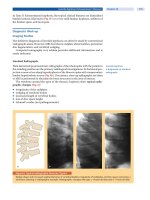
ing abdominal aortic aneurysm surgery was reported
to be 40%. In the transverse incisions studied by Fas-
siadis (using ultrasound) the incisional hernias were
found predominantly at the lateral border [23].
Schoetz found the most encouraging results in
closure of transverse incisions, 3.6% incisional hernia
incidence after continuous closure with polydioxa-
none.
No studies were found specifically comparing dif-
ferent methods of closure (materials or technique) for
the transverse incision.
Currently unpublished (submitted) results from
a randomized study (n = 150) performed at our own
institute confirmed the results that transverse incisions
(2% incisional hernia) are significantly less likely to de-
velop hernias compared to upper abdominal midline
incisions (14% incisional hernia) in the patients seen
at follow-up (
⊡
Table 15.1). Closure of the transverse
incision of the abdominal wall was achieved by closure
of the peritoneum and the posterior rectus fascia us-
ing a continuous, polyglactin 910 suture (Vicryl). The
anterior rectus sheath and the fascia of the internal and
external transverses were closed using simple inter-
rupted polygalactin 910 sutures (Vicryl).
Complications: Pain, Wound Infection
and Burst Abdomen
Armstrong et al., reporting a randomized study compar-
ing midline and transverse incisions in 60 patients, have
documented significantly reduced postoperative pain
for transverse incisions [28], a result that we confirmed
in our own (submitted) randomized trial. Halasz et al.
found a reduction in the use of analgesics in patients
after an oblique incision when compared to a parame-
dian approach [29]. A similar result was found by Gar-
cia-Valdecasas comparing oblique to midline incisions
[30]. The review by Burger et al. concluded that none
of the trials performed to date reported a significant
difference in surgical site infection rates [31].
Burst abdomen has an incidence between 0 and
2.5% and was found to be more likely after vertical in-
cisions. Pooling of data by Grantcharov and coworkers
revealed a significant difference between the incidence
of burst abdomen after vertical incision of 1% (46/4480)
and after transverse incision of 0.34% (15/4365) [32].
An odds ratio of 2.86 favouring transverse inci-
sion 95% CI 1.72–4.73 was subsequently calculated
(
⊡
Table 15.2).
Randomized Controlled Trial
The POVATI trial (ISRCTN 60734227), as initiated
by researchers from Heidelberg, Germany (Prof.
Dr. M.W. Büchler), compares the two most com-
mon incisions in general surgery, midline and trans-
verse [34].
The trial, which was started in July 2003, proposes
abdominal wall closure in a standardized way in both
groups: four Mikulicz clamps are to be placed at the
edges of the abdominal fascia and a continuous, all-
layer closure technique with two Mono Plus loops
⊡
Table 15.2. Data on burst abdomen incidence
Author Type
of publication
No. of patients Incision(s) Rate of burst
abdomen [%]
p value
Greenall [8] RCT 1292 Transverse 0 0.2453
1287 Midline 0.69
Thompson [33] Retr. 1760 Transverse 0.5 0.004
1603 Midline 2.5
Halasz [29] Retr. 3313 Transverse 0.33 0.009
3590 Midline 0.81
RCT randomized controlled trial; Retr. retrospective
Schumpelick.indd 126Schumpelick.indd 126 05.04.2007 8:50:56 Uhr05.04.2007 8:50:56 Uhr
127
V
Closure of Transverse Incisions
(Aesculap, Tuttlingen, Germany) performed, starting
from both ends of the incision with a 4:1 ratio (suture
length:wound length). Neither subcutaneous closure
nor subcutaneous drainage is proposed. Skin closure
is to be achieved with skin clips.
Primary outcome measures are the requirement of
analgesics and patient satisfaction. Secondary outcomes
are incisional hernia 1 year postoperative (diagnosed
by ultrasound). Burst abdomen, pulmonary infection
and wound infection are secondary endpoints, but are
also defined as adverse events.
Closure of the Transverse Incision:
How We Do It
Currently, hepaticopancreaticobilliary surgeons of the
Erasmus MC propose double-layered closure of trans-
verse incisions, reasoning that the cosmetic outcome
is more pleasing since, in their experience, the skin
inadvertently inverts when single-layered closure is
employed.
In detail, a USP 0 PDS loop (Ethicon, Johnson &
Johnson Amersfoort) is used to close the posterior fas-
cia in a continuous fashion starting at the lateral border
of the incision. Upon reaching the medial border of
the incision, the same loop, without interruption, is
employed to approximate the anterior fascia and the
internal and external obliques. A suture-length-to-
wound-length ratio of 4 to 1 is maintained through-
out. Subcutaneous closure is achieved in case the dead
space observed is deemed too large in the eyes of the
surgeon. For reduction of dead space interrupted
Vicryl (Ethicon, Johnson & Johnson, Amersfoort)
sutures are used. Skin closure is achieved by intra-
cutaneous, continuous suturing using Monocryl 5–0
(Ethicon, Johnson & Johnson, Amersfoort, The Nether-
lands).
Conclusion
Closure of transverse incisions can be achieved securely
using single as well as double-layered closure. Non-
absorbable or slowly absorbable sutures seem to be
advantageous in the prevention of incisional hernia,
as is continuous suturing technique. Slowly absorb-
able sutures seem to reduce the incidence of wound
pain and suture sinuses. Further research in the form of
randomized controlled trials seems warranted in light
of the lack of data on the topic of transverse closure
techniques.
References
1. Skandalakis LJ, Gadacz TR, Mansberger AR, et al. Modern
Hernia Repair: the embryological and anatomical basis of
surgery. New York: Parthenon Publishing Group, 1996
2. Guillou PJ, Hall TJ, Donaldson DR, et al. Vertical abdominal
incisions – a choice? Br J Surg 1980; 67(6): 395–399
3. Kendall SW, Brennan TG, Guillou PJ. Suture length to wound
length ratio and the integrity of midline and lateral parame-
dian incisions. Br J Surg 1991; 78(6): 705–707
4. Lacy PD, Burke PE, O’Regan M, et al. The comparison of
type of incision for transperitoneal abdominal aortic sur-
gery based on postoperative respiratory complications and
morbidity. Eur J Vasc Surg 1994; 8(1): 52–55
5. Pfannenstiel HJ. Ueber die Vortheile des suprasymphysären
Fascienquerschnitts für die gynäkologischen Koeliotomien,
zugleich ein Beitrag zu der Indikationsstellung der Opera-
tionswege. Volkmann’s Sammlung klinischer Vorträge,
Leipzig, 1900, n F. 268 (Gynäk. Nr. 97), 1735–1756
6. Luijendijk RW, Jeekel J, Storm RK, et al. The low transverse
Pfannenstiel incision and the prevalence of incisional hernia
and nerve entrapment. Ann Surg 1997; 225(4): 365–369
7. Nahai F, Hill L, Hester TR. Experiences with the tensor fascia
lata flap. Plast Reconstr Surg 1979; 63(6): 788–799
8. Greenall MJ, Evans M, Pollock AV. Midline or transverse lapa-
rotomy? A random controlled clinical trial. Part I: Influence
on healing. Br J Surg 1980; 67(3): 188–190
9. Bucknall TE, Ellis H. Abdominal wound closure a comparison
of monofilament nylon and polyglycolic acid. Surgery 1981;
89(6): 672–677
10. Cameron AE, Gray RC, Talbot RW, Wyatt AP. Abdominal
wound closure: a trial of Prolene and Dexon. Br J Surg 1980;
67(7):487–488
11. Carlson MA, Condon RE. Polyglyconate (Maxon) versus nylon
suture in midline abdominal incision closure: a prospective
randomized trial. Am Surg 1995; 61(11): 980–983
12. Cleveland RD, Zitsch RP, 3rd, Laws HL. Incisional closure in
morbidly obese patients. Am Surg 1989; 55(1): 61–63
13. Corman ML, Veidenheimer MC, Coller JA. Controlled clinical
trial of three suture materials for abdominal wall closure after
bowel operations. Am J Surg 1981; 141(4): 510–513
14. Irvin TT, Koffman CG, Duthie HL. Layer closure of laparotomy
wounds with absorbable and non-absorbable suture materi-
als. Br J Surg 1976; 63(10): 793–796
15. Kronborg O. Polyglycolic acid (Dexon) versus silk for fas-
cial closure of abdominal incisions. Acta Chir Scand 1976;
142(1):9–12
16. Krukowski ZH, Cusick EL, Engeset J, Matheson NA. Polydiox-
anone or polypropylene for closure of midline abdominal
incisions: a prospective comparative clinical trial. Br J Surg
1987; 74(9): 828–830
17. Larsen PN, Nielsen K, Schultz A, et al. Closure of the abdomi-
nal fascia after clean and clean-contaminated laparotomy.
Acta Chir Scand 1989; 155(9): 461–464
18. Leaper DJ, Allan A, May RE, et al. Abdominal wound closure:
a controlled trial of polyamide (nylon) and polydioxanone
suture (PDS). Ann R Coll Surg Engl 1985; 67(5): 273–275
19. Lewis RT, Wiegand FM. Natural history of vertical abdomi-
nal parietal closure: Prolene versus Dexon. Can J Surg 1989;
32(3):196–200
Schumpelick.indd 127Schumpelick.indd 127 05.04.2007 8:50:56 Uhr05.04.2007 8:50:56 Uhr
128
15
Abdominal Wall Closure
20. Richards PC, Balch CM, Aldrete JS. Abdominal wound closure.
A randomized prospective study of 571 patients compar-
ing continuous vs. interrupted suture techniques. Ann Surg
1983; 197(2): 238–243
21. Wissing J, van Vroonhoven TJ, Schattenkerk ME, et al. Fascia
closure after midline laparotomy: results of a randomized
trial. Br J Surg 1987; 74(8): 738–741
22. Hodgson NC, Malthaner RA, Ostbye T. The search for an ideal
method of abdominal fascial closure: a meta-analysis. Ann
Surg 2000; 231(3): 436–442
23. Fassiadis N, Roidl M, Hennig M, et al. Randomized clinical
trial of vertical or transverse laparotomy for abdominal aortic
aneurysm repair. Br J Surg 2005; 92(10): 1208–1211
24. Blomstedt B, Welin-Berger T. Incisional hernias. A comparison
between midline, oblique and transrectal incisions. Acta Chir
Scand 1972; 138(3): 275–278
25. Ellis H, Coleridge-Smith PD, Joyce AD. Abdominal inci-
sions vertical or transverse? Postgrad Med J 1984; 60(704):
407–410
26. Schoetz DJ, Jr., Coller JA, Veidenheimer MC. Closure of ab-
dominal wounds with polydioxanone. A prospective study.
Arch Surg 1988; 123(1): 72–74
27. Lord RS, Crozier JA, Snell J, Meek AC. Transverse abdominal
incisions compared with midline incisions for elective in-
frarenal aortic reconstruction: predisposition to incisional
hernia in patients with increased intraoperative blood loss.
J Vasc Surg 1994; 20(1): 27–33
28. Armstrong PJ, Burgess RW. Choice of incision and pain fol-
lowing gallbladder surgery. Br J Surg 1990; 77(7): 746–748
29. Halasz NA. Vertical Vs Horizontal Laparotomies. I. Early post-
operative comparisons. Arch Surg 1964; 88: 911–914.
30. Garcia-Valdecasas JC, Almenara R, Cabrer C, et al. Subcostal
incision versus midline laparotomy in gallstone surgery:
a prospective and randomized trial. Br J Surg 1988; 75(5):
473–475
31. Burger JW, van ‘t Riet M, Jeekel J. Abdominal incisions: tech-
niques and postoperative complications. Scand J Surg 2002;
91(4): 315–321
32. Grantcharov TP, Rosenberg J. Vertical compared with
transverse incisions in abdominal surgery. Eur J Surg 2001;
167(4):260–267
33. Thompson JB, MacLean KF, Coller FA. Role of the transverse
abdominal incision and early ambulation in the reduction
of postoperative complications. Arch Surg 1949; 59(6):
1267–1277
34. Reidel MA, Knaebel HP, Seiler CM, et al. Postsurgical pain
outcome of vertical and transverse abdominal incision:
design of a randomized controlled equivalence trial [IS-
RCTN60734227]. BMC Surg 2003; 3: 9
Discussion
Schumpelick: How should we close transverse incisions,
what is your recommendation: single or double layer?
Jeekel: I close by single layer when it is a small muscle
and when it is a big muscle I do a double layer.
Schumpelick.indd 128Schumpelick.indd 128 05.04.2007 8:50:57 Uhr05.04.2007 8:50:57 Uhr
V
16 Biological Reasons for an Incisional Hernia
J.M B
Introduction
Incisional hernia continues to represent a significant prob-
lem within the context of abdominal wall pathologies.
The incidence of incisional hernia has remained con-
stant over the past decade, despite numerous modifica-
tions in the techniques and materials used. It is a frequent
complication of abdominal surgery, with a reported inci-
dence of 2–11%. After procedures such as aortic surgery,
the rate can be as high as 16–20%. In the USA, 4 to 5 mil-
lion laparotomies are performed annually, which means
that at least 400,000 to 500,000 incisional hernias can be
expected to develop each year. Incisional hernia repair
is performed approximately 200,000 times per year. The
total financial cost of these operations could be around
2.5 billion dollars [1].
In general, the wound-healing process can be divided
into three stages: an inflammatory stage, a fibroplastic
stage and a stage of maturation. The inflammatory stage
lasts for 4–6 days, during which time the wound is pre-
pared for subsequent healing by removal of necrotic tis-
sue and bacteria. During this period, the wound has no
intrinsic strength and its integrity is entirely dependent on
the suture and the suture-holding capacity of the tissues.
This stage is followed by a fibroplastic phase character-
ized by collagen synthesis. During this second stage, the
wound rapidly gains in tensile strength by the bridging
over of collagen fibres. The fibroplastic stage is gradually
followed by a prolonged phase of maturation in which
collagen fibres are remodelled.
The tensile strength of a sutured aponeurosis after 2–3
weeks is about 20% that of unwounded tissue, and after 4
weeks is about 50%. After 6–12 months, the aponeurosis
attains about 80% of its original strength, but complete
recovery is never achieved.
Factors Contributing to the Genesis
of Incisional Hernia
Why do incisional hernias occur? Incisional her-
nias occur as the result of a biomechanical defect in
acute fascial wound healing, which affects the nor-
mal capacity of the abdominal wall to support in-
creasing tension during the postoperative recovery
period.
Most studies now support the theory that acute
fascial separation occurs early in the postoperative
period, during the course of acute wound healing at
a time when wound tensile strength is very low or ab-
sent (postoperative days 0–30), and leads to the de-
layed clinical development of abdominal wall incisional
hernias [2].
It is during this early period of acute wound healing
that the scar depends entirely on the integrity of the
suture to keep the abdominal wall closed. This integrity,
in turn, also depends on the success of the wound repair
process in each individual.
Several factors have been implicated in the aetiology
and pathogenesis of the incisional hernia [3].
Schumpelick.indd 129Schumpelick.indd 129 05.04.2007 8:50:57 Uhr05.04.2007 8:50:57 Uhr
130
16
Abdominal Wall Closure
The most frequently identified clinical risk factors
for fascial wound failure and primary incisional hernia
formation include:
▬ Type of laparotomy
▬ Suboptimal closure technique
▬ Infections
▬ Malnutrition
▬ Preoperative hypotension
▬ Jaundice, anaemia, corticosteroid therapy
▬ Biological disorders (collagen-related)
Transverse laparotomies generally show a lower inci-
dence of incisional hernia than vertical ones [4].
Many laparotomy closures are incorrectly under-
taken and basic rules such as the 4:1 Jenkins rule are
neglected [5]. In many cases, closure is undertaken by
surgeons early on in the learning curve with insufficient
training.
Infection has been directly linked to over 75% of
incisional hernias. In addition, malnutrition and sub-
stantial blood loss during surgery have been related to
a greater incidence of incisional hernia. Other factors
such as jaundice, anaemia and steroid treatment in-
terfere with the entire healing process in general and
therefore contribute to the appearance of this abdomi-
nal wall pathology.
Finally, there is also a series of factors related to
the tissue biology of each individual. These factors are
associated with the biological wound repair, or scar-
ring process. The scarring process in one subject ob-
viously differs to that in another, mainly because of
tissue components and inducers that mediate the pro-
cess.
Biological factors include the components of the ex-
tracellular matrix such as collagens and the enzymes
metalloproteinases (MMPs). Exogenous variables can
also predispose an individual to incisional hernia such
as smoking or a concurrent disease whose underlying
cause is a collagen alteration, including aortic aneurysm,
cutis laxa, Marfan’s syndrome, osteogenesis imperfecta,
and Ehlers-Danlos syndrome.
Biological Factors
The search for biological factors involved in the ap-
pearance of incisional hernia has been limited, un-
like the case for biological factors contributing to the
genesis of groin hernias. This is possibly because the
pathogenesis of incisional hernia depends on many
other factors other than those strictly classed as bio-
logical factors.
Biological factors, in an individual manner, closely
modulate the repair process at the level of the fascia;
this is the only retaining structure after a laparotomy
closure.
In fascial tissue, the mechanisms regulating the pro-
liferative and synthesizing capacity of fibroblasts have
not yet been defined. Neither do we know the reason
for the failure of a surgical wound that generates inci-
sional hernias.
To date, it has not been possible to establish a cor-
relation between the proliferative response of fascial
fibroblasts at the level of the cell cycle and wound heal-
ing failure [2].
Ischemia at the level of the fascial continuum could
arrest the cell cycle of the fibroblast as a reparatory cell.
This could occur in a technically deficient closure (when
the suture is too tight or closure is under tension) or in
cases of sustained intra-operative hypotension when the
oxygen supply to the tissues is reduced.
Notwithstanding, in the past few years some inves-
tigations have centred on those factors or diseases that
could condition the appearance of an incisional hernia
following laparotomy. Many of the factors identified
so far have also been implicated in the genesis of other
types of hernia such as groin hernias.
Experimental Models
Role of Cytokines: TGF-beta and FGFb
In a rat model, Franz et al. [6] created incisional her-
nias after performing a midline laparotomy closed
with a suture that was absorbable in the short term.
This generates a defect in the abdominal wall that pro-
duces a postlaparotomy hernia. Topical treatment of
laparotomy closures with recombinant TGF-β2 in an
aqueous medium has been noted to diminish the ap-
pearance of incisional hernia and to increase fibroblasts,
and collagen type-I and -III deposition, detected by
immunohistochemistry.
Using the same experimental model, DuBay et
al. [7] reported that by treating the fascia with FGFb
loaded in a polymer vehicle, the appearance of inci-
sional hernia was significantly reduced. In animals
treated with this growth factor, angiogenesis and col-
lagen deposition were also found to improve.
Another hypothesis proposed by the group of
Franz and Dubay [6,7], is that the aponeurotic tissue
of the abdominal wall is also dependent on mechani-
cal signals to regulate the homeostasis of the fascial
fibroblast. This mechanico-transduction theory pro-
Schumpelick.indd 130Schumpelick.indd 130 05.04.2007 8:50:57 Uhr05.04.2007 8:50:57 Uhr
131
V
Biological Reasons for an Incisional Hernia
poses that the load on soft tissue or bone is transmitted
to structural cells through the extracellular matrix, and
that there are integrin type receptors on the cell surface.
Mechanical failure or reduced mechanical signals, for
instance, when a suture fails, could lead to the impaired
kinetics and proliferative capacity of the reparative fi-
broblast.
It has been well established that during the repair
of tendons and ligaments, the mechano-transduction
pathway is important for triggering the repairing ac-
tions of fibroblasts. A wound in the fascia could show
similar behaviour.
Clinical Studies
Role of Collagen
Collagen plays a predominant role in any wound-repair
process. It constitutes the main axis of wound healing
along with the enzymes metalloproteinases (MMPs),
which balance their production and lysis.
Klinge et al. [8] observed an imbalance between col-
lagen I and III in patients with inguinal and incisional
hernia.
In cultures of fibroblasts taken from the skin of
patients with recurrent incisional hernia, Si et al. [9]
also noted an imbalance between collagen type I and
III. These authors also reported generally disorganised
levels of collagens in the extracellular matrix.
Rosch et al. [10] also described a reduction in the
collagen I/III ratio in patients with incisional hernia.
MMPs and Incisional Hernia
A balance between extracellular matrix synthesis and
degradation is important for tissue integrity, because re-
modelling occurs continuously. MMPs are the enzymes
that regulate the components of the extracellular matrix.
Changes or defects in matrix molecules may also alter
tissue architecture, impairing the proper assembly of
the matrix components and modifying the mechani-
cal properties of the tissue. Some of these enzymes
play an important role in the general scarring process
[11,12]. Thus, wounds that are difficult to repair such
as in patients with diabetes show high MMP levels. In
these patients, skin fibroblasts have been found to show
increased amounts of MMP-2 [13].
In incisional hernias, Klinge et al. [14] found re-
duced MMP-1 expression compared to controls through
Western blot analysis of fascial tissue.
Aortic Aneurysm and Incisional Hernia
The relationship among disorders in which extracel-
lular matrix components are involved, such as aortic
aneurysm, has been widely described in the litera-
ture.
Stevick et al. [15] first pointed out the link between
post-laparotomy incisional hernia and aortic aneurysm,
although Cannon et al. [16], had previously observed
a relationship between patients with inguinal hernia
and aneurysm.
In subsequent studies [17–19], a high incidence of
aortic aneurysm was correlated with a similar incidence
of incisional hernia.
The rate of incisional hernia has been reported
to be as high as 31% following midline laparotomy
for abdominal aortic-aneurysm repair [20, 21]. In a
recent randomized study performed on patients un-
dergoing surgery for aortic aneurysm, Fassiadis et al.
[22] noted a lower incidence of incisional hernia in
transverse laparotomies compared to midline proced-
ures.
Alterations to the extracellular matrix have been
reported by several authors.
In 1993, White et al. [23] reported that adventitial
elastolysis was a primary event in aneurysm forma-
tion. Later, enhanced MMP-2 and MMP-9 expression
was reported by Patel et al. [24], Skalihasan et al. [25],
and Tamarina et al. [26]. In cultured muscle cells har-
vested from the medial layer of the aortic aneurymal
wall, increased MMP-2 expression has been described
[27].
Smokers
Smokers have a high risk of incisional hernia forma-
tion independent of other recognized risk factors, pre-
sumably owing to the detrimental effect of smoking
on wound healing. Diminished collagen deposition
in surgical test wounds has been observed in smok-
ers [28].
The link between inguinal hernia, aortic aneurysm
and smoking was first suggested by Read [29]. Accord-
ing to Read, the degradation of connective tissue caused
by imbalance between proteases and their inhibitors
could also be a contributing factor. Smoking has been
related to increased proteolytic activity, activation of
neutrophils and macrophages and the release of oxi-
dants, impairing the antiprotease defence mechanism,
leading to increased collagenolysis and inappropriate
repair [30].
Schumpelick.indd 131Schumpelick.indd 131 05.04.2007 8:50:58 Uhr05.04.2007 8:50:58 Uhr
132
16
Abdominal Wall Closure
In a recent study, Sorensen et al. [31] linked smok-
ing with the appearance of incisional hernia. In this
study, the incidence of incisional hernia is four times
higher in smokers than non-smokers. A relationship
between smoking and hernia recurrence had already
been reported [32] in a study in which recurrence was
found to occur more frequently in smokers undergoing
herniorraphy.
In general terms, all the biological factors that
could induce the appearance of an incisional her-
nia are inter-related. It thus becomes obvious that in
the absence of other risk factors (infection, an inap-
propriate closure technique, malnutrition, jaundice
etc.), the biology of the individual plays a pivotal role.
Hence, when several biological risk factors are pres-
ent these could have a synergistic effect on the repair
process.
A smoker who also has a collagen disorder will
have a greater risk of developing an incisional her-
nia after a laparotomy. This would explain why her-
nia recurrence sometimes occurs after the successful
surgical repair of an incisional hernia. This event was
described in a recent report [33], in which recurrence
mechanisms of operated incisional hernias were classi-
fied.
References
1. Wedbush Morgan Securities. Biotechnology in wound care
2001; 4: 1–82
2. Franz MG, Robson MC. The use of the wound healing trajec-
tory as an outcome determinant for acute wound healing.
Wound Repair Regen 2001; 8: 511–516
3. Carlson MA. Acute wound failure. Wound healing. Surg Clin
North Am 2001; 77: 607–635
4. Grantcharov TP, Rosenberg J. Vertical compared with transverse
incisions in abdominal surgery. Eur J Surg 2001; 167: 260–267
5. Jenkins TNP. The burst abdominal wound: a mechanical ap-
proach. Br J Surg 1976; 63: 837–876
6. Franz MG, Kuhn MA, Nguyen K, Wang X, Ko F, Weig TE, Rob-
son MC. Transforming growth factor β2 lowers the incidence
of incisional hernias. J Res 2001; 97: 109–116
7. DuBay DA, Wang X, Kuhn MA, Robson MC, Franz MG. The
prevention of incisional hernia formation using a delayed-
release polymer of basic fibroblast growth factor. Ann Surg
2004; 240: 179–186
8.
Klinge U, Si ZY, Zheng H, Schumpleick V, Bhardwaj RS, Kloter-
halfen B. Abnormal collagen I to III distribution in the skin of
patient with incisional hernia. Eur Surg Res 2000; 32: 43–48
9. Si ZY, Rhanjit B, Rosch R, Mertens R, Klosterhalfen B, Klinge
U. Impaired balance of type I and type III procollagen mRNA
in cultured fibroblasts of patients with incisional hernia. Sur-
gery 2002; 131: 324–331
10. Rosch R, Junge K, Knops M, Lynen P, Klinge U, Schumpelick
V. Analysis of collagen-interacting proteins in patients
with incisional hernia. Langebecks Arch Surg 2003; 387:
427–432
11. Agren MS. Jorgensen LN, Andersen M, Viljanto J, Gottrup F.
Matrix metalloproteinase 9 level predicts optimal collagen
deposition during early wound repair in humans. Br J Surg
1998; 85: 68–71
12. Nwomeh BC, Liang HX, Cohen IK, Yager DR. MMP-8 is the
predominant collagenase in healing wounds and nonheal-
ing ulcers. J Surg Res 1999; 81: 189–195
13. Wall SJ, Sampson MJ, Levell N, Murphy G. Elevated ma-
trix metalloproteinase-2 and 3 production from human
diabetic dermal fibroblasts. Br J Dermatol 2003; 149:
13–16
14. Klinge U, Si ZY, Zheng H, Schumpelick V, Bhardwaj RS,
Klosterhalfen B. Collagen I/III and matrix metalloprotein-
ases (MMP) 1 and 13 in the fascia of patients with incisional
hernias. J Invest Surg 2001; 14: 47–54
15. Stevick CA, Long JB, Jamasbi B. Ventral hernia follow-
ing abdominal aortic reconstruction. Am Surg 1988; 51:
287–289
16 Cannon DJ, Castel L, Read RC. Abdominal aortic aneurysm,
Leriche syndrome, inguinal herniation, and smoking. Arch
Surg 1984; 119: 387–389.
17. Hall KA, Peters B, Smyth SH, Warmeke JA, Rappaport WD,
Putnam ChW, Hunter GC. Abdominal wall hernias in patients
with abdominal aortic aneurysmal versus aortoiliac occlusive
disease. Am J Surg 1995; 170: 572–576
18. Holland AJA, Castleden WM, Norman PE, Stacey MC. Inci-
sional hernias are more common in aneurysmal disease.
Eur J Vasc Endovasc Surg 1996; 12: 196–200
19. Rogers M, McCarthy, Earnshaw JJ. Prevention of incisional
hernia after aortic aneurysm repair. Eur J Vasc Endovasc Surg
2003; 26: 519–522
20. Adye B, Luna G. Incidence of abdominal wall hernia in aortic
surgery. Am J Surg 1998; 175: 400–402
21. Raffetto JD, Cheung Y, Fisher JB. Incision and abdominal wall
hernias in patients with aneurysm or occlusive aortic disease.
J Vasc Surg 2003; 37: 1150–1154
22. Fassiadis N, Roidl M, Hennig M, South LM, Andrews SM. Ran-
domized clinical trial of vertical or transverse laparotomy
for abdominal aortic aneurysm repair. Brit J Surg 2005; 92:
1208–1211
23. White JV, Haas K, Phillips S, Comerota AJ. Adventitial elas-
tolysis in primary event in aneuriysm formation. J Vasc Surg
1993; 17: 371–381
24. Patel MI, Melrose J, Ghosh P, Appleberg M. Increased syn-
thesis of matrix metalloproteinases by aortic smooth muscle
cells is implicated in the etiopathogenesis of abdominal aor-
tic aneurysms. J Vasc Surg 1996; 24: 82–92
25. Sakalihasan N, Delvenne P, Nusgens BV, Limet R, Lapière
ChM. Activated forms of MMP-2 and MMP-9 in abdominal
aortic aneurysms. J Vasc Surg 1996; 24: 127–133
26. Tamarina NA, McMillan WD, Shively VP, Pearce WH. Ex-
pression of matrix metalloproteinases and their inhibi-
tors in aneurysms and normal aorta. Surgery 1997; 122:
264–272
Schumpelick.indd 132Schumpelick.indd 132 05.04.2007 8:50:58 Uhr05.04.2007 8:50:58 Uhr
133
V
Biological Reasons for an Incisional Hernia
27. Crowther M, Goodall S, Jones JL, Bell PRF, Thompson MM.
Increased matrix metalloproteinase 2 expression in vascular
smooth muscle cells cultured from abdominal aortic aneu-
rysms. J Vasc Surg 2000; 32: 575–583
28. Jorgensen LN, Kallehave F, Christensen E, Siana JE, Gottrup
F. Less collagen production in smokers. Surgery 1998; 123:
450–455
29. Read RC. A review: The role of protease-antiprotease imbal-
ance in the pathogenesis of herniation and abdominal aortic
aneurysm in certain smokers. Postgraduate General Surg
1992; 14: 161–165
30. Read RC. Why do human beings develop groin hernias? In:
Fitzgibbons, R.J., Jr. Greeburg A.G. eds. Nyhus and Condon´s
hernia. Philadelphia: Lippincolt Williams&Wilkins 2002;
3–8
31. Sorensen LT, Hemmingsen RN, Kirkeby LT, Kallehave F, Jor-
gensen LN. Smoking is a risk factor for incisional hernia. Arch
Surg 2005; 140: 119–123
32. Sorensen LT, Friis E, Jorgensen LN, Vennits B, Ander-
sen BR, Rasmussen GI, Kjaergaard J. Smoking is a risk
factor for recurrence of groin hernia. World J Surg 2002; 26:
397–400
33. Awad ZT, Puri V, LeBlanc K, Stoppa R, Fitzgibbons Jr RJ, Iqbal
A, Filipi ChJ. Mechanisms of ventral hernia recurrence after
mesh repair and a new proposed classification. J Am Coll
Surg 2005; 201: 132–140
Discussion
Franz: In our experimental work we never found a collagen
synthesis defect in our animals. We can generate something
in the animal that looks very much like a human incisional
hernia without any recognizable biological defect, and that
is what bothers us as surgeons, that so many patients will
fail despite any easily recognizable biological defect; how-
ever, once the failure occurs and we are able to measure
postmechanical failure defects on the fibroblastic level, one
of our first surprising observations was that there never
was a defect in the collagen production either in the wound
or in the isolated fibroblast. The German group is good
about demonstrating isonomic imbalances and showing
perhaps pathology level that way, but we were never able
to measure a collagen total synthesis defect.
Kingsnorth: What has not been mentioned are two small
randomized trials using meshes prophylactically to sup-
port the wound, in aortic aneurysms and bariatric surgery.
This is probably working better than trying to supplement
the biological factors in the wound. What is your view of
prophylactic mesh in patients with high risk?
Bellon: I think that is the future ….
Schumpelick.indd 133Schumpelick.indd 133 05.04.2007 8:50:59 Uhr05.04.2007 8:50:59 Uhr
V
Introduction
“Occasional contributions have appeared on the subject of
disruption of wounds for a long time, but more than forty ar-
ticles have been found in the American literature alone during
the last few years as evidence of its importance” (Singleton
and Blocker 1939 [1]).
Postoperative abdominal incision failure remains as much
a problem and topic of controversy today as it did nearly
a century ago. The predominance of the surgical literature
on incisional hernia describes and evaluates various repair
techniques; less is written on predisposition and prevention.
In the latter subset of the literature, emphasis has been
placed upon patient-associated risk factors in the patho-
genesis of incisional failure. Over the past several decades,
however, the idea that surgeon-associated (i.e., technical)
risk factors may be important in the etiology of incisional
hernia has been gaining more acceptance [2]. The postula-
tion that the surgeon could be the most important risk fac-
tor for this complication, however, is a more radical concept.
This brief review will emphasize the role of surgeon-related
factors in the development of incisional hernia.
Dehiscence vs. Incisional Hernia:
Separate or the Same?
Abdominal wound dehiscence (variably known as
wound disruption, acute wound or fascial failure,
burst abdomen, etc.) and incisional hernia often are
thought of as two separate entities, but they prob-
ably are ends of the same continuum. In general, the
fascial disruption of wound dehiscence occurs in the
early postoperative period (within the first several
weeks); with incisional hernia, the disruption mani-
fests later. The skin remains intact in the latter, having
had ample time to heal, while in the former the skin
either disrupts with the fascia or leaks fluid. So does
an incisional hernia develop in a scar that has healed
and then weakens over time? The current data sug-
gest that a patient who acquires an incisional hernia
will have had evidence of that hernia in the early post-
operative period, i.e., during the time that a wound
dehiscence presents. This has been demonstrated in
midline incisions with the use of metal clips and plain
radiographs [3] or by measuring the distance between
the recti on CT scans [4].
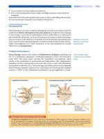
17.1 Technical Factors Associated With the Development of Incisional Hernia
M.A. C
17 Technical Pitfalls Favouring Incisional Hernia
Schumpelick.indd 135Schumpelick.indd 135 05.04.2007 8:50:59 Uhr05.04.2007 8:50:59 Uhr
136
17
Abdominal Wall Closure
This would suggest that the hernia formation begins
very early. In other words, an incisional hernia might
be thought of as a “subclinical dehiscence” in which the
fascial failure, while not catastrophic and/or eviscerat-
ing, results in a gradually widening fascial defect. Of
course, not all incisional hernias would fall under this
etiology (see later discussion about buttonhole hernias),
but the realization that postoperative abdominal wall
hernia may have a very early origin implies that its cause
could be similar to that of wound dehiscence; and the
cause of wound dehiscence in the vast majority of cases
is a technical inadequacy [5–7]. That is, the fault lies
with the surgeon.
Choice of Incision
So if the responsibility for abdominal incisional her-
nia formation is technical inadequacy, what can the
surgeon do to circumvent this? In other words, what
are the forms of the technical inadequacy? The first
(in temporal order) is the choice of incision. The best
incision the surgeon can choose which will minimize
the risk of incisional hernia is a minimal incision. If
properly closed, trocar punctures from a minimally
invasive procedure produce incisional hernia in ~1%
of cases [8], which is much less than the 10–30% rate of
herniation typically quoted for conventional incisions.
Furthermore, since emphasis is being placed on the
utilization of laparoscopic instruments with a diameter
of ≤ 5 mm, the incidence of trocar hernia most likely
will decrease.
If a laparoscopic approach is not feasible, then for
a major intra-abdominal procedure the surgeon has a
variety of incisional choices; for simplicity, these will be
classified as either vertical (most commonly midline,
through the linea alba) or transverse. There is a large
amount of historical, retrospective data which suggests
that the transverse incision has a lower incidence of
dehiscence and hernia; for an early example of this,
see Singleton and Blocker’s review of 9000 incisions
[1]. This retrospective data is influenced by various
confounding factors (e.g., use of short transverse inci-
sions for cholecystectomy vs. longer midline incisions
for emergency procedures), but the preponderance of
the data (not reviewed here) favors the transverse in-
cision.
Three randomized controlled trials comparing
hernia rates in vertical vs. midline incisions have been
published [9–11], and these provide some support for
a lower risk of incisional hernia in transverse incisions.
The most recent trial [11] found a large, statistically
significant increase in the incidence of hernia in mid-
line compared to transverse incisions in a small group
(<40) of aortic aneurysm patients. This finding needs
to be tempered by the fact that the hernia incidence in
the midline group was 94% (certainly the highest ever
recorded in a hernia trial), which suggests a problem
with suture technique (an uncontrolled variable in this
trial). Currently there are no controlled data compar-
ing transverse to midline incisions in which the suture
technique is optimized and constant.
Two UK institutions reported a very low (1% or
less) incidence of postoperative hernia with the lateral
paramedian incision in trials during the 1980s [12–17].
This is a vertical incision through the lateral portion of
the rectus sheath, about two-thirds the distance from
the medial edge of the rectus. The rectus muscle is re-
flected medially during the operation, so upon layered
closure of the rectus sheath, the muscle covers the fas-
cial incisions. This provides a splinting effect which,
the authors claim, is the basis for the robustness of the
incision. The lateral paramedian incision generally takes
longer to perform, and requires more expertise than
the midline incision. Unfortunately, there have been
no corroboratory publications from other institutions
which validate the superiority of the lateral paramedian
incision.
Abdominal Entry
The next choice the surgeon has which may influence
the risk of wound failure is the act of incising the layers
of the abdominal wall. Animal experimentation has
shown that a small amount of tissue injury (such as
delivered with a scalpel blade) is important to incite
the appropriate amount of inflammation which will
produce the strongest scar [18]. On the other hand, too
much injury (such as that delivered with coagulation
current from the cautery blade) inhibits healing because
of fascial necrosis [19]. Even more dramatic is the effect
of delayed primary or secondary wound closure which,
in animals, can increase wound breaking strength (fas-
cial or dermal) by as much as 100% at 60 days compared
to primary closure [20, 21]. The presumptive cause of
this effect is the greater fibrotic reaction inherent with
an open wound. Data from humans in this area are ab-
sent and, of course, no one would recommend delayed
primary or secondary wound closure as the standard
operating procedure for elective laparotomy closure.
The time-honored tradition of entering the abdomen
with a clean swipe of the scalpel [22], however, still
applies.
Schumpelick.indd 136Schumpelick.indd 136 05.04.2007 8:51:00 Uhr05.04.2007 8:51:00 Uhr
137
V
Technical Pitfalls Favouring Incisional Hernia
Choice of Suture Material
After the intra-abdominal procedure has been com-
pleted, the next choice the surgeon faces that may in-
fluence the risk of incisional hernia is suture material.
There is a wealth of both retrospective and controlled
data (not to be reviewed here) that scrutinizes suture
material. The bottom line is that with modern suture
material, the suture choice is of much less importance
than how the surgeon actually places it (see below).
That being said, there have been a number of meta-
analyses and systematic reviews which have favored
either nonabsorbable suture material (e.g., nylon, poly-
propylene) or slowly absorbable suture material (e.g.,
polydioxanone) in the closure of laparotomy incisions
[23–26]. The perceived detraction to using nonabsorb-
able suture is the development of buttonhole hernia [27,
28] which is a fascial defect created by the perpetual
sawing motion of the suture where it penetrates the
fascia. A patient can develop a cluster of these hernias
and end up with a so-called Swiss cheese abdomen.
Buttonhole hernia may be the reason why incisional
hernias continue to develop years out from the index
procedure [29]. It is difficult to say if the incidence
of buttonhole hernia is less with a slowly absorbable
suture.
Suture Technique: Suture-Length-
to-Wound-Length Ratio
The single most important surgeon-related factor in
the risk for incisional hernia is suture technique, which
entails items such as tissue bite, stitch interval, stitch
tension, and so on. In cases of wound dehiscence not
involving fasciitis, the most common cause of failure
is suture tearing through the fascia [5]. One possibility
suggested by this observation is that an inadequate tissue
bite during incisional closure will predispose the patient
to tissue tearing, which can result in acute wound failure
or delayed hernia. It is not surprising that in animal and
cadaver studies, a wider bite of fascia with the suture
results in a higher pull-out strength [30–32]. Further-
more, it has been shown that suture holding capacity in
experimental incisions of both the abdominal fascia and
hollow viscera actually decreases during the early post-
operative period [33], presumably because the region
immediately adjacent to the incision is biochemically
active (e.g., matrix metalloproteinase activation) and
becomes “soft” [34]. So, taking a wide bite with the
suture needle would avoid this biochemically active
wound region.
So how wide a bite should be taken? If 1 cm is bet-
ter than 5 mm, then why not 2 or 3 cm? Indeed, in
some of the early experience with wide bite closure,
some surgeons routinely placed retention sutures. For
example, Kennedy [35] informally described the per-
formance of around 30,000 abdominal incisions over
a 56-year period (between him and his mentor, Joseph
Price), and could recount only one case of dehiscence
and no hernias (!). Their technique of closure involved
through-and-through (all layers, dermis to peritoneum)
silk sutures, placed 1 inch (2.5 cm) back from the
wound edge, three for every inch of incision, and tied
loosely. They also closed the fascia with buried sutures
prior to tying the through-and-through sutures. The
silk retentions typically were removed on postoperative
day 10. Such routine retention suture placement prob-
ably would not be readily accepted today, but the above
experience is illustrative of the benefit of generous tis-
sue bites and short stitch interval on the prevention of
wound failure.
The first individual to apply some science to wide
bite closure was TPN Jenkins [6, 36]. He introduced the
concept of suture-length-to-wound-length ratio (SL:
WL), as shown in
⊡
Fig. 17.1. This applied to continu-
ous closures, and was equal to the length of suture used
to close the incision divided by length of the incision.
The suture length was dependent on two parameters:
the stitch interval (distance AB in
⊡
Fig. 17.1
) and the
tissue bite (one half of the distance TD in
⊡
Fig. 17.1
).
Jenkins determined that a SL:WL of ≥ 4 was protec-
tive of dehiscence; he had only one burst abdomen in
1500 closures in which he maintained this ratio (0.07%
T
A
D
B
⊡
Fig. 17.1. Suture-length-to-wound-length ratio [6]
Schumpelick.indd 137Schumpelick.indd 137 05.04.2007 8:51:00 Uhr05.04.2007 8:51:00 Uhr
138
17
Abdominal Wall Closure
dehiscence rate). Jenkins also applied this technique to
primary suture repair of incisional hernia and, employ-
ing SL:WL as high as 44, he achieved a relatively low
recurrence rate of 8%.
The use of SL:WL in abdominal incision closure
was popularized by Israelsson and colleagues dur-
ing the 1990s [2, 7, 37–40]. They demonstrated that
maintenance of a SL:WL greater than 4 (particularly
in regard to vertical midline incisions) minimized the
occurrence of both dehiscence and hernia. The primacy
of a SL:WL of 4 in the prevention of wound failure was
corroborated experimentally by the Aachen group [41,
42]. But, analogous to the question above, if a SL:WL
of 4 is good, would 5 or more be better? Perhaps not;
clinically it was observed that a SL:WL ≥ 5 was associ-
ated with an increased incidence of wound infection
(and subsequent wound failure), especially in obese
patients [43, 44].
Experimentally, excessively wide bites have dis-
advantages. In rat incisions closed with a constant SL:
WL of 4 [45], wounds with a relatively short stitch
interval and small tissue bite were stronger on post-
operative day 4 than wounds with a relatively long
stitch interval and large tissue bite (
⊡
Fig. 17.2
). That
is, the wounds with more stitches and smaller bites
fared better. In a study with pigs [46], closing a vertical
midline incision with wide interrupted bites through
the rectus sheath and then maintaining 20 mmHg of
intra-abdominal pressure for 3 h resulted in rectus
muscle tearing and hemorrhage with greater wound
edge separation (as marked with metal clips), as com-
pared to wounds in which stitches took only bites
of the anterior sheath. Early wound separation is, as
noted above, an early indicator of incisional hernia.
The implication of these experimental data and the
above clinical studies was that a mass stitch in wide
bite closure might be detrimental to incisional heal-
ing. So the simple concept of “more is better” in wide
bite closure may be subject to some qualifications. The
final word probably has not been heard in this arena.
Suture Technique: Tension
There are two types of tension which are relevant to
incisional healing. The first type is tension that the
surgeon (or first assistant) places on the suture during
closure. It has been shown experimentally that excessive
suture tension decreases wound strength [30, 42, 47–49]
and perfusion to the central portion of the wound [50].
Of course, inadequate tension on the suture (i.e., too
loose) will result in protrusion of intestinal loops, peri-
toneal fluid leaks, wound edge separation, and eventual
hernia. One group found that compression suture of
vertical midline incisions (in which each individual
loop of a continuous suture was tightened with 5 kg of
force) in patients resulted in fewer wound complica-
tions compared to a closure with nontightened loops
[51]. This finding is somewhat counterintuitive to the
clinical adage of “approximate, don’t strangulate.” Cur-
rently, there is no consensus on the amount of tension
to place on suture during closure.
⊡
Fig. 17.2. Role of stitch interval vs. tissue
bite in rat vertical midline wounds closed
with a constant SL:WL of 4. The wound
in C was the strongest immediately after
closure, but the wound in A and B were
stronger on postoperative day 4 [45]
a cb
Schumpelick.indd 138Schumpelick.indd 138 05.04.2007 8:51:01 Uhr05.04.2007 8:51:01 Uhr
139
V
Technical Pitfalls Favouring Incisional Hernia
The second type of tension relevant to incisional
healing is that required to bring the wound edges to-
gether, or tissue tension. This also is the tension across
the wound, or suture line, after closure has been com-
pleted. Another maxim in surgery is that suture lines
under tension will be at an elevated risk for failure; this
has been confirmed in the laboratory [47, 52]. There
are some experimental conditions, however, in which
suture line tension actually increased wound disrup-
tion strength [3, 53]; in addition, tension stimulated
granulation tissue growth in animal excisional wounds
[54, 55]. There may be some level of tissue tension that
is optimal for incisional healing; clinically, however,
this has not been defined. Furthermore, a critical level
of tissue tension beyond which the risk for incisional
failure is unacceptable also is not known.
Suture Technique: Other Issues
Perhaps less controversial in the recent literature are
choices between continuous vs. interrupted sutures and
mass vs. layered technique. There have been multiple
retrospective reviews that document the efficacy of vari-
ous combinations (running mass, interrupted layered,
etc.) which will not be reviewed here. There also have
a number of meta-analyses which have concluded that
continuous sutures are superior to interrupted [23–26,
56]. One large randomized controlled trial comparing
running vs. interrupted laparotomy closure [57] dem-
onstrated that the former had fewer wound complica-
tions (mainly dehiscence; follow-up was for 30 days).
The Smead–Jones suture technique [58], also known
as far-near near-far sutures, intermittently has been
touted (with uncontrolled clinical data) as protective
against wound failure. A variant of this technique, the
continuous double-loop suture, was shown to be acutely
stronger than other techniques in the rat; interestingly,
this technique failed in comparison to conventional
running suture in a clinical trial [59]. Retrospective data
has demonstrated that routine retention suture place-
ment (Mont Reid type [60]) at the index laparotomy
prevents acute wound failure [35, 61]. This was con-
firmed experimentally in dogs [62], but not in a clinical
randomized trial [63]. Other than the salutary effect of
closely spaced retention sutures on hernia prevention in
older retrospective data [35], the efficacy of retentions
in modern-day hernia prophylaxis is unknown.
Prophylaxis of surgical wound infection, while not
completely under control of the surgeon, should be
mentioned in an article such as this, because infection
repeatedly has been shown to be an independent risk
factor in the development of incisional hernia (data not
reviewed here). Of note, the Israelsson group has shown
that a SL:WL of 4.0–4.9 is optimal value for minimizing
wound infection risk and subsequent incisional hernia
[38, 43].
Novel Techniques for Prevention
of Incisional Hernia
Recently, the feasibility and efficacy of prophylactic
mesh placement for reinforcement of laparotomy clo-
sure has been demonstrated in one small randomized
trial of high-risk patients [64] and two small series
of bariatric [65] and aortic aneurysm [66] patients.
The optimal placement technique (e.g., sublay vs.
onlay) is not known. In regard to intestinal stomas,
there has been one small randomized trial of routine
placement of a light-weight composite mesh (Vypro)
at the time of stomal creation [67], which demon-
strated a reduction of parastomal hernia formation in
the mesh patients. Mesh reinforcement of primary hia-
tal herniorrhaphy also was efficacious in reducing her-
nia recurrence in a randomized trial [68]. Prophylactic
mesh placement is an exciting and intriguing area in
abdominal wall surgery, and needs further study.
A novel technique of laparotomy closure recently
described in animals by the Aachen group is tension
banding or the bridging technique [50, 69], in which
the fascial edges of a vertical midline incision are coated
by polylactide (slowly absorbable synthetic) U-stitches
placed into two parallel polylactide strips that have
been affixed to the anterior sheath (
⊡
Fig. 17.3). This
technique provided equivalent or better wound perfu-
sion and strength compared to conventional suturing
or onlay mesh placement. The advantage of the bridg-
⊡
Fig. 17.3. Tension banding for laparotomy closure [50]
Schumpelick.indd 139Schumpelick.indd 139 05.04.2007 8:51:01 Uhr05.04.2007 8:51:01 Uhr
140
17
Abdominal Wall Closure
ing technique has been postulated to be the avoidance
of both foreign material at the wound edge and the
strangulating effect of incisional sutures. Clinical data
are not yet available.
Recommendations
The ability to prevent both abdominal wound dehis-
cence and incisional hernia primarily lies with the sur-
geon and the technique used to close the laparotomy
incision. That being said, the technical recommenda-
tions to minimize the risk of incisional hernia after
major laparotomy which are promoted by this article
are as follows:
▬ Avoid large incisions by performing a minimally
invasive procedure whenever possible.
▬
Consider transverse incision as an alternative to the
vertical midline incision.
▬ Avoid the coagulation current of the cautery when
incising the aponeurosis.
▬
Utilize either a nonabsorbable or a slowly absorb-
able suture.
▬ In a running closure of a vertical midline incision,
maintain the suture-length-to-wound-length ratio
between 4 and 5.
▬ Avoid excessively wide suture bites which incorpo-
rate large masses of muscle and fat.
▬
Avoid incisional closure in the presence of excessive
tissue tension.
▬
Maintain adequate suture tension to coapt the fascial
edges, but do not strangulate the tissue.
▬ Choose running suture over interrupted.
▬ Minimize the risk of surgical wound infection.
▬ Consider prophylactic mesh placement for the pa-
tient at high risk for wound failure.
Acknowledgements.
Supported in part by a grant from
the United States National Institutes of Health (K08
GM00703).
References
1. Singleton A, Blocker T. The problem of disruption of abdomi-
nal wounds and postoperative hernia: a review of 9,000 con-
secutive abdominal incisions. JAMA 1939; 112(2): 122–127
2. Israelsson LA. The surgeon as a risk factor for complications
of midline incisions. Eur J Surg 1998; 164(5): 353–359
3. Pickett BP, Burgess LP, Livermore GH, Tzikas TL, Vossoughi J.
Wound healing. Tensile strength vs healing time for wounds
closed under tension. Arch Otolaryngol Head Neck Surg
1996; 122(5): 565–568
4. Burger JW, Lange JF, Halm JA, Kleinrensink GJ, Jeekel H. In-
cisional hernia: Early complication of abdominal surgery.
World J Surg 2005; 29(12): 1608–1613
5. Carlson MA. Acute wound failure. Surg Clin North Am 1997;
77(3): 607–636
6. Jenkins TPN. The burst abdominal wound: a mechanical ap-
proach. Br J Surg 1976; 63(11): 873–876
7. Israelsson LA, Jonsson T. Closure of midline laparotomy
incisions with polydioxanone and nylon: the importance
of suture technique. Br J Surg 1994; 81(11): 1606–1608
8. Tonouchi H, Ohmori Y, Kobayashi M, Kusunoki M. Trocar site
hernia. Arch Surg 2004; 139(11): 1248–1256
9. Ellis H, Coleridge-Smith PD, Joyce AD. Abdominal incisions
– vertical or transverse? Postgrad Med J 1984; 60(704):
407–410
10. Greenall MJ, Evans M, Pollock AV. Midline or transverse lapa-
rotomy? A random controlled clinical trial. Part I: Influence
on healing. Br J Surg 1980; 67(3): 188–190
11. Fassiadis N, Roidl M, Hennig M, South LM, Andrews SM. Ran-
domized clinical trial of vertical or transverse laparotomy
for abdominal aortic aneurysm repair. Br J Surg 2005;
92(10):1208–1211
12. Brennan TG, Jones NA, Guillou PJ. Lateral paramedian inci-
sion. Br J Surg 1987; 74(8): 736–737
13. Cox PJ, Ausobsky JR, Ellis H, Pollock AV. Towards no incisional
hernias: lateral paramedian versus midline incisions. J R Soc
Med 1986; 79(12): 711–712
14. Donaldson DR, Hall TJ, Zoltowski JA, Guillou PJ, Brennan TG.
Does the type of suture material contribute to the strength
of the lateral paramedian incision? Br J Surg 1982; 69(3):
163–165
15. Donaldson DR, Hegarty JH, Brennan TG, Guillou PJ, Finan PJ,
Hall TJ. The lateral paramedian incision – experience with
850 cases. Br J Surg 1982; 69(10): 630–632
16. Kendall SW, Brennan TG, Guillou PJ. Suture length to wound
length ratio and the integrity of midline and lateral parame-
dian incisions. Br J Surg 1991; 78(6): 705–707
17. Gilbert JM, Ellis H, Foweraker S. Peritoneal closure after
lateral paramedian incision. Br J Surg 1987; 74(2): 113–
115
18. Miro D, Julia MV, Sitges-Serra A. Wound breaking strength
and healing after suturing noninjured tissues. J Am Coll Surg
1995; 180(6): 659–665
19. Rappaport WD, Hunter GC, Allen R, Lick S, Halldorsson A,
Chvapil T, Holcomb M, Chvapil M. Effect of electrocautery
on wound healing in midline laparotomy incisions. Am J
Surg 1990; 160(6): 618–620
20. Fogdestam I. A biomechanical study of healing rat skin inci-
sions after delayed primary closure. Surg Gynecol Obstet
1981; 153(2): 191–199
21. Scott PG, Chambers M, Johnson BW, Williams HT. Experi-
mental wound healing: increased breaking strength and
collagen synthetic activity in abdominal fascial wounds
healing with secondary closure of the skin. Br J Surg 1985;
72(10): 777–779
22. Moynihan B. The ritual of a surgical operation. Br J Surg 1920;
8: 27–35
23. Weiland DE, Bay RC, Del Sordi S. Choosing the best ab-
dominal closure by meta-analysis. Am J Surg 1998; 176(6):
666–670
Schumpelick.indd 140Schumpelick.indd 140 05.04.2007 8:51:02 Uhr05.04.2007 8:51:02 Uhr
141
V
Technical Pitfalls Favouring Incisional Hernia
24. van ‚t Riet M, Steyerberg EW, Nellensteyn J, Bonjer HJ, Jeekel
J. Meta-analysis of techniques for closure of midline abdomi-
nal incisions. Br J Surg 2002; 89(11): 1350–1356
25. Hodgson NC, Malthaner RA, Ostbye T. The search for an ideal
method of abdominal fascial closure: a meta-analysis. Ann
Surg 2000; 231(3): 436–442
26. Rucinski J, Margolis M, Panagopoulos G, Wise L. Closure of
the abdominal midline fascia: meta-analysis delineates the
optimal technique. Am Surg 2001; 67(5): 421–426
27. Krukowski ZH, Matheson NA. ‚Button hole‘ incisional her-
nia: a late complication of abdominal wound closure with
continuous non-absorbable sutures. Br J Surg 1987; 74(9):
824–825
28. Read RC, Yoder G. Recent trends in the management of in-
cisional herniation. Arch Surg 1989; 124(4): 485–488
29. Mudge M, Hughes LE. Incisional hernia: a 10 year prospec-
tive study of incidence and attitudes. Br J Surg 1985; 72(1):
70–71
30. Sanders RJ, DiClementi D, Ireland K. Principles of abdomi-
nal wound closure. I. Animal studies. Arch Surg 1977;
112(10):1184–1187
31. Campbell JA, Temple WJ, Frank CB, Huchcroft SA. A biome-
chanical study of suture pullout in linea alba. Surgery 1989;
106(5): 888–892
32. Tera H, Aberg C. Tissue strength of structures involved in
musculo-aponeurotic layer sutures in laparotomy incisions.
Acta Chir Scand 1976; 142(5): 349–355
33. Hogstrom H, Haglund U, Zederfeldt B. Suture technique and
early breaking strength of intestinal anastomoses and lapa-
rotomy wounds. Acta Chir Scand 1985; 151(5): 441–443
34. Adamsons RJ, Musco F, Enquist IF. The chemical dimensions
of a healing incision. Surg Gynecol Obstet 1966; 123(3):
515–521
35. Kennedy JW. Tragedies of the abdominal incision. Am J Surg
1934; 25: 512–520
36. Jenkins TPN. Incisional hernia repair: a mechanical approach.
Br J Surg 1980; 67(5): 335–336
37. Israelsson LA, Jonsson T. Incisional hernia after midline
laparotomy: a prospective study. Eur J Surg 1996; 162(2):
125–129.
38. Israelsson LA, Jonsson T, Knutsson A. Suture technique and
wound healing in midline laparotomy incisions. Eur J Surg
1996; 162(8): 605–609
39. Israelsson LA, Jonsson T. Suture length to wound length ratio
and healing of midline laparotomy incisions. Br J Surg 1993;
80(10): 1284–1286
40. Israelsson LA. Continuous closure of laparotomy incisions:
aspects of suture technique. In Schumpelick V, Kingsnorth
AN, eds. Incisional hernia. Springer, Berlin Heidelberg New
York 1999. pp. 246–252
41. Hoer J, Klinge U, Schachtrupp A, Tons C, Schumpelick V. Influ-
ence of suture technique on laparotomy wound healing: an
experimental study in the rat. Langenbecks Arch Surg 2001;
386(3): 218–223
42. Hoer JJ, Junge K, Schachtrupp A, Klinge U, Schumpelick
V. Influence of laparotomy closure technique on collagen
synthesis in the incisional region. Hernia 2002; 6(3): 93–98
43. Israelsson LA, Jonsson T. Overweight and healing of midline
incisions: the importance of suture technique. Eur J Surg
1997; 163(3): 175–180
44. Millbourn D, Israelsson LA. Wound complications and stitch
length. Hernia 2004; 8(1): 39–41
45. Cengiz Y, Blomquist P, Israelsson LA. Small tissue bites and
wound strength: an experimental study. Arch Surg 2001;
136(3): 272–275
46. Cengiz Y, Gislason H, Svanes K, Israelsson LA. Mass closure
technique: an experimental study on separation of wound
edge. Eur J Surg 2001; 167(1): 60–63
47. Villegas-Alvarez F, Olvera-Duran J, Rodriguez-Aranda E, Car-
mona-Mancilla A, Vigueras-Villasenor RM, Mendez-Ramirez
I. Esophageal anastomotic failure: an experimental study.
Arch Med Res 2003; 34(3): 171–175
48. Nelson CA, Dennis C. Wound healing; technical factors in
the gain of strength in sutured abdominal wall wounds in
rabbits. Surg Gynecol Obstet 1951; 93(4): 461–467
49. Poole GV, Jr., Meredith JW, Kon ND, Martin MB, Kawamoto EH,
Myers RT. Suture technique and wound-bursting strength.
Am Surg 1984; 50(10): 569–572
50. Hoer J, Tons C, Schachtrupp A, Anurov M, Titkova S, Oettinger
A, Wetter O, Schumpelick V. Quantitative evaluation of ab-
dominal wall perfusion after different types of laparotomy
closure using laser-fluorescence videography. Hernia 2002;
6(1): 11–16
51. Mayer AD, Ausobsky JR, Evans M, Pollock AV. Compression
suture of the abdominal wall: a controlled trial in 302 major
laparotomies. Br J Surg 1981; 68(9): 632–634
52. Borgstrom S, Sandblom P. Suture technic and wound healing;
an investigation based on animal experiments. Ann Surg
1956; 144(6): 982–990
53. Thorngate S, Ferguson DJ. Effect of tension on healing of
aponeurotic wounds. Surgery 1958; 44(4): 619–624
54. Carlson MA, Longaker MT, Thompson JS. Wound splint-
ing regulates granulation tissue survival. J Surg Res 2003;
110(1):304–309
55. Carlson MA, Thompson JS. Wound splinting modulates
granulation tissue proliferation. Matrix Biol 2004; 23(4):
243–250
56. Wadstrom J, Gerdin B. Closure of the abdominal wall; how
and why? Clinical review. Acta Chir Scand 1990; 156(1):
75–82
57. Fagniez PL, Hay JM, Lacaine F, Thomsen C. Abdominal midline
incision closure. A multicentric randomized prospective trial
of 3,135 patients, comparing continuous vs interrupted poly-
glycolic acid sutures. Arch Surg 1985; 120(12): 1351–1353
58. Jones TE, Newell ET, Jr, Brubacker RE. The use of alloy steel
wire in the closure of abdominal wounds. Surg Gynecol Ob-
stet 1941; 72: 1056–1059
59. Niggebrugge AH, Trimbos JB, Hermans J, Steup WH, Van De
Velde CJ. Influence of abdominal-wound closure technique
on complications after surgery: a randomised study. Lancet
1999; 353(9164): 1563–1567
60.
Reid MR, Zinninger MM, Merrell P. Closure of the abdomen
with through-and-through silver wire sutures in cases of
acute abdominal emergencies. Am Surg 1933; 98: 890–896
61. Holman CW, Eckel JH. Prevention of wound disruption with
through-and-through silver wire stay sutures. Surg Gynecol
Obstet 1941; 72: 1052–1055
62. Miles RM, Nash JP, Gillespie H, Dacus D. Value of retention
sutures in the prevention of experimental abdominal wound
disruption. Am Surg 1967; 33(11): 898–905
Schumpelick.indd 141Schumpelick.indd 141 05.04.2007 8:51:03 Uhr05.04.2007 8:51:03 Uhr
142
17
Abdominal Wall Closure
63. Hubbard TB, Jr., Rever WB, Jr. Retention sutures in the closure
of abdominal incisions. Am J Surg 1972; 124(3): 378–380
64. Gutierrez de la Pena C, Medina Achirica C, Dominguez-
Adame E, Medina Diez J. Primary closure of laparotomies
with high risk of incisional hernia using prosthetic material:
analysis of usefulness. Hernia 2003; 7(3): 134–136
65. Strzelczyk J, Czupryniak L, Loba J, Wasiak J. The use of poly-
propylene mesh in midline incision closure following gastric
by-pass surgery reduces the risk of postoperative hernia.
Langenbecks Arch Surg 2002; 387(7 8): 294–297
66. Rogers M, McCarthy R, Earnshaw JJ. Prevention of incisional
hernia after aortic aneurysm repair. Eur J Vasc Endovasc Surg
2003; 26(5): 519–522
67. Janes A, Cengiz Y, Israelsson LA. Randomized clinical trial of
the use of a prosthetic mesh to prevent parastomal hernia.
Br J Surg 2004; 91(3): 280–282
68. Frantzides CT, Madan AK, Carlson MA, Stavropoulos GP. A
prospective, randomized trial of laparoscopic polytetrafluo-
roethylene (PTFE) patch repair vs simple cruroplasty for large
hiatal hernia. Arch Surg 2002; 137(6): 649–652
69. Hoer J, Klinge U, Anurov M, Titkova S, Oettinger A,
Schumpelick V. Tension banding closure of laparotomies:
results of an experimental study in dogs. Langenbecks Arch
Surg 2002; 387(7 8): 309–314
Discussion
Schumpelick: What about the time course of incisional
hernia development? There are interesting investigations,
for example by CT scan from Prof. Jeekel. The second
question is about suture tension: what is tension? How
do you measure it?
Carlson: The time course is difficult to say. The risk re-
mains forever. A number of people have documented
hernia formation 10 or 20 years after the operation, I
think those are typically buttonhole hernias that you see
after these years, but the risk remains as long as you
live. The suture tension remains a secret, we know it is
important but we have no way to quantify. How to stan-
dardize so that 1000 surgeons can do the same thing, we
don’t know.
Schumpelick: About 3 years ago we developed a tensiom-
eter, measuring the knotting tension of different doctors.
At a different time of the day and at different types of
operations. It showed that we use too much tension, with
a little less in the afternoon than in the morning, and in
a re-operation there is a higher tension, that means we
have to measure it. More studies concerning the problem
and validation of tension are necessary.
Jeekel: In the mentioned CT investigation it was shown
that in people who developed a hernia it was already vis-
ible on the CT scan with the fascia edges widening.
Deysine:
I heard several times to use a mesh when the
fascia has not enough strength, in other words support a
wound with a mesh. I want to caution that when you do
that you have to double the caution to keep the wound
clean, asepsis and antisepsis and antibiotics, in other
terms, the rate of wound infection will increase.
Kehlet: I think we have a great problem to translate sci-
ence into the daily clinical practice. Every lecture says
transverse incisions and suddenly you say you are not
using it and when I look around seeing fast-track surgery
they never use transverse incisions, so how can we spread
the message and why is it that you are using vertical in-
stead of transverse incisions? Here in the lecture room you
say transverse but when you go home to your operation
room you do the opposite.
Jeekel: And then, Dr Kehlet, we talk about prevention
by using a mesh instead of using the right incision, yes
that’s amazing.
Introduction
Laparoscopic surgery continues to advance in achieving
further benefits over the conventional approach for cer-
tain pathologies. In 1991 LeBlanc et al. carried out the first
laparoscopic repairs of ventral hernias [1]. Although not
originally considered to be a pathology that could benefit
from this approach, laparoscopic repair of ventral hernias
has attained wide acceptance in recent years because of
the significant advantages afforded by improvements in
prosthetic materials and in attachment methods, as well as
in the surgical technique used. The laparoscopic procedure
offers greater comfort during the postoperative period, re-
duces hospitalization time and lowers complication rates.
Even though many series still have a limited follow-up,
the technique has shown lower rates of recurrence than
the open methods, making it a procedure that solves a
long-standing challenge for the surgeon.
The relationship
17.2
Technical Pitfalls Favouring Incisional Hernia From an Expert in Laparoscopic Surgery
S. M-C
Schumpelick.indd 142Schumpelick.indd 142 05.04.2007 8:51:03 Uhr05.04.2007 8:51:03 Uhr
143
V
Technical Pitfalls Favouring Incisional Hernia
between laparoscopic surgery and ventral hernias could be
established in three senses. On the one hand, the fact that
the rate of recurrences after laparoscopic ventral hernia repair
is lower compared to an open repair, due to different factors
that could be analyzed; on the other hand, there are some
technical aspects of the laparoscopic repair of ventral hernias
that may influence in the possibility of having a recurrence.
Finally, laparoscopy, as an approach, could be involved in
the production of hernias, such as trocar site hernias or tack
hernia, as we will describe during this chapter.
Factors That May Influence a Lower
Incidence of Recurrences After Laparos-
copic Incisional Hernia Repair Compared
to an Open Repair
There are different comparative studies published in the
literature comparing laparoscopic and open ventral her-
nia repair (
⊡
Table 17.1). The purpose of these studies
was to determine whether laparoscopic intraperitoneal
prosthetic patch repair of a ventral hernia is superior to
open-mesh repair. Laparoscopic hernioplasty is as safe
and effective as the traditional open technique with
shorter length of stay and decreased hospital costs.
In these series that have been published [2–6, 8–10],
laparoscopic ventral hernioplasty compares favourably
also with respect to wound complications, hospital stay,
operative time and recurrence rate, and only one study
[7] shows that laparoscopic incisional hernia repair of
at least moderate complexity had no demonstrable ad-
vantage over the open repair.
Different questions arise after analyzing these
studies, such us why laparoscopic repair shows better
results than open conventional mesh repair and, es-
pecially why the rate of recurrences is lower. Differ-
ent factors have been related to recurrences after
open repair including wound infection and other lo-
cal wound complications, size of the hernia, obesity,
age, respiratory disease, sex (male), site of the her-
⊡
Table 17.1. Comparative studies between laparoscopic and open ventral hernia repair
Technique No.
Compl.
[%]
Operative
time [min]
Hospital
stay [days]
Infection
[%]
Recurren-
ces [%]
Holzman et
al. (1997) [2]
Open
Laparoscopic
116
120
31
23
198
128
5
1.6 d
16
15
13
10
Park et al.
(1998) [3]
Open
Laparoscopic
149
156
37
18
178
195
6.5
3.4
12
10
35
11
Ramshaw et
al. (1999) [4]
Open
Laparoscopic
174
179
26
15
182
158
2.8
1.7
13
10
20.6
12.5
Carbajo et al.
(1999) [5]
Open
Laparoscopic
130
130
50
20
112
187
9.1
2.2
18
10
17
10
De Maria et
al. (2000) [6]
Open
Laparoscopic
118
121
72
57
1–
1–
4.4
0.8
33
10
10
16
Chari et al.
(2000) [7]
Open
Laparoscopic
114
114
14
14
178
124
5.5
5
10
17
1–
1–
Robbins et al.
(2001) [8]
Open
Laparoscopic
123
131
–
–
1–
1–
1–
1–
30
16
1–
1–
Wright et al.
(2002) [9]
Open mesh
Open no m
Laparoscopic
190
119
186
28
22
24
102
170
131
2.5
1.5
1.5
13
10
19
16
19
11
MacGreevy et
al. (2003) [10]
Open
Laparoscopic
171
165
21
18
1.7 h
2.2 h
1.5
1.1
1–
1–
1–
1–
Schumpelick.indd 143Schumpelick.indd 143 05.04.2007 8:51:03 Uhr05.04.2007 8:51:03 Uhr
144
17
Abdominal Wall Closure
nia, number of times the hernia have recurred and
the presence of non-palpable defects. Some of the
advantages of laparoscopic repair are the possibility
that this approach offers to reduce the presence of
some of the factors involved on having a recurrence.
The rate of local wound complications, and especially
wound infection, is lower after a laparoscopic repair,
as has been demonstrated in different studies, decreas-
ing from 30 to 16% in some series that specifically have
measured the rate of this complication, while some se-
ries demonstrated that this rate can even be reduced to
0% [3–5] after the laparoscopic approach. We believe
the main reason for this is the fact that the presence
of seroma after an open and a laparoscopic repair is
very frequent. After an open repair the possibility of
infection of the seroma is higher since the incision
performed is localized at the top of this fluid, which is
close to the mesh. In these situations, bacteria from the
skin could easily come into contact with the seroma,
and the possibility of contamination infection of the
seroma and the mesh increases. On the other hand,
after a laparoscopic approach, trocar sites are far from
the area where the seroma and the mesh are, so this area
could be maintained under sterile conditions and the
possibility of infection decreased (
⊡
Fig. 17.4a,b).
Another factor that is involved in the presence of
hernia recurrences after an open repair is obesity. Pa-
tients who are morbidly obese traditionally have been
considered poor surgical candidates for ventral hernia
repair because of their associated comorbidities and
risk of postoperative wound infection and hernia recur-
rence. Laparoscopic repair of ventral hernias in patients
who are morbidly obese is both safe and feasible, and
can be performed with minimal morbidity. Birgisson
et al. [11] have demonstrated that this factor does not
play a role in recurrences during laparoscopic repair,
since the rate of recurrences after this approach has no
relation to the BMI of the patient.
One of the advantages of laparoscopic surgery is that
it offers the possibility to find the presence of non-pal-
pable defects that have not been detected during clinical
examinations. The laparoscopic approach allows a total
exposure of the incision once adhesiolysis is completed,
and those small defects or weakness of the anterior ab-
dominal wall can be easily detected, factors that could
be involved in the presence of further recurrences and
that are difficult to identify during an open repair. One
of the recommendations during the laparoscopic ap-
proach of incisional hernias is to expose the whole area
of the incision to detect these weak areas, which must be
covered with the mesh with a proper overlap. In fact, it
has been published [12] that 13.1% of the patients who
undergo a laparoscopic repair of an incisional hernia
have multiple defects, and the average number of defect
found is 4.8, more than the number detected during
clinical examination [13].
Technical Aspects of Laparoscopic Ventral
Hernia Repair That May Influence in Hernia
Recurrences
Incisional hernia underwent a change from conven-
tional techniques to laparoscopic approach. The rel-
evance of different factors, such as operative technique,
mesh material and fixation, concerning the outcome
following laparoscopic repair, are still under debate.
Laparoscopic repair revealed acceptable recurrence
⊡
Fig. 17.4. a Seroma after open mesh repair. Incision is at the
top of the fluid collection, which increases the possibility of
contamination infection. b Seroma after laparoscopic mesh re-
pair. Trocar site is far from the serma and the mesh, which are
under sterile conditions
a
b
Schumpelick.indd 144Schumpelick.indd 144 05.04.2007 8:51:04 Uhr05.04.2007 8:51:04 Uhr
145
V
Technical Pitfalls Favouring Incisional Hernia
rates with high patient comfort. From a surgical point
of view, the most important prognostic factor following
mesh repair is the surgeon’s experience, although some
factors can be analyzed to try to reduce the presence
of recurrences.
What Area Should Be Covered?
New hernias below original hernias have been de-
scribed as a factor of recurrence after open repair.
This factor has also been described after laparoscopic
approach [14], which has led to recommending to
cover the entire incision even in those cases in which
a weak area is not detected, since this damaged tissue
could be involved in the presence of a new hernia.
At present, it appears evident that when undertaking
laparoscopic repair of an incisional hernia, adhesio-
lysis must cover the entire area of the previous scar
in order to identify possible wall defects at this level,
other than those originally destined to be repaired. This
is precisely one of the advantages of laparoscopy over
traditional open repair. Defects that were not identified
during the clinical examination and that were the cause
of recurrence or appearance of a new defect after open
repair can be detected and repaired in the same surgical
procedure [15].
How Should the Area of Placing the Mesh Be
Prepared?
There are two factors that may influence a proper repair
of a ventral repair regarding the area where the pros-
thetic material is going to be implanted. On one hand,
the intraperitoneal fatty tissue of the anterior abdominal
wall must be removed to guarantee a proper fixation
of the mesh. This fatty tissue, including the round and
the falciform ligament, should be removed so the spiral
tacks could reach the aponeurosis of the muscle to fix
the mesh more consistently.
On the other hand, the process of removing this
fatty tissue, together with the adhesiolysis, will produce
an inflammatory reaction of the peritoneum, which
will increase the ingrowth of the prosthetic materi-
als. This fact is especially important when composite
materials with polypropylene mesh in the parietal side
are used, since this material needs this inflammatory
reaction to improve the ingrowth. This does not oc-
cur with meshes of ePTFE since this material follows a
pattern of encapsulation to be attached to the anterior
abdominal wall.
How Should Size of the Mesh Be Selected?
The two factors that should be considered during
a laparoscopic repair regarding mesh size are the
following: it is safer to avoid recurrences by using
one large mesh than two pieces of the material, since
the area where the two meshes overlap is a weak
area that has been related to the presence of recur-
rences [16].
On the other hand, it has been described that recur-
rences were reduced because of the use of an increased
overlap of the biomaterial [14]. Prostheses initially rec-
ommended were small; overlapping the defect by only
2 [17, 18] to 2.5 cm [3] in all directions, and not the
minimum of 3–5 cm currently recommended. Recently,
we have demonstrated in an experimental study that
expanded polytetrafluoroethylene ( ePTFE) prosthe-
ses decreases in size once they have been implanted
(1.63 cm out of 4 cm after 5 weeks), probably because
of the scar tissue reaction and the encapsulation process
experienced by the mesh [19], so recurrences in these
initial experiences could have happened mainly because
of the smaller size of the mesh.
How to Fix the Mesh?
One of the most interesting points currently being de-
bated is whether or not it is necessary to use sutures
and tacks or tacks alone, following the double crown
technique (
⊡
Fig. 17.5
), or other additional methods of
fixation, such as biological glues or the new method of
fixations available.
Despite the lower recurrence rate, various authors
have made efforts to analyze the causes for recurrence
in order to adequately define the laparoscopic tech-
nique and thereby achieve an even lower recurrence
rate. Initial laparoscopic ventral hernia repair series
established a direct correlation between recurrence
and the absence of transfascial sutures [9, 18, 21]. In
fact, they demonstrated that one of the essential fac-
tors to avoid recurrence is the use of these sutures
[22]. Analysis of the data derived from these early
series, data which were later the basis for recommen-
dations on the use of sutures, shows that there could
have been other factors involved in the development
of recurrence in these patients besides the use or not
of transfascial sutures: prostheses initially recom-
mended were small, the method of fixation was also
inadequate, since tacks were not yet available and mesh
patches were anchored with the old endostaplers that
did not ensure secure attachment of the material, and
Schumpelick.indd 145Schumpelick.indd 145 05.04.2007 8:51:06 Uhr05.04.2007 8:51:06 Uhr
146
17
Abdominal Wall Closure
lastly, the learning curve of these initial series could
be more directly related to the appearance of recur-
rences.
An analysis of our recurrences following the double
crown technique shows that they were not directly re-
lated to the use of sutures [20] In a series that advocates
the use of sutures, the recurrence rate ranges anywhere
from 0 to 8.3%, with a mean of 3.98% [21]. The recur-
rence rate of our series is 2.14% with a mean follow-up
of 40 months [20].
This surgical technique, double crown, has some
advantages over the use of transfascial sutures that
could be related to the presence of recurrences, since
this technique reduces the dead space between the mesh
and the anterior wall of the abdomen, which will reduce
the amount of fluid from the seroma in this area, what
will favour the ingrowth of the mesh, accelerating the
biological fixation of the mesh. On the other hand, the
inner crown of spiral tacks will guarantee a proper fixa-
tion of the mesh since the scar tissue at the edge of the
defect will offer a consistent fixation.
How Could the Seroma be Decreased?
Seroma is a frequent complication of laparoscopic or
open repair of ventral hernias. The presence of seroma
is due to different factors, one of them related to the fact
of leaving the sac in place during the procedure, since
the sac is not excised once adhesiolysis is completed. A
recent study revealed the presence of seroma in 100%
of patients when an ultrasound examination is done,
while it is diagnosed clinically in only 35% of cases [23],
this rate being very variable in the literature; but the real
incidence has not been established properly. Seroma
can produce pain and discomfort in the abdominal wall
of the patients, and could also have some influence in
recurrences for two reasons: the fluid between the mesh
and the abdominal wall will delay ingrowth of the pros-
thetic material and, on the other hand, aspiration of the
content has the risk of introducing bacteria, resulting in
infection and the recurrence of the hernia.
Different methods have been proposed to decrease
the incidence of seromas with not too good results, by
cauterizing the sac by monopolar cautery, harmonic
scalpel [24] or using argon beam. For these reasons we
have been working to reduce the presence of seroma
after the laparoscopic repair. The injection of fibrin glue
(Tissucol, Baxter Biosurgery) in the sac of the hernia,
after the repair has been completed, has reduced the
rate of seromas after the surgery in a study we are con-
ducting in our hospital. Our preliminary results show
a reduction of the presence of the seroma 1 week after
surgery from 95.2 to 66.6% and from 52.9 to 8.2% after
1 month (
⊡
Table 17.2).
Laparoscopy as a Factor in Production
of Hernias
Hernia at Trocar Sites
Incisional hernia after laparoscopic surgery is related
to trocar sites. Such hernias are attributed to the dif-
ficulty of applying standard suturing techniques to
wound closure, and to the fact that intra-operative
⊡
Fig. 17.5. Double crown technique for laparoscopic ventral
hernia repair. Tear in the peritoneum and the muscle produced
by a tack in a pig after increasing intra-abdominal pressure after
a laparoscopic incisional hernia repair. These tears could be a
tack hernia in the future
⊡
Table 17.2. Decrease in the rate of seroma after injec-
tion of fibrin glue (Tissucol, Baxter Biosurgery) in the sac
of the hernia
1 week 1 month
3 months
Seroma
without
fibrin
glue [%]
95.2 52.9 0
Seroma
with fib-
rin glue
[%]
66.6 18.3 0
Schumpelick.indd 146Schumpelick.indd 146 05.04.2007 8:51:06 Uhr05.04.2007 8:51:06 Uhr