Báo cáo y học: " Low-grade myofibroblastic sarcoma of the mandible: a case report" ppsx
Bạn đang xem bản rút gọn của tài liệu. Xem và tải ngay bản đầy đủ của tài liệu tại đây (728.17 KB, 4 trang )
Case report
Open Access
Low-grade myofibroblastic sarcoma of the mandible: a case report
Iwona Niedzielska
1
*, Tomasz Janic
1,2
and Bartlomiej Mrowiec
3
Addresses:
1
Department of Craniomaxillofacial Surgery, Medical University of Silesia, Katowice, Poland
2
Private Dental Clinic Sawdent, Sosnowiec, Poland
3
Private Dental Clinic Polmedico, Bielsko-Biala, Poland
Email: IN* - ; TJ - ; BM -
* Corresponding author
Received: 7 March 2008 Accepted: 29 January 2009 Published: 10 August 2009
Journal of Medical Case Reports 2009, 3:8458 doi: 10.4076/1752-1947-3-8458
This article is available from: />© 2009 Niedzielska et al.; licensee Cases Network Ltd.
This is an Open Access article distributed under the terms of the Creative Commons Attribution License (
/>which permits unrestricted use, distribution, and reproduction in any medium, provided the original work is properly cited.
Abstract
Introduction: Low-grade myofibroblastic sarcoma is a rare entity, which mostly develops in the soft
tissues of the head and neck. Within the oral cavity lingual lesions are the most common. It tends to
recur locally rather than to metastasise.
Case presentation: We present a 54-year-old man with a one-year history of buccal oedema. He
also had arterial hypertension and clinical examination revealed distension of the left mandibular
ramus with laminar deflection in the area of the retromolar triangle.
Conclusion: We present a rare intramandibular encapsulated lesion that caused diagnostic
difficulties. Our diagnostic methods included immunohistochemistry and molecular investigations.
We emphasise the uncommon location of this tumour type.
Introduction
Low-grade myofibroblastic sarcoma (LGMS) represents a
rare entity, which mostly develops in the soft tissues of the
head and neck [1]. Within the oral cavity lingual lesions
are the most common [2,3] and they tend to recur locally
rather than to metastasise. Diagnostic methods included
immunohistochemistry and molecular investigations.
Case presentation
In February 2006 a 54-year-old, white, Caucasian man was
seen in the Outpatient Clinic of the Craniomaxillofacial
Surgery Department in Katowice with a one-year history of
buccal oedema. He also had arterial hypertension. A
clinical examination revealed distension of the left
mandibular ramus with laminar deflection in the area of
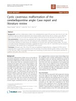
the retromolar triangle. Pantomogram and cranial X-rays
demonstrated a well-delineated osseous defect spreading
throughout the left mandibular ramus, infiltrating and
destroying the structures of the pterygopalatine fossa
(Figure 1).
Fine needle aspiration biopsy (FNAB) was non-contribu-
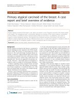
tory. A computed tomography (CT) scan of the facial area
revealed a tumorous lesion in the area of the mandibular
angle and ramus 59 × 54 mm in size which was slightly
and heterogeneously enhanced after an intravenous
contrast agent was introduced, causing bone distension
(Figure 2). The mass may possibly have infiltrated the left
masseter muscle, and it adhered closely to the hard palate
resulting in its deformation; invasion could also not be
Page 1 of 4
(page number not for citation purposes)
ruled out. It also revealed nasopharyngeal crevice defor-
mity, invasion of the subtemporal fossa and parti al
thinning of the posterior maxillary sinus wall. Tissue
density changes visible within the maxillary sinus were
possibly consistent with mucous membrane thickening.
There was no lymph node enlargement.
Tumour enucleation was performed including the perio-
steum and capsule. The tumour, which had a gelatinous
consistency, exhibited a tendency to invade the subtem-
poral fossa. Clear margins were obtained and the histology
result was low-grade myofibroblastic sarcoma. The mitotic
index was 5f.p. ¥ 10eHPF. At the time of writing, no
recurrence or metastases have been found.
Macroscopically the tumour was approximately 5 cm in
diameter, grey-white, hard and fibrous and there was no
necrosis. It was removed, along with part of the bone, and
microscopy demonstrated a tumour composed of spindle
cells arranged in interlacing bundles. The nuclei were
mostly fusiform and elongated with some round or
polymorphic nuclei also present. Most tumour cells
showed low grade atypia while polymorphonuclear cells
showed high-grade atypia. The mitotic index was low (MI
up to 5/10 HPF). The nuclei of some of the tumour cells
were hyperchromatic with tiny acidophilic nucleoli and
the cytoplasm was pale pink. No necrotic foci were seen.
There was oedema in the central part of the tumour with
pseudomyxoid and microcystic areas, and the inner and
peripheral parts showed thick bundles of collagen fibres,
local hyalinisation and the disappearance of tumour cells
and keloid-like areas.
Tumour blood vessels showed considerable variation from
tiny, round and elongated to larger ones exhibiting wall
thickening and hyalinisation. An analysis of the tumour
pattern revealed mild chronic inflammation and micro-
scopic examination also showed bone trabeculae invasion.
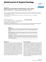
The tissue margins were clear. At immunohistochemistry,
the specimen stained positively for calponin (Figure 3),
vimentin, actin, CD99 (Figure 4), and focally for SMA
(Figure 5). CD34, S100 and desmin stainings were negative.
Oil-red stain was also negative. The histological picture was
consistent with a low-grade myofibroblastic sarcoma.
Figure 1. Cranial X-ray. The figure demonstrates a well-
delineated osseous defect spreading throughout the left
mandibular ramus, infiltrating and destroying the structures
of the pterygopalatine fossa.
Figure 2. Computed tomography scan. The figure
demonstrates a tumorous lesion in the area of the mandibular
angle and ramus, slightly and heterogeneously enhanced
after an intravenous contrast agent.
Figure 3. The histopathological picture (magnification ×250).
Strong calponin positivity of tumour cells.
Page 2 of 4
(page number not for citation purposes)
Journal of Medical Case Reports 2009, 3:8458 />Discussion
Although the patient’s age and gender were consistent with
literature reports on low-grade myofibroblastic sarcomas,
we would like to emphasise the uncommon location of
this tumour type (within the mandible) as well as a non-
typical macroscopic appearance (the presence of a
capsule). Initial histological diagnosis was inconsistent
with the clinical condition. However, problems with
differentiation between sarcoma subgroups have been
described in the literature with the most common
locations being the oral cavity (tongue) followed by the
limbs, pelvis, lungs and breasts, with a predilection for
soft, perifascial and subcutaneous tissues [2-4]. Other
locations have also been described: the salivary gland [5],
nasal skin [6] and the vulva [7]. Painless growth of a large
mass is typical of intraosseous tumours [8]. With other
locations some signs may occur such as fever, chills,
leukocytosis or meningeal irritation (cerebral tumour),
and more aggressive growth has been observed in the
abdomen. Mentzel et al. found recurrence in two, and
metastases in one, of his 18 patients [3]. Surgical resection
with clear margins is the treatment of choice. However, a
one-year survival was reported following a non-radical
resection [2]; our case of an encapsulated tumour seems to
confirm such a possibility.
Conclusions
Radiological examinations of a low-grade myofibroblastic
sarcoma of pelvic bones and limbs reveal a well-demar-
cated osteolytic lesion with no periosteal reaction [8]; our
results were similar. Diagnostic immunocytochemistry and
molecular investigations should be performed. Tumour
cell positivity is characteristic for calponin, MDM-2 and
PDGFRa. Diagnostic problems have been encountered
when differentiating from leiomyosarcoma [9] and the
prognosis depends on the malignancy grade. Guillou et al.
tried to predict the likelihood metastasis of low-grade
myofibroblastic sarcomas [10]. A high malignancy grade
(high mitotic index), a tumour size of >10 cm and a deep
location increase the tendency for metastasis.
Abbreviations
CT, computed tomography; FNAB, fine-needle aspiration
biopsy; LGMS, low-grade myofibroblastic sarcoma; MDM-
2, transformed 3T3 cell double minute 2; PDGFRa,
platelet-derived growth factor receptor, alpha polypeptide.
Consent
Written informed consent was obtained from the patient
for publication of this case report and any accompanying
images. A copy of the written consent is available for
review by the Editor-in-Chief of this journal.
Competing interests
The authors declare that they have no competing interests.
Authors’ contributions
IN, TJ and BM were all involved in the management of the
patient as well as writing the case report. All authors have
read and approved the final manuscript.
References
1. Agaimy A, Wünsch PH, Schroeder J, Gaumann A, Dietmaier W,
Hartmann A, Hofstaedter F, Mentzel T: Low-grade abdominopel-
vic sarcoma with myofibroblastic features (low-grade myofi-
broblastic sarcoma). J Clin Pathol 2008, 61:301-306.
Figure 5. The histopathological picture (magnification ×320).
Focal SMA positivity of tumour cells.
Figure 4. The histopathological picture (magnification ×400).
Focal CD99 positivity of tumour cells.
Page 3 of 4
(page number not for citation purposes)
Journal of Medical Case Reports 2009, 3:8458 />2. Laco J, Simáková E, Slezák R, Tucek L, Mottl R, Spacek J, Ryska A: Low
grade myofibroblastic sarcoma of tongue: a case report. Cesk
Patol 2006, 42:150-153.
3. MentzelT,DryS,KatenkampD,FletcherCD:Low-grade
myofibroblastic sarcoma: analysis of 18 cases in the spectrum
of myofibroblastic tumors. Am J Surg Pathol 1999, 23:1435-1436.
4. Taccagni G, Rovere E, Masullo M, Christensen L, Eyden B:
Myofibrosarcoma of the breast: review of the literature on
myofibroblastic tumors and criteria for defining myofibro-
blastic differentiation. Am J Surg Pathol 1997, 21:489-496.
5. Bisceglia M, Magro G: Low-grade myofibroblastic sarcoma of
the salivary gland. Am J Surg Pathol 1998, 22:1228-1238.
6. Chang SE, Choi JH, Sung KJ, Moon KC, Koh JK, Lee TJ, Ro JY,
Silverman JSA: A case of cutaneous low-grade myofibroblastic
sarcoma. J Dermatol 2001, 28:383-387.
7. Roth TM, Fratkin J, Woodring TC, McGehee RP: Low-grade
myofibroblastic sarcoma of the vulva. Gynecol Oncol 2004,
92:361-364.
8. Watanabe K, Ogura G, Tajino T, Hoshi N, Suzuki T: Myofibrosar-
coma of the bone: a clinicopathologic study. Am J Surg Pathol
2002, 26:393-394.
9. Eyden BP, Banerjee SS, Harris M, Mene A: A study of spindle cell
sarcomas showing myofibroblastic differentiation. Ultrastruct
Pathol 1991, 15:367-378.
10. Guillou L, Coindre JM, Bonichon F, Nguyen BB, Terrier P, Collin F,
Vilain MO, Mandard AM, Le Doussal V, Leroux A, Jacquemier J,
Duplay H, Sastre-Garau X, Costa J: Comparative study of the
National Cancer Institute and French Federation of Cancer
Centers Sarcoma Group grading systems in a population of
410 adult patients with soft tissue sarcoma. J Clin Oncol 1997,
15:350-362.
Page 4 of 4
(page number not for citation purposes)
Journal of Medical Case Reports 2009, 3:8458 />Do you have a case to share?
Submit your case report today
• Rapid peer review
• Fast publication
• PubMed indexing
• Inclusion in Cases Database
Any patient, any case, can teach us
something
www.casesnetwork.com