Báo cáo y học: " Episodic biliary obstruction due to an intrahepatic biliary cystadenoma: a case report" pptx
Bạn đang xem bản rút gọn của tài liệu. Xem và tải ngay bản đầy đủ của tài liệu tại đây (6.72 MB, 4 trang )
Case report
Open Access
Episodic biliary obstruction due to an intrahepatic biliary
cystadenoma: a case report
Pulathis N Siriwardana
1
* and Aloka Pathirana
2
Addresses:
1
University Surgical Unit, Colombo South Teaching Hospital, Kalubowila, Sri Lanka
2
Department of Surgery, Faculty of Medical Sciences, University of Sri Jayawardanapura, Sri Lanka
Email: PNS* - ; AP -
* Corresponding author
Received: 6 June 2008 Accepted: 22 January 2009 Published: 8 September 2009
Journal of Medical Case Reports 2009, 3:9032 doi: 10.4076/1752-1947-3-9032
This article is available from: />© 2009 Siriwardana and Pathirana; licensee Cases Network Ltd.
This is an Open Access article distributed under the terms of the Creative Commons Attribution License (
/>which permits unrestricted use, distribution, and reproduction in any medium, provided the original work is properly cited.
Abstract
Introduction: Biliary cystadenoma is a rare, benign neoplasm of the bile ducts with malignant
potential. Symptoms, predominantly right hypochondrial pain and the feeling of a lump or fullness are
usually due to the mass effect. Jaundice is rare. This is the fifth reported patient with an intrahepatic
biliary cystadenoma giving rise to episodic biliar y obstruction, which is usually caused by
choledocholithiasis or periampullary carcinoma. Considering the mean age of previous similar
patients (53.5, standard deviation 14.6 years), the early age of presentation is very unusual in our
patient.
Case presentation: A 25-year-old Asian woman presented with right hypochondrial pain and
episodic biliary obstruction. Contrast enhanced computed tomography revealed a cystic mass in
segment 4B and protruding into and along the left hepatic duct. Laparotomy confirmed the contrast
enhanced computed tomography findings and histology revealed an intrahepatic mucinous biliary
cystadenoma.
Conclusion: Biliary cystadenoma should be considered as a differential diagnosis in patients with
cystic liver lesions who present with episodic biliary obstruction. Due to the reported malignant
potential, radical surgery such as wide local excision of the lesion or hepatic resection is needed to
minimize the risk of local recurrence.
Introduction
Biliary cystadenoma (BCA) is a rare, benign cystic lesion of
the liver, arising from the biliary ducts, typically lined with
columnar epithelium, and usually with an “ovarian like”
cellular stroma. The cyst is usually multilocular. A majority
arise from the intrahepatic ducts, predominantly within
the left lobe [1]. They occur mostly in middle aged women.
The diameter can vary from 2 to 30 cm (mean 15 cm)
and symptoms such as right hypochondrial pain and
abdominal lump are mass effects by virtue of size [1,2].
Case presentation
A 25-year-old Sri Lankan woman was referred to our unit
with a preliminary ultrasound scan (USS) suggestive of a
Page 1 of 4
(page number not for citation purposes)
liver cyst. She had right hypochondrial pain, and several
episodes of intractable itching. Only one of these episodes
of itching was associated with clinically and biochemically
proven obstructive jaundice.
On examination, she was anicteric and did not have any
features of liver disease. Abdominal examination was
unremarkable.
Liver function tests were normal. Serum tumor markers
including carcinoembryonic antigen (CEA), a feto protein
and CA 19-9 levels were within normal limits. An USS of
the liver showed a 5.5 cm × 4 cm multiloculated
intrahepatic cyst with several smaller cysts within. The
intrahepatic ducts were slightly dilated, with the cyst
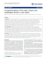
probably extending into the proximal bile duct. Contrast
enhanced computed tomography (CECT) confirmed the
presence of the cyst in segment 4B of the liver which had
an enhancing thin wall with multiple septa and no
intracystic solid component (Figure 1). CECT also revealed
intrahepatic, common hepatic and proximal common bile
duct dilatation probably due to an extension of the lesion
along the left hepatic duct. A hydroxy iminodiacetic acid
(HIDA) scan excluded a direct communication of the cyst
with the biliary tree. Serum echinococcus IgG levels were
normal.
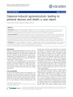
Laparotomy confirmed a lesion in the left side of the liver
with an extension into the common hepatic duct along the
left hepatic duct. The gallbladder was extrahepatic. During
left hepatectomy, the cyst extension was removed intact,
through the cut end of the left hepatic duct (Figure 2).
A synchronous cholecystectomy was performed once the
resection surface of the liver had been checked for biliary
leaks by injecting saline through the cystic duct. The
patient had an uneventful recovery.
Histology revealed a multilocular, mucinous biliary
cystadenoma lined by a single layer of glandular epithe-
lium, arising from the left intrahepatic ducts. A basement
membrane separated the epithelial lining from the under-
lying ovarian type mesenchyme. The patient is well at
24 months with no evidence of recurrent disease.
Discussion
There have been less than 100 cases of BCAs reported
worldwide. These account for less than 5% of neoplasms
originating from the bile duct [3].
BCAs are of two types, mucinous or serous. The more
common mucinous type is subdivided by the presence or
absence of a mesenchymal stroma between the inner
epithelial l ining and the outer basement membrane.
Although both types can undergo malignant transforma-
tion to biliary cystadenocarcinoma (BCAC), absence of a
mesenchymal stroma is known to be more aggressive,
especially in men [1].
Jaundice is a rare presentation of BCA, and, to the best of
our knowledge, episodic jaundice has only been reported
in four patients [4,5]. There are only eight reported cases of
intrahepatic BCA causing obstructive jaundice due to its
extension into a major duct [4-7]. The most likely cause of
episodic jaundice in this patient would have been
recurrent hemorrhage into the cyst which was extending
along the left hepatic duct to the confluence, causing a
transient rise in intracystic pressure and in turn occlusion
of the common channel of the extrahepatic biliary tree, as
reported by Taketomi et al. [7].
Pre-operative diagnosis may be difficult. Generally on USS
and CECT, they appear as focal lesions with internal septa,
hence they may be confused with other cystic hepatic
lesions such as a complicated cyst, mesenchymal hamar-
toma, undifferentiated embryonal sarcoma and cystic
metastases in addition to the malignant counterpart of
BCA and hydatid cyst [8]. While the BCAC has papillary
excrescences with solid components, hydatid cysts are
characterized by round and oval daughter cysts and “ring
like” enhancement with contrast on CECT. Mag netic
resonance imaging (MRI) also helps in diagnosis. In the
presence of intralesional hemorrhage and hyperprotei-
nous/mucinous contents, the distinction of BCA from
BCAC may be difficult [6]. In the presence of obstructive
jaundice, cholangiography, either endoscopic, percuta-
neous or magnetic resonance would show the level of
obstruction. Since this patient’s CECT revealed the cause of
Figure 1. Pre-operative contrast enhanced computed
tomography of the abdomen performed 2 months before
surgery, demonstrating the septate intrahepatic biliary
cystadenoma with extension to the left hepatic duct.
Page 2 of 4
(page number not for citation purposes)
Journal of Medical Case Reports 2009, 3 :9032 />the biliary obstruction, cholangiography was not
performed.
Treatment methods for BCA in the past have varied from
aspiration, marsupialization, internal drainage, partial
excision and enucleation [6,9]. The main concerns of
these methods are local recurrence, malignant transforma-
tion and misdiagnosis of cancer. In addition, biliary
peritonitis may occur following aspiration and marsupia-
lization [9,10]. In the past, more conservative methods
were favored probably due to poor pre-operative imaging
and the lack of understanding of BCA’s malignant
potential. Enucleation may be possible provided there is
no co-existing malignancy which can be detected by intra-
operative ultrasound scan and frozen sections [9].
However, contemporary hepatic surgery has minimal
morbidity and mortality [11]. Hence, as suggested by
previous authors [6], we endorse formal liver resection or
wide local excision as the treatment of choice for BCA.
Conclusion
This article contributes to the medical literature by adding
another reported case of episodic biliary obstruction due
to an intrahepatic BCA protruding into the common
hepatic duct, which may be considered as a very rare cause
of intermittent jaundice. Pre-operative diagnosis may be
confusing. Hence, a high degree of suspicion and a
multidisciplinary approach are needed to plan the surgical
procedure and prevent inadequate treatment. The surgical
strategy would be to radically excise the lesion. A follow-
up protocol is unavailable due to the small number of
BCAs. However, we suggest long-term evaluation with
clinical examination but more importantly with periodic
ultrasound scans at least 3 monthly during the first year,
6 monthly for 2 years and annually subsequently to
exclude local recurrence.
Consent
Written informed consent was obtained from the patient
for publication of this case report and any accompanying
images. A copy of the written consent is available for
review by the Editor-in-Chief of this journal.
Competing interests
The au thors declare that they have no competing
interests.
Figure 2. Intra-operative image showing the partially resected intrahepatic biliary cystadenoma. Partially resected left lobe
with the intrahepatic biliary cystadenoma and its extension into the left hepatic duct which is cut open and the schematic
diagram of the anatomical relationship of the intrahepatic biliary cystadenoma and the biliary ducts. The extrahepatic
gall bladder, which is not shown in the image, was removed synchronously. (A) Intrahepatic biliary cystadenoma;
(B) Polypoid extension of the intrahepatic biliary cystadenoma in the left hepatic duct (retracted upwards);
(C) Opening of the left hepatic duct through which the extension of the intrahepatic biliary cystadenoma was extracted.
Page 3 of 4
(page number not for citation purposes)
Journal of Medical Case Reports 2009, 3 :9032 />Authors’ contributions
PNS is the principal and corresponding author. PNS
actively managed the patient and was the first assistant to
the surgeon (AP). PNS contr ibuted to the paper by
performing the literature survey, and interpreting and
analyzing past cases to decide on management of the
patient. PNS wrote the manuscript and edited the
successive versions. AP is the senior author and the team
surgeon. He was a major contributor in interpreting
the cases in the literature and applying them to the
management of the patient. AP contributed to the paper
by planning the structure and editing successive versions.
Both authors read and approved the final manuscript.
Acknowledgement
We acknowledge Dr Mihiri Buddhadasa and Dr Nishantha
Liyanapathirana for the illustration and photographs.
References
1. Devaney K, Goodman ZD, Ishak KG: Hepatobiliary cystadenoma
and cystadenocarcinoma: a light microscopic and immuno-
histochemical study of 70 patients. Am J Surg Pathol 1994,
18:1078-1091.
2. Wheeler DA, Edmondson HA: Cystadenoma with mesenchymal
stroma (cms) in the liver and bile ducts. Cancer 1985, 56:1434-
1445.
3. Cahill CJ, Baily ME, Smith MGM: Mucinous cystadenoma of the
liver. Clin Oncol 1982, 8:171-177.
4. Beretta E, De Francis R, Staudacher C, Faravelli A, Primignani M,
Vecchi M, Conti E, Di Carlo V: Biliary cystadenoma: an
uncommon cause of recurrent cholestatic jaundice. Am J
Gastroenterol 1986, 81:138-140.
5. Erdogan D, Busch ORC, Rauws EAJ, van Delden OM, Gouma DJ, van
Gulik TM: Obstructive jaundice due to hepatobiliary cystade-
noma or cystadenocarcinoma. World J Gastroenterol 2006,
12:5735-5738.
6. Preetha M, Chung AYF, Lim-Tan SK, Lim DTH, Thug CH:
Intrahepatic biliary cystadenoma presenting with obstructive
jaundice. Asian J Surg 2004, 27:243-245.
7. Taketomi A, Tamada R, Takenaka K, Kawano R, Maeda T,
Sugimachi K: A case of biliary cystadenoma with obstructive
jaundice. Oncol Rep 1998, 5:833-835.
8. Choi BI, Lim JH, Han MC, Lee DH, Kim SH, Kim YI, Kim C: Biliary
cystadenoma and cystadenocarcinoma: CT and sonographic
findings. Radiology 1989, 171:57-61.
9. Lau WY, Chow CH, Leung ML: Total excision of mucinous biliary
cystadenoma. Aust NZ J Surg 1990, 60:226-228.
10. Lewis WD, Jenkins RL, Rossi RL, Munson L, ReMine SG, Cady B,
Braasch JW, McDermott WV: Surgical treatment of biliary
cystadenoma: A report of 15 cases. Arch Surg 1988, 123:563-568.
11. Jarnagin WR, Gonen M, Fong Y, DeMatteo RP, Ben-Porat L, Little S,
Corvera C, Weber S, Blumgart LImprovement in perioperative
outcome after hepatic resection: Analysis of 1,803 consecu-
tive cases over the past decade. Ann Surg 2002, 236:397-407.
Do you have a case to share?
Submit your case report today
• Rapid peer review
• Fast publication
• PubMed indexing
• Inclusion in Cases Database
Any patient, any case, can teach us
something
www.casesnetwork.com
Page 4 of 4
(page number not for citation purposes)
Journal of Medical Case Reports 2009, 3 :9032 />