Báo cáo y học: "Platypnea and orthodeoxia associated with Pneumocystis jiroveci and Cytomegalovirus pneumonia: a case report" potx
Bạn đang xem bản rút gọn của tài liệu. Xem và tải ngay bản đầy đủ của tài liệu tại đây (457.78 KB, 4 trang )
BioMed Central
Page 1 of 4
(page number not for citation purposes)
Journal of Medical Case Reports
Open Access
Case report
Platypnea and orthodeoxia associated with Pneumocystis jiroveci and
Cytomegalovirus pneumonia: a case report
Konstantinos Katsoulis
1
, Ilias Minasidis
2
, Andreas Vainas
2
,
Christoforos Bikas
1
, Theodoros Kontakiotis*
1
and Pantelis Vakianis
2
Address:
1
Pulmonary Department, General Army Hospital, Thessaloniki, Greece and
2
Nephrology Department, General Army Hospital,
Thessaloniki, Greece
Email: Konstantinos Katsoulis - ; Ilias Minasidis - ; Andreas Vainas - ;
Christoforos Bikas - ; Theodoros Kontakiotis* - ; Pantelis Vakianis -
* Corresponding author
Abstract
Introduction: Platypnea-orthodeoxia is an uncommon syndrome characterized by dyspnea and
deoxygenation accompanying a change to a sitting or standing posture from a recumbent position.
It is usually related to interatrial communications, although several other disorders associated with
platypnea-orthodeoxia syndrome have been reported. However, the precise mechanisms are
unknown.
Case presentation: We present the case of a 75-year-old Caucasian woman with chronic renal
failure due to vasculitis who was admitted with fever and respiratory failure. She was found to have
both Pneumocystis jiroveci and Cytomegalovirus pneumonia. She was HIV negative. Severe platypnea
and orthodeoxia were major features of her illness with no history of respiratory, liver or cardiac
disease. Further investigation with contrast echocardiography revealed no intracardiac or
intrapulmonary shunts. Although one case involving Pneumocystis jiroveci pneumonia and platypnea
has been previously reported, to the best of our knowledge, this is the first time that two
opportunistic pathogens have been accompanied by platypnea and orthodeoxia. As both lung bases
were predominantly affected and no obvious explanation was found, platypnea and orthodeoxia
were attributed to significant areas of low or zero ventilation/perfusion (V/Q) ratio.
Conclusion: Platypnea-orthodeoxia is a rare and usually underestimated syndrome. Intracardiac
shunts and anatomic pulmonary vascular shunts are the most common etiologic associations.
However, if a detailed examination reveals no obvious intracardiac or intrapulmonary shunting
combined with extensive pulmonary lesions, then severe V/Q mismatching should be considered
as the probable explanation.
Introduction
Platypnea-orthodeoxia is a relatively uncommon but
striking clinical syndrome characterized by dyspnea and
deoxygenation accompanying a change to a sitting or
standing posture from a recumbent position. It was first
reported in 1949 when Burchell et al. [1] described a
patient with an atrial septal defect manifesting platypnea-
orthodeoxia and subsequently described the reversal of
both following closure of a patent foramen ovale. 'Platyp-
nea' and 'orthodeoxia' were not used to describe the man-
Published: 5 December 2009
Journal of Medical Case Reports 2009, 3:9319 doi:10.1186/1752-1947-3-9319
Received: 24 September 2008
Accepted: 5 December 2009
This article is available from: />© 2009 Katsoulis et al; licensee BioMed Central Ltd.
This is an Open Access article distributed under the terms of the Creative Commons Attribution License ( />),
which permits unrestricted use, distribution, and reproduction in any medium, provided the original work is properly cited.
Journal of Medical Case Reports 2009, 3:9319 />Page 2 of 4
(page number not for citation purposes)
ifestations of this syndrome until they became commonly
accepted terms in 1969 and 1976, respectively [2,3]. Since
then, a few cases have been reported with interatrial com-
munications being the most common etiologic associa-
tions [4-6]. The precise mechanisms for both platypnea
and orthodeoxia are unknown. In several isolated case
reports, speculation over mechanisms is often geared to
whatever special features were found in the patient been
reported. We present a case of a patient with severe platyp-
nea and orthodeoxia infected with two opportunistic
pathogens and with no evidence of intracardiac or
intrapulmonary shunt.
Case presentation
A 75-year-old Caucasian woman was admitted to our hos-
pital with febrile illness accompanied by dyspnea without
other specific symptoms, such as cough or sputum. She
was normotensive, and her heart sounds were normal
with bibasal lung crepitations. An electrocardiogram dem-
onstrated sinus rhythm with a normal axis and oxygen sat-
uration in room air was 75%. Platypnea and orthodeoxia
were major features of her illness. When supine on 35%
oxygen by face mask, arterial blood gas measurements
yielded persistent hypoxemia (pO
2
: 70 mmHg, pCO
2
: 30
mmHg) with counterbalanced metabolic acidosis (HCO
3
:
18 mmol/l, pH 7.44). However, in the upright position,
she developed severe hypoxemia (pO
2
: 40 mmHg). Bio-
chemical tests showed renal failure (urea: 100 mg/dl, cre-
atinine: 2 mg/dl, hematocrit (Hct): 30%), increased
lactate dehydrogenase (LDH) levels (600 U/L) and
increased markers of inflammation (erythrocyte sedimen-
tation rate (ESR): 100 mm, CRP: >100 mg/dl), while a
chest X-ray showed a few bilateral diffuse interstitial infil-
trates, predominantly in the lower lobes.
Five months before admission, oliguric acute renal failure
was detected and kidney biopsy revealed rapidly progres-
sive glomerulonephritis with 100% crescents compatible
with Wegener's disease or nodular polyarteritis. She was
initially treated with sessions of renal dialysis and plas-
mapheresis combined with pulses of methylpred-
nisolone. Afterwards, the treatment switched to oral
methylprednisolone at 48 mg/day combined with oral
cyclophosphamide at 100 mg/day for 2 months with pro-
gressive lessening of the doses. Cyclophosphamide was
finally withdrawn due to severe side effects (leucopenia).
During the last trimester, she was in good condition under
treatment with 16 mg/day of methylprednisolone.
She was initially treated with empirical antibiotic treat-
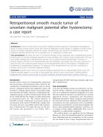
ment and underwent computed tomographic (CT) scan-
ning which showed patchy areas of ground-glass opacity
(Figure 1). With the suspicion of Pneumocystis jiroveci
pneumonia and despite a negative test for HIV, fiberoptic
bronchoscopy and bronchoalveolar lavage (BAL) were
performed. Immunostaining of the specimens was posi-
tive for P. jiroveci and several cysts were microscopically
visualized. Thus, the treatment was changed to high-dose
intravenous co-trimoxazole and prednisolone.
Although she became afebrile, the clinical presentation
deteriorated with excessive platypnea coupled with ortho-
deoxia. Sitting up was associated with a fall in her oxygen
saturation of up to 67% under oxygen administration. As
there was no evidence of liver disease or hepatopulmo-
nary syndrome, a transthoracic echocardiogram was per-
formed. Intravenously administered normal saline was
not detected in the left atrium after two or six cardiac cir-
cles excluding the presence of intracardiac or intrapulmo-
nary shunts. Due to the respiratory distress, a pulmonary
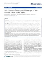
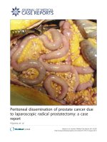
angiogram was not performed. A new CT scan revealed
further deterioration consisting of organized consolida-
tions with air bronchogram at the lung bases and air in the
mediastinum (Figure 2). On day 15, she was intubated
and admitted to the intensive care unit. At the same time,
BAL examination was positive for cytomegalovirus (CMV)
through polymerase chain reaction (PCR) (23,000 copies/
ml). Intravenous ganciclovir (5 mg/kg, twice a day) was
added to the treatment. Despite the appropriate treat-
ment, she died on day 25.
Discussion
Platypnea (increased dyspnea in the erect position
relieved by assuming a recumbent position) and orthode-
oxia (accentuated hypoxemia in the erect position,
improved by assuming a recumbent position) were the
patient's more striking symptoms. Only one case of this
syndrome associated with Pneumocystis carinii pneumonia
(the old term now replaced by jiroveci) has been previ-
Computed tomography demonstrating patchy areas of ground-glass opacityFigure 1
Computed tomography demonstrating patchy areas
of ground-glass opacity.
Journal of Medical Case Reports 2009, 3:9319 />Page 3 of 4
(page number not for citation purposes)
ously reported [7]. However, to the best of our knowledge,
the association of this syndrome with pulmonary infec-
tion by two opportunistic pathogens, including P. jiroveci,
has not been reported.
The etiologic background of this syndrome includes int-
racardiac shunts, anatomic pulmonary vascular and pul-
monary parenchymal shunts, with interatrial
communications being the most common etiologic asso-
ciation, but the precise mechanisms are not known
[5,8,9]. Other diseases associated with platypnea-ortho-
deoxia include chronic obstructive pulmonary disease
(COPD) [10], constrictive pericarditis [11], pneumonec-
tomy [12], paradoxical embolism [13] and even acute
organophosphorus poisoning [14].
In our patient, P. jiroveci and CMV were detected in BAL
specimens using reliable methods (immunostaining/
microscopic visualization and PCR, respectively) in an
immunosuppressed patient. The radiological findings
were not diagnostic as both of the above pathogens
present with similar findings. It has been noted that coin-
fection with CMV and other pathogens will be detected in
more than half of Pneumocystis-infected patients [15] and
thus the major pathogen is not easily defined. As we
found no other common pathogens and our patient con-
tinued to deteriorate despite the appropriate treatment for
P. jiroveci pneumonia, we believe that CMV was responsi-
ble for the fatal deterioration.
Our patient had no history or evidence of COPD or
chronic liver disease (for example, cirrhosis). As platypnea
and orthodeoxia were major features of her illness, further
investigation focused on the detection of probable intrac-
ardiac or intrapulmonary shunts using contrast echocardi-
ography, a widely accepted and non-invasive method
[16]. No evidence of such disorders was found, so these
symptoms probably arose as a result of significant areas of
low or zero V/Q ratio. Indeed, a CT scan indicated that the
lung bases were predominantly affected. Gravity might
increase intrapulmonary blood flow shunting through
poorly ventilated lung bases exacerbating dyspnea and
deoxygenation in the upright position. The same explana-
tion has been proposed by other authors for similar cases
[5,7].
Whether specific pathogens such as P. jiroveci and/or CMV
or severe V/Q mismatching of any etiology are responsible
for the emergence of this syndrome remains to be clari-
fied.
Conclusion
Platypnea-orthodeoxia is a rare and usually underesti-
mated syndrome. Intracardiac shunts and particularly,
interatrial communications with or without overt lung
disease as well as anatomic pulmonary vascular shunts are
the most common etiologic associations. However, if
detailed examination reveals no obvious intracardiac or
intrapulmonary shunting combined with extensive pul-
monary lesions, such as severe pneumonia even due to
opportunistic pathogens, then severe V/Q mismatching
should be considered as the probable explanation.
Abbreviations
BAL: bronchoalveolar lavage; COPD: chronic obstructive
pulmonary disease; CMV: cytomegalovirus; CRP: C-reac-
tive protein; ESR: erythrocyte sedimentation rate; Hct:
hematocrit; LDH: lactate dehydrogenase; PCR: polymer-
ase chain reaction; V/Q: ventilation/perfusion.
Consent
Written informed consent was obtained from the patient
for publication of this case report and accompanying
images. A copy of the written consent is available for
review by the Editor-in-Chief of this journal
Competing interests
The authors declare that they have no competing interests.
Authors' contributions
KK was the main author and carried out the pulmonary
investigation of the case. IM carried out the nephrological
investigation of the case. AV carried out vasculitis diagno-
sis and management of the case. CB carried out the respi-
ratory failure diagnosis and management. TK is the
corresponding author and was responsible for manuscript
preparation and the pulmonary investigation of the case.
PV carried out the renal failure diagnosis and manage-
ment of the case.
Computed tomography demonstrating organized consolida-tions with air bronchogram at the lung bases and air in the mediastinumFigure 2
Computed tomography demonstrating organized
consolidations with air bronchogram at the lung
bases and air in the mediastinum.
Publish with BioMed Central and every
scientist can read your work free of charge
"BioMed Central will be the most significant development for
disseminating the results of biomedical research in our lifetime."
Sir Paul Nurse, Cancer Research UK
Your research papers will be:
available free of charge to the entire biomedical community
peer reviewed and published immediately upon acceptance
cited in PubMed and archived on PubMed Central
yours — you keep the copyright
Submit your manuscript here:
/>BioMedcentral
Journal of Medical Case Reports 2009, 3:9319 />Page 4 of 4
(page number not for citation purposes)
References
1. Burchell HB, Hemholz HF Jr, Wood EH: Reflect orthostatic dysp-
nea associated with pulmonary hypotension. Am J Physiol 1949,
159:563-564.
2. Altman M, Robin ED: Platypnea (diffuse zone I phenomenon?).
N Engl J Med 1969, 281:1347-1348.
3. Robin ED, Lamon D, Horn BR, Theodore J: Platypnea related to
orthodeoxia caused by true vascular lung shunts. N Engl J Med
1976, 294:941-943.
4. Seward JB, Hayes DL, Smith HC, Williams DE, Piehler JM, Tajik AJ:
Platypnea-orthodeoxia: clinical profile, diagnostic workup,
management and report of seven cases. Mayo Clin Proc 1984,
59(4):221-231.
5. Robin ED, McCauley F: An analysis of platypnea-orthodeoxia
syndrome including a "new" therapeutic approach. Chest
1997, 112:1449-1451.
6. Gobart F, Rey C: Platypnea-orthodeoxia syndrome: a probably
underestimated syndrome? Chest 2001, 119:1624-1625.
7. Newton PN, Wakefield AE, Goldin R, Govan J: Pneumocystis car-
inii pneumonia with pleurisy, platypnoea and orthodeoxia.
Thorax 2003, 58:185-186.
8. Hagen PT, Scholz DG, Edwards WD: Incidence and size of patent
foramen ovale during the first ten decades of life: an autopsy
study of 965 normal hearts. Mayo Clin Proc 1984, 59:17-20.
9. Cheng TO: Mechanisms of platypnea-orthodeoxia: what
causes water to flow uphill? Circulation 2002, 105(6):47.
10. Hussain SF, Mekan SF: Platypnea-orthodeoxia: report of two
cases and review of the literature. South Med J 2004,
97(7):657-662.
11. Hashimoto M, Okawa Y, Baba H, Nishimura Y, Aoki M: Platypnea-
orthodeoxia syndrome combined with constrictive pericar-
ditis after coronary artery bypass surgery. J Thorac Cardiovasc
Surg 2006, 132(5):1225-1226.
12. Kotoulas C, Patris K, Tsintiris K, Zoumboulides A, Lazarides K,
Laoutides G: Platypnea-orthodeoxia syndrome after pneu-
monectomy relieved by mediastinal repositioning. Ann Thorac
Surg
2007, 83(4):1524-1526.
13. Delalieux S, De Greef K, Hendriks J, Lauwers P, Suys B, Van Schil P:
Orthodeoxia-platypnea syndrome presenting as paradoxical
peripheral embolism. Ann Thorac Surg 2008, 85(5):1798-1800.
14. Bouros D, Agouridakis P, Tsatsakis A, Askitopoulou E, Siafakas NM:
Orthodeoxia and platypnoea after acute organophosphorus
poisoning reversed by CPAP: a newly described cause and
review of the literature. Respir Med 1995, 89(9):625-628.
15. Fishman JA: Pneumocystis carinii. In Fishman's Pulmonary Diseases
and Disorders 3rd edition. McGraw-Hill; 1998:2313-2331.
16. Thakur CT, Nanda NC, Malhotra S, St Martin MB, Jamil F, Agrawald
D, Maheshwari S, Abrams GA, Patel B: Combined interatrial and
intrapulmonary shunting in orthodeoxia detected by tran-
soesophageal echocardiography. Echocardiography 2007,
15:101-104.