Báo cáo y học: "Menstruating from the umbilicus as a rare case of primary umbilical endometriosis: a case report" pot
Bạn đang xem bản rút gọn của tài liệu. Xem và tải ngay bản đầy đủ của tài liệu tại đây (231.44 KB, 3 trang )
BioMed Central
Page 1 of 3
(page number not for citation purposes)
Journal of Medical Case Reports
Open Access
Case report
Menstruating from the umbilicus as a rare case of primary umbilical
endometriosis: a case report
Pallavi V Bagade*
1
and Mamdouh M Guirguis
2
Address:
1
Department of Obstetrics and Gynaecology, Wansbeck General Hospital, Woodhorn Lane, Ashington NE63 9JJ, Northumberland, UK
and
2
Department of Obstetrics and Gynaecology, North Tyneside General Hospital, Rake Lane, North Sheilds NE29 8NH, Tyne and Wear, UK
Email: Pallavi V Bagade* - ; Mamdouh M Guirguis -
* Corresponding author
Abstract
Introduction: Endometriosis is a common gynecological condition and presents mainly with
involvement of the pelvic organs. Extrapelvic presentations in almost all parts of the body have been
reported in the literature. However, umbilical endometriosis that is spontaneous or secondary to
surgery is uncommon and accounts for only 0.5% to 1% of all endometriosis cases.
Case presentation: A 35-year-old Caucasian woman presented with umbilical bleeding during
periods of menstruation. Her umbilicus had a small nodule with bloody discharge. An ultrasound
was performed and a diagnosis of possible umbilical endometriosis was thus made. The nodule
shrunk in response to gonadotropin-releasing hormone analogues but continued to persist. The
patient underwent a wide local excision of the nodule with a corresponding umbilical
reconstruction. Histopathology confirmed the diagnosis of umbilical endometriosis. The patient
was asymptomatic at follow-up, but nevertheless warned of the risk of recurrence.
Conclusions: Pelvic endometriosis is a common condition, but the diagnosis of primary umbilical
endometriosis is difficult and differentials should be considered. This case strongly suggests that a
differential diagnosis of endometriosis should be considered when an umbilical swelling presents in
a woman of reproductive age.
Introduction
Endometriosis, a term first used by Sampson, is the pres-
ence of endometrial glands and stroma outside the uter-
ine cavity and musculature [1]. It affects 7% to 10% of
women in the reproductive age group [2]. It commonly
occurs in the pelvic organs, especially the ovaries, the ute-
rosacral ligaments and the pouch of Douglas. Women
with endometriosis often present with dysmenorrhea,
menorrhagia, pelvic pain and infertility.
Extragenital endometriosis is less common, but has been
described in almost every area of the female body includ-
ing the bowel, bladder, lungs, brain, umbilicus, and surgi-
cal scars [3]. Due to its varied presentations,
endometriosis remains a difficult condition to diagnose
and treat.
Umbilical endometriosis represents 0.5% to 1% of all
cases of extragenital endometriosis. It usually occurs sec-
ondary to surgical scars, but very rarely presents as pri-
mary umbilical endometriosis [4,5]. We report one such
rare case of spontaneous, primary umbilical endometrio-
sis.
Published: 10 December 2009
Journal of Medical Case Reports 2009, 3:9326 doi:10.1186/1752-1947-3-9326
Received: 13 December 2008
Accepted: 10 December 2009
This article is available from: />© 2009 Bagade and Guirguis; licensee BioMed Central Ltd.
This is an Open Access article distributed under the terms of the Creative Commons Attribution License ( />),
which permits unrestricted use, distribution, and reproduction in any medium, provided the original work is properly cited.
Journal of Medical Case Reports 2009, 3:9326 />Page 2 of 3
(page number not for citation purposes)
Case presentation
A 35-year-old Caucasian parous woman presented to the
clinic with symptoms of spontaneous and periodic bleed-
ing from the umbilicus for four months. The bleeding
would start two days before her menses and continue for
the entire duration of her period. It was accompanied by
pain and swelling in the umbilical area.
The patient had regular, heavy and painless menstrual
periods and did not wish for any treatment for such. She
had two previous spontaneous vaginal deliveries and had
no history of abdominal pain, dyspareunia or infertility.
She was not using any form of hormonal contraception.
Her medical history was not significant and she never had
any abdominal surgeries.
Clinical examination revealed that the patient had a 2 cm
× 2 cm firm nodule at the umbilicus, which appeared to
be covered by a reddish brown discharge. Suspecting that
she had an infection, the patient was swabbed and given
a five-day course of oral broad-spectrum antibiotics. She
showed up on check up two months later with no relief of
symptoms. She then underwent an ultrasound scan that
showed a 15-mm thin-walled cyst, approximately 5 mm
below the skin surface. The key clinical feature that led to
the correct diagnostic hypothesis of umbilical endometri-
osis was the temporal association of the bleeding with her
menstrual period.
The patient was offered both medical and surgical man-
agement and she opted to have depot injections of Zola-
dex (AstraZeneca UK, Goserelin acetate, 3.6 mg
subcutaneously, monthly). The swelling continued to per-
sist in spite of three doses of Zoladex, and the patient then
requested surgical excision. The risk of recurrence and scar
endometriosis were explained to her.
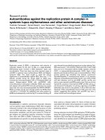
The patient successfully underwent excision of the nodule
with accompanying umbilical reconstruction. Histology
confirmed the diagnosis of endometriosis and revealed
the presence of endometriotic glands with mucinous type
metaplasia and extravasation of the mucinous secretion
into the adjacent stroma (Figure 1). No epithelial atypia
was seen and the excision appeared complete. The patient
was seen six weeks after the surgery and found to be
asymptomatic with a normal umbilicus. Before being dis-
charged, the patient was again reminded of the risk of
recurrence.
Discussion
The deposition of fragments of uterine endometrium in
the skin is a well recognized, although uncommon, phe-
nomenon (0.5% to 1% of extragenital endometriosis).
Umbilical endometriosis was first described in 1886 and
since then more than 100 cases have been described [4].
Majority of these cases occurred secondary to surgical,
commonly laparoscopy, scars. An umbilical endometri-
otic lesion without surgical history is a rare condition
[4,5]. Some case reports have also described the presence
of umbilical endometriosis during pregnancy [6].
There has been great speculation about the pathogenesis
of this phenomenon and several theories have been pro-
posed. Latcher has classified these theories into three
main categories: the embryonal rest theory, which
explains endometriosis adjoining the pelvic viscera by
Wollfian or Mullerian remnants [4,5]; the coelomic meta-
plasia theory, which states that the embryonic coelomic
mesothelium dedifferentiates into endometrial tissue
under stimulus such as inflammation or trauma [7]; and
the migratory pathogenesis theory, which explains the dis-
persion of endometrial tissue by direct extension, vascular
and lymphatic channels, and surgical manipulation. Still
others suggest cellular proliferation of endometrial cells
from initial extraperitoneal disease along the urachus
[8,9]. The real mechanism still remains a mystery.
These patients are usually in the reproductive age group
and present commonly with swelling, pain, discharge or
cyclical bleeding from the umbilicus. There may be asso-
ciated symptoms of coexistent pelvic endometriosis.
These lesions are usually bluish-black in colour and
become painful, larger and bleed about the time of men-
ses. They range in size from 0.5 cm to 3 cm, but can
enlarge to even more enormous sizes [4].
While the diagnosis is primarily clinical, magnetic reso-
nance imaging (MRI) can be useful in evaluating patients
Umbilical endometriosis: endometriotic glands with metapla-sia of the mucinous type and extravasation of the mucinous secretion into the adjacent stromaFigure 1
Umbilical endometriosis: endometriotic glands with
metaplasia of the mucinous type and extravasation of
the mucinous secretion into the adjacent stroma.
Publish with BioMed Central and every
scientist can read your work free of charge
"BioMed Central will be the most significant development for
disseminating the results of biomedical research in our lifetime."
Sir Paul Nurse, Cancer Research UK
Your research papers will be:
available free of charge to the entire biomedical community
peer reviewed and published immediately upon acceptance
cited in PubMed and archived on PubMed Central
yours — you keep the copyright
Submit your manuscript here:
/>BioMedcentral
Journal of Medical Case Reports 2009, 3:9326 />Page 3 of 3
(page number not for citation purposes)
with suspected endometriosis. Endometriomas appear
homogeneously hyperintense on T1-weighted sequences
[10]. MRI also has an advantage over laparoscopy for eval-
uating pelvic and extraperitoneal diseases, as well as
lesions concealed by adhesions.
Histological findings are characterized by irregular glan-
dular lumina embedded in the stroma with a high cellular
and vascular component resembling the stroma of func-
tional endometrium. A fairly recent study has suggested a
distinctive dermatoscopic feature in cutaneous endome-
triosis that of comprising small red globular structures
called 'red atolls' [11].
Differential diagnosis of umbilical nodules should
include pyogenic granuloma, hernia, residual embryonic
tissue, primary or metastatic adenocarcinoma (Sister
Joseph's nodule), nodular melanoma, and cutaneous
endosalpingosis.
Surgical excision of the lesion with sparing of the umbili-
cus is the preferred treatment of pelvic endometriosis [7].
In severe cases or in the presence of pelvic endometriosis,
hormonal therapy in the form of danazol or GnRH ana-
logues can be given to the patient [12]. In our case the
lesion was excised and histology confirmed the diagnosis.
Although simultaneous laparoscopy has been recom-
mended for pelvic endometriosis, this was not done
because our patient was asymptomatic. Although local
recurrence is uncommon, the patient has been warned of
the risk of scar endometriosis and of recurrence.
Conclusions
Endometriosis is a common gynaecological disease; how-
ever, primary umbilical endometriosis is very rare. Making
a diagnosis is difficult and other causes of umbilical
lesions should be considered. Surgical excision is the
standard treatment of this condition.
Abbreviations
MRI: magnetic resonance imaging; GnRH: gonadotropin
releasing hormone.
Consent
Written informed consent was obtained from the patient
for publication of this case report and any accompanying
images. A copy of the written consent is available for
review by the Editor-in-chief of this journal.
Competing interests
The authors declare that they have no competing interests.
Authors' contributions
PB was a major contributor in collecting data, writing and
preparing the manuscript. MG performed the surgical
excision and was involved in editing the manuscript. All
authors read and approved the final manuscript.
References
1. Sampson JA: Perforating hemorrhagic (chocolate) cysts of the
ovary: their importance and especially their relation to pel-
vic adenomas of the endometrial type. Arch Surg 1921, 3:245.
2. Drake TS, Grunert GM: The unsuspected pelvic factor in the
infertility investigations. Fertility and Sterility 1980, 34:27-31.
3. Markham SM, Carpenter SE, Rock JA: Extrapelvic endometriosis.
Obstet Gynecol Clin North Am 1989, 16:193-219.
4. Latcher JW: Endometriosis of the umbilicus. Am J Obstet Gynecol
1953, 66:161-168.
5. Mann LS, Clarke WR: Endometriosis of the umbilicus. Ill Med J
1964, 125:335-336.
6. Razzi S, Rubegni P, Sartini A, De Simone S, Fava A, Cobellis L, Fimiani
M, Petraglia F: Umbilical endometriosis in pregnancy: a casere-
port. Gynecol Endocrinol 2004, 18(Suppl 2):114-116.
7. Schachter LR, Tash J, Olgac S, Bochner BH: Umbilical endometri-
osis. J Urol 2003, 170:2388-2389.
8. Rubegni P, Sbano P, Santopietro R, Fimiani M: Case four: umbilical
endometriosis. Clin Exp Dermatol 2003, 28:571-572.
9. Ploteau S, Malvaux V, Draguet AP: Primary umbilical adenomyo-
tic lesion presenting as cyclical periumbilical swelling. Fertil
Steril 2007, 88(Suppl 6):1674-1675.
10. Hartigan CM, Holloway BJ: Case report: MR imaging features of
endometriosis at the umbilicus. Br J Radiol 2005, 78:755-757.
11. De Giorgi V, Massi D, Mannone F, Stante M, Carli P: Cutaneous
endometriosis: noninvasive analysis by epiluminescence
microscopy. Clin Exp Dermatol 2003, 28:315-317.
12. Purvis RS, Tyring SK: Cutaneous and subcutaneous endometri-
osis: surgical and hormonal therapy. J Dermatol Surg Oncol 1994,
20:693-695.