Báo cáo y học: " Primary hydatid cyst of the gallbladder: a case report" ppt
Bạn đang xem bản rút gọn của tài liệu. Xem và tải ngay bản đầy đủ của tài liệu tại đây (978.56 KB, 6 trang )
CAS E REP O R T Open Access
Primary hydatid cyst of the gallbladder: a case
report
Avdyl Krasniqi
1*
, Dalip Limani
1
, Lumturije Gashi-Luci
2
, Gazmend Spahija
3
, Ismail A Dreshaj
4
Abstract
Introduction: Echinococcosis, or hydatid disease, is endemic in some regions of the world, and has been a
common pathology of surgical wards in Kosovo. Primary hydatid cyst of the gallbladder is an unusual and very rare
localization of hydatid disease. So far, only five cases that fulfill the criteria of primary gallbladder hydatidosis have
been published in the English medical literature.
Case presentation: We report a case of a 39-year-old Kosovan Albanian woman referred to the Abdominal
Surgery Division of the University Clinical Center of Kosovo for “a calcified hydatid cyst of the liver with gallbladder
involvement”. Her history was significant for chronic right upper quadrant pain, characterized as intermittently
colicky pain, accompanied by nausea. The patient underwent right subcostal laparotomy. Intra-operatively, a
calcified primary hydatid cyst of the gallbladder was found. Its pericyst was tightly attached to the liver. Complete
pericystectomy with cholecystectomy followed. The histopath ology confirmed the presence of calcified hydatid
cyst of the gallbladder, and that the cyst had developed entirely extra-mucosally. Five year follow-up showed no
recurrence of disease.
Conclusion: Primary hydatid cyst of the gallbladder is a very rare clinical entity. Accurate preoperative diagnostic
localization is not always easy, particularly in centers with limited diagnostic tools.
Introduction
Hydatid disease is a zoonotic infection caused by larval
stages of dog tapeworms belonging to the genus Echino-
coc cus (family taeniidae) and is also referred to as echi-
nococcosis [1]. Three broad morphological forms of
echinococcosis are recogn ized clinically. Human cystic
echinococcosis caused by E granulosus is the most com-
mon presentation and probably accounts for more than
95% of the estimated 2-3 million annual worldwide
cases [2]. This disease continues to be a substantial
cause of morbidity and mortality in many parts of the
world [1]. Hydatidosis is endemic in Mediterranean
countries and other sheep and cattle-raising regions [3].
In Kosovo, although there is no exact data about the
incidence of human cystic echinococcosis, liver and lung
hydatid cysts continue to be a very common pathology
of surgical wards [4]. The liver (70-80%) and lungs (15-
25%) are the most frequent locations for echinococcal
cysts while occurrence in other sites is very rare [3-6]
and the real incidence of extra hepatic cysts is not
known [5].
Primary hydatid cyst of the gallbladder is an extremely
rare entity [6-8]. There are reports of the gallbladder
daughter cysts secondary to liver cysts [9]. Patients with
primary hydatid cyst of the gallbladder are those with
no previous history of hydatid disease and with no other
cysts found at the time of surgery [6]. In a recent review
of the literature through the Medline database, we
found that in the English language only five cases have
been reported. by Safioleas et al. (2004), Rigas et al.
(1979); Wani et al. (2005), and Raza et al. (2003) [5-8].
Two more cases were reported in Slavic languages
[10,11]. The aim of this case report is to highlight the
diagnostic features, routes ofdisseminationandtreat-
ment options of this rare clinical entity.
Case presentation
A 39 year old Kosovan Albanian woman was referred to
the Division of Abdominal Surgery at the University Clini-
cal Center of Kosovo for an 18-month history of ch ronic
(intermittently colicky) pain in the ri ght u pper quadrant,
* Correspondence:
1
University Clinical Center of Kosovo, Division of Abdominal Surgery, Medical
School University of Prishtina, Prishtina, Republic of Kosovo
Krasniqi et al. Journal of Medical Case Reports 2010, 4:29
/>JOURNAL OF MEDICAL
CASE REPORTS
© 2010 Krasniqi et al; licensee BioMed Central Ltd. This is an Open Access article distri buted under the terms of the Creative Commons
Attribution License ( g/licenses/by/ 2.0), which permits unrestricted use, distribution, and reproduction in
any medium, provided the original work is properly cited.
often accompanied by nausea. There was no history of
jaundice. She had been treated by her primary care physi-
cian with antispasmodics, H2 receptor blockers and anti-
biotics, without resolution. On admission, physical
examination showed n o abnormal abdominal findings
except mild tenderness in the right upper quadrant. Rou-
tine blood tests such as CBC, renal and liver panel proved
unremarkable. Chest x-ray showed no signs of cardio-
respiratory disease. Plain radiograph of the abdomen
showed a calcified opacity at the level L2-L3 vertebrae on
the right. The diagnosis, supported by ultrasound and
computed tomography, was a calcified hydatid cyst of the
liver with involvement and deformation of the gallbladder;
the architecture and the size of biliary ducts were normal.
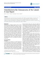
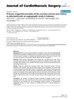
The patient underwent right subcostal laparotomy. Intra-
operatively, a calcified primary hydatid cyst of the fundus
and body of the gallbladder was found with its pericyst
attached to the liver (Figure 1). The inflammatory
response of the liver tissue against the cyst was extens ive
and formed the structural part of the posterior wall of the
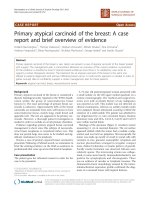
pericyst. Complete pericystectomy along with
cholecystectomy was performed. No other cysts were
found during careful exploration of the peritoneal cavity.
On opening the gallbladder, a calcified hydatid cyst
(dimensions 7 cm × 5 cm) was found, located in the body
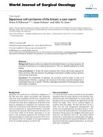
and fundus of the gallbladder (Figure 2). Macroscopically,
the hydatid cyst was part of the gallbladder. The cyst had
reduced severely the lumen of t he gallbladder and had
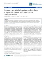
grown entirely submucosally (Figu re 3). The histopathol-
ogy confirmed the presence of calcified hydatid cyst of the
gallbladder (Figure 4) . The patient’s postoperative course
was uneventful and she was discharged in good condition
on the seventh postoperative day. She received two 21-day
courses of oral Albendazol 400 mg/day with 14 days pause
in between. At five-year follow up, she has had no recur-
rence of hydatid disease.
Discussion
Hydatid disease is a common clinical pathology in many
parts of the world. There are two clinical forms of this
disease: cystic hydatidosis caused by Echinococcus granu-
losus and alveolar hydatidosis caused by Echinococcus
Figure 1 Intraoperative view: Outer side of calcified hydatid cyst tightly attached to the liver.
Krasniqi et al. Journal of Medical Case Reports 2010, 4:29
/>Page 2 of 6
multilocularis. The mai n species pathogenic for humans
in Mediterranean and Southern European countries is
Echinococcus granulosus [1]. There has been no
reported case of alveolar hydatidosis in Kosovo [4].
Infection begins with the ingestion of tapeworm eggs,
which in the human intestine hatch into embryos that
penetrate the small bowel mucosa, enter venules and
travel via portal circulation to the liver. Hydatid cysts
most often develop in the liver. However when embryos
pass through this first filter, the second most frequent
location is the lung. Hydatid cysts can occur anywhere
in the body [3-7,12].
Unusual locations of hydatid cysts support the
hypothesis that beside portal circulation, the echino-
coccus embryos can spread via other routes, suc h as
the lymphatic system [6,8,12], the biliary tract [7,13]
and/or by disseminatio n of daughter cysts into perito-
neal or other cavities with the rupture of the primary
cyst [3,13]. Because of the small number of cases
reported, opinions about the pathogenesis of the pri-
mary gallbladder hydatid c ysts are divided depending
on the location of the cyst: in the lumen of the gall-
bladder o r on the external surface [6-8,13]. In two case
reports, Cangiotti et al. (1994) [13] and Raza et al.
(2003) [7] found cysts inside the gallbladder and
described them as a result of brood capsules dissemi-
nation through the biliary tract (i.e. through the cystic
duct). Safioleas et al. (2004) [6] reported findings in
three cases and remphasizes the idea of Rigas et al.
(1979) [8] of lymphatic rather than biliary spread in
primary gallbladder hydatid disease. In our case, the
hydatid cyst was larger than it was described in pre-
vious reports [6-8,13]. Macroscopic as well as micro-
scopic examination of the removed formation after
surgery, showed very clearly that the hydatid cyst nar-
rowed severely the lumen of the gallbladder, but that it
had grown entirely extra-mucosally ( Figure 3). In t his
case, therefore, transport of oncospheres from the
intestine to the gallbladder is more likely to have
occurred by lymphatic circulation. However, that
should be confirmed in a larger numb er of patients.
Pain, midabdominal discomfort and dyspepsia were
the main symptoms in all reported cases of primary
hydatid cyst of the gallbladder. Neither jaundice nor
anaphylactic reaction were noted in any cases [5-7].
Chronic upper abdominal low intensity pain, nausea and
intermittent biliary colic were the main complaints of
our patient. Imaging diagnostic tools such as ultrasound
and computed to mography are very helpful in the diag-
nosis of hydatid cysts [3,4,6,9,14]. Due to t he anatomic
proximity of the gallbladder to the liver, exact localiza-
tion of primary gallbladder hydatid cysts is not always
easy preoperatively, particularly in centers with limited
resources. The exact site of e xtra hepatic abdominal
hydatidosis in some cases is confirmed only in tra-opera-
tively [4-6], as in this case. The initial ultrasound and
computed tomography scans described the cystic lesion
as a hydatid cyst of the liver with involvement of the
Figure 2 The outer side of the removed cyst together with the gallbladder and a small part of the liver.
Krasniqi et al. Journal of Medical Case Reports 2010, 4:29
/>Page 3 of 6
gallbladder, only to have the diagnosis corrected during
surgery. This is the first case of a primary gallbladder
hydatid cyst in our experience of 241 patients with liver
cystic hydatidosis [15]. Gallbladder hydatid cysts should
be differentiated from liver hydatid cysts and other extra
hepatic cystic lesions [4-6]. Liver hydatidosis has a long
asymptomatic period of cystic growth [1], whereas in
the primary gallbladder hydatid cyst, biliary symptoms
begin earlier, and diagnostic imaging indicates smaller
cysts with deformation of the gallbladder [6].
Surgery is the preferred treatmen t for hydatid disease.
The goal is the eradication of the parasite without
spillage of the cyst content. In liver hydatididosis, com-
plete pericystectomy is not always possible, a nd there-
fore partial perycystectomy is the most frequent surgical
approach [3,4]. Successful total pericystectomy with cho-
lecystectomy was performed in all reported cases
[6-8,10]. We too performed perycystectomy with chole-
cystectomy. Postoperative recovery was uneventful. We
found, as did other authors [5-7], that in primary hyda-
tid cyst of the gallbladder, radical excision is easier than
it is in liver hydatidosis [3,4]. In case of exuberant
immune reaction o f liver tissues against the calcified
pericyst of gallbladder, dissection should be done v ery
Figure 3 The inner side of the cyst developed entirely extramucosally.
Krasniqi et al. Journal of Medical Case Reports 2010, 4:29
/>Page 4 of 6
carefully to avoid injuring biliary ducts that are in the
proximity to the gallbladder bed.
Conclusions
Primary hydatid cyst of the gallbladder is a very rare
clinical entity. Accurate site diagno sis was not made
pre but intra-operatively. In our experience of 241
patients treated surgically for liver hydatididosis, only
one patient (0.4%) was found to have primary gallblad-
der hydatid cyst. Careful pericystectomy with
cholecystectomy is the procedure of choice for radical
excision of primary hydatid cysts. Compare to liver
hydatidosis, t he gallbladder primary hydatid cyst has
different spread routs of parasi te embryos and a bett er
prognosis due to earlier manifestation of symptoms
leading to earlier treatment.
List of abbreviations
CBC: Complete blood count; US: Ultrasound; CT: Com-
puterized tomography.
Figure 4 Histopathologic specimen.
Krasniqi et al. Journal of Medical Case Reports 2010, 4:29
/>Page 5 of 6
Consent
Written informed consent was obtained from the patient
for publication o f this case report and accompanying
images. A copy of the written consent is available for
review by the Editor-in-Chief of this journal.
Author details
1
University Clinical Center of Kosovo, Division of Abdominal Surgery, Medical
School University of Prishtina, Prishtina, Republic of Kosovo.
2
University
Clinical Center of Kosovo, Institute of Pathology, Medical School University of
Prishtina, Prishtina, Republic of Kosovo.
3
University Clinical Center of Kosovo,
Department of Anesthesiology and Reanimation, Prishtina, Republic of
Kosovo.
4
University Hospitals & Case Medical Center, Cleveland, Ohio, USA.
Authors’ contributions
AK and DL performed the surgery and paper writing. LGL performed the
histological examination of the specimen. GS conducted anesthesia and
searched the literature. ID helped with conservative therapy and paper
writing.
All authors have read and approved the final manuscript.
Competing interests
The authors declare that they have no competing interests.
Received: 19 September 2009
Accepted: 29 January 2010 Published: 29 January 2010
References
1. Craig PS, McManus DP, Lightowlers MW, Chabalgoity JA, Garcia HH,
Gavidia CM, Gilman RH, Gonales AE, Lorca M, Naquira C, Nieto A,
Schantz PM: Prevention and control of cystic echinococcosis. Lancet Infect
Dis 2007, 7:385-94.
2. Budke CM: Global socioeconomic impact of cystic echinococcosis. Emerg
Infect Dis 2006, 12:56-64.
3. Safioleas M, Misiakos EP, Kakisis J, Manti C, Papachristodoulou A, Lambrou P,
Tsinary KK, Skalkeas G: Surgical treatment of human echinococcosis. Int
Surg 2000, 85:358-65.
4. Krasniqi A, Hoxha F, Nuhiu B, Bicaj B, Bruqi B, Tanaj H, Spahija G, Plakolli N,
Limani D: Results of different surgical methods in management of
hepatic hydatidosis. Med Arh 2006, 60:23-25.
5. Wani RA, Malik AA, Chowdri NA, Wani KA, Naqash SH: Primary extrahepatic
abdominal hydatidosis. Int J of Surgery 2005, 3:125-127.
6. Safioleas M, Stamoulis I, Theoqaris S, Moulakakis K, Makris S, Kostakis A:
Primary hydatid disease of the gallbladder: a rare clinical entity. J
Hepatopancreat Surg 2004, 11:352-6.
7. Raza MH, Harris SH, Khan R: Hydatid cyst of gallbladder. Indian J
Gastroenterol 2003, 22:67-68.
8. Rigas AM, Karatzas GM, Markidis NC, Bonikos DS, Sotiropoulou GG,
Skalkeas G: Primary hydatid cyst of the gallbladder. Br J Surg 1979, 66:406.
9. Ivanis N, Rubinic M, Gudovic A, Zeider F: Ultrasound image of an
echinococcus daughter cyst in the gallbladder. Ultraschall Med 1994,
15:269-71.
10. Putnik M, Ciric S: Hydatid cyst of the gallbladder. Srp Arh Celok Lek 1953,
81:531-2.
11. Tkebuchava GI: Calcified echinoccocosis of the gallbladder. Khirurgiia
(Mosk) 1996, 42:134-5.
12. Dioniqi G, Carrafiello G, Recaldini C, Sesso F, Boni L, Rovera F, Dionigi R:
Laparoscopic resection of a primary hydatid cyst of the adrenal gland: a
case report. J Med Case Reports 2007, 1:61.
13. Cangioti L, Muiesan P, Begni A, De Cesare V, Pouche A, Giulini S, Tibero G:
Unusual localizations of hydatid disease; an 18 years experience. Giorn
Chir 1994, 15:83-86.
14. Kapoor A, Sarma D, Gandhi D: Sonographic diagnosis of a ruptured
primary hydatid cyst of the gallbladder. J Clin Ultrasound 2000, 28:51-52.
15. Krasniqi A, Hoxha FT, Bicaj B, Spahija G, Musa R, Limani D: Surgical
treatment of large liver hydatid cysts - comparison of different methods
[abstract]. Eur Surg Res 2005, 37:s26.
doi:10.1186/1752-1947-4-29
Cite this article as: Krasniqi et al.: Primary hydatid cyst of the
gallbladder: a case report. Journal of Medical Case Reports 2010 4:29.
Submit your next manuscript to BioMed Central
and take full advantage of:
• Convenient online submission
• Thorough peer review
• No space constraints or color figure charges
• Immediate publication on acceptance
• Inclusion in PubMed, CAS, Scopus and Google Scholar
• Research which is freely available for redistribution
Submit your manuscript at
www.biomedcentral.com/submit
Krasniqi et al. Journal of Medical Case Reports 2010, 4:29
/>Page 6 of 6