báo cáo khoa học: "Turning a blind eye: the mobilization of radiology services in resource-poor regions" pot
Bạn đang xem bản rút gọn của tài liệu. Xem và tải ngay bản đầy đủ của tài liệu tại đây (342.17 KB, 8 trang )
DEBATE Open Access
Turning a blind eye: the mobilization of radiology
services in resource-poor regions
Duncan Smith-Rohrberg Maru
1,2,3*
, Ryan Schwarz
1,4
, Jason Andrews
1,2
, Sanjay Basu
1,5
, Aditya Sharma
1,6
,
Christopher Moore
1,7
Abstract
While primary care, obstetrical, and surgical services have started to expand in the world’s poorest regions, there is
only sparse literature on the essential support systems that are required to make these operations function. Diag-
nostic imaging is critical to effective rural healthcare delivery, yet it has been severely neglected by the academic,
public, and private sectors. Currently, a large portion of the world’s population lacks access to any form of diagnos-
tic imaging. In this paper we argue that two primary imaging modalities–diagnostic ultrasound and X-Ray–are ideal
for rural healthcare services and should be scaled -up in a rapid and standardized manner. Such machines, if
designed for resource-poor settings, should a) be robust in harsh environmental conditions, b) function reliably in
environments with unstable electricity, c) minimize radiation dangers to staff and patients, d) be operable by non-
specialist providers, and e) produce high-quality images required for accurate diagnosis. Few manufacturers are
producing ultrasound and X-Ray machines that meet the specifications needed for rural healthcare delivery in
resource-poor regions. A coordinated effort is required to create demand sufficient for manufacturers to produce
the desired machines and to ensure that the programs operating them are safe, effective, and financially feasible.
Diagnostic Radiology: A Neglected Essential
Service
Diagnostic radiology is a major growth industry in the
healthcare sector worldwide, butmostcitizensinrural
and impoverished areas currently lack access to any
form of imaging. While 96% of emergency departments
in the United States have CT scanners [1], large swaths
of rural populations in resource-poor countries lack
access to basic ultrasound and X-Ray. Unfortunately,
very little solid data exist that provide an accurate pic-
ture of the current global neglect [2-4]. Even where
diagnostic imaging is available, the m achines are often
unreliable; in some surveys nearly 70% of the X-Rays in
developing country setting s do not work [5-7] . Where
our organization, Nyaya Health, operates a district hos-
pital in rural western Nepal, for example, we are just
now deploying the third of three functioning X-Ray
machines for over one million people; we recently intro-
duced the first ultrasound prog ram in the same region
[8]. In fact, our motivation for this paper was the
absolute dearth of resources, companies, and implemen-
tation mechanisms as we deployed diagnostic imaging
services.
The key diagnostic imaging modalities for primary
care and emergency services in rural areas are X-Ray
and ultrasound; together, they are able to meet over
90% of the imaging needs of the population [4]. When
these modalities are not readily available, lengthy trans-
portation for appropriate diagnostic studies can signifi-
cantly delay treatment and result in gre atly increased
costs to an already marginalized patient population. In
rural western Nepal, for example, many patients must
travel over ten hours, and some over two days, to reach
an X-Ra y facility; transportation alone often costs over a
month’s income. Providing clinical care in the absence
of these essential diagnostic technologies also bears the
risk of inappropriate treatment and missed diagnoses
that can significantly impact health outcomes. These
barriers result in both under-diagnosis and delayed diag-
nosis, resulting in increased morbidity and mortal ity for
conditions such as tuberculosis, pneumonia, fractures,
and maternal complications. Tables 1 and 2 present sev-
eral common conditions in r esource-poor settings for
which diagnostic imaging services are required.
* Correspondence:
1
Nyaya Health, Bayalpata Hospital, Ridikot VDC, Achham, Nepal
Full list of author information is available at the end of the article
Maru et al. Globalization and Health 2010, 6:18
/>© 2010 Maru et al; licensee BioMed Central Ltd. This is an Open Access article distributed under the terms of the Creative Commons
Attribution License (http://c reativecommons.org/licenses/by/2.0), which permi ts unrestricted use, distr ibution, an d reproduction in
any medium, provided the original work is properly cited.
In this paper we will outline the necessa ry considera-
tions f or implementing diagnostic radiology services in
resource-poor settings, discuss modality options for
both X-ray and ultrasound, and argue that it is both
necessary and feasible to rapid ly scale up these technol-
ogies. Several pressing, competing health needs must be
considered in thinking about the character, size, an d
scope of a global imaging progra m. As we face regularly
in rural Nepal, endemic malnutrition, lack of access to
clean water, and insecure housing all are large-scale
crises that diagnostic radiology has no impact upon.
Additionally, the effectiveness of diagnostics depends
upon the availability o f therapeutics. As such, to make
the most effective use of scarce resources, the scale-up
of diagnostic imaging must coincide with an expansion
in operations research and in managerial struct ures cap-
able of overseeing the long-term maintenance, quality
assurance, and financing of imaging programs, in addi-
tion to similar capacity and infrastructure development
of basic public health services.
Developing Effective Technology Strategies
The technical requirements for diagnostic imaging in
resou rce-poor rural areas are vastly different from those
in urban tertiary-care centers. Machines designed for
resource-poor settings should a) be robust in harsh
environmental conditions, b) function reliably in envir-
onments with unstable electricity, c) minimize radiation
dangers to staff and patients, d) be operable by non-spe-
cial ists, and e) produce high-quality images required for
accurate diagnosis.
Maintenance of diagnostic imaging machines in rural
areas is critical to ensuring the long-term effectiveness
of programs. Rural health care facilities are often far
Table 1 Core Conditions Utilizing Ultrasound in Resource-Poor Settings
Type Condition Intervention Skill Level Necessity
Abdominal Cephalopelvic disproportion Cesarean section Advanced Moderate
Ectopic pregnancy Surgical management Advanced Moderate
Retained products of conception Dilation and Currettage Advanced High
Abruptio placentae Medical and surgical management Advanced High
Peripartum hemorrhage Medical management Basic Moderate
Cholecystitis Medical and surgical management Advanced High
Tuberculosis (intra-abdominal) Medical management Basic High
Hydronephrosis Medical and surgical management Basic High
Abdominal trauma Medical and surgical management Advanced High
Abdominal masses Medical and surgical management Basic High
Chest Pleural effusion Thoracentesis Advanced High
Pneumothorax Chest tube Advanced Moderate
Hemothorax Thoracentesis Advanced High
Cardiovascular Deep vein thrombosis Anticoagulation Basic High
Cardiac failure Medical management Basic Moderate
Cardiac valve disease Medical and surgical management Advanced High
Pericardial effusion Medical management and pericardiocentesis Advanced High
Orthopedic Spine, skull trauma Surgical management Advanced Moderate
Pediatric Osteomyelitis Medical management Basic Moderate
Rib, pelvis trauma Surgical management Advanced Moderate
Neurological Neonatal hemorrhage Medical management Advanced High
Neonatal infection Medical management Advanced Moderate
Procedural Intravenous Access Procedural guidance Basic Moderate
Abscess Procedural guidance Basic Moderate
Arthrocentesis Procedural guidance Basic Moderate
Paracentesis Procedural guidance Advanced High
Thoracentesis Procedural guidance Advanced High
Pericardiocentesis Procedural guidance Advanced High
Foreign Body Procedural guidance Basic Moderate
Lumbar Puncture Procedural guidance Basic Moderate
*Skill level refers to the skills required both for diagnosis and for subsequent intervention of the gene ralist practitioner who might perform sonagraphy. We
indicate the skill level as such because we assume in the resource-poor context that the generalist practitioner would be required to perform the ultrasound,
interpret the result, and perform the indicated intervention. Necessity refers to the need for the imaging modality in diagnosis and management of the condition
listed.
Maru et al. Globalization and Health 2010, 6:18
/>Page 2 of 8
away from centers where maintenance services are avail-
able; as high as 50% of all X-Ray machines in resource-
poor areas are currently non-functional [9]. To mini-
mize the risk of malfunction and disuse, machines
should be designed to function with simple maintenance
and should be accompanied with straightforward
troubleshooting manuals that can be reviewed by a non-
technician. Electronic moving parts should be
minimized, and any complex circuitry should be hous ed
in rugged casing resistant to water and physical damage.
The reality is, however, that visiting technicians will be
required on occasion. As in the case of Nyaya Health,
every effort should be made to utilize any outside tech-
nician visits to help develop local capacity (see case
study, below). This is conceptually similar to the notion
that visiting doctors are most effective when they com-
bine any direct clinical services with teaching of other
healthcare providers.
Reliable el ectricity generation is of paramount impor-
tance to r ural healthcare delivery. Many rural clinics
and hospitals do not h ave three-phase power transmis-
sion (a form of elect ricity, available in most tertiary care
center locations, where three alternating currents are
provided out o f phase of each other, instead of the sin-
gle current provided in single phase), and do not have
largeinvertersorbatterysystems capable of delivering
power beyond 5-15 kilowatts. Main ("grid”) electrical
supply is often unreliable and subject to wide voltage
fluctuations. To effectively scale-up radiology programs,
imaging equipment must be designed to operate in such
environments. A simple standard, which based on our
experiences we believe to be feasible, would be f or sys-
tems to have the power requirements of a typical laptop.
This would entail an approximately 100 watt electrical
rating supplied by a 5-15 amp outlet supplied directly to
a battery that has a life o f several hours. The battery
should be capable of being charged safely and effectively
even with wide fluctuations in voltage.
It is imperative to protect both patients and providers
from the dangerous risks of excess radiation exposure.
In resource-poor settings where appropriate room
design and architectural specifications may be more
challenging, imaging systems should be designed to
prioritize minimal radiation scattering. Guidelines for X-
Ray machines are available, having undergone significant
testing and review by several Word Health Organiza-
tion-sponsored panels [10]. The radiation risks of ultra-
sound are non-existent, which is a significant advantage
of this imaging modality.
Wepositthatgeneralistpractitioners can and should
be trained in diagnostic ima ging. An enormous shortage
of trained healthcare providers is one of the most signif-
icant barriers to effective global health delivery [11], and
diagnostic imaging is no exception. Regardless of what
imaging solutions are chosen, a major challenge is hav-
ing trained providers on-site capable of making evi-
dence-based decisions of when to use diagnostic
imaging, how to i nterpret the images, and how to adjust
treatment plans based on those interpretations. Since
typically only 3-10 images per d ay migh t be expected in
these settings [12], specialized staff would typically be
under-utilized, difficult to retain, and not cost-effective.
In this vein, task-shifting to mid-level providers [13]
including radiology technicians and nurses offers an
optimal utilization of limited resources. As we have
been doing in rural Nepal, combining task shifting with
teleradiology to gain remote consultation and quality
assurance can further optimize these resources [14].
Table 2 Core Conditions Utilizing X-Ray in Resource-Poor Settings
Type Condition Intervention Skill Level Necessity
Chest Pneumonia Medical management Basic High
Tuberculosis Medical management Basic High
Pneumothorax Chest tube placement Advanced High
Pleural effusion Thoracentesis Advanced High
Cardiac failure Medical management Advanced Moderate
Hemothorax Thoracentesis Advanced High
Chronic obstructive pulmonary disease Medical management Basic Moderate
Asthma Medical management Basic Moderate
Lung abscess Medical management Advanced High
Occupational lung diseases Medical management Basic Moderate
Limb Long bone fracture Reduction and fixation Advanced High
Small bone fracture Reduction and fixation Advanced High
Osteomyelitis Medical and surgical management Basic Moderate
Dietary deficiency diseases (scurvy, rickets) Nutrient supplementation Basic Moderate
*Skill level refers to the skills required both for diagnosis and for subsequent intervention of the gene ralist practitioner who might perform radiography. We
indicate the skill level as such because we assume in the resource-poor context that the generalist practitioner would be required to interpret the film and
perform the indicated intervention. Necessity refers to the need for the imaging modality in diagnosis and management of the condition listed.
Maru et al. Globalization and Health 2010, 6:18
/>Page 3 of 8
Finally, in spite of the modifications made to operate
effect ively in the rural environment, image quality must
be sufficient for accurate diagnosis. This latter point is
critical, particularly because there are a large number of
less expensive machines available throughout the world
but which produce compromised images and are of
questionable safety. Ongoing quality assurance and
operations research, described below, can ensure that
programs are meeting image quality standards.
Diagnostic Ultrasound
Ultrasound is a core imaging modality for point-of-
care diagnostics for the generalist physician [15-20].
Obstetric ultrasound is essential to detect high-risk
pregnancies and identify the cause of peripartum
hemorrhage. Complications during pregnancy consti-
tute some of the most common causes of maternal
mortality worldwide [21] and pose critical barriers to
achieving the UN maternal health millennial goals.
Effective obstetric imaging can be achieved b y general-
ist physicians and midwives [21], and it has been pro-
posed that generalist ultrasound plays an important
part in achieving the UN millennium goals on mater-
nal and child health [22].
Additionally, the diagnosis of a broad spectrum of
non-obstetric presentations can be assisted through
ultrasound (Table 2). These include pericardial and
pleural effusions, intra-abdominal hemmorhage, organo-
megaly, pediatric osteomyelitis, hydronephrosis, intra-
abdominal tuberculosis, and cholelithiasis [22]. Trauma
is common in rural areas, and rapid ultrasound may
effectively screen for significant thoraco-abdominal
trauma, including pneumot horax, cardiac tamp onade,
and abdominal organ injuries. These applications can
oftentimes be effectively managed by generalist physi-
cians w ho receive more advanc ed sonography training.
Further research is necessary to explore the full extent
of ultrasound’ s u se in di agnostics in resource-poor
areas. This is particularly true because the evidence is
based in wealthy settings where the sonagrapher is a
specialist with a cart-based ultrasound machine, as
opposed to most resource-poor settin gs where general-
ists are using small, portable machines. It remains to be
seenwhetherthisstrategyisabletoachievethesame
level of diagnostic accuracy as is found in wealthier
settings.
Ultrasound may be particularly helpful for procedural
guidance. Both central and peripheral intravenous access
may be aided by ultrasound. Access to fluid fill ed spaces
for diagnosis and therapy include ultrasound-guided
arthrocentesis, paracentesis, thoracentesis, and pericar-
diocentesis. Ultrasound can also determine the pr esence
and extent of an abscess pocket, and may be able to
identify and guide the removal of foreign bodies.
For ultrasound, no individual design is necessarily
superior, although recommendations do exist through
WHO manuals [24]. Portable ultrasound is likely the
most feasible in terms of transportation and mainte-
nance. Most portable ultrasounds can be powered by a
typical 5A or greater electric outlet. An approximately
3.5 MHz convex transa bdominal transducer will be the
most widely used and have the broadest public health
impact. A high frequency linear probe will be most use-
ful for procedural assistance. Cardiac and endocavity
transducers have the potential for more advanced diag-
nostic applications.
A WHO Study Group has published guidel ines avail-
able online for the training of physicians and other
health workers in diagnostic ultrasound [24]. For com-
prehensive ultrasound use, the WHO recommends that
physicians should undergo training ov er 3-6 m onths
including 300-500 ultrasound examinat ions that are tai-
lored to the local epidemiology. For non-physician pro-
viders, there is significant variation in what is feasible,
desirable, or mandated by law. The WHO Study Group
suggests 250 abdominal, 50 pelvic, 50 first trimester, 200
second/third trimester examinati ons to meet proficiency
in those areas. While these may be ideal guidelines,
more focused applications may not require as much
training, and a degree of flexibility should be allowed to
ensure that training does not interfere with treatment
access. On-the-job training, of midwives by rotating
physicians, for example, could ensure that midwives do
not have to leave their postings where they are critically
needed to obtain the necessary sonography credentials.
That said, ultrasound is a highly operator-dependent
imaging modality, and care must be taken to ensure
adequate training and continuing medical education.
Quality assurance, through additional radiologists read-
ings and feedback is a central component to maintaining
high level ultrasonagraphy. In our work with Nyaya
Health, we have used a satellite internet connection to
provide quality assurance feedback on ultrasound exami-
nations performed (see case study, below).
Diagnostic Radiography
Among other pressing global health needs, the clinical
and p ublic health need for diagnostic radiography ser-
vices globally is clear. A large number of patients pre-
senting for routine primary and emergency care suffer
from pulmonary or orthopedic conditions for which
radiography imaging is critical to diagnosis and treat-
ment (Table 2) [11-13,25]. Effectively addressing t uber-
culosis, which is rising in prevalence in some regions
and presenting in atypical manifestations secondary to
the HIV pandemic, requires readily accessible diagnostic
X-Ray to assess sputum-negative infections. Commu-
nity- and hospital-acquired pneumonias ideally warrant
Maru et al. Globalization and Health 2010, 6:18
/>Page 4 of 8
accurate X-Ray diagnosis so that antibiotics can be
appropriately prescribed. Orthopedic disease and trauma
disproportionately causes severe morbidity among the
rural poor; effective management necessitates timely
X-Ray evaluation in many cases.
The global standard for X-Ray design has been estab-
lished by several World Health Organization working
groups, which devised the World Health Imaging Sys-
tem for Radiology (WHIS-RAD) [26,27]. Originally con-
ceived in the 1970s as the Basic Radiological System and
renamed in 1993 as the WHIS-RAD, it sets the design
standards and programmatic strategies for rural
resource-poor settin gs. The system has been described
extensively elsewhere [10,28-40]. Briefly, the primary
design characteristics include: 1) fixed tube column with
tube and cassette holder at a fixed distance on a rotating
tube arm, which guarantee minimal scatter radiation; 2)
11 kW battery system that can be charged even in extre-
mely unstable electricity environments, charged by a 15
amp wall outlet at 110/220 V; 3) minimal moveable
parts that minimize servicing needs and reduce the risk
of malfunction; 4) high quality and safety largely inde-
pendent of the operator; 5) owing to minimal scatter
radiation risk, there are mini mal additional site require-
ments; 6) x-ray tube ratings suffici ent to rel iably
produce high-quality images.
Beyond the machine itself, solid image processing is a
key component o f a diagnostic radiography program.
While procurement of consumables for image proces-
sing is less of a logistical bottleneck than is the mainte-
nance of the machine itself, analogue processing
remains less desirable owi ng to challenges in storing the
films, disposing of waste, and performing distant quality
assurance on the images. S everal groups have been
developing digital retrofits of the WHIS-RAD. The up-
front costs of digital technologies are significant, but the
long-term benefits are substantial. Except in cases where
excellent maintenance and servicing can be assured,
however, we recommend having analogue as a back-up.
In Nyaya Health’s case, we chose analogue to start out
of concerns about the costs and reliability of the digital
system, though our long-term vision is to go digital (see
case study below and table 3).
For machines meeting the WHIS-RAD specifications,
several textbooks and training materials are available,
but the quality of X-Ray services, as with nearly every
other health intervention, is largely determined by
broader interventions that improve the recruitment, pro-
fessionalism, and retention of healthcare providers. With
the WHIS-RAD machine, the need for a highly-educated
radiation technologist is minimized by its ease of use.
Local healthcare providers such as health assistants or
community health workers can be trained in two-four
weeks using standardized training materials [41].
Operations and Performance Standards
A central component to effective radiology service deliv-
ery in resource-poor settings will be the developmen t of
robust data monitoring and evaluation programs. Coor-
dinated feedback programs ensure continuing medical
education for on-site staff and improved quality of care
for patients. To facilitate such assessment and program-
matic revision, programs can utilize electronic databases
with simple, low-bandwidth uploading str ategies to
engage radiology experts in separate locations. In this
manner, selective, but regular, review of images through
telemedicine collaborations can be used to improve clin-
ical quality and perform contin uing medical education
that would otherwise not be possible. Our experience in
rural Nepal has thus far been quite positive for clinical
care and staff medical education [14].
In such evaluation programs, clinical process indica-
tors (e.g. numbers of diagnostic imaging tests per-
formed, number of radiation safety checks, number of
servicing visits) and outcomes measures (e.g. accuracy of
the diagnoses, outcome of cases diagnosed with pneu-
monia, fracture, or other conditions) should be
accounted for. Ultimately, the central aim of any moni-
toring and improvement program should be to impact
public health outcomes measures, particularly in terms
Table 3 Cost of Deploying X-Ray in Rural Nepal
X-Ray Facility Capital Operating*
WHIS-RAD Machine Purchase $30,000 –
X-Ray room construction $5,000 –
Machine Transportation $7,500 –
Installation $1,500 –
Training costs $500 –
WHIS-RAD Servicing – $300
X-Ray Tube replacement** – $350
X-Ray room maintenance – $200
Technician salary – $1,000
Electricity – $50
Digital Processing
CR processor $25,000 –
Desktop (server) $400 –
Electricity – $75
Analogue Processing
Dark room construction $3,000 –
Chemical processor $1,000 –
Storage space construction $3,000 –
X-Ray developing materials – $4,000
Summary
Total costs, digital $69,900 $1,975
Total costs, analogue $51,500 $5,900
Number of years to reach cost equivalence 5
Notes: All costs in US dollars. *Operating costs calculated on a yearly basis.
**Tube replacement, since it occurs approximately every 15 years, is
annualized by dividing the cost by 15.
Maru et al. Globalization and Health 2010, 6:18
/>Page 5 of 8
of death and disability averted, of conditions in which
radiology can play a quantifiable role in reducing the
time to effective treatment.
Deploying Diagnostic Imaging Services in Rural
Nepal: The Case of Nyaya Health
Nyaya Health is a non-profit organization run by Nepal
and US-based health professionals. In collaboration with
the Nepali Ministry of Health & Population, Nyaya
operates a hospital a nd regional health program in the
district of Achham, one of the most remote and impo-
verished communities in South Asia. The district, just
emerging from a decade-long civil war, has minimal
health infrastructure; there were no allopathic physicians
for a population of 250,000 people prio r to our work
there[42].Theroll-outofdiagnosticimagingservices
proceeded step-wise , first with u ltrasound services and
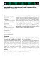
more recently x-ray. In 2008, we started an ultrasound
program using a GE LogicBook E machine provided b y
International Aid (Figure 1); this machine has an
approximately US$40,000 value. We developed protocols
for machine maintenance, appropriate use, and image
transfer to the Yale Section of Emergency Medicine for
review. The machine has been used by our physicians
and mid-level providers for both obstetric and non-
obstetric indications . Key challenges in the implementa-
tion of this program have included: a) lack of reliable
electricity, b) technical problems with the machine
without a nearby technician, c) frequent staff turnover
following training leading to gaps in utilization, and
d) damaged parts including electrical cords and plugs
requiring sourcing from an international supplier. Image
transfer has been interr upted by staff turno ver and buy-
in, and by electricity and telecommunications challenges.
While the program has faced several challenges it
continues to function today and remains an important
part of our clinical services. All protocols are publicly
available via the Nyaya Health wiki [14] and blog [8].
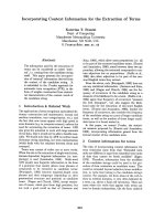
Subsequently, in 2010, Nyaya Health initiated an X-Ray
program with a WHIS-RAD machine from the Spanish
company Sedecal (Figure 2). Prior to deployment, our
team discussed and investigated options for manufacturers
and models extens ively, particularly whether to purchase
from an in-country (Nepal) or international company. We
consulted with numerous experts throughout the world,
and in fact our frustration with the current state of knowl-
edge about the scale-up of x-ray services was the primary
motivation for this piece. Ultimately we selected the
WHIS-RAD system for the reasons we have discussed
here. Implementing the x-ray program proved to be signif-
icantly more challenging than ultrasound services. Our
first challenge was delivering equipment that weighed over
750 kg to an extremely remote region with limited road
access. This was further complicated by the need to have a
technician onsite to install it; this required a team to travel
from Dehli (India) over 2 days away. Onsite challenges
included effective training of a local staff member and
developing reliable electricity to charge and operate the
WHIS-RAD system. We worked with a regional hospital
to train a mid-level provider (4 weeks) and have agreed to
a 2 year bonding period to ensure the staff member is not
drawn to an urban area where work is available and living
conditions are far better. The second major challenge
onsite was the electricity situation. Even though the
WHIS-RAD has a battery source, it requires a 110 or 220
V supply, and the main electricity grid where Nyaya works
was only supplying a degraded 170 V signal; this problem
was fixed with the addition of a relatively inexpensive vol-
tage stabilizer yet delayed implementation by several
months. Presently we are deploying analogue film
Figure 2 Nyay a Health X-Ray Installation . Note the protective
screen around the control room had yet to be installed in this
photograph.
Figure 1 Nyaya Health physician Dr. Jhapat Thapa performing
ultrasound on a pregnant patient.
Maru et al. Globalization and Health 2010, 6:18
/>Page 6 of 8
processing, though we hope to scale to digital in the
future. We have documented our work on our wiki [43]
and blog [44]. In table 3, we provide the app roximate
costs of our x-ray deployment, which we used in planning
services.
Mobilizing Radiology Services for the Rural Poor
It is feasible to achieve significant gains in access to diag-
nostic imaging. A key strategy for achieving this is to
develop a coordinated effort that can both leverage com-
panies to produce machines meeting the specifications
described here and convince donors and governments
that the endeavor is worthwhile. A survey of diagnostic
imaging services, coordinated by intergovernmental insti-
tutions such as the World Health Organization, could
help guide the financing and logistics of global diagnostic
imaging roll-out. Centralized financing, quality, and stan-
dards mechanisms such as have been achieved with the
WHO’s 3 × 5 initiative for HIV/AIDS or the Green Light
Committee for tuberculosis provide successful models
for how global radiology access could be achieved. By
collectively mobilizing resources, academic institutions,
governments, non-profit organizations, and private com-
panies can together build a global network of diagnostic
imaging services for the rural poor.
Competing and Conflicting interests
All authors have completed the Unified Competing Interest form at http://
www.icmje.org/coi_disclosure.pdf (available on request from the
corresponding author) and declare that (1) DM, RS, SB, JA have no financial
interests that may be relevant to the submitted work; (2) CM has contributed
as a paid consultant for Philips Healthcare (ultrasound division) within the last
two years. CM has contributed as a paid consultant for Sonosite, Inc.
(ultrasound manufacturer) within the last year.
Funding Sources
The authors report no funding sources for this article.
Authors’ contributions
DM conceived of the piece, drafted the initial manuscript, and read and
approved the final piece RS, JA, SB, AS, and CM provided critical comments,
edits, and literature reviews and read and approved the final piece.
Author details
1
Nyaya Health, Bayalpata Hospital, Ridikot VDC, Achham, Nepal.
2
Brigham
and Women’s Hospital, Department of Medicine, Boston, MA, USA.
3
Children’s Hospital of Boston, Department of Medicine, Boston, MA, USA.
4
Yale University School of Medicine, New Haven, CT, USA.
5
Department of
Medicine, University of California San Francisco & Division of General Internal
Medicine, San Francisco General Hospital.
6
Contra Costa Regional Health
Center, Martinez, CA, USA.
7
Department of Emergency Medicine, Yale
University School of Medicine, New Haven, CT, USA.
Received: 12 September 2010 Accepted: 14 October 2010
Published: 14 October 2010
References
1. Holm T: New Developments in basic radiographic systems. Eur J Radiol
1983, 3(1):291-293.
2. Morris MB, Chapula BT, Chi BH, Mwango A, Chi HF, Mwanza J, Manda H,
Bolton C, Pankratz DS, Stringer JS, et al: Use of task-shifting to rapidly
scale-up HIV treatment services: experiences from Lusaka, Zambia. BMC
Health Serv Res 2009, 9:5.
3. Ribeiro PS, Jacobsen KH, Mathers CD, Garcia-Moreno C: Priorities for
women’s health from the Global Burden of Disease study. Int J Gynaecol
Obstet 2008, 102(1):82-90.
4. Chen L, Kim Y, Moore CL: Diagnosis and guided reduction of forearm
fractures in children using bedside ultrasound. Pediatr Emerg Care 2007,
23(8):528-531.
5. Anomohanran O, Mokobia CE, Osakwe RA, Wawe MO: A survey of x-ray
diagnostic services in Delta State, Nigeria (1991-1994). J Radiol Prot 2002,
22(1):71-78.
6. Mettler FA, Haygood TM, Meholic AJ: Diagnostic radiology around the
world. Radiology 1990, 175(2):577-579.
7. Sungita YY: Diagnostic X-ray facilities as per quality control performances
in Tanzania. J Appl Clin Med Phys 2006, 7(4):66-73.
8. Nyaya Health. Nyaya Health Ultrasound Program Featured at
Telemedicine Symposium. [ />telemed_symposium/], Accessed August 28, 2010.
9. Palmer PES: Basic Radiological System: Manual of Radiographic
Technique. Geneva: World Health Organization 1986.
10. Cockshott WP: Diagnostic radiology: geography of a high technology.
AJR Am J Roentgenol 1979, 132(3):339-344.
11. Palmer PE: Radiology in the developing world. Br J Radiol 1984,
57(682):853-856.
12. Palmer PE: Diagnostic imaging for developing countries. WHO Chron
1985, 39(4):143-148.
13. Palmer PE: Appropriate technology for diagnostic imaging in small
hospitals. Br Med J (Clin Res Ed) 1984, 288(6428):1435-1437.
14. Nyaya Health’s Ultrasound Program. [ />UltrasoundProgram], accessed August 28, 2010.
15. Alexander JH, Peterson ED, Chen AY, Harding TM, Adams DB, Kisslo JA Jr:
Feasibility of point-of-care echocardiography by internal medicine house
staff. Am Heart J 2004, 147(3):476-481.
16. Lucas BP, Candotti C, Margeta B, Evans AT, Mba B, Baru J, Asbury JK,
Asmar A, Kumapley R, Patel M, et al: Diagnostic accuracy of hospitalist-
performed hand-carried ultrasound echocardiography after a brief
training program. J Hosp Med 2009, 4(6):340-349.
17. Martin LD, Howell EE, Ziegelstein RC, Martire C, Shapiro EP, Hellmann DB:
Hospitalist performance of cardiac hand-carried ultrasound after focused
training. Am J Med 2007, 120(11):1000-1004.
18. Martin LD, Howell EE, Ziegelstein RC, Martire C, Whiting-O’Keefe QE,
Shapiro EP, Hellmann DB: Hand-carried ultrasound performed by
hospitalists: does it improve the cardiac physical examination? Am J Med
2009, 122(1):35-41.
19. Rijken MJ, Lee SJ, Boel ME, Papageorghiou AT, Visser GH, Dwell SL,
Kennedy SH, Singhasivanon P, White NJ, Nosten F, et al: Obstetric
ultrasound scanning by local health workers in a refugee camp on the
Thai-Burmese border. Ultrasound Obstet Gynecol 2009, 34(4):
395-403.
20. Shah SP, Epino H, Bukhman G, Umulisa I, Dushimiyimana J, Reichman A,
Noble VE: Impact of the introduction of ultrasound services in a limited
resource setting: rural Rwanda 2008. BMC Int Health Hum Rights 2009, 9:4.
21. Palmer PE: Manual of Diagnostic Ultrasound. World Health Organization
1995.
22. Harris RD, Marks WM: Compact ultrasound for improving maternal and
perinatal care in low-resource settings: review of the potential benefits,
implementation challenges, and public health issues. J Ultrasound Med
2009, 28(8):1067-1076.
23. Palmer PE: The World Health Organization-Basic Radiological System.
Radiography 1985, 51(597):169-178.
24. WHO Technical Report Series #875. 1998. Training in Diagnostic
Ultrasound: Essentials, Principles, and Standards. World Health
Organization: Geneva 2009 [ />id=cL7AhS8Ru1IC], Accessed at on March 24.
25. Palmer PE, Cockshott WP: The appropriate use of diagnostic imaging.
Avoidance of the red goggle syndrome. Jama 1984, 252(19):2753-2754.
26. Holm T: X-ray apparatus specially designed for work in remote areas. Br J
Radiol 1984, 57(682):857-859.
27. Holm T: [WHO model for x-ray service is useful under Swedish primary
health care]. Lakartidningen 1985, 82(5):304-307.
Maru et al. Globalization and Health 2010, 6:18
/>Page 7 of 8
28. Palmer PE: Imaging equipment for small hospitals. Trop Geogr Med 1993,
45(3):98-102.
29. Mindel S: Role of imager in developing world. Lancet 1997,
350(9075):426-429.
30. SATCOM. Resources: Product Catalog. [ />product.do], Retrieved 3/12/08.
31. Maral Gérard : VSAT networks., Second.
32. Nepal Telecommunications Authority. [], Retrieved
3/12/08.
33. Ginde AA, Foianini A, Renner DM, Valley M, Camargo CA Jr: Availability and
quality of computed tomography and magnetic resonance imaging
equipment in U.S. emergency departments. Acad Emerg Med 2008,
15(8):780-783.
34. Partners in Health: Community health workers: Roles and functions. 2008
[ Retrieved 3/15/2008.
35. ViaSat. NewSat - Regional Broadband Services in Australia and Greater
Asia. , Retrieved 3/12/08.
36. Twinomugisha A, Aluoch S: The VSAT Buyers Guide.[ />vsat/], Retrieved 3/10/08.
37. UNICEF, Integrated Management of Childhood Illnesses (IMCI) chart
booklet. UNICEF IMCI resource. [ />health/publications/pubIMCI.htm], [Accessed March 14, 2008].
38. Sekiguchi J, Collens SR: Radiological services in rural mission hospitals in
Ghana. Bull World Health Organ 1995, 73(1):65-69.
39. Palmer PE: The technical manual and the diagnostic manual for the
basic radiological system. Diagn Imaging 1982, 51(3-4):155-165.
40. Chen L, Evans T, Anand S, Boufford JI, Brown H, Chowdhury M, Cueto M,
Dare L, Dussault G, Elzinga G, et al: Human resources for health:
overcoming the crisis. Lancet 2004, 364(9449):1984-1990.
41. Leggat PA: Basic radiological system: a case study in ‘appropriate
technology for better health’. Aust J Rural Health 1997, 5(2):87-89.
42. Nyaya Health Health Services Assessment in Five Village Development
Committee Areas Surrounding Sanfe Bagar, Achham. [http://nyayahealth.
pbwiki.com/f/HA_Achham.pdf].
43. Nyaya Health’s X-Ray Program. [ />accessed August 28, 2010.
44. Nyaya Health’s X-Ray Program Blog Posts. [ />s=x-ray], accessed August 28, 2010.
doi:10.1186/1744-8603-6-18
Cite this article as: Maru et al.: Turning a blind eye: the mobilization of
radiology services in resource-poor regions. Globalization and Health
2010 6:18.
Submit your next manuscript to BioMed Central
and take full advantage of:
• Convenient online submission
• Thorough peer review
• No space constraints or color figure charges
• Immediate publication on acceptance
• Inclusion in PubMed, CAS, Scopus and Google Scholar
• Research which is freely available for redistribution
Submit your manuscript at
www.biomedcentral.com/submit
Maru et al. Globalization and Health 2010, 6:18
/>Page 8 of 8