Manual Endourology - part 5 docx
Bạn đang xem bản rút gọn của tài liệu. Xem và tải ngay bản đầy đủ của tài liệu tại đây (391.16 KB, 12 trang )
Chapter 5 · Pediatric Endoscopy
5
45
E
⊡
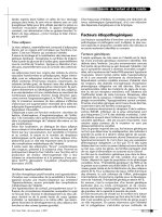
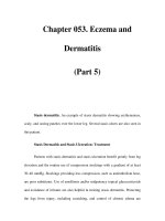
Fig. 5.10E–G. Endoscopic view after 2 months; self-
limited process of the bladder (E). Endoscopic view, urete-
ral orifice, right side (F). Ureteral groin after healing (G)
F
G
Hohenfellner_L4F-2sp.indd 45Hohenfellner_L4F-2sp.indd 45 23.06.2005 17:56:1123.06.2005 17:56:11
6
Laparoscopy for the Undescended Testicle
Ulrich Humke, Stefan Siemer, Roland Bonfig, Mark Koen
Introduction – 48
Patient Counselling and Consent – 48
Preoperative Preparation – 48
Anaesthesia – 48
Indication – 48
Limitations and Risks – 48
Contraindications – 48
Special Instruments – 48
Operative Technique (Step-by-Step) – 49
Tips and Tricks – 50
Postoperative Care – 51
Complications – 51
Do’s – 51
Dont’s – 51
References – 51
Image Gallery – 52
Hohenfellner_L4F-2sp.indd 47Hohenfellner_L4F-2sp.indd 47 23.06.2005 17:56:1323.06.2005 17:56:13
Introduction
Cryptorchidism is a frequent diagnosis in ped-
iatric urology and a well-known risk for male
infertility and testicular malignancy. About 20%
of undescended testicles are not palpable. Alt-
hough the mean age of children presented for
therapy with cryptorchidism is above 3 years,
the ideal time-point for effective preservation
of fertility is between 12 and 24 months of life.
Laparoscopy has evolved in the past years as the
method of choice for the diagnosis and treat-
ment of non-palpable testes. Clear advantages
of laparoscopy with regard to specificity and
sensitivity have been shown compared to ultra-
sonography and magnetic resonance imaging in
detecting intra-abdominal testes. The purposes
of laparoscopy for non-palpable testes are (a)
localization and evaluation of the missing testis,
(b) orchiopexy (one- or two-stage procedure)
and (c) orchiectomy (if indicated), each selec-
ted alone or in combination for the individual
case.
Patient Counselling and Consent
▬ Risk of vascular or intestinal injury during
primary trocar placement.
▬ Risk of hernia formation at the trocar site
postoperatively (depends on trocar size).
▬ Eventually intraoperative need for conversi-
on to open conventional surgery.
Preoperative Preparation
▬ Beta-HCG stimulation test only in case of
bilateral non-palpable testes.
▬ Standard bowel preparation.
Anaesthesia
▬ General anaesthesia.
Indications
▬ All cases of non-palpable testes: integrated
concept of diagnostic laparoscopy combined
with open surgery (revision of inguinal canal,
with or without orchiopexy) or combined with
therapeutic laparoscopy (staged orchiopexy
or orchiectomy for intra-abdominal testes).
▬ Suspected intersex (laparoscopy for diagno-
sis, eventually biopsy and/or orchiectomy).
Limitations and Risks
▬ Smaller body size in children implies smal-
ler space tolerances of the abdominal wall,
which makes standard trocar placement
more dangerous.
▬ Looser attachments of the peritoneum to the
extraperitoneal structures in children make
trocar penetration more difficult.
▬ A dull trocar is a potentially dangerous
instrument in children.
Contraindications
▬ Acute infectious disease.
▬ Coagulopathy.
▬ Prior abdominal surgery with suspected
adhesions.
Special Instruments
▬ Laparoscopy unit (video cart) with insuffla-
tor, light source, video camera, video moni-
tor, video recorder and electrocautery unit.
▬ Veress cannu la.
▬ Mini-laparoscope (1.9 mm) with 2.7-mm
trocar shaft, for older children 3.5 or 5-mm
laparoscopes.
▬ 3.5-mm trocars and laparoscopic forceps/
graspers/scissors for dissection, for older
children 5-mm trocars and instruments.
▬ 5- or 10-mm clipping instruments.
48 Chapter 6 · Laparoscopy for the Undescended Testicle
6
Hohenfellner_L4F-2sp.indd 48Hohenfellner_L4F-2sp.indd 48 23.06.2005 17:56:1323.06.2005 17:56:13
Operative Technique (Step-by-Step)
Placement and Removal of Trocars
▬ Supine and 10° head-down position of the
patient.
▬ Gastric tube and bladder catheter in place.
▬ Small infraumbilical skin incision reaching
the fascia.
▬ Elevation of the abdominal wall by lifting up
a skin fold or two forceps-clamps on both
sides of the umbilicus.
▬ Intraperitoneal insertion of the Veress can-
nula covered with mini-trocar (mini-laparo-
scopy set): vertical direction of puncture.
▬ Replacement of Veress cannula with mini-
telescope.
▬ Optical control of correct intraperitoneal
position of laparoscope.
▬ Thereafter start of CO
2
insufflation and crea-
tion of pneumoperitoneum (maximum pres-
sure, 12 mmHg).
▬ Inspection of peritoneal cavity and anatomi-
cal landmarks, exclusion of puncture related
iatrogenic injuries.
▬ Alternative access method: Hasson techni-
que for trocar insertion (preferred by many
pediatric urologists): Dissection and incision
of fascia and peritoneum with scissors under
direct vision. After opening of the peritoneal
cavity insertion of the trocar and fixation
with suture.
▬ Remove trocars under laparoscopic view to
exclude bleeding from the trocar canal.
▬ Remove intraperitoneal gas through the last
trocar as completely as possible, slightly com-
press the lower thoracic aperture to mobilize
gas from the upper peritoneal cavity, extract
last trocar.
▬ Close fascia with single sutures at 10-mm
trocar sites, close all skin incisions with sing-
le sutures.
Diagnostic Laparoscopy
▬ Identify anatomical landmarks: bladder
(catheter balloon visible) and urachal liga-
ment, lateral umbilical ligament, inferior
epigastric vessels, inner inguinal ring, vas
deferens, spermatic vessels.
▬ Check anatomical status relevant for cryptor-
chidism:
▬ Inner inguinal canal open (open proces-
sus vaginalis) or closed?
▬ Spermatic vessels and/or vas deferens
present, passing into the inguinal canal
or ending cranially?
▬ Testicle intra-abdominal?
▬ Testicle visible in the inguinal canal?
▬ Testicle volume? Epididymal configura-
tion?
▬ Classify anatomical findings into three thera-
peutic relevant categories:
1. All spermatic cord structures are pre-
sent and leave into the inguinal canal
(frequent condition): stop laparoscopy
and proceed with open surgery: revision
of the inguinal canal, closure of open
processus vaginalis, excision of atrophic
testicle or rudimentary testicular structu-
res (vanishing testis), alternatively orchi-
opexy of inguinal testicle.
2. Spermatic vessels and vas deferens can
be identified. They end blindly on the
psoas muscle without any testis detec-
table (vanishing testis, anorchia: rare
condition): stop laparoscopy, no further
surgery.
3. Intra-abdominal testicle present with or
without open inguinal canal (frequent
condition): proceed with laparoscopic
orchiectomy, if testicle appears small and
atrophic. Proceed with laparoscopic orchi-
opexy (one-stage procedure if testicle has
a maximal distance to the inner inguinal
ring of 2 cm) or clipping of spermatic ves-
sels as first step of two-stage orchiopexy
(Fowler Stephens manoeuvres I and II).
Chapter 6 · Laparoscopy for the Undescended Testicle
6
49
Hohenfellner_L4F-2sp.indd 49Hohenfellner_L4F-2sp.indd 49 23.06.2005 17:56:1323.06.2005 17:56:13
Primary Orchiopexy
(One-Stage Procedure)
▬ Incise retroperitoneum with a minimal 1-cm
margin laterally to the testicle and medially
alongside the vas deferens.
▬ Mobilize peritoneum carefully across sper-
matic vessels.
▬ Leave all vessels around the vas deferens and
the peritoneal plane between vas and vessels
intact. Try to avoid electrocautery as much as
possible.
▬ Mobilize the testicle carefully from the psoas
fascia towards the inguinal ring.
▬ Create new internal ring medially to the epi-
gastric vessels (shortens the overall distance
to the scrotal position).
▬ Make an incision at the lower pole of the
scrotum and provide a dartos pouch. Insert
a laparoscopic grasper, guide it through a
tunnel to the new inguinal ring, take the
mobilized testicle and pull it into the scro-
tum without forced tension.
Fowler Stephens Step I
(Clipping of Spermatic Vessels)
▬ Incise retroperitoneum bilaterally parallel to
the spermatic vessel, minimum 2 cm cranial-
ly to the upper pole of the testicle.
▬ Mobilize spermatic vessels, hold them up
with a grasper and apply two absorbable clips
without dividing them.
Fowler Stephens Step II
(Secondary Orchiopexy)
▬ Plan this procedure not before 6 months
after the first step.
▬ Dissect the clipped area of the spermatic
vessels and divide them.
▬ Incise retroperitoneum with a minimal
1-cm margin laterally to the testicle and
medially alongside the vas deferens. The
peritoneal flap remains pedicled to the vas
deferens.
▬ Leave all vessels around the vas deferens and
the peritoneal plane between vas and vessels
intact. Try to avoid electrocautery as much as
possible.
▬ Dissect gubernaculum as far distally as pos-
sible.
▬ Mobilize the testicle carefully from the psoas
fascia towards the inguinal ring.
▬ Create new internal ring medially to the epi-
gastric vessels.
▬ Make an incision at the lower pole of the
scrotum and provide a dartos pouch. Insert a
laparoscopic grasper, guide it through a tun-
nel to the new inguinal ring, take the mobili-
zed testicle and pull it into the scrotum.
Orchiectomy
▬ Indicated for small, atrophic intra-abdomi-
nal testicles.
▬ Incise retroperitoneum and dissect spermatic
vessels after clipping cranially.
▬ Mobilize testicle and vas deferens.
▬ Dissect vas deferens after coagulation.
▬ Free the testicle from remaining peritoneal
adhesions and extract it via an 5- or 10-mm
trocar with a strong grasper.
Tips and Tricks
▬ Start laparoscopy in children with mini-lapa-
roscope: risk of initial trocar injury minimi-
zed, sufficient for diagnostic purpose, change
to bigger trocars for further therapeutic lapa-
roscopy easily and safely possible.
▬ Apply gastric tube and bladder catheter
before start of operation to minimize risk of
organ injury during initial puncture of the
abdomen.
▬ Insert working trocars always under optical
guidance.
▬ Prevention of a foggy laparoscope: warm the
instrument moderately before use, clean it
intraoperatively by sweeping smoothly along
a peritoneal/intestinal surface.
▬ Remove trocars under endoscopic vision to
control bleeding.
50 Chapter 6 · Laparoscopy for the Undescended Testicle
6
Hohenfellner_L4F-2sp.indd 50Hohenfellner_L4F-2sp.indd 50 23.06.2005 17:56:1323.06.2005 17:56:13
▬ Use absorbable sutures for closure of skin
incision.
▬ Have instruments for open surgery available
in the operating room for emergency cases.
Postoperative Care
▬ Appropriate analgesia.
▬ Start of oral feeding 6 h after anaesthesia.
▬ Start of mobilization according to the child’s
activity, except after orchiopexy of an intra-
abdominal testis (bed rest minimum 24 h).
▬ Perform Duplex-sonography postoperatively
to control testicular perfusion.
▬ Give oral antiphlogistic medication to limit
postoperative swelling if necessary.
Complications
▬ Intestinal injury during initial blind trocar
placement: obvious intestinal injury has to
be revised and treated by open surgery.
▬ Vascular injury during initial blind trocar
placement: obvious vascular injury has to
be treated by immediate conversion to open
surgery.
▬ Ureteral injury during careless mobilization
of intra-abdominal testis.
▬ After orchiopexy:
▬ Loss of scrotal position due to excessive
tension.
▬ Testicular atrophy due to vascular mal-
perfusion.
Do’s
▬ Do primary one-stage orchiopexy if the
testicle is located close to the inner inguinal
ring (maximum 2 cm distance) and sper-
matic vessels appear mobile and elastic.
▬ Perform two-stage procedure if testicle is
located proximally and spermatic vessels are
too short for a one-stage procedure.
▬ Do Fowler-Stephens I laparoscopically.
▬ Do Fowler-Stephens II orchiopexy optionally
as open surgery from a small suprainguinal
incision.
Dont’s
▬ Do not perform orchiopexy under forced
tension. This will reduce testicular perfusion
and provokes retraction of testicle.
▬ Avoid torsion of the vascular/peritoneal
pedicle while pulling the testicle through the
new inguinal canal.
References
1. Lindgren BW, Franco I, Blick S, Levitt SB, Brock WA,
Palmer LS et al (1999) Laparoscopic Fowler-Stephens
orchidopexy for the high abdominal testis. J Urol
162:990–993; discussion: 994
2. Law GS, Pérez LM, Joseph DB (1997) Two-stage Fow-
ler-Stephens orchidopexy with laparoscopic clipping
of the spermatic vessels. J Urol 158:1205–1207
3. Radmayr C, Oswald J, Schwentner C, Neururer R,
Peschel R, Bartsch G (2003) Long-term outcome of
laparoscopically managed nonpalpable testes. J Urol
170:2409–2411
4. Peters CA (2004) Laparoscopy in pediatric urology.
Curr Opin Urol 14:67–73
Chapter 6 · Laparoscopy for the Undescended Testicle
6
51
Hohenfellner_L4F-2sp.indd 51Hohenfellner_L4F-2sp.indd 51 23.06.2005 17:56:1323.06.2005 17:56:13
52 Chapter 6 · Laparoscopy for the Undescended Testicle
6
Image Gallery
⊡ Fig. 6.1. Mini-laparoscopic instruments
with Veress cannula, mini-trocar and
mini-telescope (diameter of 1.9, 2.7 and
1.9 mm, respectively) for use in children
Verres canula
Trocar
Telescope
⊡ Fig. 6.2. Small, infraumbilical incision
under elevation of the periumbilical skin.
Through the incision, the abdomen may
be directly punctured with the Veress
cannula (classical approach)
⊡ Fig. 6.3. Alternatively, for safety
reasons, the peritoneum is dissected and
incised under direct vision before the tro-
car is inserted directly into the abdominal
cavity (Hasson technique)
peritoneum
Hohenfellner_L4F-2sp.indd 52Hohenfellner_L4F-2sp.indd 52 23.06.2005 17:56:1323.06.2005 17:56:13
Chapter 6 · Laparoscopy for the Undescended Testicle
6
53
⊡ Fig. 6.4. Normal, closed right inner
inguinal ring. Spermatic vessels and
vas deferens join each other in an inverse
V-shape before entering the inguinal
canal. In this case of nonpalpable right
testis, surgery proceeds with open ingui-
nal exploration
abdominal wall
right abdominal
inguinal ring
spermatic cord
bowel
⊡ Fig. 6.6. Left inner inguinal ring with
normal-sized intra-abdominal testis dis-
tally located on the external iliac vessels.
Surgery proceeds with one-stage open or
laparoscopic orchiopexy
abdominal wall
left abdominal inguinal ring
abdominal testicle
bowel
vas deferens
⊡ Fig. 6.5. Open right inner inguinal ring
with spermatic vessels and vas deferens
entering the open inguinal canal. In this
case of nonpalpable right testis, surgery
proceeds with open inguinal exploration
open inner inguinal ring
spermatic
vessels
vas deferens
Hohenfellner_L4F-2sp.indd 53Hohenfellner_L4F-2sp.indd 53 23.06.2005 17:56:1723.06.2005 17:56:17
⊡ Fig. 6.7. Intraoperative situation during
open orchiopexy of left distal intra-
abdominal testis (see
⊡ Fig. 6.6). Note
the Prentiss manoeuvre (testicle and
spermatic cord pass under the mobilized
inferior epigastric vessels to gain length
for tension-free orchiopexy)
cranial
peritoneal flap
testicle
spermatic cord
caudal
a. and v. epigastrica
54 Chapter 6 · Laparoscopy for the Undescended Testicle
6
⊡ Fig. 6.8. Intra-abdominal right testicu-
lar aplasia: blind-ending spermatic ves-
sels and blind-ending vas deferens. No
further surgery needs to be performed
blind ending vas deferens
blind ending
spermatic vessels
Hohenfellner_L4F-2sp.indd 54Hohenfellner_L4F-2sp.indd 54 23.06.2005 17:56:2123.06.2005 17:56:21
7
Transurethral Resection of Bladder Tumours
Armin Pycha, Salvatore Palermo
Introduction – 56
Indications – 56
Contraindications – 56
Preoperative Preparation – 56
Anaesthesia – 56
Instruments – 56
Patient Positioning – 57
Operative Technique (Step by Step) – 57
Resection Procedure according to Nesbit (1943) – 57
En Bloc Resection according to Mauermayer (1981) – 58
Bladder Mapping – 58
Before Finishing TUR-B – 58
After Finishing TUR-B – 59
Postoperative Care – 59
Common Complications – 59
Trouble-shooting – 59
Postoperative Complications – 60
New Developments – 60
Comments – 60
Remember – 60
Do’s – 60
Dont’s – 61
References – 61
Check – List – 62
Operation Report – 63
Image Gallery – 64
Hohenfellner_L4F-2sp.indd 55Hohenfellner_L4F-2sp.indd 55 23.06.2005 17:56:2423.06.2005 17:56:24
Introduction
As the bladder tumour is the second most com-
mon tumour of the genitourinary system, the
transurethral resection (TUR) is an intervention,
which is often performed [1]. At first manifesta-
tion, 70%–75% of bladder tumours are superfi-
cial and well differentiated. The recurrence rate
is 70% and out of these 6%–10% show a progres-
sion with an eventual lethal outcome.
The TUR of bladder tumours (TUR-B) has a
double goal: first the total removal of papillary
lesions; second to determine the depth of invasi-
on or clinical stage [1].
TUR-B is often the first step for residents in
their endourological training. From the techni-
cal point of view, new developments for video
systems, optics, electrosurgical instruments and
high-frequency (HF) generators facilitate TUR-
B procedures. Nevertheless, TUR-B is burdened
with a significant number of complications.
Indications
Any suspicious area in the bladder.
Contraindications
▬ Absolute contraindications for programmab-
le TUR-B are uncorrected coagulopathy and
active urinary tract infection.
In case of severe bleeding of bladder tumours,
there is a vital indication for TUR-B. At the same
time, the coagulopathy must be corrected by the
haematologist.
▬ Relative contraindications: anaesthetic cont-
raindications.
Preoperative Preparation
▬ Stop aspirin 1 week before operation.
▬ Rule out and treat any urinary tract infection
by urine culture and sensitivity.
▬ Thrombosis prophylaxis commencing the
evening before the operation (low-molecu-
lar-weight heparin).
▬ Rectal enema is used the day before the ope-
ration.
▬ Intravenous single-dose antibiotics at induc-
tion.
▬ Counseling and informed consent.
Anaesthesia
▬ General anaesthesia with muscle relaxation.
▬ Spinal anaesthesia.
Instruments
All instruments (1–17) used are from Karl Storz,
Tuttlingen, Germany.
▬ Latest-generation electrosurgical generator
(1)
▬ Digital video camera controller IMAGE1 (2)
with 3-CCD digital pendulum camera head
IMAGE1 P3 (3).
▬ 18" TFT-flat screen monitor with digital SDI
input (4).
▬ High-intensity 300-W Xenon light source
(5).
▬ Hopkins II Telescope 0° (6), 30° (7), and 70°
(8).
▬ Working element, passive (9).
▬ Resectoscope sheath 24-Fr single flow with
central valve (10) or resectoscope sheath
26-Fr, continuous flow, rotatable (11) visual
obturator (12).
▬ HF resection electrodes:
▬ standard vertical loop (13).
▬ Straight (longitudinal) loop (14).
56 Chapter 7 · Transurethral Resection of Bladder Tumours
7
Hohenfellner_L4F-2sp.indd 56Hohenfellner_L4F-2sp.indd 56 23.06.2005 17:56:2423.06.2005 17:56:24
▬ Roller ball electrode for coagulation,
3 mm in diameter (15).
▬ HF biopsy forceps (16).
▬ 100-ml bladder syringe (17).
▬ 18-Fr irrigation catheter.
▬ Lubricant (Instillagel®, Farco Pharma, Ger-
many).
▬ Electrolyte-free, sterile, and isotonic irrigati-
on fluid, positioned at a height of 50–60 cm
above the pubic symphysis.
Patient Positioning
▬ Lithotomy position.
▬ The thighs must be bent at an angle of 90°
from the hip to guarantee the resectionist
enough manoeuvrability.
▬ The gluteal muscles must be exactly at the
edge of the operating table.
Run through the check-list before starting the
operation.
Operative Technique (Step by Step)
▬ White balance of the video camera.
▬ Adjustment of the video zoom and focus.
▬ Enter the bladder with a visual obturator and
check the urethra.
▬ Perform a first inspection of the bladder fol-
lowing a strict protocol and compare these
findings with the records of the outpatient
clinic.
▬ Assertion of the number of lesions.
▬ Resectoscope working element with a 30°
telescope is introduced.
In reliance on localization and extensions of
the tumor, different resection techniques can
be used.
Resection Procedure according
to Nesbit (1943)
▬ The bladder is filled to half of the maximum
capacity (use of continuous-flow resectos-
cope facilitates the maintenance of optimal
bladder filling).
▬ Resection starts at the lateral border of the
tumour.
▬ String one loop strip after another in a hori-
zontal plane.
▬ On completion of one plane, the next deeper
plane follows.
▬ Resect until healthy tissue is reached.
▬ Small tumours can be cut at the level of the
pedicle, then the specimen is evacuated by
bladder washing.
▬ Thereafter, a loop-strip of the residual pedic-
le and the underlying submucosa and detru-
sor is taken and sent separately to histology.
▬ Bladder evacuation with a 100-ml syringe.
▬ Meticulous coagulation using a roller ball
electrode.
Limits
▬ Tumours on the bladder dome are technical-
ly difficult to manage using this technique.
▬ The identification of tumor base and higher
tumor planes can create problems.
▬ The loop-strips on the bottom normally
show severe fulguration artefacts compromi-
sing the histological evaluability.
Risks
▬ Often clear staging is not feasible.
▬ Exact evaluation of resection borders is often
difficult and sometimes speculative.
Tricks
▬ Resection should proceed with partially dis-
tended bladder.
▬ Take care to follow the curve of the bladder
when resecting to avoid perforation.
▬ Treat easily accessible and small tumours
first.
Chapter 7 · Transurethral Resection of Bladder Tumours
7
57
Hohenfellner_L4F-2sp.indd 57Hohenfellner_L4F-2sp.indd 57 23.06.2005 17:56:2423.06.2005 17:56:24