7Microbial Enzymes in the Biocontrol of Plant Pathogens and aEnzymes in the Environment: Activity, Ecology and Applications - Chapter 7PestsLeonid Chernin and Ilan ChetThe ppt
Bạn đang xem bản rút gọn của tài liệu. Xem và tải ngay bản đầy đủ của tài liệu tại đây (518.83 KB, 56 trang )
7
Microbial Enzymes in the Biocontrol
of Plant Pathogens and Pests
Leonid Chernin and Ilan Chet
The Hebrew University of Jerusalem, Rehovot, Israel
I. INTRODUCTION
Despite many achievements in modern agriculture, food crop production continues to be
plagued by disease-causing pathogens and pests. In many cases, chemical pesticides effec-
tively protect plants from these pathogens. However, public concerns about harmful effects
of chemical pesticides on the environment and human health have prompted a search
for safer, environmentally friendly control alternatives (1–3). One promising approach is
biological control that uses microorganisms capable of attacking or suppressing pathogens
and pests in order to reduce disease injury. Biological control of plant pathogens offers
a potential means of overcoming ecological problems induced by pesticides. It is an eco-
logical approach based on the natural interactions of organisms with the use of one or more
biological organisms to control the pathogen. Generally, biological control uses specific
microorganisms that attack or interfere with specific pathogens and pests. Because of their
specificity, different microbial biocontrol agents typically are needed to control different
pathogens and pests, or the same ones in different environments.
Agriculture benefits, and is dependent on, the resident communities of microorgan-
isms for naturally occurring biological control, but additional benefits can be achieved by
introducing specific ones when and where they are needed (4–9). Many agrochemical
and biotechnological companies throughout the world are increasing their interest and
investment in the biological control of plant diseases and pests. For plant pathogens alone,
the current list of microbial antagonists available for use in commercial disease biocontrol
includes around 40 preparations (9–11). These are all based on the practical application
of seven species of bacteria (Agrobacterium radiobacter, Bacillus subtilis, Burkholderia
cepacia, Pseudomonas fluorescens, Pseudomonas syringae, Streptomyces griseoviridis,
Streptomyces lydicus) and more than 10 species of fungi (Ampelomyces quisqualis, Can-
dida oleophila, Coniothyrium minitans, Fusarium oxysporum, Gliocladium virens, Phlebia
gigantea, Pythium oligandrum, Trichoderma harzianum, and other Trichoderma species).
The current market for biological agents is estimated at only $500 million, which is about
1% of the world’s total output for crop protection. The largest share of this market involves
biopesticides marketed for insect control (mainly products based on Bacillus thuringiensis
Copyright © 2002 Marcel Dekker, Inc.
strains that produce a protein toxin with strong insecticidal activity), and these bioinsecti-
cides represent around 4.5% of the world’s insecticide sales. Other agents used for bio-
control exist on a much smaller scale commercially. However, the biopesticides market
is expected to grow over the next 10 years at a rate of 10% to 15% per annum, vs. 1%
to 2% for chemical pesticides (12).
Several modes of action have been identified in microbial biocontrol agents, no two
of which are mutually exclusive. Biological control may be achieved by both direct and
indirect strategies. Indirect strategies include the use of organic soil amendments and com-
posts, which enhance the activity of indigenous microbial antagonists against a specific
pathogen (13), and the use of indirect modes of the microbial-biocontrol-agent action.
These include two main mechanisms. One is cross-protection, which involves the activa-
tion of physical and chemical self-defense responses (induced resistance) within the host
plant against a particular pathogen by prior inoculation of the plant with a nonvirulent
strain of that pathogen, resulting in partial or complete resistance to a variety of diseases
in several types of plants (14,15). The other is plant growth promotion by root-colonizing
bacteria and fungi that are able to stimulate plant growth and development; some of these
also are capable of inducing resistance (16–18).
The direct approach involves the introduction of specific microbial antagonists into
the soil or plant material. These antagonists need to proliferate and establish themselves
in the appropriate ecological niche in order to be active against a pathogen or a pest. A
beneficial organism used to protect plants is referred to as a biological control agent (BCA)
or, often, as an antagonist, because it interferes with the target organisms that damage the
plant. Antagonists generally are naturally occurring, mostly soil microorganisms with
some trait or characteristic that enables them to interfere with pathogen or pest growth,
survival, infection, or plant attack. Usually they have little effect on other soil organisms,
leaving the natural biological characteristics of the ecosystem more balanced and intact
than would a broad-spectrum chemical pesticide. Some BCAs have been modified geneti-
cally to enhance their biocontrol capabilities or other desirable characteristics.
There are four general direct mechanisms of biological control of plant diseases.
The first is competition with the pathogen for limited resources such as nutrients or space.
Antagonists capable of more efficiently utilizing essential resources (e.g., carbon, nitrogen,
volatile organic materials, plant residues, iron, microelements) effectively compete with
the pathogen for an ecological niche and colonization of the rhizosphere and/or phyllo-
sphere, leaving the pathogen less able to grow in the soil or to colonize the plant. Many
plant pathogens require exogenous nutrients to germinate, then penetrate and infect host
tissue successfully. Therefore, competition for limiting nutritional factors, mainly carbon,
nitrogen, and iron, may result in the biological control of plant pathogens (19,20).
The second mechanism is antibiosis, which is the inhibition or destruction of the
pathogen by a metabolic product of the antagonist. That is, the antagonist produces some
compound that is toxic or inhibitory to the pathogen, resulting in destruction of the latter’s
propagules or suppression of its activity. Antibiosis is restricted for the most part to those
interactions that involve low-molecular-weight diffusible compounds (e.g., antibiotics or
siderophores) produced by a microorganism that inhibit the growth of another microorgan-
ism (21–26). However, this definition excludes proteins or enzymes that kill the target
organism. Hence, Baker and Griffin (19) extended its scope to ‘‘inhibition or destruction
of an organism by the metabolic production of another,’’ thereby including small toxic
molecules, and volatile and lytic enzymes. The impact of antibiosis on biological control
under greenhouse and field conditions is still uncertain. Even in cases in which anti fungal
Copyright © 2002 Marcel Dekker, Inc.
metabolite production by an agent reduces disease, other mechanisms also may be op-
erating.
Hypovirulence is another mechanism that reduces virulence in some pathogenic
strains. Some natural- or laboratory-source hypovirulent strains were able to reduce the
effect of the virulent ones. Hypovirulent strains of Cryphonectria parasitica, Fusarium
spp., Rhizoctonia solani, Sclerotinia homoeocarpa, and others have been used as biocon-
trol agents of chestnut blight, wilt, rots, and other fungal diseases caused by the wild type
of these pathogens. Some of these hypovirulent strains contain a single cytoplasmic ele-
ment of double-stranded ribonucleic acid (dsRNA), which can be introduced into virulent
strains by deoxyribonucleic acid– (DNA)-mediated transformation. This may be consid-
ered a specialized form of cross-protection that is limited to the control of only established
compatible strains (27–29).
The fourth mechanism is predation/parasitism, which occurs when the BCA feeds
directly on or inside the pathogen. In this case, the antagonist is a predator or parasite of
the pathogen. When one fungus feeds on another fungus, generally it is called mycoparasi-
tism. This process results in the direct destruction of pathogen propagules or structures
(30–35).
All known BCAs utilize one or more of these general indirect or direct mechanisms.
At the product level, this includes the production of antibiotics, siderophores, and cell wall
lytic enzymes, and the production of substances that promote plant growth. Additionally,
successful colonization of the root surface is considered a key property of prospective
antagonists (9). The most effective BCAs use two or three different mechanisms. Antago-
nists also can be combined to provide multiple mechanisms of action against one or more
pathogens. An understanding of this mechanism of action is important because it provides
a wealth of information that can be useful in determining how to maintain, enhance, and
implement this form of biological control.
Numerous comprehensive reviews on specialized topics, as well as proceedings and
books describing the biocontrol activities of different microorganisms against plant patho-
gens and pests in laboratories, greenhouses, and the field, appeared in the late 1990s
(9,10,34,36–41). However, the biological control of plant diseases is not as well estab-
lished as biocontrol of insects in commercial agriculture. The latter has been a successful
approach for decades and continues to be a rapidly developing area of research. In this
chapter, we limit our discussion to enzymatic mechanisms of microbial control of plant
pathogens and pests.
II. THE ROLE OF FUNGAL ENZYMES IN THE BIOLOGICAL
CONTROL OF PLANT DISEASES
A. Gliocladium and Trichoderma Species Systems
The fungus Gliocladium virens Miller, Giddens and Foster (ϭTrichoderma virens, Miller,
Giddens, Foster, and von Ark) is a common soil saprophyte and one of the most promising
and studied fungal biocontrol agents. It originally was isolated from a sclerotium of the
plant pathogenic fungus Sclerotinia minor and then was found to be active against several
fungal plant pathogens. Trichoderma, a genus of hyphomycetes that is an anamorphic
Hypocreaceae (class Ascomycetes), also is common in the environment, especially in
soils. Many Gliocladium and Trichoderma spp. isolates obtained from natural habitats
have been used in biocontrol trials against several soil-borne plant pathogenic fungi under
Copyright © 2002 Marcel Dekker, Inc.
both greenhouse and field conditions. In particular, isolates of G. virens, G. roseum, T.
viride, T. harzianum Rafai, and T. hamatum have been reported to be antagonists of phyto-
pathogenic fungi, including Botrytis cinerea, Fusarium spp., Phytophthora cactorum,
Pythium ultimum, Pythium aphanidermatum, Rhizoctonia solani, Sclerotinia sclerotiorum,
and Sclerotium rolfsii. These cause soil-borne and foliage diseases in a wide variety of
economically important crops in a range of environmental conditions.
The antagonists kill the host by direct hyphal contact, causing the affected cells to
collapse or disintegrate; vegetative hyphae of all species have been found susceptible. The
biological and ecological characteristics and potential of these closely related genera
for the biological control of plant pathogens have been reviewed extensively
(4,9,31,34,35,42–48).
Among the biocontrol mechanisms proposed for Gliocladium and Trichoderma spp.
are competition, antibiosis, and mycoparasitism. The last mechanism is based mainly
on the activity of lytic exoenzymes (chitinases, glucanases, cellulases, and proteases) re-
sponsible for partial degradation of the host cell wall. Barnett and Binder (30) divide
mycoparasitism into necrotrophic (destructive) parasitism, which results in death and de-
struction of the host fungus, and biotrophic (balanced) parasitism, in which the develop-
ment of the parasite is favored by a living host structure. The sequential events involved in
mycoparasitism have been described in several comprehensive reviews (31–35). Briefly,
mycoparasitism is a complex process that involves ‘‘recognition’’ of the host, positive
chemotropic growth, attachment, and de novo synthesis of a set of cell-wall-degrading
enzymes that aid the parasite in penetrating the host and completing its destruction. Lec-
tins, the sugar-binding proteins or glycoproteins of nonimmune origin, which agglutinate
cells and are involved in interactions between the cell surface components and its extracel-
lular environment, have been shown to play a role in the recognition and contact between
necrotrophic mycoparasites of Gliocladium and Trichoderma spp. and soil-borne patho-
genic fungi. This contact, in turn, initiates a signal transduction cascade toward the second,
most important step of mycoparasitism, the induction of lytic enzymes able to degrade
fungal cell walls.
Most fungi attacked by Gliocladium and Trichoderma spp. have cell walls that con-
tain chitin as a structural backbone and laminarin (β-1,3-glucan) as a filling material. The
other minor cell wall components are proteins and lipids. The ability to produce lytic
enzymes has been shown to be a crucial property of these and other mycoparasitic fungi.
Several contemporary reviews discuss the role of, in particular, chitinolytic enzymes of
Trichoderma spp. in fungal mycoparasitism and biocontrol activity (33,49–51). In the last
few years, the enzymatic patterns of various strains of Trichoderma and Gliocladium spp.
have been determined, the corresponding genes cloned, and their products characterized.
Some of these enzymes have been studied in more detail, with the goal of understanding
their role in fungal biocontrol activity and principles of their expression regulation. In
general, fungal cell-wall-degrading enzymes produced by G. virens and Trichoderma spp.
are strong inhibitors of spore germination and hyphal elongation in a number of phyto-
pathogenic fungi. The excretion of lytic enzymes enables Trichoderma spp. to degrade
the target fungal cell wall and utilize its nutrients (52–55).
A considerable amount of recent research has been devoted to studying the indi-
vidual lytic systems produced by Trichoderma spp. Most of the studies on the expres-
sion and regulation of these lytic enzymes have been performed in liquid cultures supple-
mented with different C sources (e.g., chitin, glucose, β-1,4-linked N-acetylglucosamine
[GlcNAc], fungal cell walls) and their antifungal effects determined in vitro. These growth
Copyright © 2002 Marcel Dekker, Inc.
conditions facilitated the identification of the lytic enzymes induced in Trichoderma spp.
to hydrolyze the polymers constituting the fungal cell walls. However, they did not reflect
the exact conditions existing during the antagonistic interactions between Trichoderma
spp. and its hosts. Thus, using T. harzianum–R. solani and T. harzianum–S. rolfsii interac-
tions as model systems, Elad et al. (52) revealed lysed sites and penetration holes in the
hyphae of the host fungus caused by the antagonist’s attachment and coiling around it
(Fig. 1). In the presence of the protein synthesis inhibitor cycloheximide, antagonism was
prevented and enzymatic activity reduced. These observations suggested that the lytic
enzymes whose synthesis de novo was induced as a result of early stages of interaction
with the target phytopathogen excreted by Trichoderma spp. degrade R. solani and S.
rolfsii cell walls at the interaction sites. According to more recent data obtained by electron
microscopy of the interaction between T. harzianum and the arbuscular mycorrhizal fungus
Glomus intraradices, chitinolytic degradation was seen only in areas adjacent to the sites
of Trichoderma spp. penetration. The interaction between T. harzianum and G. intrara-
dices involves the following events: (i) recognition and local penetration of the antagonist
into mycorrhizal spores, (ii) active proliferation of antagonist cells in mycorrhizal hyphae,
and (iii) release of the antagonist through moribund hyphal cells (56).
1. Chitinolytic Enzymes
Chitin, an unbranched insoluble homopolymer consisting of GlcNAc units, is the second
(after cellulose) most common biodegradable polysaccharide in nature, being the main
structural component of cell walls of most fungi and arthropods (insects, nematodes, and
other invertebrates) including many agricultural pests (57–59). Many species of bacteria,
streptomycetes and other actinomycetes, fungi, and plants produce chitinolytic enzymes
that catalyze the hydrolysis of chitin. Chitinases produced by various microbes differ con-
siderably in their molecular masses, high-temperature optima, and degrees of stability,
probably because of glycosylation; they generally are active in a rather wide pH range.
In recent years, soil-borne microorganisms that produce chitinases have become consid-
ered as potential biocontrol agents against fungal pathogens, insects, and nematodes that
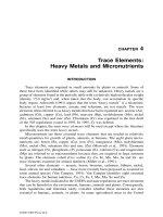
Figure 1 Scanning electron micrograph of Trichiderma spp. hyphae interacting with those of S.
rolfsii. Hypha of S. rolfsii, from which a coiling hypha of T. harzianum was removed, showing
digested zone with penetration sites caused by the antagonists (ϫ5, 500). (From Ref. 52.)
Copyright © 2002 Marcel Dekker, Inc.
causediseasesanddamageinagriculturalcrops.Chitinasesalsoplayanimportantphysio-
logicalandecologicalroleinecosystemsasrecyclersofchitin,bygeneratingCandN
sources.Someproducersofchitinases,includingTrichodermaspp.,arealsosourcesof
mycolyticenzymepreparations(51,59,60).
ChitinolyticenzymesaredefinedasenzymesthatcleaveabondbetweentheC1
andC4oftwoconsecutiveGlcNAcunits.Onthebasisofaminoacidsequencesimilarities,
allchitinaseshavebeengroupedintofamilies18,19,and20,underthemainclassof
glycosylhydrolases.Mostofthemicrobialchitinasesbelongtofamily18(61,62).Even
withinthesamefamily,chitinasesshowwidelydifferingpropertieswithrespecttosub-
stratespecificity,reactionspecificity,andpHoptimum.Thechitinolyticenzymesaredi-
videdintothreeprincipaltypesdependingontheiractiononchitinsubstrates.According
tothenomenclaturesuggestedbySahaiandManocha(59),endochitinases(EC3.2.1.14)
aredefinedasenzymescatalyzingtherandomhydrolysisof1,4-βlinkagesofGlcNAcat
internalsitesovertheentirelengthofthechitinmicrofibril.Theproductsofthereaction
aresoluble,low-molecular-massmultimersofGlcNAcsuchaschitotetraose,chitotriose,
anddiacetylchitobiose.Exochitinases,alsotermedchitobiosidasesorchitin-1,4-β-chito-
biosidases(63),catalyzetheprogressivereleaseofdiacetylchitobioseunitsinastepwise
fashionasthesoleproductfromthechitinchains,suchthatnomonosaccharidesoroligo-
saccharidesareformed.
Thethirdtypeofchitinolyticenzymeischitobiasealsotermedashexosaminidase
(EC3.2.1.52)orN-acetyl-β-1,4-d-glucosaminidase(EC3.2.1.30)belongstofamily20
andalsoactsinexosplittingmodeondiacetylchitobioseandhigheranalogsofchitin,
includingchitotrioseandchitotetraose,toproduceGlcNAcmonomers.Rapidandspecific
methodshavebeendevelopedfordetectionandquantitativeassaysofN-acetyl-β-gluco-
saminidase,chitobiosidase,andendochitinaseinsolutionsusingp-nitrophenyl-N-acetyl-
β-d-glucosaminide,p-nitrophenyl-β-d-N,N′-diacetylchitotriose,andp-nitrophenyl-β-d-
N,N′,N″-triacetylchitotrioseorcolloidalchitinassubstrates,respectively(64).Procedures
alsoaredescribedforthedirectassayofthesethreeenzymesaftertheirseparationby
sodiumdodecylsulfate(SDS)-polyacrylamidegelelectrophoresis(PAGE)inwhichthe
enzymesarevisualizedasfluorescentbandsbyusinganagaroseoverlaycontaining4-
methyl-umbelliferylderivativesofN-acetyl-β-d-glucosaminide,β-d-N,N′-diacetyl-
chitobioside,orβ-d-N,N,N″-triacetylchitotriose,respectively(65).
AsetofchitinolyticenzymessecretedbyvariousstrainsofT.harzianum(e.g.,TM,
T-Y,39.1,CECT2413,P1ϭT.atroviride),whengrownonchitinasthesoleCsource,
consistsofN-acetylglucosaminidases,endochitinases,andexochitinases(chitobiosidases).
Intotal,10separatedchitinolyticenzymeswerelistedbyLorito(50);onlyonestepin
themicroparasiticprocessofT.harzianum,whichisthedissolutionofthecellwallof
thetargetfungusbyenzymeactivity,mayinvolvemorethan20separategenesandgene
productssynergisticonetoanother(Table1).TwoN-acetylglucosaminidaseswithappar-
ent molecular masses of 102 to 118 kD (depending on the isolate and the procedure used)
and 72 to 73 kD (ϭNAG1) have been described by Ulhoa and Peberdy (66), Lorito et
al. (67), and Haran et al. (68). The 102-kD enzyme (CHIT102) is the only chitinase of
T. harzianum to be expressed constitutively when the fungus is grown with glucose instead
of chitin as the sole C source (69). Four endochitinases—CHIT31, CHIT33, CHIT52, and
CHIT42 (ϭECH42)—have been reported by De La Cruz et al. (70), Ulhoa and Peberdy
(66), Harman et al. (63), and Haran et al. (68). Additionally, a glycosylated chitobiosidase
of 40 kD is secreted by strain P1 when grown on crab-shell chitin as the sole C source
(63), and a 28-kD exochitinase releasing GlcNAc only was purified from the culture filtrate
Copyright © 2002 Marcel Dekker, Inc.
Table 1 Examples of Lytic Enzymes Produced by Mycoparasitic Fungi which May Be
Involved in Disease Biocontrol
Molecular mass Encoding
Enzyme (kDa) gene Fungus/strain Reference
N-Acetylglucosaminidase 102–118 ND Trichoderma harzianum (66, 68)
(EC 3.2.1.30) (TM, 39.1)
N-Acetylglucosaminidase 72–73 nag1 T. harzianum (TM, P1) (67, 68, 88)
(EC 3.2.1.30)
Endochitinase (EC 52 ND T. harzianum (TM) (68)
3.2.1.14)
Endochitinase (EC 41–42 ech42 T. harzianum (39.1, P1, (63, 70, 78,
3.2.1.14) CEST2413); G. vir- 79, 84,
ens (41) 106)
Exochitinase (chitibiosi- 40 ND T. harzianum (P1) (63)
dase)
Endochitinase (EC 37 ND T. harzianum (CEST 68, 70)
3.2.1.14) 2413, TM)
Endochitinase (EC 33 chit33 T. harzianum (CEST (68, 70)
3.2.1.14) 2413, TM)
Proteinase 31 prb1 T. harzianum (55)
β-1,3-endoglucanase (EC 78 bgn13.1 T. harzianum (109)
3.2.1.6; EC 3.2.1.39) (CECT2413)
β-1,3-endoglucanase 17 ND T. harzianum (113)
(CECT2413)
β-1,3-endoglucanase 36 ND T. harzianum (39.1) (110)
β-1,3-exoglucanase (EC 77–110 lam1.3 T. harzianum (P1, T-Y, (67, 111,
3.2.1.58) IMI1206040) 112)
β-1,6-endoglucanase 43 ND T. harzianum (117, 118)
(CECT2413)
β-1,4-endoglucanase 51 egl1 T. longibrachiarum (290)
β-1,3-exoglucanase 84 exgA Ampelomyces quis- (141)
qualis
Endochitinase 40 ND Fusarium chlamy- (130)
dosporum
β-1,3-glucanase ND ND Trametes versicolor, (131)
Pleurotus eryngii
β-1,3-glucanase, β-1,6- ND ND Penicillium purpuro- (132)
glucanase, chitinase genum
β-1,3-glucanase ND ND Tilletiopsis spp. (136)
of strain T. harzianum T198. This particular enzyme displayed activity on a wide array
of chitin substrates of more than two GlcNAc units in length (71).
Lorito et al. (72,73) studied the antifungal activities of a 42-kD endochitinase and
a 40-kD chitobiosidase from T. harzianum strain P1 in bioassays against nine different
fungal species. Both spore germination and germ-tube elongation were inhibited in all
chitin-containing fungi. The degree of inhibition was proportional to the level of chitin
in the cell wall of the target fungus. Combining the two enzymes resulted in a synergistic
increase in antifungal activity. A variety of synergistic interactions have been found when
different enzymes were combined or associated with biotic or abiotic antifungal agents.
Copyright © 2002 Marcel Dekker, Inc.
The levels of inhibition obtained by using enzyme combinations were, in some cases,
comparable with those of commercial fungicides. Moreover, the antifungal interaction
between enzymes and common fungicides allowed up to 200-fold reductions in the re-
quired chemical doses. These two enzymes, separately or in combination, substantially
improved the antifungal ability of a biocontrol strain of Enterobacter cloacae (74). In an
in vitro bioassay, different classes of cell-wall-degrading enzymes (glucan 1,3-β-glucosi-
dase [EC 3.2.1.58], N-acetyl-β-glucosaminidase, endochitinase, and chitin 1,4-β-chitobio-
sidase) produced by T. harzianum and G. virens inhibited spore germination of B. cinerea.
The addition of any chitinolytic or glucanolytic enzyme to the reaction mixture synergisti-
cally enhanced the antifungal properties of five different fungitoxic compounds against
B. cinerea (73). Some of the combinations showed a high level of synergism, suggesting
that the interaction between membrane-affecting compounds and cell-wall-degrading en-
zymes could be involved in biocontrol processes and plant self-defense mechanisms (75).
A correlation between high capacity to produce chitinolytic enzymes and the superior
biocontrol potential of the mycoparasitic fungi was also reported by Lima et al. (76). In
general, chitinolytic enzymes from Trichoderma spp. appeared to be more effective in
vitro against a number of fungal plant pathogens than were similar enzymes from plants
or bacteria (72).
The ech42 chitinase gene was shown to be highly conserved within the genus
Trichoderma (77) and its product, the 42-kD chitinase, is believed to be one of the most
crucial for mycoparasitic interactions between Trichoderma spp. and target pathogens. A
similar endochitinase was purified from G. virens (78). Carsolio et al. (79) cloned and
characterized ech42 (previously named ThEn42) encoding a 42-kD endochitinase in the
biocontrol strain T. harzianum IMI206040. Expression of the complementary deoxyribo-
nucleic acid (cDNA) clone in Escherichia coli produced bacteria with chitinase activity.
This chitinase displayed lytic activity on B. cinerea cell walls in vitro. The ech42 gene
was assigned to a double-chromosomal band (chromosome V or VI) upon electrophoretic
separation and Southern analysis of the chromosomes. Expression of ech42 was strongly
enhanced during direct interaction of the mycoparasite with a phytopathogenic fungus
when confronted in vitro and when it was grown in minimal medium containing chitin
as sole C source. Similarly, light-induced sporulation resulted in high levels of transcript,
suggesting developmental regulation of the gene. T. virens strains in which the 42-kD
chitinase gene was disrupted or constitutively overexpressed were constructed through
genetic transformation. The resulting transformants were stable and showed patterns simi-
lar to those of the wild-type strain with respect to growth rate, sporulation, antibiotic
production, colonization efficiency on cotton roots, and growth/survival in soil. However,
biocontrol activities of the ‘‘disrupted’’ and constitutively overexpressed strains were sig-
nificantly decreased and enhanced, respectively, against cotton seedling disease incited
by R. solani when compared with those of the parental strain (80).
However, several recently reported experiments have put into question the role of
CHIT42 endochitinase as the only key enzyme in mycoparasitism. The biocontrol strain
T. harzianum P1, recently attributed to T. atroviride (81), was genetically modified by
targeted disruption of the single-copy ech42 gene. A mutant, lacking the 42-kD endochi-
tinase but retaining the ability to produce other chitinolytic and glucanolytic enzymes of
this strain expressed during mycoparasitic activity, was unable to clear a medium contain-
ing colloidal chitin but grew and sporulated similarly to the wild type. In vitro antifungal
activity of the ech42-disruptant culture filtrate against B. cinerea and R. solani was reduced
by about 40% relative to that of the wild type, but its activity in protecting against P.
Copyright © 2002 Marcel Dekker, Inc.
ultimum and R. solani in biocontrol experiments was the same or even better than that of
strain P1. In contrast, the mutant’s antagonism against B. cinerea on bean leaves was
significantly reduced compared with that of strain P1. These results indicate that the antag-
onistic interaction between strain P1 and various fungal hosts is based on different mecha-
nisms (82).
Corresponding results were obtained with several transgenic T. harzianum strains
carrying multiple copies of ech42, and the corresponding gene disruptants were con-
structed. The level of extracellular endochitinase activity when T. harzianum was grown
under inductive conditions increased up to 42-fold in multicopy strains relative to that of
the wild type, whereas gene disruptants exhibited practically no activity. However, no
major differences in the efficacies of the strains generated as biocontrol agents against R.
solani or S. rolfsii were observed in greenhouse experiments (83). One possible explana-
tion for these results is that other enzymes of Trichoderma’s chitinolytic system are suffi-
cient to control these fungal phytopathogens and that the lack of a certain protein can be
compensated for by altering the levels of other proteins with similar activity. In view of
the results showing efficient synergism between different chitinolytic enzymes produced
by the same Trichoderma sp. isolate, it is not surprising that overexpression of one of
these enzymes does not necessarily lead to an increase in biocontrol activity. Moreover,
to achieve the highest level of antagonism toward target pathogens, a combination of
several enzymes gives a better effect than the overproduction of only one of them.
Several groups have reported cloning genes ech42 (79,84–86), chit33 (87), and nag1
(88). Very little is known, however, about the regulation of these genes and the roles of
the corresponding enzymes in fungi during mycoparasitism. Generally, products of chitin
degradation are thought to induce chitinolytic enzyme expression, and easily metaboliz-
able C sources serve as repressors (59,89,90). Fungal cell walls, colloidal chitin, and C
starvation have been shown to be inducers of the cloned chitinase genes (79,84,87,88,91).
To study the regulation of chitinolytic enzyme synthesis during the Trichoderma
sp.–host mycoparasitic interaction, more specific confrontation assays (dual culture) on
plates were developed (53,69,92). The differential expression of chitinolytic enzymes dur-
ing the interaction of T. harzianum with S. rolfsii and the role of fungal–fungal recognition
in this process were studied by Inbar and Chet (92). A change in the chitinolytic enzyme
profile was detected during the interaction between the fungi grown in dual culture on
synthetic medium. Before contact with one another, both fungi contained a protein with
constitutive 1,4-β-N-acetylglucosaminidase activity. As early as 12 h after contact, the
chitinolytic activity in S. rolfsii disappeared, while that in T. harzianum (a protein with
a molecular mass of 102 kD, CHIT102) greatly increased. After 24 h of interaction, the
activity of CHIT102 diminished concomitantly with the appearance of a 73-kD 1,4-β-N-
acetylglucosaminidase, which became clear and strong at 48 h. This phenomenon did not
occur if the S. rolfsii mycelium was autoclaved prior to incubation with T. harzianum,
suggesting its dependence on vital elements from the host. Cycloheximide inhibited this
phenomenon, indicating that de novo synthesis of enzymes takes place in Trichoderma
spp. during these stages of the parasitism. A biomimetic system based on the binding of
a purified surface lectin from the host S. rolfsii to nylon fibers was used to dissect the
effect of recognition. An increase in CHIT102 activity was detected, suggesting that the
induction of chitinolytic enzymes in Trichoderma sp. is an early event that is elicited by
the recognition signal (i.e., lectin–carbohydrate interactions). Experiments with T. harzia-
num and the host lectin–covered nylon threads indicated that mere physical contact with
the host triggers both the mycoparasitism-specific coiling of Trichoderma sp. hyphae
Copyright © 2002 Marcel Dekker, Inc.
around the host and chitinase formation (32,92). It is postulated that recognition is the
first step in a cascade of antagonistic events that trigger the parasitic response in Tricho-
derma spp.
These observations were extended by Haran et al. (69), who showed that the expres-
sion of the various N-acetylglucosaminidases and endochitinases during mycoparasit-
ism can be regulated in a very specific and finely tuned manner that is affected by the
host. When strain T. harzianum T-Y antagonized S. rolfsii,theN-acetylglucosaminidase
CHIT102 was the first to be induced. As early as 12 h after contact, its activity diminished,
and the other N-acetylglucosaminidase, CHIT73, was expressed at high levels. However,
when T. harzianum antagonized R. solani, the chitinase expression patterns differed con-
siderably. Twelve hours after contact, CHIT 102 activity was elevated, and the activities
of three additional endochitinases, at 52 kD (CHIT 52), 42 kD (CHIT 42), and 33 kD
(CHIT 33), were detected. As the antagonistic interaction proceeded, CHIT102 activity
decreased, whereas the activities of the endochitinases gradually increased.
Similarly, Carsolio et al. (79) detected the induction of ech42 gene transcription
only 24 h after contact of T. harzianum with B. cinerea. These data suggested that chitinase
formation takes place during the later stages of the host–mycoparasite interaction, for
example, to T. harzianum in penetration of the host hyphae. Therefore, chitinase induction
generally has been regarded as a consequence of, rather than a prerequisite for, mycopara-
sitism. Krishnamurthy et al. (93) reported that differential induction of chitinase isoforms
in vitro might depend on C sources in the growth medium. Nevertheless, in vivo the
differential expression of T. harzianum chitinases may influence the overall antagonistic
ability of the fungus against a specific host.
The specific and unique role of the 102-kD enzyme in triggering the expression of
other chitinolytic enzymes was questioned by Zeilinger et al. (94). To monitor chitinase
expression during mycoparasitism of strain T. harzianum P1 (ϭT. atroviride) in situ,
strains were constructed containing fusions of the green fluorescent protein to the 5′-
regulatory sequences of the Trichoderma nag1 and ech42 genes. Confronting these strains
with R. solani led to induction of gene expression before or after physical contact in the
cases of genes ech42 and nag1, respectively. Separating the two fungi abolished ech42
expression, indicating that macromolecules are involved in its precontact activation. No
ech42 expression was triggered by culture filtrates of R. solani or placement of T. harzia-
num on plates previously colonized by R. solani. Instead, high expression occurred upon
incubation of T. harzianum with the supernatant of R. solani cell walls digested with
culture filtrates or purified CHIT42. The results indicate that ech42 is expressed before
contact of T. harzianum with R. solani and its induction is triggered by soluble chitooligo-
saccharides produced by constitutive activity of CHIT42 and/or other chitinolytic en-
zymes. Therefore, ech42 expression, in contrast to that of nag1, is a relatively early event,
taking place prior to physical hyphal contact of the fungus with its host (R. solani). This
indicates that this enzyme could be involved in the very early stages of the mycoparasitic
process. Furthermore, the involvement of chitinase activity in the induction of ech42 gene
expression pre contact has been demonstrated by the effect of the chitinase inhibitor allo-
samidin, an actinomycete-derived metabolite. Expression of the 73-kD exochitinase nag1
gene was observed only after contact of Trichoderma spp. with its host and was most
active during overgrowth of R. solani. Therefore, different mechanisms of induction may
occur for ech42 and nag1, and nag1 gene expression and may depend on products gener-
ated by CHIT42 activity. The results support the earlier suggestion by Lora and associates
(95) that constitutive chitinases may partially degrade the cell walls of the host, thereby
Copyright © 2002 Marcel Dekker, Inc.
generating oligosaccharides containing GlcNAc that may act in turn as elicitors for the
general antifungal response of Trichoderma sp. Although Zeilinger et al. (94) did not
determine the number or expression patterns of other chitinase genes during this process,
the ability of R. solani cell walls to induce ech42 expression clearly was shown. The
authors suggested that low constitutive activity of CHIT42 or some other chitinase triggers
the induction of ech42 when the host is at close range. A major role for CHIT42 in the
induction process is implied by the fact that it generated the most strongly inducing mix-
ture from R. solani cell walls. However the authors did not exclude the possibility that
other chitinases, e.g., the 102-kD N-acetyl-β-d-glucosaminidase or CHIT33, as shown
previously by Haran et al. (69) and Garcia et al. (84), respectively, also may be produced
constitutively and act in a similar manner. This implies that chitinolytic enzymes not only
are involved in the destruction of the host cell wall but also may play a role during the
initial stages of mycoparasitism.
Cortes et al. (96) also studied whether physical contact between the mycoparasite
and its host is necessary to induce expression of the Trichoderma sp. hydrolytic enzymes
during the parasitic response. Dual cultures of Trichoderma sp. and a host, with and with-
out contact, were used to study the mycoparasitic response in Trichoderma spp. Northern
analysis showed a high level of expression of genes encoding a proteinase (prb1) and an
endochitinase (ech42) in dual cultures, even when contact with the host was prevented
by cellophane membranes. Neither gene was induced during the interaction of Trichod-
erma sp. with lectin-coated nylon fibers, even through the latter do induce hyphal coiling
and appressorium formation (92). Therefore, the signal involved in triggering the produc-
tion of these hydrolytic enzymes is independent of the recognition mediated by this lectin–
carbohydrate interaction. The results showed that induction of prb1 and ech42 is contact-
independent, and a diffusible molecule produced by the host is the signal that triggers
expression of both genes in vivo. Furthermore, a molecule that is resistant to heat and
protease treatment, obtained from R. solani cell walls, induced expression of both genes.
Thus, this molecule is involved in regulating the expression of hydrolytic enzymes during
mycoparasitism by T. harzianum (96). The antagonism observed in dual cultures, however,
is not necessarily correlated with the fungus’s chitinolytic activity. Thus, similarities as
well as variations were observed in the abilities of various isolates of G. virens and Tri-
choderma longibrachiatum to invade the test pathogens R. solani, S. rolfsii, and P. apha-
nidermatum in dual culture. Although all the isolates produced enhanced levels of lytic
enzymes, no correlation was observed between this attribute and the hyperparasitic poten-
tial of the various isolates in dual culture (97). Therefore, the relevance and role of en-
zymes and toxic metabolite(s) of these mycoparasitic fungi in their antagonism toward
plant pathogens can vary among independent isolates and should be reassessed for each
individual case. Moreover, the ability of lytic enzymes to provide biocontrol depends on
both the type of plant being protected and the fungal pathogen. Thus, chitinase production
does not appear to play a major role in protecting wood against fungal strains (98). Further
characterization of the full chitinolytic system of Trichoderma sp. at the gene level should
clarify which singular of these enzymes is really responsible for precontact gene expres-
sion. This, in turn, will help in understanding the relevance of this mechanism to biocon-
trol.
Studies on the regulation of ech42 and nag1 gene expression have been reported
by Lorito et al. (99) and Mach and colleagues (81). Competition experiments, using oligo-
nucleotides containing functional and nonfunctional consensus sites for binding of the C
catabolite repressor Cre1, provided evidence that the complex from nonmycoparasitic my-
Copyright © 2002 Marcel Dekker, Inc.
celia involves the binding of Cre1 to both fragments of the ech42 promoter. The presence
of two and three consensus sites for the binding of Cre1 in the two ech42 promoter frag-
ments used is consistent with these findings. In contrast, formation of the protein–DNA
complex from mycoparasitic mycelia is unaffected by the addition of the competing oligo-
nucleotides and hence does not involve Cre1. The addition of equal amounts of protein
of cell-free extracts from nonmycoparasitic mycelia converted the mycoparasitic DNA–
protein complex into a nonmycoparasitic complex. The addition of purified Cre1 ::glutathi-
one S-transferase protein to mycoparasitic cell-free extracts produced the same effect.
These findings suggest that ech42 expression in T. harzianum is regulated by (i) binding
of Cre1 to two single sites in the ech42 promoter, (ii) binding of a ‘‘mycoparasitic’’
protein–protein complex to the ech42 promoter near the Cre1 binding sites, and (iii) func-
tional inactivation of Cre1 upon mycoparasitic interaction to allow formation of the myco-
parasitic protein–DNA complex (99,100). Using a reporter system based on the Aspergil-
lus niger glucose oxidase goxA gene, Mach et al. (81) showed ech42 gene expression
during growth on fungal (B. cinerea) cell walls or after prolonged C starvation, indepen-
dent of the use of glucose or glycerol as a C source, suggesting that relief of C catabolite
repression is not involved in induction during starvation. In addition, ech42 gene transcrip-
tion was triggered by physiological stresses, such as low temperature, high osmotic pres-
sure, or addition of ethanol. This corresponds to the finding that the ech42 promoter con-
tains four copies of a putative stress-response element CCCCT, also found in yeasts. The
nag1 gene expression was triggered by growth on chitin, GlcNAc, and the cell walls of
B. cinerea used as a C source but, in contrast to ech42, also by a number of the chitin
degradation products (chitooligomers) when added to mycelia pregrown on different C
sources. The application of new techniques for examining the activities of the mycopara-
site (fusion[s] of ech42 or nag1 with novel reporter genes such as green fluorescent protein
or A. niger goxA) offers the possibility of revealing for the first time that (i) ech42 tran-
scription is induced before Trichoderma sp. physically contacts its host (94) and (ii) differ-
ent regulatory signals are involved in triggering the expression of the 42-kD endochitinase
and the 73-kD N-acetyl-β-d-glucosaminidase. This last enzyme revealed high similarity
to N-acetyl-glucosaminidases from other eukaryotes, such as Candida albicans, and inver-
tebrate and vertebrate animal tissues; the greatest similarity was to the corresponding gene
from the silkworm (88).
The pattern of chitinolytic enzymes production can be an important marker for Tri-
choderma sp. strain identification and classification. The identification of Trichoderma
sp. strains is important for their application as biocontrol agents. Schikler et al. (101) used
a two-dimensional analysis in which extracellular proteins of T. harzianum strains T-35,
Y, and TM were separated first according to their isoelectric point and then according to
their molecular mass. Chitinase activities were detected in situ after the second separation.
Each of the three strains exhibited a unique pattern of three to five different chitinases
(one or two N-acetyl-β-glucosaminidases, and two or four endochitinases). These unique
profiles can be used to differentiate among strains within this species, a requirement for
specific biocontrol applications. Random amplification of polymorphic DNA (RAPD) was
applied to characterize 34 strains of seven species of Trichoderma, including T. hamatum,
T. harzianum, and T. viride isolated from various fungal sources. The RAPD patterns
of T. viride strains were highly variable; isolates of T. harzianum proved to be more
uniform; T. hamatum demonstrated remarkable intraspecific divergence. These three types
comprised certain pairs of strains that have become promising participants in a strain-
improving program since their strong genetic affinities offer good chances for combining
their contrasting biocontrol traits (102).
Copyright © 2002 Marcel Dekker, Inc.
2.Glucanases
β-1,3-glucan,orlaminarin,isapolymerofd-glucoseinaβ-1,3configuration,arranged
ashelicalcoils.Fungalcellwallscontainmorethan60%laminarin.Whereaschitinis
arrangedinregularlyorderedlayers,laminarinfibrilsarearrangedinanamorphicmanner.
Therearechemicalbondsbetweenthelaminarinandchitin,andtogethertheyforma
complexnetofglucanandGlcNAcoligomers(103).Laminarinishydrolyzedmainlybyβ-
1,3-glucanases,alsoknownaslaminarinases.Theseenzymes,describedinfungi,bacteria,
actinomycetes,algae,mollusks,andhigherplants,arefurtherclassifiedasexo-andendo-
β-glucanases.Exo-β-1,3-glucanases(β-1,3-glucanglucanohydrolase,[EC3.2.1.58])hy-
drolyzelaminarinbysequentiallycleavingglucoseresiduesfromthenonreducingendsof
polymersoroligomers.Consequently,thesolehydrolysisproductsareglucosemonomers.
Endo-β-1,3-glucanases(β-1,3-glucanglucanohydrolase[EC3.2.1.6orEC3.2.1.39])
cleaveβ-1,3linkagesatrandomsitesalongthepolysaccharidechain,releasingsmaller
oligosaccharides.Bothenzymetypesarenecessaryforthefulldigestionoflaminarin
(104).Theseenzymeshaveseveralfunctionsinfungiincludingnutritioninsaprotropism,
mobilizationofβ-glucansunderconditionsofC-andenergy-sourceexhaustion,anda
physiologicalroleinmorphogeneticprocessesduringfungaldevelopmentanddifferentia-
tion(105).
Glucanaseshavebeensuggestedasanothergroupofkeyenzymesinvolvedinthe
mycoparasitismofGliocladiumandTrichodermaspp.againstfungalplantpathogens(Ta-
ble1).Thesubstrateoftheseenzymes,β-1,3-glucan,isoneofthemajorcomponentsof
fungal cell walls along with chitin. Aside from the β-1,3-glucanases, the Trichoderma
spp. also produce β-1,6-glucanases under specific growth conditions, and these enzymes
hydrolyze minor structure polymers of fungal cells walls, β-1,6-glucans, which are thought
to play an important role in the antagonistic action of Trichoderma spp. against a wide
range of fungal plant pathogens (53). However, similarly to chitinases, glucanases are
produced by Trichoderma sp. when it is grown in the presence of not only isolated fungal
cell walls but chitin as well (106,107). Isolated plasma membranes of B. cinerea provide
useful tools to study synergism between cell-wall-hydrolytic chitinases and glucanases of
T. harzianum during the antagonism with phytopathogenic fungi. The data obtained in
this system showed that cell wall synthesis is a major target of mycoparasitic antagonism
by T. harzianum. Inhibition of the resynthesis of the host cell wall β-glucans sustained
the disruptive action of β-glucanases and enhanced fungicidal activity. Therefore, cell
wall turnover was considered a major target of mycoparasitic antagonism (100).
Large interstrain and interspecies differences exist in the production levels of both
the laminarinase and chitinase enzymes by Trichoderma sp. isolates. Total activities of
the enzymes were greater when isolates were cultured in malt medium, but specific chi-
tinase and laminarinase activities were higher under low-nutrient conditions. Glucose ap-
pears to inhibit the formation of all of the inducible β-1,3-glucanases and chitinase, al-
though this effect was not common to all Trichoderma sp. isolates for the latter enzyme
(108). Similarly to chitinolytic enzyme production, the same strain of Trichoderma sp.
can produce several extracellular β-1,3-glucanases. T. harzianum CECT 2413 was shown
to produce at least three extracellular β-1,3-glucanases. The most basic 78-kD extracellular
enzyme, named BGN13.1, was expressed when either fungal cell wall polymers or auto-
claved mycelia from different fungi were used as the C source. The enzyme is specific
for β-1,3 linkages and has an endolytic mode of action.
Sequence comparison shows that this β-1,3-glucanase, first described for filamen-
tous fungi, belongs to a family different from that of its previously described bacterial,
yeast, and plant counterparts. BGN13.1 hydrolyzes yeast and fungal cell walls; it is re-
Copyright © 2002 Marcel Dekker, Inc.
pressed by glucose and induced by either fungal cell wall polymers or autoclaved yeast
cells and mycelia. A gene encoding the BGN13.1 endo-β-1,3-glucanase has been cloned
and sequenced. Its structural analysis suggests that the enzyme contains a hydrophobic
leader peptide that may be cleaved by an endoproteinase (109). A 36-kD endo-β-1,3-
glucanase, purified from T. harzianum 39.1, was active toward glucans containing β-1,3-
linkages and hydrolyzed laminarin to form oligosaccharides (110). At least seven extracel-
lular β-1,3-glucanases ranging from 60 to 80 kD were produced by strain T. harzianum
IMI206040 upon induction with laminarin or a soluble β-1,3-glucan or in the presence
of different glucose polymers and fungal cell walls. The level of secreted β-1,3-glucanase
activity was proportional to the amount of glucan present in the inducer. The properties
of this complex group of enzymes suggest they have different roles in host cell wall lysis
during mycoparasitism (111).
A novel 110-kD extracellular β-1,3-exoglucanase, LAM1.3, was purified from T.
harzianum strain T-Y grown with laminarin. The corresponding gene, lam1.3, was cloned
and the deduced amino acid sequence of the LAM1.3 enzyme showed high homology to
EXG1, a β-1,3-exoglucanase of the phytopathogenic fungus Cochliobolus carbonum, and
lower homology to BGN13.1 (112). Further studies of the β-1,3-glucanase system of T.
harzianum strain T-Y revealed at least five different enzymes with molecular masses of
30 to 200 kD. In contrast to other β-1,3-glucanases, whose production is repressed by
glucose and induced by a variety of polysaccharides as sole C source (109,111–113), the
largest enzyme, Gβ-1,3-200, was the most abudant when strain T-Y was grown with no
C source and was repressed by GlcNAc or malic acid (114). β-1,3-glucanases in T. harzia-
num are found in the periplasm, bound to cell walls, or secreted into the growth medium
(115), and regulation of the enzymes’ expression is considered a key step in β-glucan
biodegradation and consequently in mycoparasitism.
Total β-1,3-glucanase activity has been found to be induced by different polysaccha-
rides or by fungal cell walls and repressed by high glucose concentrations (109,113).
Moreover, different fungal cell walls have been shown to induce different levels of β-
1,3-glucanase activity and different enzyme patterns were observed when T. harzianum
was grown on different C-source-containing media (109,111). The interaction between T.
harzianum and the soil-borne plant pathogen P. ultimum (which is exceptional in that the
cell walls contain β-[1,3]-[1,6]-d-glucans and cellulose instead of chitin as major structural
components) and studied by electron microscopy and gold cytochemistry, revealed marked
alteration of the β-1,3-glucan component of the Pythium sp. cell wall. This suggested that
β-1,3-glucanases played a key role in the process (116). By specific detection of their
activity in gels, different Trichoderma sp. strains grown under different growth conditions
excreted the β-1,6-glucanase isozymes (107,116–118).
Despite considerable evidence that Trichoderma spp. produce chitinolytic enzymes
and glucanases in vitro, much less is known about what happens in vivo under natural
conditions, and no definitive evidence has shown the presence or activity of chitinases or
endoglucanase in the rhizosphere (the zone immediately adjacent to the plant root) associ-
ated with a soil-borne fungal pathogen. Most studies have been performed on plates or
in liquid cultures supplemented with different C sources, and these theories have not been
fully studied in vivo. de Soglio et al. (119) detected chitobiosidase, endochitinase, endo-
β-1-3-glucanase, and N-acetylglucosaminidase simultaneously in the roots of soybean
seedlings and in cell-free culture filtrates of T. harzianum isolate Th008. With the excep-
tion of that of endochitinase, activity of these enzymes also was associated with R. solani
isolate 2B-12, causal agent of soybean root rot. In greenhouse experiments, soybean seeds
Copyright © 2002 Marcel Dekker, Inc.
inoculatedwithT.harzianumTh008wereplantedinasoilmixtureinfestedwithR.solani
2B-12.Fifteendaysafteremergence,therhizospherewasassayedforchitinolyticenzymes
andendoglucanase.OnlytheN-acetylglucosaminidaseandendochitinaseactivitiesinthe
rhizospheresamplesweresignificantlyelevatedabovethoseofthecontrols.Itwasdeter-
minedthatT.harzianumTh008wasthesourceoftheendochitinaseintherhizosphere.
Theresultsindicatedthattheprobablesourceofthedetectableendochitinaseactivityin
rhizosphereextractsisthebiocontrolagentratherthansoybeanrootorthepathogen.A
positivecorrelationwasfoundbetweendiseaseindexandtotalprotein(milligramsper
gram[mg/g]soil)inrhizospheresamplesandinN-acetylglucosaminidaseactivityinrhizo-
sphereextracts.ThisfindingsuggeststhereleaseofN-acetylglucosaminidaseintotherhi-
zosphereresultsfromaresponseofrootcellstothepathogen.
3.Cellulases
Trichodermaspp.produceenzymesinthecellulolytic(exo-andendo-β-1-4-glucanase,
β-1-4-glucosidase)andhemicellulolytic(especiallyxylanaseandβ-xylosidase)complexes
thatareeffectiveindegradingnaturallignocelluloses.Chitinandβ-(1,3)-glucanarethe
twomajorstructuralcomponentsofmanyplantpathogenicfungi,exceptoomycetes,
whichcontaincelluloseintheircellwallandhavenoappreciablelevelsofchitin.There-
fore,thebiologicalcontrolofsucheconomicallyimportantplantpathogenicoomycetes
asPythiumspp.canbeprovidedbyabiocontrolagentabletoproducecellulases(Table
1).
Cellulose, a linear, essentially insoluble β-1,4-glucosidically linked homopolymer
of about 8,000 to 12,000 glucose units, is used as an energy source by numerous diverse
microorganisms, including fungi and bacteria, which produce cellulases. Among the best-
characterized of these systems are the inducible cellulases of the saprophytic fungus
Trichoderma reesei (ϭT. longibrachiarum), which include 1,4-β-d-glucan cellobiohydro-
lases (EC 3.2.1.91), endo-1,4-β-d-glucanases (EC 3.2.1.4), and 1,4-β-d-glucosidases (EC
3.2.1.21) (120).
There have been indications that endo-1,3-β-glucanase (EC 3.2.1.6) and endo-1,4-
β-d-glucanase activity of T. harzianum isolate T3 is induced in sphagnum peat moss culti-
vations and dual culture experiments by the presence of P. ultimum. Further, P. ultimum
stimulated the germination of Trichoderma sp. conidia. Low concentrations of purified
17-kD endo-1,3-β-glucanase and 40- and 45-kD cellulases were able to inhibit the germi-
nation of encysted zoospores and elongation of germ tubes of a plant-pathogenic Pythium
sp. isolate. A strong synergistic effect was observed on the inhibition of cyst germination
by a combination of endo-1,3-β-glucanase and fungicide (Fongarid). Finally, in a time-
course study of colonization of the rhizosphere of cucumber seedlings, the active fungal
mycelial biomass of a GUS-transformant of T. harzianum isolate T3 increased over 4
weeks. Trichoderma sp. appeared to colonize healthy roots only superficially, whereas
the mucilage of the root hairs and of distal parts of the wounded areas or broken parts of
the roots was extensively colonized (113).
The interaction between T. harzianum and P. ultimum has been studied by electron
microscopy and further investigated by gold cytochemistry. Early contact between the
two fungi was accompanied by the abnormal deposition of a cellulose-enriched material
at sites of potential antagonist penetration. The antagonist displayed the ability to penetrate
this barrier, indicating that cellulolytic enzymes had been produced. However, the presence
of cellulose in the walls of severely damaged Pythium sp. hyphae indicated that cellulolytic
enzymes were not the only critical factors involved in the antagonistic process. The marked
Copyright © 2002 Marcel Dekker, Inc.
alterationoftheβ-1,3-glucancomponentofthePythiumsp.cellwallsuggestedthatβ-
1,3-glucanasesplayedakeyroleintheprocess(118).
4.Proteases
Proteaseproductioniscommoninmicroorganisms,includingfungi,amongwhichTrichod-
ermaspp.arewell-knownproducers(121).ProteaseactivityofT.harzianumcanbeinduced
byautoclavedmycelia,afungalcellwallpreparation,orchitin;however,theinductiondoes
notoccurinthepresenceofglucoseandincreaseswhentheliquidculturemediumcontains
organicnitrogensources(122).Rodriguez-Kabanaetal.(123)providedevidencethatT.
virideproteolyticactivityisinvolvedinthebiocontrolofS.rolfsii.Agene,prb1,ofT.
harzianumIMI206040wasclonedanditsproductwasbiochemicallycharacterizedasa
31-kDbasicserineproteinase(Prb1)(55).Thatwasthefirstreportofcloningamycoparasi-
tism-relatedgene(Table1).Thisproteasewassuggestedtoprovidethemycoparasitewith
nutrients, since it was involved in the degradation of pathogen cell walls and membranes
and release of the proteins from the lysed pathogen (124). Strong expression of this protease
was observed during mycoparasitic interactions with R. solani (125).
Foliage diseases have been some of the most difficult to control with biological
agents because of the severe environment on the leaf surface. Until recently, most research
on the biological control of aerial plant diseases was focused on the control of bacterial
pathogens (10). The last decade, however, has seen increased activity in the development
of biocontrol agents for foliar fungal pathogens. The strain T. harziaum T39, known as
an efficient biocontrol agent of B. cinerea, which causes gray mold, a foliage disease of
grapes and some other crops, was found to produce protease in liquid culture medium
and directly on the surface of bean leaves. On the latter surface, the protease obtained
from liquid culture medium of T. harzianum isolates resulted in a 56% to 100% reduction
in disease severity. The hydrolytic enzymes endo- and exopolygalacturonase produced by
B. cinerea were shown to be targets of the proteolytic activity secreted by strain T39.
Since T39 was found to be a poor producer of chitinase and β-1,3-glucanase in vitro and
these enzymes were not detected on leaves treated with T39, protease is suggested to be
the key enzyme involved in biocontrol of B. cinerea by this T. harzianum isolate (126).
Other observations, however, have brought the role of proteases in Trichoderma sp.
strain biocontrol activity into question. Methods for measuring protease activity from fungi
based on the use of four chromogenic substrates were developed by Mischke (127). Diges-
tion of azoalbumin, a water-soluble substrate, resulted in a level of dye release closely
proportional to enzyme activity. Water-insoluble substrates were advantageous for time-
course studies, and azocoll was more sensitive to digestion and easier to handle than
powder azure. The optimal pH was 7 for measurements of extracellular protease activity
from the Trichoderma sp. strains. The addition of calcium or serine protease inhibitors
did not affect crude protease activity. The optimized protocol was used to demonstrate
that the specific activity of proteases produced by the strains of Trichoderma sp. tested
is not correlated to their known biocontrol ability.
B. Lytic Enzymes Involved in the Biocontrol Activity of Other Fungi
Besides Trichoderma spp., several other fungi exhibit the role of cell-wall-lytic enzymes in
biocontrol activity (Table 1). One example is Mucarales sp., which can suppress Fusarium
oxysporum f.sp. lycopersici via degradation of the fungal cell wall (128,129). A 40-kD
endochitinase was purified from the culture filtrate of Fusarium chlamydosporum. The
Copyright © 2002 Marcel Dekker, Inc.
purified chitinase inhibited the germination of Puccinia arachidis uredospores and also
lysed the walls of uredospores and germ tubes. Results from these experiments indicated
that F. chlamydosporum chitinase plays an important role in the biocontrol of groundnut
rust (130). The β-1,3-glucanase activity of two soil-borne fungal biocontrol agents, Tra-
metes versicolor and Pleurotus eryngii, was shown to contribute to the degradation of the
hyphal cell walls of F. oxysporum f.sp. lycopersici race 2, containing glucan as the princi-
pal component of its cell walls. The lack of cellulase and xylanase activities (acting on
plant cell wall polysaccharides) in T. versicolor suggests this species to be a better alterna-
tive for the potential control of diseases caused by Fusarium spp. (131). β-1,3-glucanase,
β-1,6-glucanase, and chitinase were shown responsible for biocontrol activity of the fun-
gus Penicillium purpurogenum against the plant pathogens Monilinia laxa and F. oxy-
sporum f.sp. lycopersici on peach and tomato. These lytic activities were inducible by
cell walls and live mycelium of M. laxa but not of F. oxysporum f.sp. lycopersici, whereas
crude enzyme preparations produced lysis of hyphae and spores of both these fungal patho-
gens. A relationship was found between the severity of the lytic effects on the fungi myce-
lia in vitro and the decrease in disease incidence caused by these pathogens in vivo (132).
Similarly, correlation analyses between the extracellular enzymatic activities of different
isolates of Talaromyces flavus and their ability to antagonize S. rolfsii indicated that myco-
parasitism by T. flavus and biological control of S. rolfsii were related to the former’s
chitinase activity (133).
Transposon mutagenesis and subsequent in vivo assays have shown that the biocontrol
ability of a Stenotrophomonas maltophilia strain against P. ultimum is mediated by chitinase
and protease production (134). In a dual culture with R. solani, the mycoparasitic fungus
Schizophyllum commune markedly enhanced production of endo-β-1,3(4)-glucanase com-
pared with that of cultures of the mycoparasite alone (135). β-1,3-glucanase of Tilletiopsis
spp. was shown to be responsible for biocontrol of powdery mildew by this yeast (136).
Ampelomyces quisqualis Ces. has been reported as a biotrophic mycoparasite and
biocontrol agent of many fungi that cause powdery mildew (137,138). The anatomical
characteristics of the mycoparasitic interaction between the fungus and its hosts, the Erysi-
phales, have been studied intensively (139); however, the enzymatic basis of A. quisqualis
mycoparasitism is less clear. In vitro, the fungus constitutively produces several extracellu-
lar enzymes, among them a β-1,3-glucanase (140). Very recently, the exgA gene encoding
an 84-kD endo-1,3-glucanase in strain A. quisqualis 10, a very efficient biocontrol agent
of powdery mildew, was isolated and sequenced (141). The predicted polypeptide deduced
from exgA showed 46%, 42%, and 30% identity to amino acid sequences of exo-β-1,3-
glucanases produced by T. harzianum and C. carbonum, and of T. harzianum BGN13.1
endo-β-1,3-glucanase, respectively. All of these glucanases have a putative hydrophobic
leader sequence of 33, 35, and 48 amino acids for T. harzianum endo-β-1,3-glucanase,
T. harzianum exo-β-1,3-glucanase, and A. quisqualis exo-β-1,3-glucanase, respectively.
These leader sequences end with the amino acids Lys–Arg and can be cleaved by an
endoprotease (109,141). exgA was shown to be expressed during the late stages of myco-
parasitism when the mycoparasite forms pycnidia, and transcription was induced by fungal
cell wall components. The crude preparation of EXGA from A. quisqualis was able to
lyse cell walls of Sphaerotheca fusca, a causative agent of powdery mildew (141). The
differences in modes of mycoparasitism between Trichoderma spp. and A. quisqualis,
considered necrotrophic and biotrophic mycoparasites, respectively, can be explained par-
tially by the differt patterns of lytic enzymes produced by the fungi.
The role of cellulolytic enzymes in fungal mycoparasitism was shown by light and
Copyright © 2002 Marcel Dekker, Inc.
electron microscopic studies of interactions between the mycoparasitic oomycete Pythium
oligandrum and the plant pathogenic oomycete P. ultimum (142). Localization of the host-
wall cellulose component showed that cellulose was altered at potential penetration sites.
At least two distinct mechanisms were suggested to be involved in the process of oomycete
and fungal attack by P. oligandrum: (i) mycoparasitism, mediated by intimate hyphal
interactions, and (ii) antibiosis, with alteration of the host hyphae prior to contact with
the antagonist. However, the possibility that the antagonistic process relies on the dual
action of antibiotics and hydrolytic enzymes appears plausible (142).
More evidence of the role of cellulolytic enzymes in fungal antagonism was obtained
by studying the mode of action of a species of the antagonistic fungus Microsphaeropsis
against Venturia inaequalis. Cytological observations indicated that the antagonistic inter-
action between the two fungi is likely to involve a sequence of events, including (i) attach-
ment and local penetration of Microsphaeropsis sp. into V. inaequalis hyphae, (ii) induc-
tion of host structural response at sites of potential antagonist entry, (iii) alteration of host
cytoplasm, and (iv) active multiplication of antagonistic cells in pathogen hyphae, leading
to host-cell breakdown and release of the antagonist. The use of gold-complexed β-1,4-
exoglucanase and a wheat germ agglutinin/ovomucoid gold complex to localize cellulosic
β-1,4-glucan and chitin monomers, respectively, resulted in regular labeling of V. inaequa-
lis cell walls. This finding supports other studies refuting the classification of ascomycetes
as solely a glucan-chitin group. At an advanced stage of parasitism, the labeling pattern
of cellulose and chitin, which clearly showed that the level of integrity of these compounds
was affected, suggested the production of cellulolytic and chitinolytic enzymes by Micros-
phaeropsis sp. Wall appositions formed in V. inaequalis in response to antagonist attack
contained both cellulose and chitin. However, penetration of this newly formed material
was frequently successful (143).
In some cases, the interaction between cell-wall lytic enzymes produced by the an-
tagonist and the pathogen can help the latter overcome the antagonist’s attack. Cell-free
culture filtrates of R. solani isolate 2B-12, causal agent of soybean (Glycine max) root
rot, inhibited the growth of the biocontrol agent soil-borne T. harzianum isolate Th008 and
the rhizosphere-competent bacterium Bacillus megaterium strain B153-2-2. The pathogen
secretes endoproteinase, exochitinase, glucanase, and phospholipase, all of which poten-
tially are detrimental to the cell wall/membrane integrity of the biocontrol agents. Com-
pared to R. solani 2B-12, the T. harzianum isolate produced more extracellular endochitin-
ase and endoproteinase, both of which can disrupt the cell wall and membrane structure
of R. solani (144). Metabolites produced by R. solani and P. ultimum strains may reduce
the density of Trichoderma sp. strain mycelial growth and the production of antagonist
conidia on agar media (145). Similarly two isolates of T. harzianum (T39, a biocontrol
agent, and NCIM 1185) reduced the level of hydrolytic enzymes produced by B. cinerea
both in vitro and in vivo and inhibited infection caused by B. cinerea (146).
C. Involvement of Fungal Enzymes in Induced Resistance
To protect themselves against diseases, plants have defense mechanisms known as induced
systemic resistance (ISR) that can be induced, prior to disease development, by pathogens,
nonpathogens, and certain chemical compounds (147,148). The general plant’s defense
response consists of induction and accumulation of low-molecular-weight proteins, called
pathogenesis-related (PR) proteins, and depositions of structural polymers such as callose
and lignin. Acidic PR proteins, including acidic β-1,3-glucanases and chitinases, act
Copyright © 2002 Marcel Dekker, Inc.
againstfungalandbacterialpathogensatanearlystageoftheinfectionprocess;basicβ-
1,3-glucanasesandchitinasesmayinteractwithpathogensatalaterstageofinfection
(149).Anothergroupofenzymesincludesperoxidases,whichplayakeyroleintheplant
resistanceprocess,sincetheyareinvolvedinsynthesisofphenoliccompoundsandforma-
tionofstructuralbarriers(150).EvidencewaspresentedthatT.harzianumstrain39may
participateininducedplantdefenseagainstfoliagediseasecausedbyB.cinerea(151).
SignificantincreaseoftheactivityofthemostwidelyrecognizedPRproteins,chitinase,
β-1,3-glucanase,cellulase,andperoxidase,wasobservedincucumberrootstreatedbyT.
harzianumstrainT-203.Theexpressedchitinaseisozymeswerederivedfromboththe
plantdefensesystemandthefungus.Twoproteinswithapparentmolecularweightsof
102and73kDwereclassifiedasexochitinasesrelatedtothemycoparasiticsystemthat
consistsofsixknownchitinaseisozymesofT.harzianum.Aproteinwithanapparent
molecularweightof33kDhasbeensuggestedasbeingofplantorigin.Allofthese
hydrolyticactivitiesreachedtheirmaximaat72hafterinoculation,indicatingtheactiva-
tionofageneraldefenseresponseintheplant(152).
Besidescell-walllyticenzymes,afewexamplesshowingtheinvolvementofother
enzymesinthebiocontrolactivityofTrichodermasp.andotherfungalantagonistsof
plantpathogenshavebeenfound.AxylanaseproducedbyT.viridehasinduceddefense
responses,includingethylenebiosynthesisandnecrosis,inNicotianatabacumcv.Xanthi
leaves.Thesensitivityoftheleavestoxylanaseandethylenewasinfluencedbytissue
age:youngleaveswererelativelyinsensitivetoboth;matureleaveswererelativelyinsensi-
tivetoxylanasebutbecameverysensitivetoxylanaseaftertreatmentwithethylene;sen-
escingleavesweremoresensitivetoxylanasethanwereyoungormatureleaves.Asecond
ethylenetreatmentoftobaccoplants,afterlossoftheeffectsoftheinitialtreatment,re-
storedtheenhancedsensitivityofthetissuestoxylanase.Thecontinualpresenceofethyl-
enewasrequiredtomaintainitseffects,andthetimingoftheinductionandsubsequent
lossofethylene’seffectswerecloselycoordinatedatthemolecularandwholetissuelevels
(153).Glucose-oxidaseactivitymayplayaroleintheantagonismofT.flavusagainst
V.dahliaebyretardinggerminationandhyphalgrowthandmelanizingnewlyformed
microsclerotia(133).
III.BACTERIALENZYMESINTHEBIOCONTROLOFPLANT
PATHOGENSANDPESTS
A.LyticEnzymesofSoil-BorneandRhizosphericBacteria
inPlant-PathogenBiocontrol
Chitinaseactivityhasbeenfoundinawidevarietyofbacteria(59).Bacteriaproduce
chitinasetodigestchitin,primarilytoutilizeitasaCandenergysource.Theabilityto
producelyticenzymesisawidelydistributedpropertyofsoil,marine,andrhizosphere
bacteria.Manyofthesearepotentialbiocontrolagentsofchitin-containingplantpatho-
gens.ThelistofsuchbacterialantagonistsincludesAeromonascaviae(154),Chromobac-
teriumviolaceum(155,156),Enterobacteragglomerans(157),Paenibacillussp.and
Streptomycessp.(158),Pseudomonasfluorescens(159,160),Pseudomonasstutzeri(161),
Serratiamarcescens(162,163),Serratialiquefaciens(164),andSerratiaplymuthica
(164,165)(Fig.2,Table2).Considerableinteresthasbeenfocusedontheroleandproduc-
tion of cell-wall-degrading enzymes in bacteria and the ability of chitinolytic bacteria to
protect plants against diseases and pests. Antifungal properties of chitinolytic soil bacteria
Copyright © 2002 Marcel Dekker, Inc.
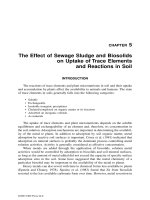
Figure 2 Clearing zones of colloidal chitin formed by chitinases produced by chitinolytic strains
E. agglomerans (1), A. caviae (2), and S. marcescens (3).
may enable them to compete successfully with fungi for chitin. Moreover, the production
of chitinase may be part of a lytic system that enables the bacteria to use living hyphae
rather than chitin as the actual growth substrate since chitin is an important constituent
of most fungal cell walls.
A strain of S. marcescens, isolated from the rhizosphere of plants grown in soil
infested with S. rolfsii Sacc., was found to be an effective biocontrol agent under green-
house conditions against this pathogen and R. solani Kuhn. A chitinase(s) produced by
the bacterium caused degradation of S. rolfsii hyphae in vitro, which provides evidence
that this enzyme has a role in biocontrol (163). S. marcescens was shown to produce
several chitinolytic enzymes, including endochitinases of 58 kD (ChiA), 54 kD (ChiB),
48 kD (C1), 36 kD (C2) and 22 kD and a 94-kD chitobiase (166–170,306). The structural
genes encoding some of these enzymes have been cloned and characterized (162,171,172).
S. marcescens mutants in which chiA had been inactivated were used to prove the impor-
tance of the ChiA chitinase for biocontrol activity toward Fusarium sp. on pea seedlings
(162). Shapira et al. (173) demonstrated the involvement of S. marcescens ChiA in the
control of S. rolfsii via genetic engineering: the enzyme produced by an E. coli strain
carrying the chiA gene of S. marcescens cloned under the control of a strong and regulated
promoter caused rapid and extensive bursting of the pathogenic fungus’s hyphal tip. A
recombinant E. coli expressing the chiA gene from S. marcescens was effective in reducing
disease incidence caused by S. rolfsii and R. solani. In addition to S. marcescens, other
Serratia species have been found to be efficient biocontrol agents. Strains of Serratia spp.
have been isolated from the rhizosphere of oilseed rape. The percentage of Serratia sp.
in this microenvironment was determined to be 12.4% of the total antifungal bacteria. S.
liquefaciens, S. plymuthica, and S. rubidaea also were found. All of the isolates showed
antifungal activity against different phytopathogenic fungi in vitro, albeit at different effi-
ciencies. The antifungal mechanisms of 18 selected strains were investigated. The direct
antifungal effect may be based on antibiosis and the production of lytic enzymes (chi-
tinases and β-1,3-glucanases). Potent siderophores are secreted by the strains to improve
iron availability. No strain was able to produce cyanide. In addition, most of the strains
secrete the plant growth hormone indole acetic acid (IAA), which can directly promote
root growth. The mechanisms were specific for each isolate (164). Other strains of S.
Copyright © 2002 Marcel Dekker, Inc.
Table 2 Examples of Lytic Enzymes Produced by Bacterial Biocontrol Agents.
Mol. masse, Encoding
Producer Enzyme kDa gene Reference
Aeromonas caviae Endochitinase 94 chiA (154, 174)
Bacillus cereus Chitobiosidase 36 ND (194)
Chromobacte- Endochitinase 52 ND (155)
rium violaceum
–‘‘– Endochitinase 37 ND (155)
Enterobacter ag- Endochitinase 58 chiA (157, 176)
glomerans
–‘‘– β-N-acetylglucosaminidase 89 ND (157)
–‘‘– β-N-acetylglucosaminidase 67 ND –‘‘–
–‘‘– Chitobiosidase 50 ND –‘‘–
E. asburiae, Cellulase ND ND (195)
Kurthia zopfii Chitinase 72 ChiSH-1 (215)
Pseudomonas Cellulase, Endochitinase, ND ND (159, 160, 184,
fluorescens β-1,3-glucanase 195)
P. stutzeri Endochitinase, β-1,3-glu- ND ND (161)
canase
P. cepacia β-1,3-glucanase ND ND (183)
Serratia marces- Endochitinase 58 chiA (162, 166–172)
cens
–‘‘– Endochitinase 54 chiB –‘‘–
–‘‘– β-N-acetylglucosaminidase 98 ND –‘‘–
Serratia plymuth- β-N-acetylglucosaminidase ND ND (164, 165)
ica
–‘‘– Endochitinase ND (164, 165)
–‘‘– Chitobiosidase ND (164, 165)
Streptomyces coe- Chitinase ND ND (185)
licolor, S. hal-
stedi
S. lydicus Chitinase ND ND (213)
S. violaceusniger Chitinase, β-1,3-glucanase ND ND (214)
Xanthomonas mal- Endochitinase ND ND (187)
tophilia
plymuthica isolated from the rhizosphere of oilseed rape showed antifungal activity against
the phytopathogenic fungus V. dahliae var. longisporum in vitro. One of these isolates,
C48, produced several chitinolytic enzymes (one N-acetyl-β-d-glucosaminidase, one chi-
tobiosidase, and one endochitinase) but no antifungal antibiotics, siderophores, or gluca-
nases. A C48 mutant, deficient in chitinolytic activity, not only lost inhibitory activity on
plates but was unable to protect oilseed rape from Verticillium sp. wilt. Therefore, the
chitinolytic activity was suggested to be exclusively responsible for strain C48’s antifungal
activity (165).
A chitinolytic strain of A. caviae, isolated from roots of healthy bean plants growing
in soil artificially infested with S. rolfsii, was able to control R. solani and F. oxysporum
f.sp. vasinfectum in cotton and S. rolfsii in beans under greenhouse conditions (154). The
strain produced an extracellular ca. 94-kD chitinase with a high degree of similarity to
Copyright © 2002 Marcel Dekker, Inc.
Figure 3 Detection of chitinolytic activity (A) and Coomassie blue staining (B) of extracellular
proteins produced by an E. agglomerans strain grown on minimal media with chitin, after separation
by SDS-PAGE. Chitinolytic activity was detected with the 4-methylumbelliferyl-β-d-N,N′-diacetyl-
chitobioside (4-MU-(GlcNAc)
2
). The bands on lane B corresponding to chitinolytic enzymes visible
on lane A are indicated by arrows.
the ChiA endochitinase of S. marcescens (174). A soil-borne chitinolytic E. agglomerans
strain IC1270 was found to be a strong antagonist of about 30 species of plant-pathogenic
bacteria and fungi in vitro and an efficient biocontrol agent of several diseases caused by
soil-borne fungal pathogens (157,175). The strain produced and excreted a complex of
chitinolytic enzymes consisting of two N-acetyl-β-d-glucosaminidases with apparent mo-
lecular masses of 89 and 67 kD and a 58-kD endochitinase. Additionally, a 50-kD chitobio-
sidase was observed in two other strains of E. agglomerans tested in this work (157). The
chitinolytic activity was induced when the strains were grown in the presence of colloidal
chitin as the sole C source; the observed chitinolytic enzymes seemed to be the most
abundant proteins secreted by the bacteria under this condition (Fig. 3). The chiA gene
of the 58-kD endochitinase was cloned from strain IC1270 in E. coli. The nucleotide
sequences of this gene showed an 86.8% identity with the corresponding gene chiA of S.
marcescens. A database search revealed that the deduced Chia_Entag protein amino acid
sequence was 87.7%, 71.9%, 52.2%, and 32.2% identical to Chia_Serma, Chia_Aerca
from A. caviae, Chia_Altso from an Alteromonas sp., and Chi1_Bacci from Bacillus circu-
lans, respectively. These comparisons suggest that the levels of diversity among various
chitinases correlate with the evolutionary distances between the bacteria that produce
them. Thus, the chitinases of S. marcescens and E. agglomerans (both Enterobacteriaceae)
are closer to those of A. caviae (the Vibrionaceae family) than to those of Alteromonas
sp. (a group of aerobic marine bacteria) or those of the Gram-positive Bacillus circulans.
The antifungal activity of the endochitinase secreted by strain IC1270 has been de-
monstrated in vitro by inhibition of F. oxysporum spore germination. The ChiA_Entag-
producing E. coli strain decreased the disease incidence of root rot caused by R. solani
on cotton under greenhouse conditions (176).
In addition to its chitinolytic activity, the strain IC1270 produces an antibiotic pyrrol-
nitrin {3-chloro-4-(2′-nitro-3′-chlorophenyl)pyrrole} with a wide range of activity against
many phytopathogenic bacteria and fungi in vitro (177). This antibiotic also was shown
to be important to biocontrol activity of several Pseudomonas and Serratia spp. rhizo-
sphere strains (164,178,179). However, the Tn5 mutants of strain IC1270, one of which
Copyright © 2002 Marcel Dekker, Inc.
is deficient in chitinolytic enzyme production but still possesses antibiotic activity, and
the other of which is deficient in both of these activities, were equally unable to protect
cotton against root rot caused by R. solani (157). These observations raised doubts as to
whether pyrrolnitrin can be considered the main compound responsible for biocontrol
activity of this E. agglomerans strain toward R. solani in the rhizosphere or whether it
needs to be combined with cell-wall lytic enzymes to provide the host strain with biocon-
trol capacity. The mode of activity of pyrrolnitrin is not yet completely understood, al-
though direct interference of pyrrolnitrin or its synthetic derivatives with fungal plasma
membranes has been demonstrated (180,181). On the basis of these data, the ability of
E. agglomerans IC1270 to produce pyrrolnitrin in combination with chitinases would be
advantageous in attacking fungal phytopathogens.
Secreted chitinolytic activity of soil-borne C. violaceum C-61 has been shown to
be important for this strain’s ability to suppress damping off of cucumber and eggplant
caused by R. solani (155). Tn5 mutants that cannot produce two of the four chitinase
isoforms are unable to inhibit mycelial growth of R. solani on plates, and their ability to
suppress the disease was much lower than that of the parental strain. Production of six
chitinolytic enzymes in another C. violaceum strain, ATCC31532, was shown to be con-
trolled by a two-component quorum-sensing mechanism (156).
Rhizosphere pseudomonads are receiving increasing attention as protectors of plants
against soil-borne fungal pathogens (6,9,182). Many of these strains have been defined by
Kloepper and coworkers (16,18) as plant-growth-promoting bacteria. Enzymatic activities
important for bacterial biocontrol capacity occasionally have been reported among Pseu-
domonas spp. strains, but compared to the extensive work on these enzymes in other
bacteria and fungi, very little has been done to explore these enzymes’ role in the biocon-
trol provided by the producer strain. Lim et al. (161) presented probably the first piece
of evidence that Pseudomonas sp. strains can produce cell-wall lytic enzymes important
for the bacterium’s biocontrol activity. P. stutzeri strain YPL-1 isolated from the rhizo-
sphere of ginseng was found to produce β-1,3-glucanase (laminarase) and chitinase activi-
ties. These extracellular lytic enzymes markedly inhibited mycelial growth and also caused
lysis of F. solani mycelia and germ tubes. Abnormal hyphal swelling and retreat were
caused by the lysing agents from P. stutzeri YPL-1, and a penetration hole was formed
on the hyphae in the region of interaction with the bacterium; the walls of this region
were lysed rapidly, causing leakage of protoplasm. In several biochemical tests with cul-
ture filtrates of P. stutzeri YPL-1 and in mutational analyses of antifungal activities of
reinforced or defective mutants, the authors found that the bacterium’s anti–F. solani
mechanism may involve a lytic enzyme rather than a toxic substance or antibiotic. Since
that report, several groups have succeeded in isolating lytic-enzyme-producing bacteria
with biocontrol activity. A β-1,3-glucanase-producing strain of Pseudomonas cepacia,
isolated on a synthetic medium with laminarin as sole C source, significantly decreased
the incidence of diseases caused by R. solani, S. rolfsii, and P. ultimum. The biocontrol
ability of this Pseudomonas sp. strain was correlated with the induction of the β-1,
3-glucanase by different fungal cell walls in synthetic medium (183). Strain PF-21 of
P. fluorescens, isolated from the rhizosphere of rice and producing chitinase and β-1,3-
glucanase, was found to be very effective in inhibiting the growth of R. solani in vitro
and in controlling rice sheath blight under greenhouse conditions. A significant relation-
ship between the antagonistic activity of P. fluorescens and its level of chitinase production
was observed (184).
In fact, chitinolytic pseudomonads are distributed widely in the environment: be-
Copyright © 2002 Marcel Dekker, Inc.
tween 0.01% and 0.5% of the total aerobic counts isolated from airtight stored cereal grain
were chitinolytic bacteria (185). Among them Gram-negative bacteria, mainly Pseudomo-
nadaceae, constituted approximately 80% of the chitinolytic population. However, only
4% of the chitinolytic isolates exibited antagonism toward fungi (185). Several chitinolytic
respesentatives of the Pseudomonadaceae family (Pseudomonas spp. and Xanthomonas
spp.) with wide ranging antifungal activity were described by Andreeva and coworkers
(186). A chitinolytic strain of X. maltophilia was shown to suppress Magnaporthe poae,
the causal agent of summer patch on Kentucky bluegrass, efficiently in growth chamber
studies (187). An endochitinase constitutively produced in low-glucose medium by several
P. fluorescens strains was suggested as an antagonistic mechanism toward R. solani (160).
To understand better the relationship between chitinolytic and antifungal properties of
bacteria that occur naturally in soils, i.e., without artificial selection, three inner dune sites
along the Dutch coast, two of which were lime-poor and one lime-rich, were selected as
a natural source of chitinolytic bacteria. These bacteria constituted up to 5.7% of the total
amount of culturable bacteria of these dune sites. Among them, Pseudomonas spp. were
the most abundant at the lime-poor sites, whereas Xanthomonas and Cytophaga spp. were
important at the lime-rich site. The percentage of bacterial isolates that were antagonistic to
fungal dune strains (Chaetomium globosum, Fusarium culmorum, F. oxysporum, Idriella
[Microdochium] bolleyi, Mucor hiemalis, Phoma exigua, Ulocladium sp.) was consider-
ably higher for chitinolytic strains than for nonchitinolytic ones. However, in many cases
the inhibition of fungal growth was not accompanied by bacterial chitinase production,
indicating that other cell-wall-degrading enzymes (β-1,3-glucanase and protease) and/or
antibiotics may also be involved in the antagonistic activities of chitinolytic bacteria
against fungi (188).
B. Biocontrol Potential of Lytic-Enzyme-Producing
Bacterial Endophytes
The term endophytic is applied to bacteria living inside a plant without causing any
visible symptoms. The best-characterized microbial endophytes are nonpathogenic fungi,
for which much compelling evidence of plant/microbe mutualism has been provided.
Some endophytic fungi are thought to produce compounds that render plant tissues less
attractive to herbivores, whereas other strains may increase host plant drought resis-
tance. In return, fungal endophytes are thought to benefit from the comparatively nutrient-
rich, buffered environment inside plants (189). However, endophytic fungi constitute
only part of the nonpathogenic microflora found naturally inside plant tissues. Bacterial
populations exceeding 1 ϫ 10
7
colony forming units (cfu) g
Ϫ1
plant matter have been
reported within tissues of various plant species. Despite their discovery more than four
decades ago, bacterial endophytes are much less known than are their fungal counterparts
(190).
Compared to use of soil-borne and rhizospheric bacteria, only a few indications
support the possibility of using endophytic bacteria as biocontrol agents. Even less is
known about the role of endophytic exoenzymes in bacterial antagonism to plant patho-
gens. Nevertheless, data obtained with plant species of agricultural and horticultural im-
portance indicate that some endophytic bacterial strains stimulate host plant growth by
acting as biocontrol agents, either through direct antagonism of microbial pathogens or
through induction of systemic resistance to disease-causing organisms. Other endophytic
bacterial strains may protect crops from plant-parasitic nematodes and insects (191). Re-
Copyright © 2002 Marcel Dekker, Inc.
gardless of the mechanism(s) involved, bacterial endophytes appear to be part of a special
type of mutualistic plant/microorganism symbiosis that warrants further study. Evidence
has been presented that plants can be protected from pathogens by manipulating these
naturally occurring microorganisms, and the potential of endophytes as biocontrol agents
has been explored (192,193). Endophytic Bacillus cereus strain 65 isolated from Sinapis
sp. was found to excrete a 36-kD chitobiosidase that exibited antifungal activity in a F.
oxysporum spore-germination bioassay (194). The ability to produce cellulases that cause
hydrolysis of wall-bound cellulose near bacterial cells was described for a systemic cotton-
plant-colonizing bacterium, Enterobacter asburiae JM22, and a cortical root–colonizing
P. fluorescens 89B-61, a plant-growth-promoting strain with biocontrol potential against
various pathogens (195).
C. Genetic Systems for Regulating the Production of Enzymes
and Secondary Metabolites Involved in Biocontrol Activity
of Gram-Negative Bacteria
In many Gram-negative bacteria, including plant-growth-promoting pseudomonads, three
types of control elements are involved in the production of some secondary metabolites
and enzymes that are synthesized at the end of exponential growth or during the stationary
phase and are involved in biological control. These are (i) two-component global regula-
tory systems that mediate transduction of environmental signals into the cells, (ii) sigma-
factor-mediated transcription by RNA polymerase, and (iii) a diffusible N-acyl-homoser-
ine lactone (N-acyl-HSL) quorum sensing signals.
Signaling pathways involve a two-component design consisting of a transmembrane
sensor kinase (designated LemA, ApdA,orGacS) and a cognate cytoplasmic response
regulator protein (GacA). The sensor kinase, when activated by a signal, phosphorylates
its own conserved histidine residue, which then serves as a histidine protein kinase (HPK),
a phosphoryl donor to an aspartate in the response-regulator protein. Two-component sys-
tems seem to be a common way for bacteria to sense and respond to their environment:
when triggered by some environmental signals, the sensor phosphorylates the regulator.
The phosphorylated regulator functions as a transcriptional activator of target genes
(159,196–198). The genes gacA, encoding the response regulator (159,196), and apdA
(also called lemA, repA, pheN,orgacS) (198,199), encoding the cognate sensor kinase,
are highly conserved among Pseudomonas spp. When mutations in gacA and apdA occur,
a similar pleiotropic phenotype develops, but production of several antibiotics, an extracel-
lular protease(s), and a tryptophan side-chain oxidase disappears (198). In vitro, all of
these compounds are synthesized at the end of exponential growth or during the stationary
phase. In response to starvation or upon entry into the stationary phase, gram-negative
bacteria undergo a process of differentiation that leads to the development of a cellular
state with markedly enhanced tolerance to a variety of individual stresses.
Besides the gacA-apdA system of global regulation, the stationary-phase sigma fac-
tor σ
S
(σ
38
), encoded by the rpoS gene, plays a critical role as a regulator of the production
of secondary metabolites responsible for the biocontrol potential of P. fluorescens (200).
The two-component regulatory system and σ
S
interact or operate through independent
regulatory circuits; however, the GacA-ApdA system influences σ
S
accumulation and the
stress response of stationary-phase cells of Pseudomonas spp. (201). The importance of
another sigma factor, σ
D
(σ
70
) encoded by the rpoD gene, for the production control of
a number of secondary metabolites and biocontrol activity was demonstrated in P. fluo-
Copyright © 2002 Marcel Dekker, Inc.