Enzymes in the Environment: Activity, Ecology and Applications - Chapter 12 ppsx
Bạn đang xem bản rút gọn của tài liệu. Xem và tải ngay bản đầy đủ của tài liệu tại đây (565.72 KB, 20 trang )
12
Microbes and Enzymes in Biofilms
Jana Jass
Umea
˚
University, Umea
˚
, Sweden
Sara K. Roberts
University of Illinois–Chicago, Chicago, Illinois
Hilary M. Lappin-Scott
Exeter University, Exeter, England
I. INTRODUCTION
Microbial enzymes and their activities have been studied primarily in pure liquid cultures
under laboratory conditions. However, in natural environments microorganisms grow at
interfaces as attached (sessile) mixed communities rather than as suspended planktonic
populations (1). Studies of microbial enzymes in soil go some way to recognizing this,
but data interpretation has often been difficult because the methodologies do not easily
differentiate between enzymes associated with surface-attached populations and those
loosely attached or free in the liquid phase. It is the aim of this chapter to discuss the
biological characteristics of these sessile microbial populations with particular reference
to their enzyme activities.
II. WHAT IS A BIOFILM?
Microorganisms attached to a surface are collectively referred to as a biofilm and are of
current interest because they are different in their phenotype and physiological characteris-
tics from the planktonic populations. Early research on biofilms was conducted in the
1920s and 1930s, primarily by Claude ZoBell (2–4), who was one of the first people
to note that bacteria existed as what he termed attached films. Three of ZoBell’s many
observations (3,4) include that bacteria attach rapidly to surfaces; planktonic bacteria are
not covered in ‘‘sticky’’ material, but sessile bacteria are; and these organisms, once asso-
ciated with a surface, secrete a ‘‘cementing’’ substance. It is significant that many of the
problems encountered by biofilm researchers today are the same as those from as long as
60 years ago; they include understanding the mechanisms underlying attachment, detach-
ment (3), microbial interactions, population diversity, biofilm structure, and growth (5).
There have been many proposed definitions of a biofilm over the years (6,7), the
most useful is given by Costerton and associates (8), who defined the biofilm as bacteria
attached to surfaces and aggregated in a hydrated polymeric matrix of their own synthesis.
Copyright © 2002 Marcel Dekker, Inc.
However,itisimportanttobeawarethatinmanyinstances,particularlyinnaturalenviron-
ments,biofilmsconsistnotonlyofbacteriabutoffungi(9,10),yeasts(11),algae(12),
andprotozoa(13).Nonetheless,muchofthepublishedliteratureconcentratesonbacterial
biofilms,althoughwiththeincreasedemergenceofinfections(11)andproblemassociated
withfungalbiofilms(10)theimportanceofstudyingmorecomplexmixedbiofilmscon-
tainingbothprokaryotesandeukaryoteshasrecentlybeenrealized(14).
III.BIOFILMLIFECYCLE
Itisofteneasiertounderstandwhatabiofilmisintermsoftheeventsthatleadtoits
formation.Moststudiesofbiofilmsinnaturalenvironmentshaveconcentratedonevents
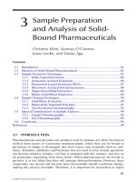
atsolid–liquidinterfaces,thecolonizationofasubmergedabioticsurfaceisdepictedin
Fig.1.Uponimmersionofanonbiologicalmaterial,suchasglassorsilica,thesurface
becomescoatedrapidlywithalayerofproteinacousmaterialcalledaconditioninglayer
(15–17).Ionsandothernutrientsourcesaccumulateattheinterface,givingrisetohigher
microenvironmentconcentrationsthatwillattractmicroorganismsfromthenutrient-and
energy-starvedliquidphasetothesurface(18).Bacteria,whichareoftenthefirstcoloniz-
ers,begintosynthesizecopiousamountsofexopolysaccharide(EPS)materialinitiated
Figure1Aschematicillustratingthelifecycleofabiofilm.
Copyright © 2002 Marcel Dekker, Inc.
upon contact with a surface (19,20). The microbial cells become embedded within this
matrix, grow, and divide to form microcolonies. Other microorganisms present in the
surrounding environment are recruited into the biofilm at all stages of biofilm development
to form complex functioning communities (14,21,22). Bryers and Characklis (23) pro-
posed a three-step colonization process that is widely accepted by many authors: initial
biofilm formation, exponential accumulation of cells and biomass, and steady state. This
pattern of colonization dictates a typical sigmoidal growth curve. Steady state is reached
when attachment of cells is equal to detachment of cells as a consequence of such processes
as predation, sloughing, and erosion.
Although biofilms are complex and dynamic and differ from environment to envi-
ronment, they all have three primary common features. First, a biofilm is associated with
an interface at which the cells accumulate. The solid–liquid interface is most frequently
studied and well characterized, however, biofilms may also form at air–liquid (24,25),
solid–air (26), and, in some cases; when a phase separation occurs, liquid–liquid inter-
faces. Second, a biofilm contains a number of microbial cells of one or more species at
an interface. A single attached microorganism does not constitute a biofilm although opin-
ions differ as to how dense the attached organisms must be to constitute, a biofilm (27).
Third, the sessile microorganisms produce an extracellular polymer matrix within which
they are embedded. This matrix, often composed of EPS synthesized by the bacteria,
may contain materials and components trapped from their surrounding environment. For
example, biofilms in natural water habitats contain particles of sediments and plant mate-
rial trapped within the matrix. In addition, the EPS matrix is believed to be important in
a variety of biofilm functions, which are discussed in the following sections. Studies have
also shown that biofilm bacteria are more resistant to antimicrobial regimes than their
planktonic counterparts (8). The exopolymer matrix may contribute to the increased resis-
tance to antimicrobial agents by either ionically binding the compounds or physically
reducing penetration of the agent through the structure, although other factors may be
involved (28–30).
IV. FUNCTION OF BIOFILM STRUCTURE AND ARCHITECTURE
It would be naive to assume that a biofilm community is simply defined as microorganisms
residing and growing at an interface. Microbes are, in fact, components of complex com-
munities continuously responding to both their immediate microenvironment and their
surrounding habitat. This is reflected in the range of biofilm structures: from thin layers
of attached cells, as seen with monocultures of some Pseudomonads or smooth colony
variants of Vibrio cholerae (31), to more complex forms of attached communities con-
taining multiple species interacting with each other (22,32).
Biofilm structure (three-dimensional) and architecture (microbial organization) are
strongly connected to the functions and survival of the microorganisms within. Research
has shown that there are many conditions that contribute to biofilm architecture; these can
be categorized as physical factors (i.e., flow rates, hydrodynamic forces, and viscosity),
chemical factors (i.e., nutrient availability and EPS composition), and biological factors
(i.e., competition and predation) (33). In practice it is difficult to separate the influences
of these categories, as there is overlap between them. The combination of species specific-
ity and physical, chemical, and biological factors influence biofilm structure in such a
manner that it is virtually impossible (and probably unrealistic) to agree on a standard
Copyright © 2002 Marcel Dekker, Inc.
Table 1 Factors That Influence Biofilm Structure
Factor Examples of variables Reference
Surface Hydrophobicity Bos et al. (15)
Roughness Lewandowski et al. (34)
Electrochemical properties Geesey et al. (35)
Hydrodynamics Mass transfer Lewandowski et al. (36)
Flow rate/velocity Stoodley et al. (37,38)
Nutrients Concentration Stoodley et al. (37,38)
Mass transfer Xu et al. (39)
Availability deBeer and Stoodley (40)
Møller et al. (41)
Exopolymeric matrix Exopolysaccharide Sutherland (42, 43)
production Skillman et al. (44)
Ecology Consortia Stoodley et al. (37)
Predation Caron (45)
Rogers et al. (46)
Cell signaling Davies et al. (47)
Source: Adapted From Ref. 13.
biofilm model. In practice, different models are available for different growth conditions,
based on a consensus of variables that influence biofilm architecture (Table 1). With ad-
vances in imaging technology, such as confocal scanning laser microscopy (48), real-
time image capture (49), and fluorescent staining (41,50,51), our understanding of biofilm
structure is increasing rapidly. Some researchers believe that biofilm structure and in-
creased resistance to antimicrobial regimes are attributable to the production of chemical
signals (52).
An increasing number of microorganisms, including bacteria and fungi, are found
to produce a range of molecules that regulate their population density; these are called
quorum sensing or cell–cell communication molecules (53). Many gram-negative bacteria
produce N-acylhomoserine lactones (AHL-s) as sensor molecules (54); however, other
substances have been implicated in signaling including 3-hydroxypalmatic acid methyl
ester produced by the plant pathogen Ralstonia solanacearum (55). Gram-positive organ-
isms (e.g. streptomyces spp.) produce different signal molecules such as small posttransla-
tionally modified peptides or other compounds related to AHLs such as γ-buytrolactones
(56). These small diffusible molecules accumulate at high cell densities within the biofilm
and, at a critical concentration, activate a genetic response in the microorganisms. The
response is not always restricted to the same species producing the sensing molecules;
other bacterial species or even eukaryotic cells (fungi, plant, or animal cell cultures) may
respond to these chemical signals (57,58). Davies et al. (47) reported that the quorum
sensing system of Pseudomonas aeruginosa that affects biofilm formation is composed
of a two-gene cascade systems, lasR-lasI and rhlR-rhlI. The lasI and rhlI gene products
are involved in the synthesis of two different AHL molecules, N-(3-oxododecanoyl)-l-
homoserine lactone and N-buytryl-l-homoserine lactone, respectively (47). The AHL mol-
ecules are required to activate the transcriptional regulators (products of lasR and rhlR)
in a sequential order, where the gene product of lasR activates the rhlR-rhlI system and
a number of virulence factors and secondary metabolites. Mutants lacking both lasI and
Copyright © 2002 Marcel Dekker, Inc.
rhlIorjustlasIgeneproductswereabletoadheretoaglasssurfacebutwerenotable
todifferentiateintothickmultilayeredbiofilms.Thissystemalsoregulatestheexpression
ofotherfactors(59),suchastypeIVpiliinP.aeruginosa(twitchingmotion),whichhave
alsobeenfoundtoinfluencethedifferentiationofadherentmonolayerstothickbiofilm
structures(60).Anincreasingnumberofbacteriaarebeingfoundtobeassociatedwith
newdensity-dependentcommunicationmolecules,bothinthelaboratoryandinsitu
(53,61).Forexample,thepresenceofAHLswasdetectedinnaturallyoccuringaquatic
biofilmsonstonesbyintroducingAgrobacteriumtumefaciensA136withalacZfusion
asanindicatororganism(61).However,itwouldbenai
¨
vetoassumethatadhesionand
biofilmformationrestsolelyontheproductionofchemicalsignals(52,62).Otherresearch
hasshownthatalthoughAHLsplayanimportantroleintheaccumulationofcellsonthe
surfaceandtheformationofbiofilms,theoverallstructureofbiofilmsgrowinginaqueous
environmentsduringtheearlystagesofcolonizationisdeterminedlargelybytheflow
conditions(37,63).
Therearetwomaindelimitingfactorsthatinfluencethestructureofabiofilmin
aqueousenvironments:flowrateandnutrientavailability.Flowcanbecategorizedas
laminarorturbulent.Laminarflowisthesmoothflowoffluidthroughapipeorduct.In
contrast,whenflowbecomeserraticandirregularitisdescribedasturbulent.Lewandow-
skiandWalser(64)foundthatthethicknessofamixedculturebiofilmwasatamaximum
nearthetransitionbetweenlaminarandturbulentflows.However,manydifferentbiofilm
structureshavebeenobserved,oftenexplainedbyexaminingthemasstransferproperties
ofthebulkliquid.Inaturbulentsystemthereisgoodmixingofnutrients,andthebulk
liquidcomesintocontactwithlargeproportionsofthebiofilmwhereuptakeofnutrients
cantakeplace.Incomparison,underlaminarflowconditionsthereispoormixingof
nutrientsinthebulkliquid,limitingnutrientavailability.Indeed,LewandowskiandWalser
(64)hypothesizedthattherewasanoptimalflowratebelowwhichbiofilmaccumulation
waslimitedbymasstransferandabovewhichbiofilmaccumulationwaslimitedbycontin-
ualcelldetachment.Manyoftherecentconfocalmicroscopestudieshaveshownthata
biofilmconsistsofmicrocoloniesofbacteriainadenseEPSmatrixwithlessdenseintersti-
tialvoidsorwaterchannels(38,65,66).Usingmicroelectrodes(50)ithasbeendemon-
stratedthattheseinterstitialvoidscontaingreaterconcentrationsofnutrientsthanthe
microcoloniesandthuscanactastransportchannelsfornutrientsandtheremovalofby-
products,makingthemanessentialstructureinanybiofilm(66).Others(67)haveshown
thattherewerefewerchannels,whichwerelessdefinedinamaturingbiofilm.Reduction
ofthesechannelswoulddecreasethemasstransportcharacteristicswithinthebulkliquid
phase,therebycontrollinggrowthrateofthemicrobeswithinthebiofilmduetoreduced
nutrientand,possibly,oxygenavailability(68).Inthelaboratoryundernutrient-richcon-
ditions,bacterialmonoculturesmayformthinlayerbiofilms;however,eventhesebio-
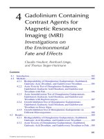
filmsarenotuniformintheirstructure.Forexample,thinlayeredbiofilmsproduced
byP.aeruginosaoftencontainbacteriadistributedoverasurfaceinterdispersedwith
uncolonizedregions(Figs.2and3),andthesespacesareasimportanttoabiofilmas
the regions containing the bacteria. Dalton et al. (69) showed that a marine bacterium,
Psychrobacter sp, SW5, produced a tightly packed multilayered biofilm on a hydrophobic
surface (silanized glass). In contrast, the biofilm formed on a hydrophilic surface (glass)
was composed of multicellular chains arranged in a more open architecture with greater
distances between the chains of bacteria. The more open biofilm structure may improve
nutrient flux and availability; however, it may have a negative effect on other processes
such as plasmid transfer, nutrient exchange, and effects of signaling molecules (70).
Copyright © 2002 Marcel Dekker, Inc.
Figure 2 A scanning electron micrograph of a P. aeruginosa biofilm formed on a silastic surface
over 48 h. This biofilm has thick regions visible here and areas that are only sparsely covered with
cells.
Figure 3 A transmission electron micrograph of a cross section of a P. aeruginosa biofilm on a
silastic surface demonstrating the cell distribution and biofilm thickness.
Copyright © 2002 Marcel Dekker, Inc.
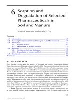
Figure 4 A schematic illustrating some of the variability in biofilm formation under different
flow conditions. In aerial view: B, biofilm clusters, shading, biofilm thickness; S, streamer structures;
R, ripples; dashed arrows, oscillation with flow; bold arrows, direction of flow around the channels
and biofilm clusters. (Based on Ref. 38.)
Stoodley et al. (38) showed that a mixed culture biofilm grown under laminar flow
conditions was ‘‘patchy’’ in that it consisted of rounded clusters of cells up to 100 µm
in diameter separated by interstitial voids containing only a thin dispersion of single cells
on the surface. Biofilms grown in turbulent flow conditions were also patchy but consisted
of migratory ripple-like patches and elongated tapered colonies termed streamers, which
oscillated in the direction of flow (38). Figure 4 is a schematic representation of the differ-
ent structures under these flow conditions. In addition to flow dynamics, the biofilm struc-
ture was affected by changing nutrient conditions. When the glucose concentration was
increased from 40 to 400 mg L
Ϫ1
, there was a parallel increase in biofilm thickness from
30 to 130 µm over a 2-day period (38). However, 10 hours after the addition of glucose,
migratory ripple-like structures had disappeared and the streamers became rounded to
form larger porous structures. When the glucose concentration was reduced to the original
concentration, the migratory ripple formation was again observed after 2 days. This may
be indicative of the biofilm responding to a decrease in nutrient availability, thereby in-
creasing its surface area and thus contact with the bulk fluid.
V. WHERE ARE BIOFILMS FOUND?
Biofilms are ubiquitous and may be beneficial or detrimental, depending on where they
are found. Beneficial biofilms are those actively employed in processes such as wastewater
and drinking water treatment (71). Slimy adherent microbial populations on the surface
Copyright © 2002 Marcel Dekker, Inc.
ofrocks(tricklingfilter)orassociatedwitharotatingbiologicalcontactor(biodisk)
areusedintheremovaloftheorganiccarbonduringsewagetreatment(72).Wastewater
ispassedoverthesurfacecontainingtheadherentmicrobialcommunities,formedof
primarilyslimeproducingZooglearamigeraandotherbacteria(72).ThethickEPS
matrixcanretainalargenumberofotherorganismstoproduceaconsortiumthatisable
toabsorbandutilizethedissolvedorganiccarbonpresentinthewater.Similarsystems
havebeenusedforbiodegradationandremediationofindustrialwastewaters.Intheenvi-
ronment,naturalselectionfavorsmicrobialcommunitiesthatcansurviveandgrowby
utilizingthewasteasnutrients.However,thisisoftenaslowprocess.Studiesareunder
wayintomethodsforincreasingthepopulationofbiodegradingorganismsatcontam-
inatedsitesbyenrichmenttechniquesandimmobilizationoftheorganismstosubstrata.
Usingimmobilizedcommunitiesinbiofilmsismoreadvantageousbecausehighercon-
centrationsoftoxiccompoundscanbeappliedandtheyarelesssusceptibletowashout
underhighfloworloading(73).Indrinkingwaterpurificationsystems,sandfilterscon-
tainingmicrobialcommunitiesareusedtoremovepotentialpathogensbytrappingthem
withintheEPSmatrixofthebiofilm(72).Inmostinstances,however,considerableprob-
lemsareassociatedwithbiofilmgrowthorbiofoulinginindustrialprocessesandcost
industryasignificantamountofmoneytodevelopcontrolregimens(74).Inwaterdistribu-
tionsystems,biofilmscausecorrosionanddegradethequalityofthewaterthroughmicro-
bialby-products.Biofilmsmayalsoharborpathogensthatputconsumersandworkersat
risk.Inthefoodanddrinkindustry,biofilmscausecontaminationandspoilageofthe
product.
Oneofthemostcommonoccurrencesofabiofilmcommunityisdentalplaque,
whichhasbeenstudiedfornearly300years(75).Over500differentmicrobialspecies
havebeenidentifiedindentalplaque(76).Whereasnormalmicrobialfloracanexistin
themouthwithoutcausinganyproblems,whenpathogensarepresentthereisapotential
forperiodontaldisease.Thisbiofilmexemplifiescooperationandcoexistenceinacomplex
microbialconsortiuminresponsetocontinualenvironmentalchanges.Oneexampleof
thiswithinthedentalbiofilmisthepresenceoftheobligateanaerobeFusobacterium
nucleatum,whichaggregateswithbothaerobesandanaerobeswithinamicrobialpopula-
tion.Thepresenceofthisorganismaidsinthesurvivalofobligateanaerobesbypromoting
aggregationinassociationwithaerobesthatremoveoxygenfromtheimmediateenviron-
ment,therebycreatingalocalizedanaerobicregion.Bradshawandassociates(76)found
thatwithoutF.nucleatumpresentinthemicrobialconsortium,theanaerobicpopulation
wassignificantlydecreased.Therefore,withinthisparticularmicrobialcommunity,bacte-
riainteractwitheachothertocreatesuitablemicroenvironmentsthatsupportthegrowth
ofadiversemicrobialpopulationthatoftenwouldnotsurviveasmonoculturesinthe
sameenvironment(76).
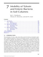
Otherfrequentlystudiedbiofilmsarethosefoundinaquatichabitats,includingfresh-
water,groundwater,andmarineenvironments,wherethemicroorganismsareattachedto
abioticorbioticsurfaces(Fig.5).Thesebiofilmsincludealargenumberofbacteriaand
unicellular marine organisms. However, there are many other habitats that are currently
being investigated with respect to microbial adhesion and biofilms, including soil particles
(77,78), plant surfaces (25,79), and animal guts (80). Microbial adhesion and physiological
processes are much more difficult to investigate in these habitats because of their diversity
and range in conditions. These habitats are divided into aquatic and nonaquatic environ-
ments and are discussed separately.
Copyright © 2002 Marcel Dekker, Inc.
Figure 5 A scanning electron micrograph of a biofilm formed on a glass slide immersed in pond
water. This multispecies biofilm demonstrates the diverse population, variable structure, and debris
present within a natural biofilm.
VI. AQUATIC ENVIRONMENTS
In fresh alpine rivers, there are nearly 1000 times more bacteria attached to surfaces
(square centimeters) than are present as planktonic cells (ml) (1,81). Biofilms composed
of bacteria and algae have been found on sediments and rock surfaces in both freshwater
and marine ecosystems. The organisms synthesize large amounts of exopolymer material,
creating a complex matrix that aids in sediment cohesion and stability in intertidal sedi-
ments (82). In other instances, when the river is polluted and has high organic matter
content, these biofilms may become so thick that they clog the river beds, creating drainage
problems and stagnation (78).
Microorganisms in aquatic environments adhere to inorganic rocks and clay particles
as well as biological/organic surfaces. Although at times biofilms are also found on living
marine animals (83) and plants (84), their surfaces have mechanisms that resist microbial
adhesion and often remain free of biofilms. In some cases, however, a biofilm on a plant
or animal surface is in a symbiotic relationship whereby the microorganisms enhance the
growth of the higher organisms. In the highly integrated rhizobia–legume symbiosis, bio-
film formation is preceded by recognition and attachment of the microorganisms to the
root surface. Root colonization is often multifunctional in that the organisms aid in nutrient
acquisition and also provide a protective environment for the plant. For example, the
colonization of mangrove roots is believed not only to help with nitrogen fixation and
solublization of phosphorus but also to protect mangroves growing in saline or brackish
waters (85).
Copyright © 2002 Marcel Dekker, Inc.
Microbialmatsareexamplesofthicklylayeredbiofilmsofphotosyntheticmicro-
organismsattachedtorocksandsedimentparticlesinaqueoushabitats(25).Theyare
oftenfoundunderextremeenvironmentalconditions.Forexample,inthevicinityofdeep
seahydrothermalvents,microorganismswithinbiofilmssurviveextremetemperatures
(86,87).Hotspringsareanotherextremehabitatwherebothhightemperaturesandsulfide
concentrationsharbormatscontaininglayersprimarilycomposedofArchaea,including
sulfate-reducingpurplebacteria(e.g.,Chloroflexisspp.,Chromatiumspp.,Thiopediaro-
seopersicinia)inassociationwithcyanobacteria(25).Additionalextremeenvironments
wheremicrobialmatsmaybefoundincludehypersalinelakes(88),terrestrialdesertswith
cyclicaldroughtanddesiccation,sodalakesandacidthermalwaterscontainingextreme
pHconditions,andregionswithhighlevelsofultraviolet(UV)irradiation(88).Themicro-
bialspeciesthatarefoundintheseextremeenvironmentsarelimitedtoprimarilycyano-
bacteria(e.g.,OscillatoriaandSpirulinaspp.)andotherssuchasDesulfovibriospp.,Beg-
giatoaspp.,andThiovulumspp.,withdifferingandvaryingdegreesoftolerance(89).
Althoughmatsareprimarilycomposedofprokaryotes,otherorganisms,suchastheeukar-
yoticCyanidiumsp.,havebeenfoundatpHlevelsbelow4.5(89).Studieshaveshownthat
mostoftheorganismswithinamatareoftennotphysiologicallyadaptedtotheextreme
environmentbutgrowthwithinlayersofathickbiofilmhelpsthemsurviveandfinda
suitablemicroniche(89).Microbialmatsareagoodexampleoftheprotectivenatureof
biofilmgrowthandthemethodwithwhichstratificationcanencouragenutrientavailability
andcycling(90).
Biofilmshavebeenobservedatotheraquaticinterfacesbesidesthoseatasolid–
liquidinterface.Forexample,instagnantwaters,biofilmsaresometimesfoundattheair–
liquidinterfaceandareoftenseenasbrownorgreenlayerscomposedofalgaeandother
aquaticmicroorganisms.Anotherexampleisthewaxytypebiofilmattheair–liquidinter-
faceformedfromtherugosephenotypeofVibriocholeraeisolatedfromstarvationme-
dium(91).Theinterfacebetweenjetfuelsandwatercanalsoharborbiofilmgrowth,such
asthefungusCladosporiumresinae(92).
VII.NONAQUATICENVIRONMENTS
Althoughbiofilmshaveoftenbeenstudiedinaquaticenvironments,morerecentstudies
haveshownthatmicroorganismswithinthickEPSmatricesorbiofilmsarealsofoundin
nonaquaticenvironmentssuchastherhizosphere(Chapter4),soil,andsubsurfaceenviron-
ments(93,94).Oneofthemorecomplexenvironmentsisthesoilecosystem,withitsmany
differentparticlesandporespaces(95).Microorganismsinthesoiladheretosurfacessuch
asinorganicsolidparticles,humicmatter,plantmaterial(roots),andmicrofauna.Plants
providelargeamountsofcarbonandothernutrientstoencouragemicrobialgrowthinthe
vicinityoftheroots,and,inturn,themicroorganismsfixnitrogen,assisttheplantin
adsorptionofnutrientsfromthesoil,andprotecttherootsagainstpathogens.Another
exampleofanonaquaticbiofilmisthecolonizationoftheleavesofplants—thephyllo-
sphere(96;Chapter6).Thesebiofilmsconsistofadiversepopulationofmicroorganisms,
including gram-positive and gram-negative bacteria, yeasts, and filamentous fungi, sup-
ported within extensive exopolymer matrices (96,25).
The primary component of biofilms is the EPS matrix produced by the bacteria. In
nonaquatic environments, the EPS matrix is of primary importance for microbial survival
since they experience intermittent flux of nutrients and water. Roberson and Firestone
Copyright © 2002 Marcel Dekker, Inc.
(93) reported that a soil Pseudomonas sp. produced more exopolysaccharide and less pro-
tein under low-water conditions than when growing in a water-rich environment. The
polysaccharide adsorbs large amounts of water, thus reducing the rate of drying and pro-
tecting the cells from desiccation. This suggests that biofilms are important for the survival
of microorganisms in the soil. Mucoid strains of Escherichia coli, Acinetobacter calcoace-
ticus, and Erwinia stewartii exhibited up to 35% greater survival under desiccating condi-
tions compared with isogenic nonmucoid mutants (97). The EPS matrices not only are
necessary for the microbes to survive low relative humidity and desiccation but may also
have a role in plant microbe symbiosis and plant pathogenesis (98). These polysaccharides
have also been shown to be virulence factors on plant pathogens such as Pseudomonas,
Erwinia, and Xanthomonas spp., however, they are also important in the symbiosis of
Rhizobia spp. (99). Both events depend on bacterial adhesion to surfaces and the formation
of a temporary biofilm.
VIII. ENZYMES IN BIOFILMS
A number of different processes that occur within a microbial biofilm contribute to the
creation of a heterogenous, dynamic environment. These processes, among many others,
include cycling and exchange of nutrients, plasmid transfer, communication via chemical
signals, and frequent deterioration of the surface (corrosion or degradation). The biofilm
microorganisms respond by having different physiological characteristics, metabolic activ-
ity, and growth rates from those of unattached organisms growing outside the biofilm
(e.g., soil aqueous phase, river water) (100).
Differences in the enzyme activities occurring within biofilms could account for
some of the differences between biofilm and planktonic cells. Although there have been
only a few reports on enzyme activities within biofilms, studies undertaken on samples
from natural environments suggest that many biofilm processes would be mediated by
enzymes. The bioremediation of pollutants by bacteria attached to soil particles, on the
surface of biodegradable materials, and on rocks in rivers or trickling filters exemplifies
enzymatic activity while bacteria are within biofilms. Potential substrates concentrated/
precipitated at the surface or diffusing into the biofilm are accessible to the attached micro-
organisms in a way that is not possible for planktonic bacteria. Vetter and Deming (101)
suggested that bacteria degrade both particulate and dissolved organic carbon by secreting
extracellular enzymes, as they are too large for direct uptake into the cell. In natural aquatic
environments it is difficult to imagine that these enzymes may have any substantial effects
except when localized near the carbon source, such as in adherent cells and biofilms (101).
This is supported by laboratory studies in which bacterial enzyme activity was important
in colloidal organic matter degradation by a biofilm community in a reactor (102). This
book identifies many processes in natural environments, and since biofilms are often found
in those environments, the enzymatic processes described would therefore be relevant
here.
Biofilms that accumulate on suspended particles in rivers, lakes, and marine systems
are considered beneficial to their environments as they play an essential role of purification
by removing suspended, settled, and dissolved organic material. Freeman et al. (103) de-
scribed river biofilms as a trophic link between dissolved nutrients in the water column
and the higher trophic levels of the ecosystem. Natural biofilms are able to biodegrade
organic compounds and transform inorganic compounds as part of their natural metabolic
Copyright © 2002 Marcel Dekker, Inc.
pathway. It is assumed that in a natural environment, such as the soil habitat, bacteria and
fungi do form biofilms and therefore the degradation of environmental pollutants occurs
in a biofilm state. The role of bacterial–fungal biofilms in the degradation of environmental
pollutants is not well understood. It is thought that the success of fungi in the role of
biodegradation is mainly attributable to the production of extracellular enzymes. Manga-
nese peroxidases and lignin peroxidases produced by Phanerochaete chrysosporium play
a key role in the detoxification and decolorization of pulp bleach plant effluent (104). A
1993 report has shown that enzymes produced by fungi and bacteria would act simulta-
neously in the biodegradation of pesticides under field conditions (105). Levanon (105)
described how the mineralization of alachlor and atrazine was mainly due to fungal activity
and that the mineralization of carbofuran and malathion was mainly due to bacterial activ-
ity. The bacterial enzyme identified to degrade malathion was a carboxylesterase. Fungi
release many hydrolytic enzymes into the soil, and these enzymes are capable of hydrolyz-
ing pesticides. Degradation by fungal enzymes may be due to less specific enzymes, as
in the case of lignin-degrading enzyme systems. However, the ability of fungi to degrade
a wide range of pesticides is believed to be related to the structural similarity of lignin
to the pesticides.
The degradation of inorganic minerals and the precipitation of toxic metals result
from enzymatic activity of microorganisms within biofilms. The degradation of mining
tailings by Thiobacillus ferrooxidans occurs when reduced iron and sulfur compounds in
ores or wastes are converted to sulfuric acid and iron (III) (106). The oxidation of these
metals is a chemical process enhanced by microbial biocatalysts, that is, enzymes that
encourage the flow of electrons (107,108). With Thiobacillus ferrooxidans, these enzymes
are believed to be located on the cell envelope; therefore, it is important that the minerals
are closely associated with the bacterium (109). Although the details of the bacterial–
mineral interaction are not fully understood, it is known that the bacterium colonizes the
mineral with the aid of its lipopolysaccharide (110). Studies have shown that even in
environments that solubilize metals, bacteria precipitate and accumulate metals within
their matrix (110). This is a natural system for cleaning the surrounding environment of
toxic metals so that other organisms and bacteria may survive.
Some surfaces act as both a colonization surface and a nutrient source. Often en-
zymes are required to degrade substances to a soluble and low-molecular-weight form that
can be utilized by the bacterium as a carbon and nitrogen source. Enzymatic degradation of
organic materials is often preceded by bacterial attachment and biofilm formation. A pri-
mary example is the hydrolysis of cellulose by anaerobic, thermophylic Clostridium ther-
mocellum (111). This organism produces an extracellular protein complex called a cellulo-
some that has a dual purpose; it both binds to and degrades cellulose. The cellulosome
produces catabolic enzymes that solubilize cellulose substances, primarily to the disaccha-
ride cellobiose, which then can be taken up by the bacterium as a nutrient source (111).
It is presumed that biofilm formation is required to position the bacterium close to the
substrate, thereby concentrating the enzyme for hydrolytic activity. Similar studies have
been undertaken on other cellulose-degrading microorganisms such as Fibrobacter succi-
nogenes and Ruminococcus albus (112).
In many cases the microorganism uses the degraded compounds as a nutrient source;
however, this process also liberates nutrients and ions for other bacteria in the biofilm
community as well as for higher organisms such as plants or animals. For example, bio-
films in the guts of ruminant animals help enzymatically degrade feed particles into sub-
stances that the animal is able to utilize (80). It is likely that each insoluble substance,
Copyright © 2002 Marcel Dekker, Inc.
such as cellulose, amylose, starch, and proteins, has its own biofilm population. The bacte-
ria produce the appropriate suite of extracellular degradative enzymes that convert the
substances into soluble nutrients available for the uptake by both bacteria and animal (80).
Many bacteria and fungi grow in association with a surface, and this process often
results in the enzymic deterioration of that surface. The surface can therefore act as a
substrate. For example, polyvinyl chloride (PVC) is largely composed of carbon, hydro-
gen, and chlorine, although these are unlikely to be available to a colonizing population;
the added UV stabilizers and colourant may well influence attachment. A 1999 report
(113) has shown that plasticizers increase the adhesion of the deteriogenic fungus Aureo-
basidium pallulans to PVC. The loss of plasticizers in PVC due to microbial degradation
results in brittleness, shrinkage, and ultimate failure of the PVC in its intended application
(113). The production of extracellular esterase by A. pallulans is thought to be instrumental
the biodeterioration of PVC (114). Stachybotrys chartarum (atra) is a saprophytic green–
black fungus that is found in many habitats and grows particularly well in high-cellulose
material, damp wallpaper, fiber board, lint, and dust. It produces potent macrocyclic thri-
chothecene toxins (satratoxins H, G, F; roridin E; verrucarin J; and trichoverrols A and
B), and is associated with chronic health problems, and is implicated in ‘‘sick building
syndrom.’’
Dental biofilms are highly complex consortia that cooperate via nutrient cycling. The
aerobic or facultative organisms often aid the survival of anaerobic bacteria by utilizing the
oxygen (115); thus the anaerobes are able to survive transient levels of oxygen. They
also produce degrading enzymes that can catalyze the degradation of polysaccharides,
glycoproteins, and complex macromolecules encountered in the oral cavity to smaller
compounds usable by the microflora. For example, some of the enzymes known to be
produced by dental microorganisms for the hydrolysis of polysaccharide components in-
clude β-galactosidase, β-N-acetylglucosaminidase, β-N-acetylgalactosaminidase, α- and
β-mannosidase, and α-fucosidase (116). Other bacteria then produce proteolytic enzymes,
such as glyprodiamino peptidase, which catalyzes the degradation of proteins into peptides
and amino acids. The degradation of these complex molecules requires cooperative and
synergistic enzymic interactions within the microbial community and, inevitably, bacterial
diversity. In turn, the resultant nutrients released support a diverse range of organisms
with different nutrient requirements (116).
Microorganisms are attracted to an inert interface as a result of the increased concen-
tration of nutrients at that interface, including some ions and larger molecules such as
amino acids and glycoproteins, which may not be easily transported into the cell. Dissolved
organic matter, ions (e.g., NH
4
ϩ
) and metals that serve as energy sources for chemolitho-
trophs (e.g., Fe
2ϩ
,Mn
2ϩ
) may initially become adsorped at a surface and also by ionic and
physical entrapment within the exopolymer. Both these processes concentrate nutrients in
the proximity of the microorganisms. Extracellular enzymes produced by some microor-
ganisms hydrolyze and degrade organic ions into soluble products available for their or
other organisms’ use. For example, an exoprotease producing Pseudomonas sp. strain S9
was used to determine adhesion by taking advantage of its degradation of ribulose-1,5-
bisphosphate carboxylase adsorbed to the surface (117). In many cases, enzymes within
biofilms are one of the normal consequence of microbial metabolism. The microenviron-
ments within a biofilm may provide the conditions in which enzyme induction and secre-
tion are stimulated. The activity of some of these enzymes outside the biofilm may be
restricted as a result of the comparatively low concentrations of substrates, inducers, and
other effector molecules outside the biofilm.
Copyright © 2002 Marcel Dekker, Inc.
Biofilm maintenance or thickness may be controlled by specific enzymes produced
by the bacterium in response to environmental stimuli. Boyd and Chakrabarty (118) dis-
covered a P. aeruginosa strain that produces alginate lyase and degrades alginate. This
may be a mechanism through which bacteria are detached from a biofilm. Similarly, Strep-
tococcus mutans produces a protein called surface protein-releasing enzyme that catalyzes
the release of bacteria from biofilms (119). This may be one method whereby organisms
become dissociated from the biofilm environment when nutrients are depleted.
IX. CONCLUSIONS
Direct evidence for the production of specific enzymes within biofilms is sparse, possibly
because of the limitations of research techniques. However, there is no doubt that extracel-
lular enzymes, either secreted or arising from dead and lysing cells, are found in the
biofilm matrix. Biofilms represent a dynamic and heterogeneous environment; therefore,
localized (and low) concentrations of enzymes have been difficult to detect. With the
current development of more sensitive microscopy methods, reporter gene technology,
molecular biology, and nanotechnology, investigating single-cell and small population
responses to environmental stimuli is possible. A number of useful reviews (120,121) and
textbooks (122–124) have been published on current techniques to study microbial adhe-
sion and biofilm formation in natural environments. This has provided a large resource
for future studies into enzymes within biofilms. Indirect evidence suggests that there are
a number of different processes that involve microbial enzymes and that they occur within
biofilms but do not occur in planktonic cells. It is assumed that biofilm cells produce
enzymes in response to environmental stimuli in order to maintain the integrity of the
overall structure, composition, and activity of the biofilm.
This chapter has shown biofilms to have three primary functions with respect to
enzyme production and activity. First, biofilm formation places the bacterium close to the
substrate or the substratum where it is concentrated for which an enzyme may be specific.
In this case, the bacterium may be producing enzymes still associated with the cell wall
(i.e., mural enzymes) requiring attachment to the substrate for catalysis to occur. Second,
a biofilm matrix may concentrate the enzyme so that it may reach concentrations at which
the activity becomes relevant. Third, the biofilm may create an environment that induces
the production of a specific enzyme.
REFERENCES
1. GG Geesey, R Mutch, JW Costerton, RB Green. Sessile bacteria: An important component
of the microbial population in small mountain streams. Limnol Oceanogr 23:1214–1223,
1978.
2. CE ZoBell, EC Allen. Attachment of marine bacteria to submerged slides. Proc Soc Exp
Biol Med 30:1409–1411, 1933.
3. CE ZoBell, EC Allen. The significance of marine bacteria in the fouling of submerged sur-
faces. J Bacteriol 29:239–251, 1935.
4. CE ZoBell. The effect of solid surfaces upon bacterial activity. J Bacteriol 46:39–56, 1935,
1943.
Copyright © 2002 Marcel Dekker, Inc.
5. HM Lappin-Scott. Claude E. ZoBell-his life and contributions to biofilm microbiology. In: CB
Bell, M Brylinsky, P Johnson-Green, eds. Proceedings of the 8th International Symposium on
Microbial Ecology, Atlantic Canada Society for Microbiol Ecology, Halfax, 2000, pp 43–50.
6. WG Characklis, KC Marshall. Biofilms: A basis for an interdisciplinary approach. In: WG
Characklis, KC Marshall, eds. Biofilms. New York: John Wiley & Sons, 1990, pp 3–15.
7. JW Costerton, Z Lewandowski, DE Caldwell, DR Korber, HM Lappin-Scott. Microbial bio-
films. Annu Rev Microbiol 49:711–745, 1995.
8. JW Costerton, PS Stewart, EP Greenberg. Bacterial biofilms: A common cause of persistent
infections. Science 284:1318–1322, 1999.
9. MV Jones. Fungal biofilms: Eradication of a common problem. In: J Wimpenny, P Handley,
P Gilbert, HM Lappin-Scott, eds. The Life and Death of Biofilm. Cardiff: BioLine, 1995,
pp 157–160.
10. JD Elvers, K Leeming, CP Moore, HM Lappin-Scott. Bacterial-fungal biofilms in flowing
water photo-processing tanks. J Appl Microbiol 84:607–618, 1998.
11. KW Millsap, HC van der Mei, R Bos, HJ Busscher. Adhesive interactions between medically
important yeasts and bacteria. FEMS Microbiol Rev 21:321–336, 1998.
12. KE Cooksey. Bacterial and algal interactions in biofilms. In: LF Melo et al., eds. Biofilms:
Science and Technology. Netherlands: Kluwer Academic, 1992, pp 163–173.
13. J Rogers, CW Keevil. Immunogold and fluorescein labelling of Legionella pneumophila
within an aquatic biofilm visualized by episcopic differential interference contrast micros-
copy. Appl Environ Microbiol 58:2326–2330, 1992.
14. P Gilbert, DG Allison. Dynamics in microbial communities: Alamarkian perspective. In: J
Wimpenny, P Gilbert, J Walker,M Brading, R Bayston, eds. Biofilms: The good, the bad
and the ugly. Cardiff: BioLine, 1999, pp 263–268.
15. R Bos, HC van der Mei, HJ Busscher. Physico-chemistry of initial microbial adhesive interac-
tion: Its mechanisms and methods for study. FEMS Microbiol Rev 23:179–230, 1999.
16. HJ Busscher, R Bos, HC van der Mei. Initial microbial adhesion is a determinant for the
strength of biofilm adhesion. FEMS Microbiol Lett 128:229–234, 1995.
17. DJ Bradshaw, PD Marsh, K Watson, K Schilling. The effect of the conditioning films on
adhesion. In: J Wimpenny, P Handley, P Gilbert, HM Lappin-Scott, eds. The Life and Death
of Biofilm. Cardiff: BioLine, 1995, pp 47–52.
18. PC Griffith, M Fletcher. Hydrolysis of protein and model dipeptide substrates by attached
and non-attached marine Pseudomonas sp. Strain NCIMB 2021. Appl Environ Microbiol 57:
2186–2191, 1991.
19. P Vandevivere, DL Kirchman. Attachment stimulates exopolysaccharide synthesis by a bac-
terium. Appl Environ Microbiol 59:3280–3286, 1993.
20. DG Davies, AM Chakrabarty, GG Geesey. Exopolysaccharide production in biofilms: Sub-
stratum activation of alginate gene expression by Pseudomonas aeruginosa. Appl Environ
Microbiol 59:1181–1186, 1993.
21. P Gilbert, DG Allison, AE Jacob, D Korber, G Wolfaardt, I Foley. Immigration of planktonic
Enterococcus faecalis cells into mature E. faecalis biofilms In: J Wimpenny, P Handley, P
Gilbert, HM Lappin-Scott, M Jones, eds. Biofilms Community Interactions and Control.
Cardiff: BioLine, 1997, pp 133–141.
22. LC Skillman, IW Sutherland, MV Jones. Co-operative biofilm formation between two species
of Enterobacteriaceae. In: J Wimpenny, P Handley, P Gilbert, HM Lappin-Scott, M Jones,
eds. Biofilms Community Interactions and Control. Cardiff: BioLine, 1997, pp 119–127.
23. J Bryers, WG Characklis. Early fouling biofilm formation in a turbulent flow system: Overall
kinetics. Water Res 15:483–491, 1981.
24. SN Wai, Y Mizunoe, S-I Yoshida. How Vibrio cholerae survive during starvation. FEMS
Microbiol Lett 180:123–131, 1999.
25. TD Brock. Life at high temperatures. Yellowstone Association, Yellowstone National Park,
1994, p 9.
Copyright © 2002 Marcel Dekker, Inc.
26. CE Morris, J-E Monier, M-A Jaques. Methods for observing microbial biofilms directly on
leaf surfaces and recovering them for isolation of culturable microorganisms. Appl Environ
Microbiol 63:1570–1576, 1997.
27. PS Handley. Is there a universal biofilm structure? J Wimpenny, P Handley, P Gilbert, HM
Lappin-Scott, eds. The Life and Death of Biofilm. Cardiff: BioLine, 1995, pp 21–25.
28. P Gilbert, DG Allison. Biofilms and their resistance towards antimicrobial agents. In: HN
Newman, M Wilson, eds. Dental plaque revisited: Oral biofilms in health and disease.
Cardiff: BioLine, 1999, pp 125–143.
29. DG Alison, P Gilbert. Modification by surface association of antimicrobial susceptibility of
bacterial populations. J Ind Microbiol 15:311–317, 1995.
30. WW Nichols. Biofilm permeability to antibacterial agents. In: J Wimpenny, W Nichols, D
Stickler, HM Lappin-Scott, eds. Bacterial Biofilms in Medicine and Industry. Cardiff: Bio-
Line, 1993, pp 141–149.
31. FH Yildiz, GK Schoolnik. Vibrio cholerae 01 El Tor: Identification of a gene cluster required
for the rugose colony type, exopolysaccharide production, chlorine resistence, and biofilm
formation. Proc Natl Acad Sci USA 96:4028–4033, 1999.
32. HM Dalton, PE March, KC Marshall. Community interaction in marine bacterial biofilms.
In: J Wimpenny, P Handley, P Gilbert, HM Lappin-Scott, M Jones, eds. Biofilms community
interactions and control. Cardiff: BioLine, 1997, pp 129–132.
33. MG Brading, J Jass, HM Lappin-Scott. Dynamics of bacterial biofilm formation. In: HM
Lappin-Scott, JW Costerton, eds. Microbial biofilms. Cambridge: Cambridge University
Press, 1995, pp 46–63.
34. Z Lewandowski, W Dickinson, W Lee. Electrochemical interactions of biofilms with metal
surfaces. Water Sci Technol 36:295–302. 1997.
35. GG Geesey, RJ Gillis, R Avci, D Daly, M Hamilton, E Shope, G Harkin. The influence of
surface features on bacterial colonisation and subsequent changes of 316L stainless steel.
Corrosion Sci 38:73–95, 1996.
36. Z Lewandowski, P Stoodley. Flow induced vibrations, drag force and pressure drop in con-
duits covered with biofilm. Water Sci Technol 32:19–26, 1995.
37. P Stoodley, F Jørgensen, P Williams, HM Lappin-Scott. The role of hydrodynamics and
AHL signalling molecules as determinants of the structure of Ps. aeruginosa biofilms. In: J
Wimpenny, P Gilbert, J Walker, M Brading, R Bayston, eds. Biofilms: The Good, the Bad
and the Ugly. Cardiff: BioLine, 1999, pp 223–230.
38. P Stoodley, I Dodds, J Boyle, HM Lappin-Scott. Influence of hydrodynamics and nutrients
on biofilm structure. J Appl Microbiol 85:19S–28S, 1999.
39. KD Xu, P Stewart, F Xia, CT Huang, GA McFeters. Spatial physiological heterogeneity in
Pseudomonas aeruginosa Biofilm is determined by oxygen availability. Appl Environ Micro-
biol 64:4035–4039, 1998.
40. D de Beer, P Stoodley. Relation between the structure of an aerobic biofilm and transport
phenomena. Water Sci Technol 32:1–18, 1995.
41. S Møller, C Sternberg, JB Andersen, BB Christensen, JL Ramos, M Givskov, S Molin. In
situ gene expression in mixed-culture biofilms: Evidence of metabolic interactions between
community members. Appl Environ Microbiol 64:721–732, 1998.
42. I Sutherland. Biofilm matrix polymers. In: HN Newman, M Wilson, eds. Dental Plaque Re-
visited: Oral Biofilms in Health and Disease. Cardiff: BioLine, 1999, pp 49–62.
43. I Sutherland. Microbial biofilm exopolysaccharides—superglue or velcro? In: J Wimpenny,
P Handley, P Gilbert, HM Lappin-Scott, M Jones, eds. Biofilms Community Interactions
and Control. Cardiff: BioLine. 1997, pp 33–40.
44. LC Skillman, IW Sutherland, MV Jones. The role of exopolysaccharides in dual species
biofilm development. J Appl Microbiol 85:S13–S18, 1999.
45. DA Caron. Grazing of attached bacteria by heterotrophic microflagellates. Microb Ecol 13:
203–218, 1987.
Copyright © 2002 Marcel Dekker, Inc.
46. J Rogers, AB Dowsett, PJ Dennis, JV Lee, CW Keevil. Influence of plumbing materials on
biofilm formation and growth of Legionella pneumophila in potable water systems. Appl
Environ Microbiol 60:1842–1851, 1994.
47. DG Davies, MR Parsek, JP Pearson, BH Iglewski, JW Costerton, EP Greenberg. The involve-
ment of cell-to-cell signals in the development of bacterial biofilm. Science 280:295–298,
1998.
48. JR Lawrence, DR Korber, BD Hoyle, JW Costerton, DE Caldwell. Optical sectioning of
microbial biofilms. J Bacteriol 173:6558–6567, 1991.
49. P Stoodley, Z Lewandowski, JD Boyle, HM Lappin-Scott. Structural deformation of bacterial
biofilms caused by short-term fluctuations in fluid shear: and in situ investigation of biofilm
rheology. Biotech Bioeng 65:83–92, 1999.
50. C-T Huang, FP Yu, GA McFeters, PS Stewart. Nonuniform spatial patterns of respiratory
activity within biofilms during disinfection. Appl Environ Microbiol 61:2252–2256, 1995.
51. PJ Robinson, JT Walker, CW Keevil, J Cole. Reporter genes and fluorescent probes for
studying the colonisation of biofilms in a drinking water supply line by enteric bacteria.
FEMS Microbiol Lett 129:183–188, 1995.
52. HG MacLehose, P Gilbert, D Allison. Homoserine lactones and biocide susceptibility: A
cautionary note. In: J Wimpenny, P, Gilbert, J Walker, M Brading, R Bayston, eds. Biofilms:
The Good, the Bad and the Ugly. Cardiff: BioLine, 1999, pp 231–236.
53. WC Fuqua, SC Winans, EP Greenberg. Quorum sensing in bacteria: The LuxR-LuxI family
of cell density-responsive transcriptional regulators. J Bacteriol 176:269–275, 1994.
54. MTG Holden, SR Chhabra, R de Nys, P Stead, NJ Bainton, PJ Hill, M Manefield, N Kumar,
M Labatte, D England, S Rice, M Givskov, GPC Salmond, GSAB Stewart, BW Bycroft, S
Kjelleberg, P Williams. Quorum-sensing cross talk: isolation and chemical characterisation
of cyclic dipeptides from Pseudomonas aeruginosa and other Gram-negative bacteria. Mol
Microbiol 33:1254–1266, 1999.
55. AB Flavier, SJ Clough, MA Schell, TP Denny. Identification of 3-hydroxypalmitic acid
methyl ester as a novel autoregulatory controlling virulence in Ralstonia solanacearum.Mol
Microbiol 26:251–259, 1997.
56. S Horinouchi, T Beppu. A-factor as a microbial hormone that controls cellular differentiation
and secondary metabolism in Streptomyces griseus. Mol Microbiol 12:859–864, 1994.
57. B Baker, P Zambryski, B Staskawicz, SP Dinesh-Kumar. Signalling in plant-microbe interac-
tions. Science 276:726–733, 1997.
58. B Rodleas, JK Lithgow, F Wisniewski-Dye, A Hardman, A Wilkinson, A Economou, P
Williams, JA Downie. Analysis of quorum-sensing-dependent control of rhizosphere-ex-
pressed (rhi) genes in Rhizobium leguminosarum bv. viciae. J Bacteriol 181:3816–3823,
1999.
59. A Glessner, RS Smith, BH Iglewski, JB Robinson. Roles of Pseudomonas aeruginosa las
and rhl quorum sensing systems in control of twitching motility. J Bacteriol 181:1623–1629,
1999.
60. GA O’Toole, R Kolter. Flagellar and twitching motility are necessary for Pseudomonas aeru-
ginosa biofilm development. Mol Microbiol 30:295–304, 1998.
61. RJC McClean, M Whieley, DJ Stickler, WC Fuqua. Evidence of autoinducer activity in
naturally occurring biofilms. FEMS Microbiol Lett 154:259–263, 1997.
62. MJ Lynch, S Swift, D Kirke, CER Dodd, CW Keevil, G Stewart, P Williams. Investigation
of quorum sensing in Aeromonas hydrophilia biofilms formed on stainless steel. In: J Wim-
penny, P Gilbert, J Walker, M Brading, R Bayston, eds. Biofilms: The Good, the Bad and
the Ugly. Cardiff: BioLine, 1999, pp 209–222.
63. MG Brading, J Boyle, HM Lappin-Scott. Biofilm formation in laminar flow using Pseudomo-
nas fluorescens EX101. J Ind Microbiol 15:297–304, 1995.
64. Z Lewandowski, G Walser. Influence of hydrodynamics on biofilm accumulation. Environ-
mental engineering proceedings, EE Div/ASCE, Reno, 1991, pp 619–624.
Copyright © 2002 Marcel Dekker, Inc.
65. P Stoodley, JD Boyle, I Dodds, HM Lappin-Scott. Consensus model of biofilm structure.
In: J Wimpenny, P Handley, P Gilbert, HM Lappin-Scott, M Jones, eds. Biofilms community
interactions and control. Cardiff: BioLine, 1997, pp 1–10.
66. JW Costerton, Z Lewandowski, D deBeer, D Caldwell, D Korber, G James. Biofilms, the
customized microniche. J Bacteriol 176:2137–2142, 1994.
67. SK Roberts, HM Lappin-Scott, K Leeming. The control of bacterial-fungal biofilms. In: J
Wimpenny, P Gilbert, J Walker, M Brading, R Bayston, eds. Biofilms: the Good, the Bad
and the Ugly. Cardiff: BioLine. 1999, pp 93–104.
68. WG Characklis, MH Turakhia, N Zelver. Transport and interfacial transport phenomena. In:
WG Characklis, KC Marshall, eds. Biofilms. New York: John Wiley & Sons, 1990, pp. 265–
340.
69. HM Dalton, LK Poulsen, P Halasz, ML Angles, AE Goodman, KC Marshall. Substratum-
induced morphological changes in a marine bacterium and their relevance to biofilm struc-
ture. J Bacteriol 176:6900–6906, 1994.
70. M Hausner, S Wuertz. High rates of conjugation in bacterial biofilms as determined by quan-
titative in situ analysis. Appl Environ Microbiol 65:3710–3717, 1999.
71. J Boyle. Biofilms in natural waters. In: J Wimpenny, W Nichols, D Stickler, HM Lappin-
Scott, eds. Bacterial Biofilms and Their Control in Medicine and Industry. Cardiff: BioLine,
1994, pp 37–40.
72. LM Prescott, JP Harley, DA Klein. Microbiology. 2nd ed. Oxford: Wm C Brown Communi-
cations, 1993, pp 832–838.
73. RC Wyndham, KJ Kennedy. Microbial consortia in industrial wastewater treatment. In: HM
Lappin-Scott, JW Costerton, eds. Microbial Biofilms. Cambridge: Cambridge University
Press, 1995, pp 183–195.
74. HM Lappin-Scott, JW Costerton. Bacterial biofilms and surface fouling. Biofouling 1:323–
342, 1989.
75. SK Roberts, C Bass, M Brading, HM Lappin-Scott, P Stoodley. Biofilm formation and struc-
ture: What’s new? In: HN Newman, M Wilson, eds. Dental Plaque Revisited: Oral Biofilms
in Health and Disease. Cardiff: BioLine, 1999, pp 15–36.
76. DJ Bradshaw, PD Marsh, GK Watson, C Allison. Interspecies interactions in microbial com-
munities. In: J Wimpenny, P Handley, P Gilbert, HM Lappin-Scott, M Jones, eds. Biofilms
Community Interactions and Control. Cardiff: BioLine, 1997, pp 63–71.
77. A
˚
Aakra, M Hesselsoe, LR Bakken. Surface attachment of ammonia-oxidizing bacteria in
soil. Microb Ecol 39:222–235, 2000.
78. TJ Battin, D Sengschmitt. Linking sediment biofilms, hydrodynamics, and river bed clogging:
evidence from a large river. Microb Ecol 37:185–196, 1999.
79. I Carmichael, IS Harper, MJ Coventry, PWJ Taylor, J Wan, MW Hickey. Bacterial colonisa-
tion and biofilm development on minimally processed vegetables. J Appl Microbiol 85:45s–
51s, 1999.
80. K-J Cheng, TA McAllister, JW Costerton. Biofilms of the ruminant digestive tract. In: HM
Lappin-Scott, JW Costerton, eds. Microbial Biofilms. Cambridge: Cambridge University
Press, 1995, pp 221–232.
81. GG Geesey, WT Richardson, HG Yeomans, RT Irvin, JW Costerton. Microscopic examina-
tion of natural sessile bacterial populations from an alpine stream. Can J Microbiol 23:1733–
1736, 1977.
82. ML Yallop, DM Paterson, P Wellsbury. Interrelationships between rates of microbial produc-
tion, exopolymer production, microbial biomass, and sediment stability in biofilms of inter-
tidal sediments. Microb Ecol 39:116–127, 2000.
83. J Jolly, HM Lappin-Scott, J Anderson, CD Clegg. Scanning electron microscopy of the gut
microflora of two earthworms: Lumbricus terrestris and Octolasion cyaneum. Microbial Ecol
26:235–245, 1993.
84. M Givskov, R de Nys, M Manefield, L Gram, R Maximilien, L Eberl, S Molin, PD Steinberg,
Copyright © 2002 Marcel Dekker, Inc.
S Kjelleberg. Eukaryotic interference with homoserine lactone mediated prokaryotic signal-
ling. J Bacteriol 178:6618–6622, 1996.
85. ME Puente, G Holguin, BR Glick, Y Basham. Root-surface colonization of black mangrove
seedlings by Asperillium halopraeferens and Azospirillum brasilense in seawater. FEMS Mi-
crobiol Ecol 29:283–292, 1999.
86. J Guezennec, O Ortega-Morales, G Raguenes, G Geesey. Bacterial colonization of artificial
substrate in the vicinity of deep-sea hydrothermal vents. FEMS Microbiol Ecol 26:89–99,
1998.
87. D Wynn-Williams, C Elli-Evans, R Leakey. Microbial ecology in Antarctica. SGM Q No-
vember, 1992, pp 99–104.
88. C Kruschel, RW Castenholz. The effect of solar UV and visible irradiance on the vertical
movement of cyanobacteria in microbial mats of hypersaline waters. FEMS Microbiol Ecol
27:53–72, 1998.
89. F Garcia-Pichel, M Mechling, RW Castenholz. Diel migration of microorganisms within a
benthic, hypersaline mat community. Appl Environ Microbiol 60:1500–1511, 1994.
90. D Krekeler, A Teske, H Cypionka. Strategies of sulfate-reducing bacteria to escape oxygen
stress in a cyanobacterial mat. FEMS Microbiol Ecol 25:89–96, 1998.
91. SN Wai, Y Mizunoe, A Takade, S-I Kawabata, S-I Yoshida. Vibrio cholerae 01 strain TSI-
4 produces the exopolysaccahride material that determine colony morphology, stress resis-
tance, and biofilm formation. Appl Environ Microbiol 64:3648–3655, 1998.
92. LM Prescott, JP Harley, DA Klein. Microbiology. 2nd ed. Oxford: Wm C Brown Communi-
cations, 1993, pp 906–907.
93. EB Roberson, MK Firestone. Relationship between desiccation and exopolysaccaride pro-
duction in a soil Pseudomonas sp. Appl Environ Microbiol 58:1284–1291, 1992.
94. RC Foster. Polysaccharides in soil fabrics. Science 214:665–667, 1981.
95. RG Burns. Microbial and enzymic activities in soil. In: WG Characklis, PA Wilderer, eds.
Structure and Function of Biofilms. New York: John Wiley & Sons, 1989, pp 333–349.
96. CE Morris, J-M Monier, M-A Jacques. A technique to quantify the population size and com-
position of biofilm components in communities of bacteria in the phyllosphere. Appl Environ
Microbiol 64:4789–4795, 1998.
97. T Ophir, DL Gutmick. A role for exopolysaccharides in the protection of microorganisms
from desiccation. Appl Environ Microbiol 60:740–745, 1994.
98. M Wilson, SE Lindow. Effect of phenotypic plasticity on epiphytic survival and colonization
by Pseudomonas syringae. Appl Environ Microbiol 59:410–416, 1993.
99. JA Leigh, DL Coplin. Exopolysaccharide in plant-bacteria interaction. Annu Rev Microbiol
46:307–346, 1992.
100. C Freeman, MA Lock. [
3
H] Thymidine incorporation as a measure of bacterial growth within
intact river biofilms. Sci Total Environ 138:161–167, 1993.
101. YA Vetter, JW Deming. Growth rates of marine bacterial isolates on particulate organic
substrates solubilized by freely released extracellular enzymes. Microb Ecol 37:86–94, 1999.
102. T Larsen, P Herremoes. Degradation mechanisms of colloidal organic matter in biofilm reac-
tors. Wat Res 28:1443–1452, 1994.
103. C Freeman, PJ Chapman, K Gilman, MA Lock, B Reynolds, HS Wheater. Ion exchange
mechanisms and the entrapment of nutrients by river biofilms. Hydrobiologia 297:61–65,
1995.
104. FC Michel, SB Dass, EA Grulke, CA Reddy. Role of manganese perioxidases and lignin
perioxidases of Phanerochaete chrysosporium in the decolorization of kraft bleach plant ef-
fluent. Appl Environ Microbiol 57:2368–2375, 1991.
105. D Levanon. Roles of fungi and bacteria in the mineralization of the pesticides atrazine,
alachlor, malathion and carbofuran in soil. Soil Biol Biochem 25:1097–1105, 1993.
106. F Baldi, T Clark, SS Pollack, GJ Olson. Leaching of pirites of various reactivities by Thioba-
cillus ferrooxidans. Appl Environ Microbiol 58:1853–1856, 1992.
Copyright © 2002 Marcel Dekker, Inc.
107. PLAM Corstjens, JPM de Vrind, P Westbroek, EW de Vrind-de Jong. Enzymatic iron oxida-
tion by Leptothrix discophora: Identification of an iron-oxidising protein. Appl Environ Mi-
crobiol 58:450–454, 1992.
108. G Southam, FG Ferris, TJ Beveridge. Mineralized bacterial biofilms in sulphide tailings and
in acid mine drainage systems. In: HM Lappin-Scott, JW Costerton, eds. Microbial Biofilms.
Cambridge: Cambridge University Press, 1995, pp 148–170.
109. G Southam, TJ Beveridge. Enumeration of Thiobacilli within pH-neutral and acidic mine
tailings and their role in the development of secondary mineral soil. Appl Environ Microbiol
58:1904–1912, 1992.
110. G Southam, TJ Beveridge. Examination of lipopolysaccharide (o-antigen) populations of
Thiobacillus ferrooxidans from two mine tailings. Appl Environ Microbiol 59:1283–1288,
1993.
111. EA Bayer, E Morag, Y Shoham, J Tormo, R Lamed. The cellulosome: A cell surface organ-
elle for the adhesion to and degradation of cellulose. In: M Fletcher, ed. Bacterial Adhesion:
Molecular and Ecological Diversity. New York. Wiley-Liss, 1996, pp 155–182.
112. J Gong, CW Forsberg. Factors affecting adhesion of Fibrobacter succinogenes subsp. Succi-
nogenes S85 and adherence-defective mutants to cellulose. Appl Environ Microbiol 55:
3039–3044.
113. JS Webb, HC Van der Mei, M Nixon, IM Eastwood, M Greenhalgh, S Read, GD Robson,
PS Handley. Plasticizers increase adhesion of the deteriogenic fungus Aureobasidium pullu-
lans to polyvinyl chloride. Appl Environ Microbiol 65:3575–3581, 1999.
114. JS Webb, M Nixon, IM Eastwood, M Greenhalgh, GD Robson, PS Handley. Fungal colonisa-
tion and biodeterioration of plasticized polyvinyl chloride. Appl Environ Microbiol 66:3194–
3200, 2000
115. DJ Bradshaw, PD Marsh, GK Watson, C Allison. Role of Fusobacterium nucleatum and
coaggregation in anaerobe survival in planktonic and biofilm oral microbial communities
during aeration. Infect Immun 66:4729–4732, 1998.
116. DJ Bradshaw, KA Holmer, PD Marsh, D Beighton. Metabolic cooperation in oral microbial
communities during growth on mucin. Microbiol 140:3407–3412, 1994.
117. M-O Samuelsson, DL Kirchman. Degradation of adsorbed protein by attached bacteria in
relation to surface hydrophobicity. Appl Environ Microbiol 56:3643–3648, 1991.
118. A Boyd, AM Chakrabarty. Role of alginate lyase in cell detachment of Pseudomonas aerugi-
nosa. Appl Environ Microbiol 60:2355–2359, 1994.
119. SF Lee, YH Li, GH Bowden. Detachment of Streptococcus mutans biofilm cells by an endog-
enous enzymatic activity. Infect Immun 64:1035–1038, 1996.
120. JC Rayner, HM Lappin-Scott. Experimental biofilms and their applications on the study of
environmental processes. In: C Edwards, ed. Environmental monitoring of bacteria. Clifton
NJ: Humana Press, 1999 pp 279–306.
121. L Hall-Stoodley, JC Rayner, P Stoodley, HM Lappin-Scott. Establishment of experimental
biofilms using the modified Robbins device and flow cells. In: C Edwards ed. Environmental
monitoring of bacteria. Clifton, NJ: Humana Press, 1999 pp 307–319.
122. HA Yuehuei, RJ Friedman. eds. Handbook of bacterial adhesion. Clifton NJ: Humana Press,
2000.
123. H-C Flemming, U Szewzyk, T Griebe ed. Biofilms; Investigative methods and applications.
Lancaster: Technomic, 2000.
124. R Doyle. Biofilms. Vol. 310. Methods in Enzymology, Biofilms. San Diego: Academic Press,
1999.
Copyright © 2002 Marcel Dekker, Inc.