TOXICOLOGICAL CHEMISTRY AND BIOCHEMISTRY - CHAPTER 3 pps
Bạn đang xem bản rút gọn của tài liệu. Xem và tải ngay bản đầy đủ của tài liệu tại đây (599.41 KB, 20 trang )
C
HAPTER
3
Biochemistry
3.1 BIOCHEMISTRY
Most people have had the experience of looking through a microscope at a single cell. It may
have been an amoeba, alive and oozing about like a blob of jelly on the microscope slide, or a cell
of bacteria, stained with a dye to make it show up more plainly. Or it may have been a beautiful
cell of algae with its bright green chlorophyll. Even the simplest of these cells is capable of carrying
out a thousand or more chemical reactions. These life processes fall under the heading of
bio-
chemistry
, the branch of chemistry that deals with the chemical properties, composition, and
biologically mediated processes of complex substances in living systems.
Biochemical phenomena that occur in living organisms are extremely sophisticated. In the
human body, complex metabolic processes break down a variety of food materials to simpler
chemicals, yielding energy and the raw materials to build body constituents, such as muscle, blood,
and brain tissue. Impressive as this may be, consider a humble microscopic cell of photosynthetic
cyanobacteria only about a micrometer in size, which requires only a few simple inorganic chemicals
and sunlight for its existence. This cell uses sunlight energy to convert carbon from CO
2
, hydrogen
and oxygen from H
2
O, nitrogen from NO
3
–
, sulfur from SO
4
2–
, and phosphorus from inorganic
phosphate into all the proteins, nucleic acids, carbohydrates, and other materials that it requires to
exist and reproduce. Such a simple cell accomplishes what could not be done by human endeavors
even in a vast chemical factory costing billions of dollars.
Ultimately, most environmental pollutants and hazardous substances are of concern because of
their effects on living organisms. The study of the adverse effects of substances on life processes
requires some basic knowledge of biochemistry. Biochemistry is discussed in this chapter, with an
emphasis on the aspects that are especially pertinent to environmentally hazardous and toxic
substances, including cell membranes, deoxyribonucleic acid (DNA), and enzymes.
Biochemical processes not only are profoundly influenced by chemical species in the environ-
ment, but they largely determine the nature of these species, their degradation, and even their
syntheses, particularly in the aquatic and soil environments. The study of such phenomena forms
the basis of
environmental biochemistry
.
1
3.1.1 Biomolecules
The biomolecules that constitute matter in living organisms are often polymers with molecular
masses of the order of a million or even larger. As discussed later in this chapter, these biomolecules
may be divided into the categories of carbohydrates, proteins, lipids, and nucleic acids. Proteins
and nucleic acids consist of macromolecules, lipids are usually relatively small molecules, and
carbohydrates range from relatively small sugar molecules to high-molecular-mass macromolecules,
such as those in cellulose.
L1618Ch03Frame Page 59 Tuesday, August 13, 2002 5:54 PM
Copyright © 2003 by CRC Press LLC
The behavior of a substance in a biological system depends to a large extent upon whether the
substance is hydrophilic (water-loving) or hydrophobic (water-hating). Some important toxic sub-
stances are hydrophobic, a characteristic that enables them to traverse cell membranes readily. Part
of the detoxification process carried on by living organisms is to render such molecules hydrophilic,
therefore water soluble and readily eliminated from the body.
3.2 BIOCHEMISTRY AND THE CELL
The focal point of biochemistry and biochemical aspects of toxicants is the
cell
, the basic
building block of living systems where most life processes are carried. Bacteria, yeasts, and some
algae consist of single cells. However, most living things are made up of many cells. In a more
complicated organism the cells have different functions. Liver cells, muscle cells, brain cells, and
skin cells in the human body are quite different from each other and do different things. Cells are
divided into two major categories depending upon whether or not they have a nucleus:
eukaryotic
cells have a nucleus, and
prokaryotic
cells do not. Prokaryotic cells are found in single-celled
bacteria. Eukaryotic cells compose organisms other than bacteria.
3.2.1 Major Cell Features
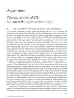
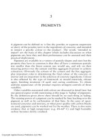
Figure 3.1 shows the major features of the
eukaryotic cell
, which is the basic structure in which
biochemical processes occur in multicelled organisms. These features are as follows:
•
Cell membrane
, which encloses the cell and regulates the passage of ions, nutrients, lipid-soluble
(fat-soluble) substances, metabolic products, toxicants, and toxicant metabolites into and out of
the cell interior because of its varying
permeability
for different substances. The cell membrane
protects the contents of the cell from undesirable outside influences. Cell membranes are composed
in part of phospholipids that are arranged with their hydrophilic (water-seeking) heads on the cell
membrane surfaces and their hydrophobic (water-repelling) tails inside the membrane. Cell mem-
branes contain bodies of proteins that are involved in the transport of some substances through
the membrane. One reason the cell membrane is very important in toxicology and environmental
biochemistry is because it regulates the passage of toxicants and their products into and out of the
cell interior. Furthermore, when its membrane is damaged by toxic substances, a cell may not
function properly and the organism may be harmed.
•
Cell nucleus
, which acts as a sort of “control center” of the cell. It contains the genetic directions
the cell needs to reproduce itself. The key substance in the nucleus is DNA.
Chromosomes
in the
Figure
3.1
Some major features of the eukaryotic cell in animals (left) and plants (right).
Nucleus
Mitochondria
Lysosome
Ribosome
Cell membrane
Golgi body
Vacuole
Vacuole
Cell wall
Chloroplast
Starch grain
Mitochondria
L1618Ch03Frame Page 60 Tuesday, August 13, 2002 5:54 PM
Copyright © 2003 by CRC Press LLC
cell nucleus are made up of combinations of DNA and proteins. Each chromosome stores a separate
quantity of genetic information. Human cells contain 46 chromosomes. When DNA in the nucleus
is damaged by foreign substances, various toxic effects, including mutations, cancer, birth defects,
and defective immune system function may occur.
•
Cytoplasm
, which fills the interior of the cell not occupied by the nucleus. Cytoplasm is further
divided into a water-soluble proteinaceous filler called
cytosol
, in which are suspended bodies
called
cellular organelles
, such as mitochondria or, in photosynthetic organisms, chloroplasts.
•
Mitochondria
, “powerhouses” that mediate energy conversion and utilization in the cell. Mito-
chondria are sites in which food materials — carbohydrates, proteins, and fats — are broken down
to yield carbon dioxide, water, and energy, which is then used by the cell for its energy needs.
The best example of this is the oxidation of the sugar glucose, C
6
H
12
O
6
:
C
6
H
12
O
6
+ 6O
2
→
6CO
2
+ 6H
2
O + energy
This kind of process is called
cellular respiration
.
•
Ribosomes
, which participate in protein synthesis.
•
Endoplasmic reticulum
, which is involved in the metabolism of some toxicants by enzymatic
processes.
•
Lysosome
, a type of organelle that contains potent substances capable of digesting liquid food
material. Such material enters the cell through a “dent” in the cell wall, which eventually becomes
surrounded by cell material. This surrounded material is called a
food vacuole
. The vacuole merges
with a lysosome, and the substances in the lysosome bring about digestion of the food material.
The digestion process consists largely of
hydrolysis reactions
in which large, complicated food
molecules are broken down into smaller units by the addition of water.
•
Golgi bodies
, which occur in some types of cells. These are flattened bodies of material that serve
to hold and release substances produced by the cells.
•
Cell walls
of plant cells. These are strong structures that provide stiffness and strength. Cell walls
are composed mostly of cellulose, which will be discussed later in this chapter.
•
Vacuoles
inside plant cells that often contain materials dissolved in water.
•
Chloroplasts
in plant cells that are involved in photosynthesis (the chemical process that uses energy
from sunlight to convert carbon dioxide and water to organic matter). Photosynthesis occurs in these
bodies. Food produced by photosynthesis is stored in the chloroplasts in the form of
starch grains
.
3.3 PROTEINS
Proteins
are nitrogen-containing organic compounds that are the basic units of life systems.
Cytoplasm, the jelly-like liquid filling the interior of cells, is made up largely of protein. Enzymes,
which act as catalysts of life reactions, are made of proteins; they are discussed later in the chapter.
Proteins are composed of
amino acids
(Figure 3.2) joined together in huge chains. Amino acids are
organic compounds that contain the carboxylic acid group,
–
CO
2
H, and the amino group,
–
NH
2
. They
are sort of a hybrid of carboxylic acids and amines (see Sections 1.8.1 and 1.8.2). Proteins are polymers,
or
macromolecules
, of amino acids containing from approximately 40 to several thousand amino acid
groups joined by peptide linkages. Smaller molecule amino acid polymers, containing only about 10
to about 40 amino acids per molecule, are called
polypeptides
. A portion of the amino acid left after
the elimination of H
2
O during polymerization is called a
residue
. The amino acid sequence of these
residues is designated by a series of three-letter abbreviations for the amino acid.
Natural amino acids all have the following chemical group:
RCC
O
OH
H
N
HH
L1618Ch03Frame Page 61 Tuesday, August 13, 2002 5:54 PM
Copyright © 2003 by CRC Press LLC
In this structure the –NH
2
group is always bonded to the carbon next to the –CO
2
H group. This is
called the “alpha” location, so natural amino acids are alpha-amino acids. Other groups, designated as
R, are attached to the basic alpha-amino acid structure. The R groups may be as simple as an atom of
H found in glycine, or they may be as complicated as the structure of the R group in tryptophan:
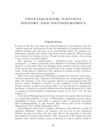
Figure
3.2
Amino acids that occur in proteins. Those marked with an asterisk cannot be synthesized by the
human body and must come from dietary sources.
COH
O
C
NH
2
H
H COH
O
C
NH
2
H
OH
C
H
H
3
C
COH
O
C
NH
2
H
C
H
H
Glycine (gly) Serine (ser)
COH
O
C
NH
2
H
CHO
H
H
COH
O
C
NH
2
C
HH
H
CH
CH
3
CH
3
COH
O
C
NH
2
CC
H
H
HH
H
SH
3
C
Isoleucine (ile)*
Methionine (met)*
COH
O
C
NH
2
H
CHS
H
H
COH
O
C
NH
2
H
CH
3
H
CC
H
H
H
3
C
COH
O
C
NH
2
H
C
H
HH
H
CH
2
N
O
C
COH
O
C
NH
2
C
H
H
3
C
H
3
C
H
COH
O
C
NH
2
CC
H
H
HO
H
2
N
COH
O
C
NH
2
H
H
H
C
H
H
CC
H
H
C
H
H
H
3
N
+
Tyrosine (tyr)
HO C OH
O
C
NH
2
H
C
H
H
COH
O
C
NH
2
H
C
O
HO C
H
H
COH
O
C
NH
2
H
H
3
C
O
OHC
H
H
H
H
H
H
H
H
C
CC
C
N
Cysteine (cys)
Glutamine (gin)
Valine (val)* Lysine (lys)*
Tryptophan (try)*
COH
O
C
NH
2
H
C
H
H
H
N
Phenylalanine (phe)*
Alanine (ala)
COH
O
C
NH
2
H
C
H
HNNH
+
H
Histidine (his)
Proline (pro)
Leucine (leu)*
Aspartic acid (asp)
Asparagine (asn)
COH
O
C
NH
2
H
H
H
C
H
H
CHO
O
C
Glutamic acid (glu)
COH
O
C
NH
2
H
H
H
C
H
H
CNC
H
H
H
C
NH
2
+
H
2
N
Arginine (arg)
Threonine (thr)*
HCC
O
OH
H
N
HH
+
HC C
O
O
–
H
NH
H
H
Glycine
Zwitterion
form
L1618Ch03Frame Page 62 Tuesday, August 13, 2002 5:54 PM
Copyright © 2003 by CRC Press LLC
As shown in Figure 3.2, there are 20 common amino acids in proteins. These are shown with
uncharged
–
NH
2
and
–
CO
2
H groups. Actually, these functional groups exist in the charged
zwitterion
form, as shown for glycine above.
Amino acids in proteins are joined together in a specific way. These bonds constitute the
peptide
linkage
. The formation of peptide linkages is a condensation process involving the loss of water.
For example, consider the condensation of alanine, leucine, and tyrosine shown in Figure 3.3. When
these three amino acids join together, two water molecules are eliminated. The product is a
tri
peptide
since there are three amino acids involved. The amino acids in proteins are linked as shown for
this tripeptide, except that many more monomeric amino acid groups are involved.
Proteins may be divided into several major types that have widely varying functions. These are
listed in Table 3.1.
Figure
3.3
Condensation of alanine, leucine, and tyrosine to form a tripeptide consisting of three amino acids
joined by peptide linkages (outlined by dashed lines).
Table
3.1
Major Types of Proteins
Type of Protein Example Function and Characteristics
Nutrient Casein (milk protein) Food source; people must have an adequate supply
of nutrient protein with the right balance of amino
acids for adequate nutrition
Storage Ferritin Storage of iron in animal tissues
Structural Collagen (tendons), keratin (hair) Structural and protective components in organisms
Contractile Actin, myosin in muscle tissue Strong, fibrous proteins that can contract and cause
movement to occur
Transport Hemoglobin Transport inorganic and organic species across cell
membranes, in blood, between organs
Defense — Antibodies against foreign agents such as viruses
produced by the immune system
Regulatory Insulin, human growth hormone Regulate biochemical processes such as sugar
metabolism or growth by binding to sites inside cells
or on cell membranes
Enzymes Acetylcholine esterase Catalysts of biochemical reactions (see Section 3.6)
+ +
Alanine
Leucine
Tyrosine
CH
2
NC
O
OH
H
CHH
OH
CH
2
NC
O
OH
H
CHH
C
H
H
3
CCH
3
CH
2
NC
O
OH
CH
3
H
CC
O
OH
H
CHH
OH
NCH
2
NC
O
CH
3
H
N
CH
3
H
3
C
H
C
HHC
HO
CC
HH
L1618Ch03Frame Page 63 Tuesday, August 13, 2002 5:54 PM
Copyright © 2003 by CRC Press LLC
3.3.1 Protein Structure
The order of amino acids in protein molecules, and the resulting three-dimensional structures
that form, provide an enormous variety of possibilities for
protein structure
. This is what makes
life so diverse. Proteins have primary, secondary, tertiary, and quaternary structures. The structures
of protein molecules determine the behavior of proteins in crucial areas such as the processes by
which the body’s immune system recognizes substances that are foreign to the body. Proteinaceous
enzymes depend on their structures for the very specific functions of the enzymes.
The order of amino acids in the protein molecule determines its primary structure.
Secondary
protein structures
result from the folding of polypeptide protein chains to produce a maximum
number of hydrogen bonds between peptide linkages:
Further folding of the protein molecules held in place by attractive forces between amino acid side
chains gives proteins a
secondary structure
, which is determined by the nature of the amino acid
R groups. Small R groups enable protein molecules to be hydrogen-bonded together in a parallel
arrangement, whereas large R groups produce a spiral form known as an
alpha-helix
.
Tertiary structures
are formed by the twisting of alpha-helices into specific shapes. They are
produced and held in place by the interactions of amino side chains on the amino acid residues
constituting the protein macromolecules. Tertiary protein structure is very important in the processes
by which enzymes identify specific proteins and other molecules upon which they act. It is also
involved with the action of antibodies in blood, which recognize foreign proteins by their shape
and react to them. This is what happens in the phenomenon of disease immunity, where antibodies
in blood recognize specific proteins from viruses or bacteria and reject them.
Two or more protein molecules consisting of separate polypeptide chains may be further
attracted to each other to produce a
quaternary structure
.
Some proteins are
fibrous proteins
, which occur in skin, hair, wool, feathers, silk, and tendons.
The molecules in these proteins are long and threadlike and are laid out parallel in bundles. Fibrous
proteins are quite tough and do not dissolve in water.
An interesting fibrous protein is keratin, which is found in hair. The cross-linking bonds between
protein molecules in keratin are –S–S– bonds formed from two HS– groups in two molecules of
the amino acid cysteine. These bonds largely hold hair in place, thus keeping it curly or straight.
A “permanent” consists of breaking the bonds chemically, setting the hair as desired, and then
reforming the cross-links to hold the desired shape.
Aside from fibrous protein, the other major type of protein form is the
globular protein
. These
proteins are in the shape of balls and oblongs. Globular proteins are relatively soluble in water. A
typical globular protein is hemoglobin, the oxygen-carrying protein in red blood cells. Enzymes
are generally globular proteins.
CO
NHOC
HN
Illustration of hydrogen bonds between
N and O atoms in peptide linkages, which
constitutes protein secondary structures
Hydrogen bonds
Hydrogen bonds
L1618Ch03Frame Page 64 Tuesday, August 13, 2002 5:54 PM
Copyright © 2003 by CRC Press LLC
3.3.2 Denaturation of Proteins
Secondary, tertiary, and quaternary protein structures are easily changed by a process called
denaturation
. These changes can be quite damaging. Heating, exposure to acids or bases, and even
violent physical action can cause denaturation to occur. The albumin protein in egg white is
denatured by heating so that it forms a semisolid mass. Almost the same thing is accomplished by
the violent physical action of an egg beater in the preparation of meringue. Heavy metal poisons
such as lead and cadmium change the structures of proteins by binding to functional groups on the
protein surface.
3.4 CARBOHYDRATES
Carbohydrates
have the approximate simple formula CH
2
O and include a diverse range of
substances composed of simple sugars such as glucose:
High-molecular-mass
polysaccharides
, such as starch and glycogen (animal starch), are biopoly-
mers of simple sugars.
Photosynthesis in a plant cell converts the energy from sunlight to chemical energy in a
carbohydrate, C
6
H
12
O
6
. This carbohydrate may be transferred to some other part of the plant for
use as an energy source. It may be converted to a water-insoluble carbohydrate for storage until it
is needed for energy. Or it may be transformed to cell wall material and become part of the structure
of the plant. If the plant is eaten by an animal, the carbohydrate is used for energy by the animal.
The simplest carbohydrates are the
monosaccharides
. These are also called
simple sugars
.
Because they have six carbon atoms, simple sugars are sometimes called
hex
oses. Glucose (formula
shown above) is the most common simple sugar involved in cell processes. Other simple sugars
with the same formula but somewhat different structures are fructose, mannose, and galactose.
These must be changed to glucose before they can be used in a cell. Because of its use for energy
in body processes, glucose is found in the blood. Normal levels are from 65 to 110 mg of glucose
per 100 ml of blood. Higher levels may indicate diabetes.
Units of two monosaccharides make up several very important sugars known as
disaccharides
.
When two molecules of monosaccharides join together to form a disaccharide,
C
6
H
12
O
6
+ C
6
H
12
O
6
→
C
12
H
22
O
11
+ H
2
O (3.4.1)
a molecule of water is lost. Recall that proteins are also formed from smaller amino acid molecules
by condensation reactions involving the loss of water molecules. Disaccharides include sucrose (cane
sugar used as a sweetener), lactose (milk sugar), and maltose (a product of the breakdown of starch).
Polysaccharides
consist of many simple sugar units hooked together. One of the most important
polysaccharides is
starch
, which is produced by plants for food storage. Animals produce a related
material called
glycogen
. The chemical formula of starch is (C
6
H
10
O
5
)
n
, where
n
may represent a
number as high as several hundred. What this means is that the very large starch molecule consists
CC
C
C
CO
H
CH
2
OH
H
OH
H
H
OH
H
OH
HO
Glucose molecule
L1618Ch03Frame Page 65 Tuesday, August 13, 2002 5:54 PM
Copyright © 2003 by CRC Press LLC
of many units of C
6
H
10
O
5
joined together. For example, if
n
is 100, there are 6 times 100 carbon
atoms, 10 times 100 hydrogen atoms, and 5 times 100 oxygen atoms in the molecule. Its chemical
formula is C
600
H
1000
O
500
. The atoms in a starch molecule are actually present as linked rings,
represented by the structure shown in Figure 3.4. Starch occurs in many foods, such as bread and
cereals. It is readily digested by animals, including humans.
Cellulose is a polysaccharide that is also made up of C
6
H
10
O
5
units. Molecules of cellulose are
huge, with molecular weights of around 400,000. The cellulose structure (Figure 3.5) is similar to
that of starch. Cellulose is produced by plants and forms the structural material of plant cell walls.
Wood is about 60% cellulose, and cotton contains over 90% of this material. Fibers of cellulose
are extracted from wood and pressed together to make paper.
Humans and most other animals cannot digest cellulose. Ruminant animals (cattle, sheep, goats,
moose) have bacteria in their stomachs that break down cellulose into products that can be used
by the animal. Chemical processes are available to convert cellulose to simple sugars by the reaction
(C
6
H
10
O
5
)
n
+ nH
2
O → nC
6
H
12
O
6
(3.4.2)
cellulose glucose
where n may be 2000 to 3000. This involves breaking the linkages between units of C
6
H
10
O
5
by
adding a molecule of H
2
O at each linkage, a hydrolysis reaction. Large amounts of cellulose from
wood, sugar cane, and agricultural products go to waste each year. The hydrolysis of cellulose
enables these products to be converted to sugars, which can be fed to animals.
Carbohydrate groups are attached to protein molecules in a special class of materials called
glycoproteins. Collagen is a crucial glycoprotein that provides structural integrity to body parts.
It is a major constituent of skin, bones, tendons, and cartilage.
3.5 LIPIDS
Lipids are substances that can be extracted from plant or animal matter by organic solvents,
such as chloroform, diethyl ether, or toluene (Figure 3.6). Whereas carbohydrates and proteins are
Figure 3.4 Part of a starch molecule showing units of C
6
H
10
O
5
condensed together.
Figure 3.5 Part of the structure of cellulose.
CC
C
C
CO
H
CH
2
OH
H
O
OH
H
H
OH
H
O
CC
C
C
CO
H
CH
2
OH
H
C
OH
H
H
OH
H
O
C
C
C
CO
H
CH
2
OH
H
OH
H
H
OH
H
O
CC
C
C
CO
H
CH
2
OH
H
O
OH
H
H
OH
O
H
C
OC
C
C
CC
OH
CH
2
OH
H
C
OH
H
C
O
H
H
C
CO
H
CH
2
OH
H
OH
H
H
OH
O
H
H
L1618Ch03Frame Page 66 Tuesday, August 13, 2002 5:54 PM
Copyright © 2003 by CRC Press LLC
characterized predominately by the monomers (monosaccharides and amino acids) from which
they are composed, lipids are defined essentially by their physical characteristic of organophilicity.
The most common lipids are fats and oils composed of triglycerides formed from alcohol glycerol,
CH
2
(OH)CH(OH)CH
2
(OH), and a long-chain fatty acid such as stearic acid, CH
3
(CH
2
)
16
C(O)OH
(Figure 3.7). Numerous other biological materials, including waxes, cholesterol, and some vitamins
and hormones, are classified as lipids. Common foods, such as butter and salad oils, are lipids.
Long-chain fatty acids, such as stearic acid, are also organic soluble and are classified as lipids.
Lipids are toxicologically important for several reasons. Some toxic substances interfere with
lipid metabolism, leading to detrimental accumulation of lipids. Many toxic organic compounds
are poorly soluble in water, but are lipid soluble, so that bodies of lipids in organisms serve to
dissolve and store toxicants.
An important class of lipids consists of phosphoglycerides (glycerophosphatides). These com-
pounds may be regarded as triglycerides in which one of the acids bonded to glycerol is ortho-
Figure 3.6 Lipids are extracted from some biological materials with a soxhelet extractor (above). The solvent
is vaporized in the distillation flask by the heating mantle, rises through one of the exterior tubes
to the condenser, and is cooled to form a liquid. The liquid drops onto the porous thimble containing
the sample. Siphon action periodically drains the solvent back into the distillation flask. The extracted
lipid collects as a solution in the solvent in the flask.
Condenser
Cooling
water in
Cooling
water out
Rising solvent vapor
Porous thimble
containing sample
Siphon back to
solvent reservoir
Heating mantle
Condensed solvent
Boiling solvent
L1618Ch03Frame Page 67 Tuesday, August 13, 2002 5:54 PM
Copyright © 2003 by CRC Press LLC
phosphoric acid. These lipids are especially important because they are essential constituents of
cell membranes. These membranes consist of bilayers in which the hydrophilic phosphate ends of
the molecules are on the outside of the membrane and the hydrophobic “tails” of the molecules
are on the inside.
Waxes are also esters of fatty acids. However, the alcohol in a wax is not glycerol; it is often
a very long chain alcohol. For example, one of the main compounds in beeswax is myricyl palmitate,
in which the alcohol portion of the ester has a very large hydrocarbon chain. Waxes are produced
by both plants and animals, largely as protective coatings. Waxes are found in a number of common
products. Lanolin is one of these. It is the “grease” in sheep’s wool. When mixed with oils and
water, it forms stable colloidal emulsions consisting of extremely small oil droplets suspended in
water. This makes lanolin useful for skin creams and pharmaceutical ointments. Carnauba wax
occurs as a coating on the leaves of some Brazilian palm trees. Spermaceti wax is composed largely
of cetyl palmitate,
which is extracted from the blubber of the sperm whale. It is very useful in some cosmetics and
pharmaceutical preparations.
Steroids are lipids found in living systems that all have the ring system shown in Figure 3.8
for cholesterol. Steroids occur in bile salts, which are produced by the liver and then secreted into
the intestines. Their breakdown products give feces its characteristic color. Bile salts act on fats in
the intestine. They suspend very tiny fat droplets in the form of colloidal emulsions. This enables
the fats to be broken down chemically and digested.
Some steroids are hormones. Hormones act as “messengers” from one part of the body to
another. As such, they start and stop a number of body functions. Male and female sex hormones
are examples of steroid hormones. Hormones are given off by glands in the body called endocrine
glands. The locations of the important endocrine glands are shown in Figure 3.9.
Figure 3.7 General formula of triglycerides, which make up fats and oils. The R group is from a fatty acid and
is a hydrocarbon chain, such as –(CH
2
)
16
CH
3
.
(C
30
H
61
)COC
H
H
O
(C
15
H
31
)
Alcohol portion Fatty acid portion
of ester of ester
Cetyl palmitateCOC
H
H
(C
15
H
31
)(C
15
H
31
)
O
L1618Ch03Frame Page 68 Tuesday, August 13, 2002 5:54 PM
Copyright © 2003 by CRC Press LLC
3.6 ENZYMES
Catalysts are substances that speed up a chemical reaction without themselves being consumed
in the reaction. The most sophisticated catalysts of all are those found in living systems. They bring
about reactions that could not be performed at all, or only with great difficulty, outside a living
organism. These catalysts are called enzymes. In addition to speeding up reactions by as much as
10- to a 100 million-fold, enzymes are extremely selective in the reactions they promote.
Enzymes are proteinaceous substances with highly specific structures that interact with partic-
ular substances or classes of substances called substrates. Enzymes act as catalysts to enable
biochemical reactions to occur, after which they are regenerated intact to take part in additional
reactions. The extremely high specificity with which enzymes interact with substrates results from
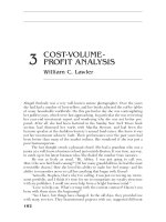
their “lock and key” action, based on the unique shapes of enzymes, as illustrated in Figure 3.10.
This illustration shows that an enzyme “recognizes” a particular substrate by its molecular
structure and binds to it to produce an enzyme–substrate complex. This complex then breaks apart
to form one or more products different from the original enzyme, regenerating the unchanged
enzyme, which is then available to catalyze additional reactions. The basic process for an enzyme
reaction is, therefore,
Figure 3.8 Steroids are characterized by the ring structure shown above for cholesterol.
Figure 3.9 Locations of important endocrine glands.
H
3
C
H
3
C
HO
CH
3
C
H
CH
2
CH
2
CH
2
CH
CH
3
CH
3
Cholesterol, a typical steroid
Ovaries
(female)
Testes
(male)
Adrenal
Thymus
Thyroid
Parathyroid
Pituitary
L1618Ch03Frame Page 69 Tuesday, August 13, 2002 5:54 PM
Copyright © 2003 by CRC Press LLC
enzyme + substrate enzyme–substrate complex enzyme + product (3.6.1)
Several important things should be noted about this reaction. As shown in Figure 3.10, an enzyme
acts on a specific substrate to form an enzyme–substrate complex because of the fit between their
structures. As a result, something happens to the substrate molecule. For example, it might be split
in two at a particular location. Then the enzyme–substrate complex comes apart, yielding the
enzyme and products. The enzyme is not changed in the reaction and is now free to react again.
Note that the arrows in the formula for enzyme reaction point both ways. This means that the
reaction is reversible. An enzyme–substrate complex can simply go back to the enzyme and the
substrate. The products of an enzymatic reaction can react with the enzyme to form the enzyme–sub-
strate complex again. It, in turn, may again form the enzyme and the substrate. Therefore, the same
enzyme may act to cause a reaction to go either way.
Some enzymes cannot function by themselves. In order to work, they must first be attached to
coenzymes. Coenzymes normally are not protein materials. Some of the vitamins are important
coenzymes.
Enzymes are named for what they do. For example, the enzyme given off by the stomach,
which splits proteins as part of the digestion process, is called gastric proteinase. The “gastric”
part of the name refers to the enzyme’s origin in the stomach. “Proteinase” denotes that it splits
up protein molecules. The common name for this enzyme is pepsin. Similarly, the enzyme produced
by the pancreas that breaks down fats (lipids) is called pancreatic lipase. Its common name is
Figure 3.10 Representation of the “lock and key” mode of enzyme action, which enables the very high specificity
of enzyme-catalyzed reactions.
Enzyme
Substrate
+
Enzyme-substrate complex
+
Products Regenerated enzyme
→
←
→
←
L1618Ch03Frame Page 70 Tuesday, August 13, 2002 5:54 PM
Copyright © 2003 by CRC Press LLC
steapsin. In general, lipase enzymes cause lipid triglycerides to dissociate and form glycerol and
fatty acids.
The enzymes mentioned above are hydrolyzing enzymes, which bring about the breakdown
of high-molecular-weight biological compounds by the addition of water. This is one of the most
important reactions involved in digestion. The three main classes of energy-yielding foods that
animals eat are carbohydrates, proteins, and fats. Recall that the higher carbohydrates that humans
eat are largely disaccharides (sucrose, or table sugar) and polysaccharides (starch). These are formed
by the joining together of units of simple sugars, C
6
H
12
O
6
, with the elimination of an H
2
O molecule
at the linkage where they join. Proteins are formed by the condensation of amino acids, again with
the elimination of a water molecule at each linkage. Fats are esters that are produced when glycerol
and fatty acids link together. A water molecule is lost for each of these linkages when a protein,
fat, or carbohydrate is synthesized. In order for these substances to be used as a food source, the
reverse process must occur to break down large, complicated molecules of protein, fat, or carbo-
hydrate to simple, soluble substances that can penetrate a cell membrane and take part in chemical
processes in the cell. This reverse process is accomplished by hydrolyzing enzymes.
Biological compounds with long chains of carbon atoms are broken down into molecules with
shorter chains by the breaking of carbon–carbon bonds. This commonly occurs by the elimination
of –CO
2
H groups from carboxylic acids. For example, pyruvic decarboxylase enzyme acts upon
pyruvic acid,
(3.6.2)
to split off CO
2
and produce a compound with one less carbon. It is by such carbon-by-carbon
breakdown reactions that long-chain compounds are eventually degraded to CO
2
in the body, or
that long-chain hydrocarbons undergo biodegradation by the action of spill bacteria on spilled
petroleum. Oxidation and reduction are the major reactions for the exchange of energy in living
systems. Cellular respiration, discussed in Section 3.2, is an oxidation reaction in which a carbo-
hydrate, C
6
H
12
O
6
, is broken down to carbon dioxide and water with the release of energy:
C
6
H
12
O
6
+ 6O
2
→ 6CO
2
+ 6H
2
O + energy (3.6.3)
Actually, such an overall reaction occurs in living systems by a complicated series of individual
steps. Some of these steps involve oxidation. The enzymes that bring about oxidation in the presence
of free O
2
are called oxidases. In general, biological oxidation–reduction reactions are catalyzed
by oxidoreductase enzymes.
In addition to the types of enzymes discussed above, there are many enzymes that perform
miscellaneous duties in living systems. Typical of these are isomerases, which form isomers of
particular compounds. For example, there are several simple sugars with the formula C
6
H
12
O
6
.
However, only glucose can be used directly for cell processes. The other isomers are converted to
glucose by the action of isomerases. Transferase enzymes move chemical groups from one
molecule to another, lyase enzymes remove chemical groups without hydrolysis and participate in
the formation of C=C bonds or addition of species to such bonds, and ligase enzymes work in
conjunction with adenosine triphosphate (ATP), a high-energy molecule that plays a crucial role
in energy-yielding, glucose-oxidizing metabolic processes, to link molecules together with the
formation of bonds such as carbon–carbon or carbon–sulfur bonds.
Pyruvic acid Acetaldehyde
Pyruvate
decarboxylase
+CO
2
CC
H
H
H
O
H
CCCH
H
H
OH
OO
L1618Ch03Frame Page 71 Tuesday, August 13, 2002 5:54 PM
Copyright © 2003 by CRC Press LLC
Enzyme action may be affected by many different things. Enzymes require a certain hydrogen
ion concentration to function best. For example, gastric proteinase requires the acid environment
of the stomach to work well. When it passes into the much less acidic intestines, it stops working.
This prevents damage to the intestine walls, which would occur if the enzyme tried to digest them.
Temperature is critical. Not surprisingly, the enzymes in the human body work best at around
98.6°F (37°C), which is the normal body temperature. Heating these enzymes to around 140°F
permanently destroys them. Some bacteria that thrive in hot springs have enzymes that work best
at relatively high temperatures. Other “cold-seeking” bacteria have enzymes adapted to near the
freezing point of water.
One of the greatest concerns regarding the effects of surroundings on enzymes is the influence
of toxic substances. A major mechanism of toxicity is the alteration or destruction of enzymes by
agents such as cyanide, heavy metals, or organic compounds, such as insecticidal parathion. An
enzyme that has been destroyed obviously cannot perform its designated function, whereas one
that has been altered either may not function at all or may act improperly. Toxicants can affect
enzymes in several ways. Parathion, for example, bonds covalently to the nerve enzyme acetylcho-
linesterase, which can then no longer serve to stop nerve impulses. Heavy metals tend to bind to
sulfur atoms in enzymes (such as sulfur from the amino acid cysteine, shown in Figure 3.2), thereby
altering the shape and function of the enzyme. Enzymes are denatured by some poisons, causing
them to “unravel” so that the enzyme no longer has its crucial specific shape.
3.7 NUCLEIC ACIDS
The essence of life is contained in deoxyribonucleic acid (DNA), which stays in the cell
nucleus, and ribonucleic acid (RNA), which functions in the cell cytoplasm. These substances,
which are known collectively as nucleic acids, store and pass on essential genetic information that
controls reproduction and protein synthesis.
The structural formulas of the monomeric constituents of nucleic acids are given in Figure 3.11.
These are pyrimidine or purine nitrogen-containing bases, two sugars, and phosphate. DNA mol-
ecules are made up of the nitrogen-containing bases adenine, guanine, cytosine, and thymine;
phosphoric acid (H
3
PO
4
); and the simple sugar 2-deoxy-β-D-ribofuranose (commonly called deox-
yribose). RNA molecules are composed of the nitrogen-containing bases adenine, guanine, cytosine,
and uracil; phosphoric acid (H
3
PO
4
); and the simple sugar β-D-ribofuranose (ribose).
The formation of nucleic acid polymers from their monomeric constituents may be viewed as
the following steps.
• Monosaccharide (simple sugar) + cyclic nitrogenous base yields nucleoside:
CC
C
C
C
N
C
C
C
N
NH
2
O
HH
H
CH
2
HO
H
H
HO
H
H
H
O
Deoxyctidine formed by the
dimerization of cytosine and
deoxyribose with the elimin-
ation of a molecule of H
2
O.
L1618Ch03Frame Page 72 Tuesday, August 13, 2002 5:54 PM
Copyright © 2003 by CRC Press LLC
• Nucleoside + phosphate yields phosphate ester nucleotide.
Figure 3.11 Constituents of DNA (enclosed by ) and of RNA (enclosed by
|||||
).
C
N
C
C
C
N
O
O
H
H
H
H
CH
3
C
N
C
C
C
N
O
O
H
H
H
N
C
C
C
N
C
N
C
N
H
H
H
NH
2
N
C
C
C
N
C
N
C
NH
H
O
H
2
N
H
CC
C
C
CH
2
HO
H
H
HO
H
OH
OH
H
O
CC
C
C
CH
2
HO
H
H
HO
H
H
OH
H
O
P
O
-
O
-
-
O
O
Occur in both DNA
and RNA
Uracil (U)
Thymine (T)
Adenine (A)
Guanine(G)
Phosphate
2-Deoxy-β-D-
ribofuranose
C
N
C
C
C
N
NH
2
O
HH
H
H
Cytosine (C)
β-D-Ribofuranose
Occur only in DNA
Occur only in RNA
Nucleotide formed by the
bonding of a phosphate
group to deoxyctidine
P
O
-
-
O
O
CC
C
C
C
N
C
C
C
N
NH
2
O
HH
H
CH
2
O
H
H
HO
H
H
O
H
O
L1618Ch03Frame Page 73 Tuesday, August 13, 2002 5:54 PM
Copyright © 2003 by CRC Press LLC
• Polymerized nucleotide yields nucleic acid, as shown by the structure below. In the nucleic acid
the phosphate negative charges are neutralized by metal cations (such as Mg
2+
) or positively charged
proteins (histones).
Molecules of DNA are huge, with molecular weights of greater than 1 billion. Molecules of
RNA are also quite large. The structure of DNA is that of the famed double helix. It was figured
out in 1953 by James D. Watson, an American scientist, and Francis Crick, a British scientist. They
received the Nobel Prize for this scientific milestone in 1962. This model visualizes DNA as a so-
called double α-helix structure of oppositely wound polymeric strands held together by hydrogen
bonds between opposing pyrimidine and purine groups. As a result, DNA has both a primary and
a secondary structure; the former is due to the sequence of nucleotides in the individual strands of
DNA, and the latter results from the α-helix interaction of the two strands. In the secondary structure
of DNA, only cytosine can be opposite guanine and only thymine can be opposite adenine and
vice versa. Basically, the structure of DNA is that of two spiral ribbons “counterwound” around
each other, as illustrated in Figure 3.12. The two strands of DNA are complementary. This means
that a particular portion of one strand fits like a key in a lock with the corresponding portion of
another strand. If the two strands are pulled apart, each manufactures a new complementary strand,
so that two copies of the original double helix result. This occurs during cell reproduction.
The molecule of DNA is like a coded message. This “message,” the genetic information
contained in and transmitted by nucleic acids, depends on the sequence of bases from which they
are composed. It is somewhat like the message sent by telegraph, which consists only of dots,
dashes, and spaces in between. The key aspect of DNA structure that enables storage and replication
of this information is the famed double helix structure of DNA mentioned above.
Portions of the DNA double helix may unravel, and one of the strands of DNA may produce
a strand of RNA. This substance then goes from the cell nucleus out into the cell and regulates the
synthesis of new protein. In this way, DNA regulates the function of the cell and acts to control
life processes.
Segment of the DNA polymer
showing linkage of two nucleotides
P
O
-
O
O
CC
C
C
C
N
C
C
C
N
NH
2
O
HH
H
CH
2
O
H
H
O
H
H
C
C
H
O
P
O
-
O
N
C
CC
C
C
N
C
N
C
N
NH
2
H
H
CH
2
O
H
HH
HO
H
O
L1618Ch03Frame Page 74 Tuesday, August 13, 2002 5:54 PM
Copyright © 2003 by CRC Press LLC
3.7.1 Nucleic Acids in Protein Synthesis
When a new cell is formed, the DNA in its nucleus must be accurately reproduced from the
parent cell. Life processes are absolutely dependent upon accurate protein synthesis, as regulated
by cell DNA. The DNA in a single cell must be capable of directing the synthesis of up to 3000
or even more different proteins. The directions for the synthesis of a single protein are contained
in a segment of DNA called a gene. The process of transmitting information from DNA to a newly
synthesized protein involves the following steps:
• The DNA undergoes replication. This process involves separation of a segment of the double
helix into separate single strands, which then replicate such that guanine is opposite cytosine (and
vice versa) and adenine is opposite thymine (and vice versa). This process continues until a
complete copy of the DNA molecule has been produced.
• The newly replicated DNA produces messenger RNA (mRNA), a complement of the single strand
of DNA, by a process called transcription.
• A new protein is synthesized using mRNA as a template to determine the order of amino acids in
a process called translation.
3.7.2 Modified DNA
DNA molecules may be modified by the unintentional addition or deletion of nucleotides or
by substituting one nucleotide for another. The result is a mutation that is transmittable to offspring.
Mutations can be induced by chemical substances. This is a major concern from a toxicological
viewpoint because of the detrimental effects of many mutations and because substances that cause
mutations often cause cancer as well. DNA malfunction may result in birth defects, and the failure
to control cell reproduction results in cancer. Radiation from x-rays and radioactivity also disrupts
DNA and may cause mutation.
Figure 3.12 Representation of the double helix structure of DNA showing the allowed base pairs held together
by hydrogen bonding between the phosphate–sugar polymer “backbones” of the two strands of
DNA. The letters stand for adenine (A), cytosine (C), guanine (G), and thymine (T). The dashed
lines represent hydrogen bonds.
A
T
CG
T
A
CG
CG
CG
T
A
CG
T
A
L1618Ch03Frame Page 75 Tuesday, August 13, 2002 5:54 PM
Copyright © 2003 by CRC Press LLC
3.8 RECOMBINANT DNA AND GENETIC ENGINEERING
As noted above, segments of DNA contain information for the specific syntheses of particular
proteins. Within the last two decades it has become possible to transfer this information between
organisms by means of recombinant DNA technology, which has resulted in a new industry based
on genetic engineering. Most often the recipient organisms are bacteria, which can be reproduced
(cloned) over many orders of magnitude from a cell that has acquired the desired qualities.
Therefore, to synthesize a particular substance, such as human insulin or growth hormone, the
required genetic information can be transferred from a human source to bacterial cells, which then
produce the substance as part of their metabolic processes.
The first step in recombinant DNA gene manipulation is to lyze (open up) a donor cell to
remove needed DNA material by using enzyme action to cut the sought-after genes from the donor
DNA chain. These are next spliced into small DNA molecules. These molecules, called cloning
vehicles, are capable of penetrating the host cell and becoming incorporated into its genetic material.
The modified host cell is then reproduced many times and carries out the desired biosynthesis.
Early concerns about the potential of genetic engineering to produce “monster organisms” or
new and horrible diseases have been largely allayed, although caution is still required with this
technology. In the environmental area, genetic engineering offers some hope for the production of
bacteria engineered to safely destroy troublesome wastes and to produce biological substitutes for
environmentally damaging synthetic pesticides.
3.9 METABOLIC PROCESSES
Biochemical processes that involve the alteration of biomolecules fall under the category of
metabolism. Metabolic processes may be divided into the two major categories of anabolism
(synthesis) and catabolism (degradation of substances). An organism may use metabolic processes
to yield energy or to modify the constituents of biomolecules.
3.9.1 Energy-Yielding Processes
Organisms can gain energy by the following three processes:
• Respiration, in which organic compounds undergo catabolism that requires molecular oxygen
(aerobic respiration) or that occurs in the absence of molecular oxygen (anaerobic respiration).
Aerobic respiration uses the Krebs cycle to obtain energy from the following reaction:
C
6
H
12
O
6
+ 6O
2
→ 6CO
2
+ 6H
2
O + energy
• About half of the energy released is converted to short-term stored chemical energy, particularly
through the synthesis of ATP nucleoside. For longer-term energy storage, glycogen or starch
polysaccharides are synthesized, and for still longer term energy storage, lipids (fats) are generated
and retained by the organism.
• Fermentation, which differs from respiration in not having an electron transport chain. Yeasts
produce ethanol from sugars by fermentation:
C
6
H
12
O
6
→ 2CO
2
+ 2C
2
H
5
OH
• Photosynthesis, in which light energy captured by plant and algal chloroplasts are used to syn-
thesize sugars from carbon dioxide and water:
L1618Ch03Frame Page 76 Tuesday, August 13, 2002 5:54 PM
Copyright © 2003 by CRC Press LLC
6CO
2
+ 6H
2
O + h
ν
→ C
6
H
12
O
6
+ 6O
2
Plants cannot always get the energy that they need from sunlight. During the dark they must
use stored food. Plant cells, like animal cells, contain mitochondria in which stored food is converted
to energy by cellular respiration.
Plant cells, which use sunlight for energy and CO
2
for carbon, are said to be autotrophic. In
contrast, animal cells must depend on organic material manufactured by plants for their food. These
are called heterotrophic cells. They act as “middlemen” in the chemical reaction between oxygen
and food material, using the energy from the reaction to carry out their life processes.
SUPPLEMENTARY REFERENCES
Bettelheim, F.A. and March, J., Introduction to Organic and Biochemistry, Saunders College Publishing, Fort
Worth, TX, 1998.
Chesworth, J.M., Stuchbury, T., and Scaife, J.R., An Introduction to Agricultural Biochemistry, Chapman &
Hall, London, 1998.
Garrett, R.H. and Grisham, C.M., Biochemistry, Saunders College Publishing, Philadelphia, 1998.
Gilbert, H.F., Ed., Basic Concepts in Biochemistry, McGraw-Hill, Health Professions Division, New York,
2000.
Kuchel, P.W., Ed., Schaum’s Outline of Theory and Problems of Biochemistry, McGraw-Hill, New York, 1998.
Lea, P.J. and Leegood, R.C., Eds., Plant Biochemistry and Molecular Biology, 2nd ed., John Wiley & Sons,
New York, 1999.
Marks, D.B., Biochemistry, Williams & Wilkins, Baltimore, 1999.
Meisenberg, G. and Simmons, W.H., Principles of Medical Biochemistry, Mosby, St. Louis, 1998.
Switzer, R.L. and Garrity, L.F., Experimental Biochemistry, W.H. Freeman and Co., New York, 1999.
Voet, D., Voet, J.G., and Pratt, C., Fundamentals of Biochemistry, John Wiley & Sons, New York, 1998.
Vrana, K.E., Biochemistry, Lippincott Williams & Wilkins, Philadelphia, 1999.
Wilson, K. and Walker, J.M., Principles and Techniques of Practical Biochemistry, Cambridge University
Press, New York, 1999.
QUESTIONS AND PROBLEMS
1. What is the toxicological importance of lipids? How do lipids relate to hydrophobic (water-
disliking) pollutants and toxicants?
2. What is the function of a hydrolase enzyme?
3. Match the cell structure on the left with its function on the right:
A. Mitochondria 1. Toxicant metabolism
B. Endoplasmic reticulum 2. Fills the cell
C. Cell membrane 3. DNA
D. Cytoplasm 4. Mediate energy conversion and utilization
E. Cell nucleus 5. Encloses the cell and regulates the passage of materials into and out
of the cell interior
4. The formula of simple sugars is C
6
H
12
O
6
. The simple formula of higher carbohydrates is C
6
H
10
O
5
.
Of course, many of these units are required to make a molecule of starch or cellulose. If higher
carbohydrates are formed by joining together molecules of simple sugars, why is there a difference
in the ratios of C, H, and O atoms in the higher carbohydrates, compared to the simple sugars?
5. Why does wood contain so much cellulose?
6. What would be the chemical formula of a trisaccharide made by the bonding together of three
simple sugar molecules?
L1618Ch03Frame Page 77 Tuesday, August 13, 2002 5:54 PM
Copyright © 2003 by CRC Press LLC
7. The general formula of cellulose may be represented as (C
6
H
10
O
5
)
x
. If the molecular weight of a
molecule of cellulose is 400,000, what is the estimated value of x?
8. During 1 month, a factory for the production of simple sugars, C
6
H
12
O
6
, by the hydrolysis of
cellulose processes 1 million kg of cellulose. The percentage of cellulose that undergoes the
hydrolysis reaction is 40%. How many kilograms of water are consumed in the hydrolysis of
cellulose each month?
9. What is the structure of the largest group of atoms common to all amino acid molecules?
10. Glycine and phenylalanine can join together to form two different dipeptides. What are the
structures of these two dipeptides?
11. One of the ways in which two parallel protein chains are joined together, or cross-linked, is by
way of an –S–S– link. What amino acid to you think might be most likely to be involved in such
a link? Explain your choice.
12. Fungi, which break down wood, straw, and other plant material, have what are called “exoenzymes.”
Fungi have no teeth and cannot break up plant material physically by force. Knowing this, what
do you suppose an exoenzyme is? Explain how you think it might operate in the process by which
fungi break down something as tough as wood.
13. Many fatty acids of lower molecular weight have a bad odor. Speculate as to the reasons why
rancid butter has a bad odor. What chemical compound is produced that has a bad odor? What
sort of chemical reaction is involved in its production?
14. The long-chain alcohol with ten carbons is called decanol. What do you think would be the formula
of decyl stearate? To what class of compounds would it belong?
15. Write an equation for the chemical reaction between sodium hydroxide and cetyl stearate. What
are the products?
16. What are two endocrine glands that are found only in females? Which of these glands is found
only in males?
17. The action of bile salts is a little like that of soap. What function do bile salts perform in the
intestine? Look up the action of soaps, and explain how you think bile salts may function somewhat
like soap.
18. If the structure of an enzyme is illustrated as
how should the structure of its substrate be represented?
19. Look up the structures of ribose and deoxyribose. Explain where the “deoxy” came from in the
name deoxyribose.
20. In what respect are an enzyme and its substrate like two opposite strands of DNA?
21. For what discovery are Watson and Crick noted?
22. Why does an enzyme no longer work if it is denatured?
L1618Ch03Frame Page 78 Tuesday, August 13, 2002 5:54 PM
Copyright © 2003 by CRC Press LLC