Applications in Musculoskeletal Disorders - part 10 ppsx
Bạn đang xem bản rút gọn của tài liệu. Xem và tải ngay bản đầy đủ của tài liệu tại đây (743.91 KB, 29 trang )
Taylor’s Musculoskeletal
Problems and Injuries
Taylor’s Musculoskeletal
Problems and Injuries
A Handbook
Robert B. Taylor, M.D. Editor
Professor Emeritus
Department of Family Medicine
Oregon Health & Science University
School of Medicine
Portland, Oregon
Associate Editors
Alan K. David, M.D. Scott A. Fields, M.D.
Professor and Chairman Professor and Vice Chairman
Department of Family and Department of Family Medicine
Community Medicine Oregon Health & Science University
Medical College of Wisconsin School of Medicine
Milwaukee, Wisconsin Portland, Oregon
D. Melessa Phillips, M.D. Joseph E. Scherger, M.D., M.P.H.
Professor and Chairman Clinical Professor
Department of Family Medicine Department of Family and
University of Mississippi Preventive Medicine
School of Medicine University of California,
Jackson, Mississippi San Diego School of Medicine
San Diego, California
With 53 Illustrations
Robert B. Taylor, M.D.
Professor Emeritus
Department of Family Medicine
Oregon Health & Science University
School of Medicine
Portland, OR 97239-3098, USA
Associate Editors
Alan K. David, M.D. Scott A. Fields, M.D.
Professor and Chairman Professor and Vice Chairman
Department of Family and Department of Family Medicine
Community Medicine Oregon Health & Science University
Medical College of Wisconsin School of Medicine
Milwaukee, WI 53226-0509, USA Portland, OR 97201-3098, USA
D. Melessa Phillips, M.D. Joseph E. Scherger, M.D., M.P.H.
Professor and Chairman Clinical Professor
Department of Family Medicine Department of Family and Preventive Medicine
University of Mississippi School University of California, San Diego
of Medicine School of Medicine
Jackson, MS 39216-4500, USA San Diego, California 92103-0801, USA
Library of Congress Control Number: 2005935915
ISBN-10: 0-387-29171-7 Printed on acid-free paper.
ISBN-13: 978-0387-29171-0
© 2006 Springer Science+Business Media, LLC
All rights reserved. This work may not be translated or copied in whole or in part without the
written permission of the publisher (Springer Science+Business Media, LLC, 233 Spring Street,
New York, NY 10013, USA), except for brief excerpts in connection with reviews or scholarly
analysis. Use in connection with any form of information storage and retrieval, electronic
adaptation, computer software, or by similar or dissimilar methodology now known or hereafter
developed is forbidden.
The use in this publication of trade names, trademarks, service marks and similar terms, even if they
are not identified as such, is not to be taken as an expression of opinion as to whether or not they
are subject to proprietary rights.
While the advice and information in this book are believed to be true and accurate at the date
of going to press, neither the authors nor the editors nor the publisher can accept any legal
responsibility for any errors or omissions that may be made. The publisher makes no warranty,
express or implied, with respect to the material contained herein.
Printed in the United States of America. (SPI/EB)
987654321
springer.com
Preface
After more than a quarter century as a primary care educator, I am
convinced that our graduates enter practice inadequately trained in
the diagnosis and management of musculoskeletal problems and
injuries. One reason for this perceived deficiency is the relatively
short duration of primary care training—typically three years for
family medicine, general internal medicine, and general pediatrics.
During this time, there are just not enough months to teach all a cli-
nician needs to know about diseases and trauma involving the mus-
culoskeletal system. This inadequacy is compounded by the
sometimes quirky nature of the problems: that is, for example, the
increased risk of nonunion in a fracture of the carpal navicular
(scaphoid) bone or the maneuver that can magically reduce a child’s
radial head subluxation.
The chapters in this book are from the edited reference book Family
Medicine: Principles and Practice, 6th edition, which is widely used
by family physicians in the United States and abroad. The publisher
and I believe that, in addition to family physicians, the chapters in this
book will also be useful to other clinicians providing broad-based
care: general internists, general pediatricians, emergency physicians,
nurse practitioners, and physician assistants. When compared to the
large, comprehensive book, this volume will be preferred by some
readers because of the physically smaller size and perhaps by the
lower cost.
In selecting chapters to include in the book, I have included
problems involving all areas of the skeleton and related musculature,
in both children and adults. Athletic injuries are included because,
after all, primary care clinicians manage most sports injuries. I have
included a chapter on acute lacerations, which often accompany other
types of injuries. In addition to sprains, strains, and fractures, there are
chapters covering illnesses affecting the musculoskeletal system:
various types of arthritis, fibromyalgia, and the complex regional pain
syndrome.
I hope you find this book useful in daily practice; comments are
welcome.
Robert B. Taylor, M.D.
Portland, Oregon, USA
vi Preface
Clinical Practice
Notice
Everyone involved with the preparation of this book has worked very
hard to assure that information presented here is accurate and that it
represents accepted clinical practices. These efforts include confirm-
ing that drug recommendations and dosages discussed in this text
are in accordance with current practice at the time of publication.
Nevertheless, therapeutic recommendations and dosage schedules
change with reports of ongoing research, changes in government rec-
ommendations, reports of adverse drug reactions, and other new
information.
A few recommendations and drug uses described herein have Food
and Drug Administration (FDA) clearance for limited use in restricted
settings. It is the responsibility of the clinician to determine the FDA
status of any drug selection, drug dosage, or device recommended to
patients.
The reader should check the package insert for each drug to deter-
mine any change in indications or dosage as well as for any precau-
tions or warnings. This admonition is especially true when the drug
considered is new or infrequently used by the clinician.
The use of the information in this book in a specific clinical setting or
situation is the professional responsibility of the clinician. The authors,
editors, or publisher are not responsible for errors, omissions, adverse
effects, or any consequences arising from the use of information in this
book, and make no warranty, expressed or implied, with respect to the
completeness, timeliness, or accuracy of the book’s contents.
Preface . . . . . . . . . . . . . . . . . . . . . . . . . . . . . . . . . . . . . . . . . v
Clinical Practice Notice . . . . . . . . . . . . . . . . . . . . . . . . . . . vii
Contents. . . . . . . . . . . . . . . . . . . . . . . . . . . . . . . . . . . . . . . . ix
Contributors. . . . . . . . . . . . . . . . . . . . . . . . . . . . . . . . . . . . . xi
1 Disorders of the Back and Neck . . . . . . . . . . . . . . . . . . . . . . 1
Walter L. Calmbach
2 Disorders of the Upper Extremity . . . . . . . . . . . . . . . . . . . . 35
Ted C. Schaffer
3 Disorders of the Lower Extremity . . . . . . . . . . . . . . . . . . . . 59
Kenneth M. Bielak and Bradley E. Kocian
4 Osteoarthritis. . . . . . . . . . . . . . . . . . . . . . . . . . . . . . . . . . . . 89
Alicia D. Monroe and John B. Murphy
5 Rheumatoid Arthritis and Related Disorders . . . . . . . . . . . . 97
Joseph W. Gravel Jr., Patricia A. Sereno,
and Katherine E. Miller
6 Selected Disorders of the Musculoskeletal System . . . . . . 127
Jeffrey G. Jones and Doug Poplin
7 Musculoskeletal Problems of Children . . . . . . . . . . . . . . . 147
Mark D. Bracker, Suraj A. Achar, Todd J. May,
Juan Carlos Buller, and Wilma J. Wooten
8 Osteoporosis . . . . . . . . . . . . . . . . . . . . . . . . . . . . . . . . . . . 181
Paula Cifuentes Henderson and Richard P. Usatine
9 Gout . . . . . . . . . . . . . . . . . . . . . . . . . . . . . . . . . . . . . . . . . 197
James F. Calvert, Jr.
10 Athletic Injuries . . . . . . . . . . . . . . . . . . . . . . . . . . . . . . . . 205
Michael L. Tuggy and Cora Collette Breuner
11 Care of Acute Lacerations . . . . . . . . . . . . . . . . . . . . . . . . . 233
Bryan J. Campbell and Douglas J. Campbell
12 Selected Injuries . . . . . . . . . . . . . . . . . . . . . . . . . . . . . . . . 261
Allan V. Abbott
Index. . . . . . . . . . . . . . . . . . . . . . . . . . . . . . . . . . . . . . . . . 281
x Contents
Walter L. Calmbach, M.D., Associate Professor of Family and
Community Medicine, Director of Sports Medicine Fellowship and
South Texas Ambulatory Research Network (STARNET), University
of Texas Health Science Center, San Antonio, Texas
Disorders of the Back and Neck
James F. Calvert Jr, M.D., Associate Professor of Family Medicine,
Oregon Health & Science University School of Medicine, Portland;
Cascades East Family Practice Residency Program, Klamath Falls,
Oregon
Gout
Bryan J. Campbell, M.D., Assistant Professor of Family and
Preventive Medicine, University of Utah School of Medicine, Salt
Lake City, Utah
Care of Acute Lacerations
Douglas J. Campbell, M.D., Community Attending Physician, Good
Samaritan Regional Family Practice Center, Yavapai Regional
Medical Center, Prescott, Arizona
Care of Acute Lacerations
Joseph W. Gravel, Jr, M.D., Assistant Clinical Professor of Family
Medicine and Community Health, Tufts University School of
Medicine, Boston; Director, Tufts University Family Practice
Residency Program, Malden, Massachusetts
Rheumatoid Arthritis and Related Disorders
Paula Cifuentes Henderson, M.D., Clinical Instructor of Family
Medicine, University of California – Los Angeles School of
Medicine, Los Angeles, California
Osteoporosis
Jeffrey G. Jones, M.D., M.P.H., Medical Director, St. Francis
Traveler’s Health Center, Indianapolis, Indiana
Selected Disorders of the Musculoskeletal System
Bradley E. Kocian, M.D., Sports Medicine Fellow, University of
Tennessee -Knoxville Medical Center, Knoxville, Tennessee
Disorders of the Lower Extremity
xii Contributors
Todd J. May, D.O., Lieutenant Commander, Medical Corps, United
States Naval Hospital, Camp Pendleton, California
Musculoskeletal Problems of Children
Katherine E. Miller, M.D., Assistant Clinical Professor of Family
Medicine and Community Health, Tufts University School of
Medicine, Boston; Faculty, Tufts University Family Practice
Residency Program, Malden, Massachusetts
Rheumatoid Arthritis and Related Disorders
Alicia D. Monroe, M.D., Associate Professor of Family Medicine,
Brown Medical School, Providence; Memorial Hospital of Rhode
Island, Pawtucket, Rhode Island
Osteoarthritis
John B. Murphy, M.D., Professor of Family Medicine, Brown Medical
School, Providence, Rhode Island
Osteoarthritis
Doug Poplin, M.D., M.P.H., Medical Director, Saint Francis
Occupational Health Center, Indianapolis, Indiana
Selected Disorders of the Musculoskeletal System
Ted C. Schaffer, M.D., Clinical Assistant Professor, Department of
Family Medicine and Clinical Epidemiology, University of Pittsburgh
School of Medicine; Director, UPMC – St. Margaret Hospital Family
Practice Residency Program, Pittsburgh, Pennsylvania
Disorders of the Upper Extremity
Patricia A. Sereno, M.D., M.P.H., Assistant Clinical Professor of
Family Medicine and Community Health, Tufts University School of
Medicine, Boston; Hallmark Family Health Center, Malden,
Massachusetts
Rheumatoid Arthritis and Related Disorders
Michael L. Tuggy, M.D., Clinical Assistant Professor of Family
Medicine, University of Washington School of Medicine; Director,
Swedish Family Medicine Residency Program, Seattle, Washington
Athletic Injuries
Contributors xiii
Richard P. Usatine, M.D., Professor of Clinical Family Medicine and
Assistant Dean of Student Affairs, University of California – Los
Angeles School of Medicine, Los Angeles, California
Osteoporosis
Wilma J. Wooten, M.D., M.P.H., Associate Clinical Professor of
Family and Preventive Medicine, University of California-San Diego
School of Medicine, La Jolla, California
Musculoskeletal Problems of Children
xiv Contributors
2 Walter L. Calmbach
is the most expensive cause of work-related disability.
8
Risk factors
for the development of low back pain include heavy lifting and twist-
ing, bodily vibration, obesity, and poor conditioning; however, low
back pain is common even among patients without these risk factors.
1
In cases of more severe back pain, occupational exposures are
much more significant, including repetitive heavy lifting, pulling, or
pushing, and exposures to industrial and vehicular vibrations. If even
temporary work loss occurs, additional important risk factors include
job dissatisfaction, supervisor ratings, and job environment (i.e., bor-
ing, repetitive tasks).
1
Factors associated with recurrence of low
back pain include traumatic origin of first attack, sciatic pain, radi-
ographic changes, alcohol abuse, specific job situations, and psy-
chosocial stigmata.
Of patients with acute low back pain, only 1.5% develop sciatica
(i.e., painful paresthesias and/or motor weakness in the distribution of
a nerve root). However, the lifetime prevalence of sciatica is 40%, and
sciatica afflicts 11% of patients with low back pain that lasts for more
than two weeks.
9,10
Sciatica is associated with long-distance driving,
truck driving, cigarette smoking, and repeated lifting in a twisted pos-
ture. It is most common in the fourth and fifth decades of life, and
peaks in the fourth decade. Most patients with sciatica, even those
with significant neurological abnormalities, recover without surgery.
11
Only 5% to 10% of patients with persistent sciatica require surgery.
5,12
Despite the incidence and prevalence of low back pain and sciatica,
the major factor responsible for its societal impact is disability.
12
The
National Center for Health Statistics estimates that 5.2 million
Americans are disabled with low back pain, of whom 2.6 million are
permanently disabled.
13
Between 70% and 90% of the total costs due
to low back pain are incurred by the 4% to 5% of patients with tem-
porary or permanent disability.
12
Risk factors for disability due to low
back pain include poor health habits, job dissatisfaction, less appeal-
ing work environments, poor ratings by supervisors, psychological
disturbances, compensable injuries, and history of prior disability.
12
These same factors are associated with high failure rates for treat-
ments of all types.
Natural History
Recovery from nonspecific low back pain is usually rapid.
Approximately one third of patients are improved at one week, and two
thirds at seven weeks. However, recurrences are common, affecting 40%
of patients within six months. Thus, “acute low back pain” is increasingly
perceived as a chronic medical problem with intermittent exacerbations.
14
Low back pain may originate from many structures, including par-
avertebral musculature, ligaments, the annulus fibrosus, the spinal
nerve roots, the facet joints, the vertebral periosteum, fascia, or blood
vessels. The most common causes of back pain include musculoliga-
mentous injuries, degenerative changes in the intervertebral discs and
facet joints, spinal stenosis, and lumbar disc herniation.
14
The natural history of herniated lumbar disc is usually quite favor-
able. Only about 10% of patients who present with sciatica have suf-
ficient pain at six weeks that surgery is considered. Sequential
magnetic resonance imaging (MRI) shows gradual regression of the
herniated disc material over time, with partial or complete resolution
in two thirds of patients by six months.
14
Acute disc herniation has
changed little from its description in the classic article of Mixter and
Barr: the annulus fibrosus begins to deteriorate by age 30, which leads
to partial or complete herniation of the nucleus pulposus, causing irri-
tation and compression of adjacent nerve roots.
5,15,16
Usually this her-
niation is in the posterolateral position, producing unilateral
symptoms. Occasionally, the disc will herniate in the midline, and a
large herniation in this location can cause bilateral symptoms. More
than 95% of lumbar disc herniations occur at the L4–L5 or L5–S1 lev-
els.
10
Involvement of the L5 nerve root results in weakness of the great
toe extensors and dorsiflexors of the foot, and sensory loss at the dor-
sum of the foot and in the first web space. Involvement of the S1 nerve
root results in a diminished ankle reflex, weakness of the plantar flex-
ors, and sensory loss at the posterior calf and lateral foot.
Among patients who present with low back pain, 90% recover within
six weeks with or without therapy.
17
Even in industrial settings, 75% of
patients with symptoms of acute low back pain return to work within
one month.
17
Only 2% to 3% of patients continue to have symptoms at
six months, and only 1% at one year. However, symptoms of low back
pain recur in approximately 60% of cases over the next two years.
Demographic characteristics such as age, gender, race, or ethnicity
do not appear to influence the natural history of low back pain.
Obesity, smoking, and occupation, however, are important influ-
ences.
18
Adults in the upper fifth quintile of height and weight are
more likely to report low back pain lasting for two or more weeks.
9,18
Occupational factors that prolong or delay recovery from acute low
back pain include heavier job requirements, job dissatisfaction, repe-
titious or boring jobs, poor employer evaluations, and noisy or
unpleasant working conditions.
16
Psychosocial factors play an impor-
tant role in the natural history of low back pain, modulating response
to pain, and promoting illness behavior. The generally favorable nat-
ural history of acute low back pain is significantly influenced by a
1. Disorders of the Back and Neck 3
variety of medical and psychosocial factors that the practicing physi-
cian must be familiar with in order to counsel patients regarding prog-
nosis and treatment.
Clinical Presentation
History
Low back pain is a symptom that has many causes. When approaching
the patient with low back pain, the physician should consider three
important issues. Is a systemic disease causing the pain? Is the patient
experiencing social or psychosocial stresses that may amplify or pro-
long the pain? Does the patient have signs of neurological compromise
that may require surgical evaluation?
14
Useful items on medical history
include: age, fever, history of cancer, unexplained weight loss, injec-
tion drug use, chronic infection, duration of pain, presence of night-
time pain, response to previous therapy, whether pain is relieved by
bed rest or the supine position, persistent adenopathy, steroid use, and
previous history of tuberculosis.
14
Factors that aggravate or alleviate
low back pain should also be elicited. Nonmechanical back pain is usu-
ally continuous, whereas mechanical back pain is aggravated by
motion and relieved by rest. Low back pain that worsens with cough
has traditionally been associated with disc herniation, although recent
data indicate that mechanical low back pain also worsens with cough.
The presence of leg weakness or leg paresthesias in a nerve root dis-
tribution is consistent with disc herniation. Bowel or bladder inconti-
nence with or without saddle paresthesias suggests the cauda equina
syndrome; this is a surgical emergency and requires immediate refer-
ral to a surgeon. Hip pain can mimic low back pain, and is often
referred to the groin, the anterior thigh, or the knee, and is worsened
with ambulation. Patients with osteoarthritis or degenerative joint dis-
ease report morning stiffness, which improves as the day progresses.
Patients with spinal stenosis report symptoms suggestive of spinal
claudication, that is, neurological symptoms in the legs that worsen
with ambulation. Spinal claudication is differentiated from vascular
claudication in that the symptoms of spinal claudication have a slower
onset and slower resolution. A history of pain at rest, pain in the
recumbent position, or pain at night suggests infection or tumor as a
cause for low back pain. Osteoporosis is a consideration among post-
menopausal women or women who have undergone oophorectomy.
These patients report severe, localized, unrelenting pain after even
“minor” trauma. Patients who present writhing in pain suggest the
presence of an intra-abdominal process or vascular cause for the pain,
such as abdominal aortic aneurysm.
4 Walter L. Calmbach
Physical Examination
The initial examination is fairly detailed. With the patient standing
and appropriately gowned, the examining physician notes the stance
and gait, as well as the presence or absence of the normal curvature of
the spine (e.g., thoracic kyphosis, lumbar lordosis, splinting to one
side, scoliosis). The range of motion of the back is documented,
including flexion, lateral bending, and rotation. Intact dorsiflexion
and plantar flexion of the foot is determined by observing heel-walk
and toe-walk. Intact knee extension is determined by observing the
patient squat and rise, while keeping the back straight.
With the patient seated, a distracted straight-leg raising test is
applied. With the hip flexed at 90 degrees, the flexed knee is brought
to full extension. A positive straight-leg raising test reproduces the
patient’s paresthesias in the distribution of a nerve root at Ͻ60
degrees of knee extension. Sensation to light touch and pinprick are
examined and motor strength of hip and knee flexors is tested. The
deep tendon reflexes are tested [knee jerk (L4), ankle jerk (S1)]
and long tract signs are elicited by applying Babinski’s maneuver
(Table 1.1).
With the patient in the supine position, the straight-leg raising test
is repeated. With the hip and knee extended, the leg is raised (i.e., the
1. Disorders of the Back and Neck 5
Table 1.1. Motor, Sensory, and Deep Tendon Reflex Patterns
Associated with Commonly Affected Nerve Roots
Deep tendon
Nerve root Motor reflexes Sensory reflexes reflexes
C5 Deltoid Lateral arm Biceps jerk (C5,C6)
C6 Biceps, Lateral forearm Brachioradialis
brachioradialis,
wrist extensors
C7 Triceps, wrist Middle of hand, Triceps jerk
flexors, MCP middle finger
extensors
C8 MCP flexors Medial forearm —
T1 Abductors and Medial arm —
adductors of
fingers
L4 Quadriceps Anterior thigh Knee jerk
L5 Dorsiflex foot Dorsum of foot Hamstring reflex
and great toe (L5, S1)
S1 Plantarflex foot Lateral foot, Ankle jerk
posterior calf
MCP ϭ metacarpophalangeal.
hip is flexed). A positive test reproduces the patient’s paresthesias in
the distribution of a nerve root. Isolated low back pain does not indi-
cate a positive straight-leg raising test. The crossed straight-leg rais-
ing test (i.e., reproduction of the patient’s symptoms by straight-leg
raising of the contralateral leg) is very specific for acute disc hernia-
tion, and suggests a large central disc herniation. The examining
physician should realize that the straight-leg raising test is sensitive
but not specific, whereas the crossed straight-leg raising test is spe-
cific but not sensitive.
14
Hip range of motion is then tested, and pain
radiation to the groin, anteromedial thigh, or knee is documented.
A more detailed examination may be necessary in selected patients.
If significant pathology is suspected in a male patient, the cremasteric
reflex is tested; i.e., application of a sharp stimulus at the proximal
medial thigh should normally cause retraction of the ipsilateral scro-
tum. With the patient in the prone position, the femoral stretch test is
applied. While the hip and knee are in extension, the knee is flexed,
placing increased stretch on the femoral nerve, which includes
elements from the L2, L3, and L4 nerve roots (i.e., the prone knee-
bending test). The hamstring reflex is tested by striking the semi-
tendinosus and semimembranosus tendons at the medial aspect of the
popliteal fossa. The hamstring reflex involves both the L5 and S1
nerve roots. Thus, an absent or decreased hamstring reflex in the pres-
ence of a normal ankle jerk response (S1) implies involvement of the
L5 nerve root (Table 1.1). Sensation in the area between the upper
buttocks is tested, as well as the anal reflex and anal sphincter tone
(S2, S3, S4).
The clinical diagnosis of acute disc herniation requires repeated
physical examination demonstrating pain or paresthesias localized
to a specific nerve root, with reproduction of pain on straight-leg
raising tests, and muscle weakness in the nerve-appropriate root
distribution.
Diagnosis
Radiology
Plain Radiographs. Plain radiographs are usually not helpful in diag-
nosing acute low back pain, because they cannot demonstrate soft tis-
sue sprains and strains, or an acute herniated disc. However, plain
radiographs are useful in ruling out conditions such as vertebral frac-
ture, spondylolisthesis, spondylolysis, infection, tumor, or inflamma-
tory spondyloarthropathy
5,19
(Fig. 1.1). In the absence of neurologic
deficits, plain radiographs in the evaluation of low back pain should be
reserved for patients over 50 years of age, patients with a temperature
6 Walter L. Calmbach
Ͼ38°C, patients with anemia, a history of trauma, previous cancer,
pain at rest, or unexplained weight loss, drug or alcohol abuse, steroid
use, diabetes mellitus, or any other reason for immunosuppression.
20
For selected patients, initial plain radiographs of the spine in the early
evaluation of acute low back pain should include anteroposterior and
lateral views of the lumbar spine.
15
Oblique views are used to rule out
1. Disorders of the Back and Neck 7
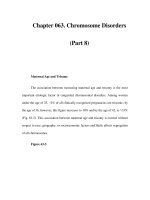
Fig. 1.1. Radiologic studies of the lumbar spine. (A) Plain radi-
ograph demonstrating a compression fracture of the L2 vertebral
body due to multiple myeloma. (B) CT scan demonstrating
nucleus pulposus herniating posteriorly into the spinal canal. (C)
MRI demonstrating an enhancing intramedullary metastatic
lesion in the cauda equina at the L1 level.
spondylolysis, particularly when evaluating acute low back pain in
young athletic patients active in sports such as football, wrestling,
gymnastics, diving, figure skating, or ballet.
21
If the patient’s pain
fails to improve after four to six weeks of conservative therapy, radi-
ographs should be obtained; such patients may be at risk for vertebral
infection, cancer, or inflammatory disease.
22
For patients 65 years of age and older, diagnoses such as cancer,
compression fracture, spinal stenosis, and aortic aneurysm become
more common. Osteoporotic fracture may occur even in the absence
of trauma. Because hormone replacement therapy and other medica-
tions may prevent further fractures, early radiography is recom-
mended for older patients with back pain.
14
Radiographic abnormalities are nonspecific and are observed
equally in patients with and without symptoms of low back pain.
23
Clinical correlation is essential before symptoms of low back pain can
be attributed to radiographic abnormalities.
CT, MRI, and Myelogram. Computed tomography (CT), myelo-
gram, and magnetic resonance imaging (MRI) each have a specific role
in evaluating a select subset of patients with low back pain. Physicians
must be aware that many asymptomatic patients demonstrate disc
bulging, protrusion, and even extrusion.
5,24
For example, 30% to 40%
of CT scans and 64% of MRIs demonstrate abnormalities of the inter-
vertebral disc in asymptomatic patients.
7,24
CT or MRI should be reserved for patients in whom there is strong
clinical suggestion of underlying infection or cancer, progressive or
persistent neurological deficit, or cauda equina syndrome therapy.
5,14
CT or MRI should be considered for patients who show no response
to a four- to six-week course of conservative therapy.
5
CT and MRI
are equally effective in detecting disc herniation and spinal stenosis,
but MRI is more sensitive in detecting infection, metastatic cancer,
and neural tumors.
14
Myelography is useful in differentiating significant
disc herniation from incidental disc bulging not responsible for the
patient’s signs or symptoms, but has largely been replaced by nonin-
vasive techniques such as MRI or CT.
15
CT myelography is some-
times used in planning surgery.
14
Ancillary Tests
Because plain radiographs are not highly sensitive for detection of
early cancer or vertebral infection, tests such as erythrocyte sedimen-
tation rate (ESR) and complete blood count (CBC) should be obtained
for selected patients.
14,25
8 Walter L. Calmbach
Differential Diagnosis
Osteoarthritis
Osteoarthritis of the vertebral spine is common in later life, and is
especially prevalent in the cervical and lumbar spine (also see Chapter 4).
Typically, the pain of osteoarthritis of the spine is worse in the morn-
ing, increases with motion, but is relieved by rest. It is associated with
morning stiffness, and a decreased range of motion of the spine in the
absence of systemic symptoms. The severity of symptoms does not
correlate well with radiographic findings, and patients with severe
degenerative changes on plain radiographs may be asymptomatic,
whereas patients with symptoms suggestive of osteoarthritis of the
spine may have minimal radiologic findings. In some patients, exten-
sive osteophytic changes may lead to compression of lumbar nerve
roots or may even cause cauda equina syndrome.
Spinal Stenosis
Spinal stenosis is a common cause of back pain among older adults.
Symptoms usually begin in the sixth decade, and over time the
patient’s posture becomes progressively flexed forward. The mean
age of patients at the time of surgery for spinal stenosis is 55 years,
with an average symptom duration of 4 years.
10
The symptoms of
spinal stenosis are often diffuse because the disease is usually bilat-
eral and involves several vertebrae. Pain, numbness, and tingling may
occur in one or both legs. Pseudoclaudication is the classic symptom
of spinal stenosis. Pseudoclaudication is differentiated from vascular
claudication in that pseudoclaudication has a slower onset and a
slower resolution of symptoms.
7
Symptoms are usually relieved with flexion (e.g., sitting, pushing a
grocery cart) and exacerbated by back extension. Plain radiographs
often show osteophytes at several levels, but as mentioned earlier,
caution must be used in ascribing back pain to these degenerative
changes. CT or MRI may be used to confirm the diagnosis.
Electromyography (EMG) or somatosensory evoked potentials may
be used to differentiate the pain of spinal stenosis from peripheral
neuropathy. The natural history of spinal stenosis is such that patients
tend to remain stable or slowly worsen. Symptoms evolve gradually,
but about 15% of patients improve over a period of about four years,
70% remain stable, and 15% experience worsening symptoms.
14
Nonoperative therapy for spinal stenosis includes leg strengthening
and avoidance of alcohol to reduce the risk of falls, and physical activ-
ity such as walking or using an exercise bicycle is also recom-
mended.
27
Decompressive laminectomy may be necessary for
1. Disorders of the Back and Neck 9
selected patients with spinal stenosis who have persistent severe pain.
Although treatment for spinal stenosis must be individualized, recent
reports suggest that patients treated surgically have better outcomes at
four years than patients treated nonsurgically, even after adjusting for
differences in baseline characteristics.
28
However, at four-year follow-
up, 30% of patients still have severe pain and 10% have undergone
reoperation.
28
Osteoporosis
Osteoporosis is a common problem among seniors, affecting up to
25% of women over 65. Decreased bone mineral density in the verte-
bral body is associated with an increased risk for spinal compression
fractures. In primary care settings, 4% of patients who present with
acute low back pain have compression fractures as the cause.
14
Pain
symptoms are worse with prolonged sitting or standing, and usually
resolve over three to four months as compression fractures heal.
6
African-
American and Mexican-American women have only one fourth as
many compression fractures as European-American women.
5
Patients
with compression fractures due to osteoporosis usually have no neu-
rological complaints and do not suffer from neural compression. Plain
radiographs document a loss of vertebral body height due to com-
pression fractures. Laboratory tests are normal in primary osteoporo-
sis, and any abnormalities should prompt a search for secondary
causes of osteoporosis. The diagnosis of primary osteoporosis is made
on clinical grounds, i.e., diffuse osteopenia, compression fractures, and
normal laboratory findings.
29,30
Neoplasia
Multiple myeloma is the most common primary malignancy of the
vertebral spine. However, metastatic lesions are the most common
cause of cancers of the spine, arising from breast, lung, prostate, thy-
roid, renal, or gastrointestinal tract primary tumors. Both Hodgkin’s
and non-Hodgkin’s lymphomas frequently involve the vertebral spine.
Because the primary site of the tumor is often overlooked, back pain
is the presenting complaint for many cancers. In primary care settings,
0.7% of patients who present with low back pain have cancer as the
cause.
10,25
Findings significantly associated with cancer as the cause
of low back pain include age Ͼ50 years, previous history of cancer,
pain lasting Ͼ1 month, failure to improve with conservative therapy,
elevated ESR, and anemia.
25
Patients report a dull constant pain that
is worse at night, and not relieved by rest or the recumbent position.
Typical radiographic changes may be absent early in the course of
vertebral body tumors. A technetium bone scan is usually positive due
10 Walter L. Calmbach
to increased blood flow and reactive bone formation; however, in mul-
tiple myeloma and metastatic thyroid cancer, the bone scan may be
negative.
31
Greater diagnostic specificity and improved cost-effective-
ness can be achieved by using a higher cut-off point for the ESR (e.g.,
Ͼ50 mm/hr) combined with either a bone scan followed by MRI as
indicated, or MRI alone.
32
Symptomatic cancer of the lumbar spine is
an ominous sign with a potential for devastating morbidity due to
spinal cord injury.
33
Early recognition and treatment are essential if
irreversible cord damage is to be avoided.
Posterior Facet Syndrome
The posterior facet syndrome is caused by degenerative changes in the
posterior facet joints. These are true diarthrodial joints that sometimes
develop degenerative joint changes visible on plain radiographs.
Degenerative changes in the posterior facet joints cause a dull achy
pain that radiates to the groin, hip, or thigh, and is worsened with
twisting or hyperextension of the spine.
34
Steroid injection into the
posterior facet joints to relieve presumed posterior facet joint pain is
a popular procedure, but the placebo effect of injection in this area is
significant and controlled studies have failed to demonstrate benefit
from steroid injections.
35,36
The presence of degenerative changes in
the facet joints on plain radiographs does not imply that the posterior
facets are the cause of the patient’s pain. Caution must be used in
ascribing the patient’s symptoms to these degenerative changes.
Historically, the posterior facet syndrome was diagnosed by demon-
strating pain relief after injection of local anesthetic into the posterior
facet joints, but recent studies cast doubt on the validity of this proce-
dure.
7,34
Several factors have been proposed to identify subjects who
might benefit from lidocaine injection into lumbar facet joints: pain
relieved in the supine position, age Ͼ65, and low back pain not wors-
ened by coughing, hyperextension, forward flexion, rising from flex-
ion, or extension-rotation.
37
However, a recent systematic review
concluded that although facet joint injection provided some short-
term relief, this benefit was not statistically significant; therefore, con-
vincing evidence is lacking regarding the effects of facet joint
injection therapy on low back pain.
38
Ankylosing Spondylitis
Ankylosing spondylitis is a spondyloarthropathy most commonly
affecting men under 40 years of age. Patients present with mild to mod-
erate low back pain that is centered in the back and radiates to the poste-
rior thighs. In its initial presentation, the symptoms are vague and the
diagnosis is often overlooked. Pain symptoms are intermittent, but
1. Disorders of the Back and Neck 11
decreased range of motion in the spine remains constant. Early signs
of ankylosing spondylitis include limitation of chest expansion, ten-
derness of the sternum, and decreased range of motion and flexion
contractures at the hip. Inflammatory involvement of the knees or hips
increases the likelihood of spondylitis.
39
The radiological hallmarks of
ankylosing spondylitis include periarticular destructive changes, oblit-
eration of the sacroiliac joints, development of syndesmophytes on the
margins of the vertebral bodies, and bridging of these osteophytes by
bone between vertebral bodies, the so-called bamboo spine.
Laboratory analysis is negative for rheumatoid factor, but the ESR is
elevated early in the course of the disease. Tests for human leukocyte
antigen (HLA)-B27 are not recommended because as many as 6% of
an unselected population test positive for this antigen.
15
Visceral Diseases
Several visceral diseases may present with back pain as a chief symp-
tom.
5
These include nephrolithiasis, endometriosis, and abdominal
aortic aneurysm. Abdominal aortic aneurysm causes low back pain by
compression of surrounding tissues or by extension or rupture of the
aneurysm. Patients report dull steady back pain unrelated to activity,
which radiates to the hips or thighs. Patients with an acute rupture or
extension of the aneurysm report severe tearing pain, diaphoresis, or
syncope, and demonstrate signs of circulatory shock.
29
Cauda Equina Syndrome
The cauda equina syndrome is a rare condition caused by severe com-
pression of the cauda equina, usually by a large midline disc hernia-
tion or a tumor.
14
The patient may report urinary retention with
overflow incontinence, as well as bilateral sciatica, leg weakness, and
sensory loss in a saddle distribution. Patients with these findings rep-
resent a true surgical emergency, and should be referred immediately
for surgical treatment and decompression.
Psychosocial Factors
Psychological factors are frequently associated with complaints of
low back pain, influencing both patient pain symptoms and therapeu-
tic outcome.
40
Features that suggest psychological causes of low back
pain include nonorganic signs and symptoms, dissociation between
verbal and nonverbal pain behaviors, compensable cause of injury,
joblessness, disability-seeking, depression, anxiety, requests for nar-
cotics or other psychoactive drugs, and repeated failure of multiple
treatments.
41
Prolonged back pain may be associated with failure of
previous treatment, depression, or somatization.
14
Substance abuse,
12 Walter L. Calmbach
job dissatisfaction, pursuit of disability compensation and involve-
ment in litigation are also associated with persistent unexplained
symptoms.
8
Management
Nonspecific Low Back Pain
For most patients, the best recommendation is rapid return to normal
daily activities. However, patients should avoid heavy lifting, twist-
ing, or bodily vibration in the acute phase.
14
A four- to six-week trial
of conservative therapy is appropriate in the absence of cauda equina
syndrome or a rapidly progressive neurological deficit (Table 1.2).
Bed Rest
Bed rest does not increase the speed of recovery from acute back pain,
and sometimes delays recovery.
42,43
Symptomatic relief from back
pain may benefit from one or two days of bed rest, but patients should
be told that it is safe to get out of bed even if pain persists.
14
Medications
Anti-inflammatories. Nonsteroidal anti-inflammatory drugs (NSAIDs)
are effective for short-term symptomatic relief in patients with acute
low back pain.
44
There does not seem to be a specific type of NSAID
that is clearly more effective than others.
44
Therapy is titrated to pro-
vide pain relief at a minimal dose, and is continued for four to six
weeks. NSAIDs should not be continued indefinitely, but rather pre-
scribed for a specific period.
3
Muscle Relaxants. Although evidence for the effectiveness of mus-
cle relaxants is scant, the main value of muscle relaxants is less for
muscle relaxation than for their sedative effect. Diazepam (Valium),
cyclobenzaprine (Flexeril), and methocarbamol (Robaxin) are com-
monly used as muscle relaxants, and carisoprodol (Soma) has docu-
mented effectiveness.
3
Muscle relaxants should be prescribed in a
time-limited fashion, usually less than two weeks. Muscle relaxants
and narcotics are not recommended for patients who present with
complaints of chronic low back pain (i.e., low back pain of greater
than three months’ duration).
5
Unproven Treatments
Traction is not recommended for the treatment of acute low back
pain.
45
No scientific evidence supports the efficacy of corsets or
braces in the treatment of acute low back pain, and these treatments
1. Disorders of the Back and Neck 13