The Tourniquet Manual: Principles and Practice - part 4 docx
Bạn đang xem bản rút gọn của tài liệu. Xem và tải ngay bản đầy đủ của tài liệu tại đây (232.61 KB, 12 trang )
Pressure just sufficient to occlude the underlying blood vessels results in a block
of nerve conduction in 15–45 minutes. At a cuff pressure of 150 mm Hg, sensory
loss and paralysis develop at the same rate as when a pressure of 300 mm Hg is
used. This indicates that ischaemia rather than mechanical pressure is the under-
lying cause of such conduction block, which is rapidly reversible and physiological.
When the cuff is inflated to a higher pressure, there is a risk of mechanical damage
to the nerve fibres, resulting in a longer-lasting conduction block – a local demy-
elinating block, which has been called “tourniquet paralysis”.
33, 34
The underlying
force seems to be the pressure gradient within the nerve between its compressed
and uncompressed portions, the displacements being away from the region of
high pressure towards the uncompressed region beyond the edge of the cuff
tourniquet.
The biological basis of localised conduction blocks induced by direct pressure has
been analysed extensively in a series of experimental studies.
33–35
These experiments
were carried out on baboons, with a tourniquet cuff pressure of about 1000 mm Hg
for 90–180 minutes. When single teased fibres were examined within a few hours or
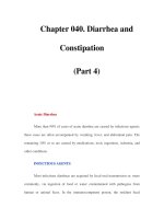
days, they showed a specific morphological phenomenon: under each border zone
of the compressed segment, the nodes of Ranvier had been displaced along each
fibre, so that the paranodal myelin was stretched on one side of the node and invagi-
nated on the other. The whole picture is strongly reminiscent of an intussusception,
as it occurs in the bowel. The underlying force seemed to be the pressure gradient
within the nerve between its compressed and uncompressed portions. In each case,
the displacement was away from the region of high pressure towards the uncom-
pressed region beyond the edge of the cuff (Figure 2.10).
The result was localised degenerative changes of the damaged myelin (paranodal
demyelination). Only large myelinated fibres were affected. In these experiments,
a cuff pressure of 1000 mm Hg maintained for one to three hours produced paral-
ysis of distal muscles lasting for up to three months. There was a significant
correlation between the duration of compression and the duration of the sub-
sequent conduction block. The effects of the block correspond with the type of
1111
2
3
4
5
611
7
8
9
1011
11
2
3111
4
5
6
7
8
9
2011
1
1
2
3
4
5
6
7
8
9
3011
1
1
2
3
4
5
6
7
8
9
4011
1
211
28
The Tourniquet Manual ➀➋➂➃➄➅➆
Figure 2.10 Diagram to show the direction of displacement of nodes of Ranvier in relation to the cuff. Reprinted
with permission from Ochoa, J, Fowler, TJ, Gilliatt, RW (1972). Anatomical changes in peripheral nerves compressed by a pneu-
matic tourniquet.
Journal of Anatomy
113: 433–455.
nerve injury classified by Seddon in 1943 as neurapraxia.
36
Gilliatt in 1980 showed
by direct recordings from the exposed nerve “a double conduction block” affecting
the large myelinated fibres as two separate regions of the nerve trunk corresponding
in position to both edges of the cuff, while the intermediate region showed little
or no change in conduction.
37
2.5 Effects on the Skin
On the whole, the skin is resilient and unaffected in the vast majority of cases of
tourniquet use. Damage at the site of the tourniquet may be caused by pressure
necrosis or friction burns. Such burns are thought to be caused by spirit-based anti-
septic solutions that seep beneath the tourniquet and are held against the skin
under pressure (see Chapter 5).
38
Friction burns may result during operations on the thigh due to a fully inflated
tourniquet cuff slipping down and away from the plaster wool padding.
39
An inves-
tigation on the effects produced by commonly used antiseptic paints and a known
chemical irritant, anthralin, was carried out on the upper arms and forearms of volun-
teers.
40
Site-related variations in anthralin-induced inflammation were observed, but
there was no demonstrable effect of either pressure or ischaemia on the inflam-
matory response. It was not possible to keep the tourniquets in place for longer
than half an hour because it would have been too painful for the volunteers to
tolerate the pain of ischaemia. It was concluded that burns under tourniquets are
likely to be idiosyncratic reactions, and their further investigation required detailed
examination of individuals affected by chemical burns.
2.6 Systemic and Local Effects of the Application
of a Tourniquet
There have been few reports describing the systemic effects of reperfusing the
ischaemic limb.
41, 42
Complete arrest of the circulation to the limb produces acidosis
and changes in levels of potassium,
43, 44
which in theory could result in effects on
the rhythm of the heart when the tourniquet is released. Although changes in the
acid–base status of the blood leaving the limb have been described, the state of
the blood reaching the heart after the release of a tourniquet has received little
attention.
45
An animal and clinical study was undertaken to establish whether any
biochemical changes in the limb are reflected in the right atrium. In addition, the
time taken for the ischaemic limb to recover was investigated.
46
2.6.1 Animal Experiments
An infant-size Kidde tourniquet cuff 5 cm wide was applied to the experimental limb
of a rhesus monkey and inflated to a pressure of 300 mm Hg for a predetermined
29
➀➋➂➃➄➅➆ Effect of the Tourniquet on the Limb
time from one to five hours. At regular intervals during the period when the tourni-
quet was in place, samples were taken over a period of one minute from the cannula
in the right atrium to establish control values for acid–base status and potassium
levels. After the release of the tourniquet, further samples were taken simultane-
ously from both the internal jugular route and the femoral vein for periods as long
as two hours.
Whenever possible, all samples were measured immediately for pCO
2
, pH, excess of
base, and standard bicarbonate. If this was not possible, samples were stored in ice
for no longer than 30 minutes.
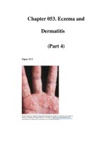
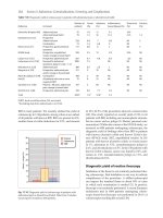
When the tourniquet was released, samples taken from the right side of the heart
showed little or no change in acid–base status. The longer the tourniquet had been
in place, the greater were the biochemical changes in the limb (Figure 2.11). The
readings for pH, potassium and pCO
2
in the right atrium immediately before the
release of the tourniquet were taken as 100%. Each subsequent reading taken from
the atrium and the femoral vein was then expressed as a percentage of the initial
reading.
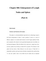
The results obtained were plotted on semilogarithmic paper. The best-fit line for
each variable was drawn for the samples for the heart and limb. Recovery time for
the limb was measured at the point where the initial slope of the curve for the limb
intersected with the line for readings from the right side of the heart. This was
plotted against the time for which the tourniquet had been inflated (Figure 2.12).
After one hour with the tourniquet, recovery occurred in the limb within 20 minutes.
For tourniquet periods of two to four hours, recovery of all variables was complete
with 40 minutes. However, after five hours of tourniquet use, recovery for potas-
sium and standard bicarbonate occurred within one hour and 40 minutes, but pH
returned to the level of the blood in the right atrium after two hours and 40 minutes.
1111
2
3
4
5
611
7
8
9
1011
11
2
3111
4
5
6
7
8
9
2011
1
1
2
3
4
5
6
7
8
9
3011
1
1
2
3
4
5
6
7
8
9
4011
1
211
30
The Tourniquet Manual ➀➋➂➃➄➅➆
Figure 2.11 Mean initial
readings from the first sample
of blood from the limb after
release of the tourniquet
plotted against the time for
which the tourniquet was
inflated.
Reprinted with permission
from Klenerman, L, Biswas, M,
Hulands, GH, Rhodes, AM (1980).
Systemic and local effects of the
application of a tourniquet.
Journal
of Bone and Joint Surgery
62B:
385–388.
2.6.2 Clinical Studies
Patients who were about to undergo total knee replacement or a high tibial
osteotomy for rheumatoid arthritis or osteoarthritis were informed of the studies and
consented to participate. All patients received appropriate premedication of
papaveretum and atropine. Anaesthesia was induced with thiopentone, an intra-
venous injection of pancuronium was given, and intubation was carried out.
Anaesthesia was maintained with nitrous oxide, oxygen and phenoperidine, and
occasionally halothane (less than 0.5%). Ventilation was adjusted for a standard
paCO
2
of 5.4 kPa. A cannula was passed via the right internal jugular vein into the
atrium and its position checked by looking for atrial oscillations; 5% dextrose solution
was infused. An intravenous drip of Hartmann’s solution was set up in one forearm.
The electrocardiogram was displayed continuously, and the temperature was moni-
tored by a nasopharyngeal probe. An Esmarch bandage was used to exsanguinate
the site of operation, and a 10-cm Kidde tourniquet cuff was inflated to occlude the
arterial flow at a pressure of twice the pre-induction systolic pressure. During the
operation, several samples were taken from the internal jugular cannula to establish
baseline values for blood analysis from the central venous pool. At the end of the
operation, pressure dressings were applied to the limb while the tourniquet was
still inflated. Samples of blood were taken from the atrium via the internal jugular
cannula and also from the femoral vein of the operated limb by direct needle stab
just before releasing the tourniquet. When the tourniquet was released, samples
were taken simultaneously from the femoral needle and the internal jugular cannula
for a period of approximately 15 minutes and then intermittently from the jugular
cannula for approximately two hours. These samples were analysed as described
above.
There were nine patients (three men, six women), of average age 68 years (range
51–80 years). The tourniquet was inflated for periods ranging from 70 to 186 minutes.
31
➀➋➂➃➄➅➆ Effect of the Tourniquet on the Limb
Figure 2.12 Estimated recovery
time for each variable in the
blood supply in the limb
subjected to ischaemia in
relation to the time for which
the tourniquet was used.
Reprinted with permission from
Klenerman, L, Biswas, M, Hulands,
GH, Rhodes, AM (1980). Systemic
and local effects of the application
of a tourniquet.
Journal of Bone
and Joint Surgery
62B: 385–388.
2.6.3 Results of Investigations
There were only minor fluctuations in the three variables – potassium, bicarbonate
and pH – in the samples taken from the right atrium. These transiently reflected the
marked changes that occurred in the blood from the limb. No cardiac dysrhythmias
were detected on monitoring.
Neither the patients nor the experimental animals showed evidence of nerve palsies.
In a limb that has been rendered ischaemic, metabolites accumulate as a result of
hypoxia in the tissues. Theoretically, a rapid influx of some of these products, e.g.
potassium, into the coronary circulation is likely to produce cardiac dysfunction. In
these studies, although the potassium levels in the blood leaving the limb were
raised, at no time was a significant rise detected in the right atrium either in the
animals or in the patients. The most likely explanation for this is a dilutional effect
due to the larger volume of blood contained in the venous side of the circulation
(50% of the circulating blood volume is accommodated on the venous side, but
only 15% is in the arterial system). Similarly, the fall in pH in the venous blood leaving
the acidotic limb was not reflected in the acid–base status of the blood samples
from the right atrium. Again, the effect of dilution is a factor here, but in addition
there is the efficient buffering capacity of the blood. A criticism of the sampling
technique used could be based on the well-known streaming effect of blood from
the venae cavae. This is well documented in relation to the measurement of venous
oxygen in estimations of cardiac output. However, the authors were not aware of
work showing that this effect was also applicable to other biochemical measure-
ments. Although streaming within the atrium cannot be discounted, it is unlikely to
be an important factor as the results were consistent. These findings are essentially
in agreement with those described in patients undergoing operations under tourni-
quet with lumbar epidural anaesthesia.
45
In the animal studies, it was found that the acid–base balance in the limb returned
to normal within 20 minutes of the release of a tourniquet that had been in place
for one hour, and within 40 minutes after four hours of ischaemia. The practice of
releasing the tourniquet at two hours for a period of five to ten minutes to allow a
“breathing period” therefore does not seem appropriate.
The investigations that have been described were undertaken in healthy animals
and fit patients who did not suffer from cardiovascular disease. When, as is not
uncommon, the buffering capacity is reduced by anaemia, hypovolaemia, metabolic
acidosis or pre-existing vascular disease, there is likely to be a reduction in the
normal range of safety. In addition, under certain conditions a compromised
myocardium may be sensitised to catecholamines by anaesthetic agents. In these
circumstances, the period for which a tourniquet is used should be reduced to
the minimum and full cardiovascular monitoring must be available. The changes
noted in the acid–base balance indicate that a period of three hours under a
tourniquet is safe. This coincides with findings made in histological studies of the
ischaemic muscle.
26
1111
2
3
4
5
611
7
8
9
1011
11
2
3111
4
5
6
7
8
9
2011
1
1
2
3
4
5
6
7
8
9
3011
1
1
2
3
4
5
6
7
8
9
4011
1
211
32
The Tourniquet Manual ➀➋➂➃➄➅➆
2.7 Haemodynamic Changes
The haemodynamic changes associated with the application and release of a tourni-
quet are minimal in healthy adults, but they may not be tolerated by patients with
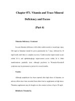
poor cardiac reserve. In a series of patients who were monitored for changes in
central venous pressure (CVP) and systolic blood pressure, it was found that the
main rise in CVP with the application of bilateral tourniquets was 14.5 cm H
2
O.
47
This
was maintained in 80% of patients until the tourniquets were released (Figure 2.13).
It is likely that this was due to an increase of approximately 15% of circulating blood
volume – about 700–800 ml of blood. In comparison, the CVP values when single
tourniquets were applied showed that the circulation could deal with the smaller
autotransfusion of blood more easily. The mean systolic pressure change was ±18.5
mm Hg when the tourniquets were inflated. On deflation, the mean fall below the
blood pressure at the start of surgery was 43.5 mm Hg. The initial rise in blood pres-
sure either was sustained or fell gradually to the level before a tourniquet was
applied, and then had a further dramatic fall within three minutes of release of the
tourniquet. In a review of the records of 500 patients who had surgery under a
tourniquet, the frequency of intraoperative hypertension (defined as a 30% increase
in either systolic or diastolic pressure compared with the first pressure recording
after incision) was 11%. The probability of hypertension was increased if the patient
was elderly, had cardiac enlargement as shown by X-ray or electrocardiogram (ECG),
or had nitrous oxide and narcotic anaesthesia. Pre-existing hypertension, increased
serum creatinine concentration, anaemia, or treatment with hypertensive drugs were
not associated strongly with intraoperative hypertension.
48
Patients with head
injuries and multiple sites of trauma may have marked increases in intracranial pres-
sure when lower limb tourniquets are released.
49
Using transoesophageal echocardiography during 59 total knee replacements, it
was found that showers of echogenic material traversed the right atrium, right
ventricle and pulmonary artery after the tourniquets were deflated.
50
This was
observed in various degrees in all patients and lasted for 3–15 minutes. The mean
peak intensity occurred within 30 seconds (range 24–45 seconds) after the tourni-
quet was released. Only three patients had evidence of clinical pulmonary embolism.
These findings are similar to those described by Parmet and colleagues in a smaller
series of 29 patients.
51
This group aspirated a 3 × 6-mm fresh thrombus from a central
catheter in one patient. Another patient, who had a Greenfield filter in the inferior
vena cava to prevent emboli reaching the lungs from the legs following previous
thromboembolism, showed very little echogenic material, indicating that the
filter acted as an effective block. Inadequate exsanguination of the limb under-
going surgery coupled with stasis and cooling may contribute to fresh thrombus
formation. Nevertheless, these 29 patients had echogenic material with clinically
adequate exsanguination. Bone cement activation of the coagulation cascade could
also form fresh clot. It is likely that the pulmonary circulation is often exposed to
embolic material during normal everyday life and that the lungs are able to clear
small emboli.
33
➀➋➂➃➄➅➆ Effect of the Tourniquet on the Limb
2.8 Limb Blood Flow in the Presence of a
Tourniquet
The blood supply to the limbs of rhesus monkeys was studied with 50- diameter
microspheres labelled with
51
Cr and by the washout of
22
Na injected into the tissues.
One limb, upper or lower, was exsanguinated and the circulation was occluded with
a pneumatic tourniquet. The opposite limb was used as a control. The blood to the
occluded limb was found to be less than 1% of the flow to the control limb. The
venous return was less than 0.2% of that of the control limb. It was concluded that a
limb with a tourniquet in place is virtually isolated from the circulation and the
amount of blood reaching the tissues probably via the intramedullary circulation is
likely to be of no significance to relieve the ischaemia.
52
Added support for the isola-
tion of the limb from normal blood flow is provided by the work of Santavirta and
colleagues, who studied tissue oxygen levels in rabbits.
53
The tourniquet was in place
for 60, 80 or 120 minutes. The baseline PO
2
in the tibialis anterior muscle was 22.6±0.6
mm Hg. While the tourniquet was in place, the oxygen tension dropped to minimal
values between 9.2±0.5 and 10.7±0.6 mm Hg in the three groups rendered ischaemic
for 60, 80 and 120 minutes, but the tissue microclimate never reached fully anoxic
conditions. This minimal value was reached in 19–26 minutes and then remained con-
stant during the remainder of the time that the tourniquet was in place, but it never
reached zero. The decline of PO
2
and recovery after release of the tourniquet was
independent of tourniquet time. Continuous oxygen during the experiment had no
influence on the PO
2
.
1111
2
3
4
5
611
7
8
9
1011
11
2
3111
4
5
6
7
8
9
2011
1
1
2
3
4
5
6
7
8
9
3011
1
1
2
3
4
5
6
7
8
9
4011
1
211
34
The Tourniquet Manual ➀➋➂➃➄➅➆
Figure 2.13 Changes in
central venous pressure and
blood pressure with a
tourniquet in place and
after release.
Reproduced
with permission from Bradford,
EMW (1968). Haemodynamic
changes associated with the
application of lower limb
tourniquets.
Anaesthesia
24:
190–197.
2.9 Hyperaemia and Swelling of a Limb After
Release of a Tourniquet
Using monkeys, a quantitative study was carried out to measure the effect of a
tourniquet on the lower limb on peak flow, the amount of swelling, and the time
for recovery. The disappearance of acute swelling is related to the period of
ischaemia. As the duration of the tourniquet increased, no significant change in
peak flow was demonstrated. The swelling that results from a tourniquet for one
hour is overcome rapidly, but the effects are much more obvious for tourniquet
times of two and three hours. When attempting to obtain haemostasis after release
of a tourniquet, surgeons should remember that for a one-hour period of ischaemia,
the hyperaemia falls to one-half in about five minutes, but that it takes 12 and 25
minutes, respectively, for this to take place after two and three hours of tourniquet
use.
54
These times are of relevance to breathing periods. The onset of hyperaemia is
related to the changes brought about by the effects of free oxygen radicals (see
Chapter 3).
2.10 Haematological Effects
At the end of orthopaedic operations, there is a pronounced increase in fibrinolytic
activity in the blood from the systemic circulation, as well as from the operated limb,
whereas there is only a small systemic increase after surgery on the leg without a
tourniquet. The vasa vasorum are probably the main source of plasminogen acti-
vator in the vasculature and may be stimulated to respond maximally by complete
ischaemia; the increase in fibrinolytic activity does not appear to be related to the
duration of the application of a tourniquet.
55
However, there is no difference in the
incidence of deep vein thrombosis in surgery on the lower limbs with and without
a tourniquet.
56
The increase in fibrinolytic activity is short-lived; it is maximal at 15
minutes and returns to preoperative levels within 30 minutes of the release of the
tourniquet. It then falls below the preoperative levels, where it remains for at least
48 hours. The tourniquet appears to alter the timing of a short period of increased
fibrinolytic activity without altering the overall pattern. It is unlikely that this would
alter the incidence of deep vein thrombosis, but it may affect the degree of bleeding
after release of the tourniquet.
57
2.11 Temperature Changes
An increased core body temperature occurs during the application of arterial tourni-
quets, probably because of reduced metabolic heat transfer from the central to the
peripheral compartments and from decreased heat loss from distal skin. When the
tourniquet is released, there is a transient decrease in core temperature as a result of
redistribution of body heat from the return of hypothermic venous blood flow from
35
➀➋➂➃➄➅➆ Effect of the Tourniquet on the Limb
the tourniquet limb into the systemic circulation.
58
A marked rise in temperature may
cause the anaesthetist concern about the possibility of malignant hyperthermia. An
association between the use of tourniquets for limb surgery and a progressive
increase in body temperature of greater than one degree with bilateral tourniquets
has been reported in children.
59
With a tourniquet in place, the limb cools gradually; during the course of an oper-
ation, the temperature may drop by 3–4 °C. Part of the cooling is counterbalanced
by the effects of the lights and drapes in the operation theatre. There may be obvious
drying out of the issues exposed, which should always be kept moist with Hartman’s
solution or normal saline.
2.12 Tourniquet Pain
When a tourniquet is applied to the arms of volunteers, they experience a vague,
dull pain in the limb, which is associated with an increase in blood pressure. The
average pain tolerance is 31 minutes, increasing to 45 minutes with sedation.
Prolonged tourniquet inflation during general anaesthesia causes an increase in
heart rate and blood pressure, which commonly leads the anaesthetist to increase
the depth of anaesthesia. A cutaneous neural mechanism is thought to be respon-
sible for the tourniquet pain, and the rise in blood pressure follows a humoral
response to the pain. Tourniquet pain and the associated hypertension can also
complicate spinal or epidural anaesthesia despite adequate sensory anaesthesia of
the dermatome underlying the tourniquet.
Tourniquet pain is thought to be transmitted by unmyelinated, slow-conducting
C-fibres, which are normally inhibited by fast pain impulses conducted by myelinated
A-delta-fibres. Mechanical compression causes loss of conduction due to ischaemia.
Large A-delta nerve fibres are blocked, leaving C-fibres still functioning.
16
Summary
The effect of a tourniquet on the tissues beneath and distal to it have been described.
Nerves are vulnerable to high pressures, and muscle is vulnerable to prolonged
ischaemia. Based on a study of the ultrastructure of muscle and biochemical changes
in the limb subjected to ischaemia in relation to their return to normal, three hours
is the upper limit of safety for a tourniquet to be kept in place.
References
1 American Heart Association (1967). Report of a subcommittee of the postgraduate education committee:
recommendations for human blood pressure determination by sphygmomanometers. Circulation XXXVI;
980–988.
1111
2
3
4
5
611
7
8
9
1011
11
2
3111
4
5
6
7
8
9
2011
1
1
2
3
4
5
6
7
8
9
3011
1
1
2
3
4
5
6
7
8
9
4011
1
211
36
The Tourniquet Manual ➀➋➂➃➄➅➆
2 Pedowitz, RA, Gershuni, DH, Botte, MJ, et al. (1993). The use of lower tourniquet inflation pressures in
extremity surgery facilitated by curved and wide tourniquets and an integrated cuff inflation system.
Clinical Orthopaedics and Related Research 287: 237–243.
3 Klenerman, L, Hulands, G (1979). Tourniquet pressures for the lower limb. Journal of Bone and Joint Surgery
61B: 124.
4 Lieberman, JR, Staheli, LT, Dales, MC (1997). Tourniquet pressures on paediatric patients: a clinical study.
Orthopaedics 20: 1143–1147.
5 Shaw, JA, Murray, DG (1982). The relationship between tourniquet pressure and underlying soft tissue
pressure in the thigh. Journal of Bone and Joint Surgery 64A: 1148–1151.
6 Neimkin, RJ, Smith, RJ (1983). Double tourniquet with linked mercury manometers for hand surgery. Journal
of Hand Surgery 8A: 938–941.
7 Graham, B, Breault, MJ, McEwen, JA, McGraw, RW (1993). Occlusion of arterial flow in the extremities at
subsystolic pressure through the use of wide cuffs. Clinical Orthopaedics and Related Research 286: 257–260.
8 Rydevik, BJ, Lundborg, G, Olmarker, K, Myers RR (2001). Biomechanics of peripheral nerves and spinal
nerve roots. In Nordin, M, Frankel, VH, eds. Basic Biomechanics of the Musculoskeletal System, 3rd edn.
Philadelphia: Lippincott, Williams & Wilkins.
9 Lundborg, G (1988). Nerve Injury and Repair. Edinburgh: Churchill Livingstone, p. 83.
10 Yousif, NJ, Grunert, BK, Forte, RA, et al. (1993). A comparison of upper and forearm tourniquet tolerance.
Journal of Hand Surgery 18B: 639–641.
11 Hutchinson, DT, McClinton, MA (1993). Upper extremity tourniquet tolerance. Journal of Hand Surgery 18A:
206–210.
12 Odensson, A, Finsen, V (2002). The position of the tourniquet on the upper limb. Journal of Bone and Joint
Surgery 84B: 202–204.
13 Michelson, JD, Perry, M (1996). Clinical safety and efficiency of calf tourniquets. Foot and Ankle International
17: 573–575.
14 Lichtenfeld, NS (1992). The pneumatic tourniquet with ankle block anaesthesia for foot surgery. Foot and
Ankle International 13: 344–349.
15 Finsen, V, Kasseth, A (1997). Tourniquets in forefoot surgery. Less pain when placed at the ankle. Journal
of Bone and Joint Surgery 79B: 99–101.
16 Kam, PCA, Kanaugh, R, Yoong, FFY (2001). The arterial tourniquet: pathophysiological consequences and
anaesthetic implications. Anaesthesia 56: 534–545.
17 Bruner, JW (1951). Safety factors in the use of the pneumatic tourniquet for haemostasis in surgery of the
hand. Journal of Bone and Joint Surgery 33A: 221–224.
18 Boyes, JH (1964). Bunnell’s Surgery of the Hand. Philadelphia: J.B. Lippincott and Co., p. 133.
19 Parkes, A (1973). Ischaemic effects of external and internal pressure of the upper limb. The Hand 5: 105–112.
20 Harman, JW, Gwian, RP (1949). The recovery of skeletal muscle fibres from acute ischaemia by histologic
and chemical methods. American Journal of Pathology 24: 741–745.
21 Dahlback, LO (1970). Effects of temporary tourniquet ischaemia on striated muscle fibres and motor end-
plates. Morphological and histological studies in the rabbit and electromyographical studies in man.
Scandinavian Journal of Plastic and Reconstructive Surgery Suppl 7.
22 Moore, DH, Ruska, H, Copenhaver, WN (1956). Electromicroscopic and histochemical observations of muscle
degeneration after tourniquet. Journal of Biophysical and Biochemical Cytology 2: 755–764.
23 Tountas, CP, Bergman, RA (1977). Tourniquet ischaemia: ultrastructural and histochemical observations of
ischaemic human muscle and of monkey muscle and nerve. Journal of Hand Surgery 2: 31–37.
24 Strock, PE, Majino, G (1969). Microvascular changes in acutely ischaemic rat muscle. Surgery, Gynaecology
and Obstetrics 129: 1213–1224.
25 Barnard, RJ, Edgerton, VR, Furukaws, T, Peter, JB (1971). Histochemical, biochemical and contractile prop-
erties of red, white and intermediate fibres. American Journal of Physiology 220: 410–414.
26 Patterson, S, Klenerman, L (1979). The effect of pneumatic tourniquets on the ultrastructure of skeletal
muscle. Journal of Bone and Joint Surgery 61B: 178–183.
27 McAlllister, LP, Munger, BL, Neel, JR (1977). Electron microscopic observations and acid phosphate activity
in the ischaemic rat heart. Journal of Molecular and Cellular Cardiology 9: 353–364.
28 Patterson, S, Klenerman, L, Biswas, M, Rhodes, A (1981). The effect of pneumatic tourniquets on skeletal
muscle physiology. Acta Orthopaedica Scandinavica 52: 171–175.
29 Mohler, LR, Pedowitz, RA, Lopez, MA, Gershuni, DH (1999). Effects of tourniquet compression on neuro-
muscular function. Clinical Orthopaedics and Related Research 359: 213–220.
30 Grace, PA (1994). Ischaemic–reperfusion injury. British Journal of Surgery 81: 637–647.
37
➀➋➂➃➄➅➆ Effect of the Tourniquet on the Limb
31 Leif, A (1973). Cell swelling a factor in ischaemic tissue injury. Circulation 8: 455–458.
32 Herald, J, Cooper, L, Machart, J (2002). Tourniquet induced restriction of the quadriceps muscle mecha-
nism. Journal of Bone and Joint Surgery 84B: 856–857.
33 Fowler, TJ, Danta, G, Gilliatt, RW (1972). Recovery of nerve conduction after pneumatic tourniquet, obser-
vations on the hindlimb of the baboon. Journal of Neurology, Neurosurgery and Psychiatry 35: 638–647.
34 Ochoa, J, Fowler TJ, Gilliatt, RW (1972). Anatomical changes in peripheral nerves compressed by a pneu-
matic tourniquet. Journal of Anatomy 113: 433–455.
35 Rudge, P, Ochoa, J, Gilliatt, RW (1974). Acute peripheral nerve compression in the baboon. Journal of
Neurological Science 23: 403–420.
36 Seddon, H (1943). Three types of nerve injury. Brain 66: 237–288.
37 Gilliatt, RW (1980). Acute compression block. In Sumner, AJ, ed. The Physiology of Peripheral Nerve Disease.
Philadelphia: W.B. Saunders, pp. 287–315.
38 Dickinson, JC, Bailey, BN (1988). Chemical burns beneath tourniquets. British Medical Journal 297: 1513.
39 Choudhary, S, Koshy, C, Ahmed, J, Evans, J (1998). Friction burns to thigh caused by tourniquet. British
Journal of Plastic Surgery 51: 142–143.
40 Parslew, R, Braithwaite, J, Klenerman, L, Friedmann, P (1997). An investigation into the effect of ischaemia
and pressure on irritant inflammation. British Journal of Dermatology 136: 734–736.
41 Dery, R, Pelletier, J, Jacques, A, et al. (1965). Metabolic changes induced in the limb during tourniquet
ischaemia. Canadian Anaesthetic Society Journal 12: 367–368.
42 Solonen, KA, Takkanen, L, Narvenen, S, Gordin, R (1968). Metabolic changes in the upper limb during
tourniquet ischaemia. Acta Orthopaedica Scandinavica 39: 20–22.
43 Stock, W, Bohn, HJ, Isselhard, W (1971). Metabolic changes in rat skeletal muscle after acute arterial occlu-
sion. Vascular Surgery 5: 249–255.
44 Wilgis, EFS (1971). Observations on the effects of tourniquet ischaemia. Journal of Bone and Joint Surgery
53A: 1343–1346.
45 Modig, J, Kolstad, K, Wigren, A (1978). Systemic reactions to tourniquet ischaemia. Acta Anaesthesiologica
Scandinavica 22: 609–614.
46 Klenerman, L, Biswas, M, Hulands, GH, Rhodes, AM (1980). Systemic and local effects of the application of
a tourniquet. Journal of Bone and Joint Surgery 62B: 385–388.
47 Bradford, EMW (1968). Haemodynamic changes associated with the application of lower limb tourniquets.
Anaesthesia 24: 190–197.
48 Kaufman, RD, Walts, LF (1982). Tourniquet induced hypertension. British Journal of Anaesthesia 54: 333–336.
49 Sparling, RJ, Murray, AW, Choksey, M (1993). Raised intracranial pressure associated with raised hyper-
tension after tourniquet removal. British Journal of Neurosurgery 7: 75–78.
50 Berman, AT, Parmet, JR, Harding, SP, et al. (1998). Emboli observed with the use of transoesophageal
echocardiography immediately after tourniquet release during total knee arthroplasty with cement. Journal
of Bone and Joint Surgery 89A: 389–396.
51 Parmet, JL, Berman, AT, Horrow, JC, et al. (1993). Thromboembolism coincident with tourniquet deflation
during total knee arthroplasty. Lancet 341: 1057–1058.
52 Klenerman, L, Crawley, J (1977). Limb blood flow in the presence of a tourniquet. Acta Orthopaedica
Scandinavica 48: 291–295.
53 Santavirta, J, Hockerstedt, K, Niinikoski, J (1978). Effect of pneumatic tourniquet on muscle oxygen tension.
Acta Orthopaedica Scandinavica 49: 415–419.
54 Klenerman, L, Crawley, J, Lowe, A (1982). Hyperaemia and swelling of a limb upon release of a tourniquet.
Acta Orthopaedica Scandinavica 53: 209–213.
55 Klenerman, L, Mackie, I, Charabarti, R, et al. (1977). Changes in haemostatic system after application of a
tourniquet. Lancet 1: 970–972.
56 Angus, PD, Nakielny, R, Gordrum, DT (1983). The pneumatic tourniquet and deep venous thrombosis.
Journal of Bone and Joint Surgery 65B: 336–339.
57 Price, AJ, Jones, NAG, Webb, PJ, et al. (1980). Do tourniquets prevent deep vein thrombosis? Journal of
Bone and Joint Surgery 62B: 529.
58 Estebe, JP, Le Naoures, A, Malledant, Y, Ecoffey, C (1996). Use of the pneumatic tourniquet induces changes
in central temperature. British Journal of Anaesthesia 77: 786–788.
59 Bloch, EC (1986). Hypothermia resulting from tourniquet application in children. Annals of the Royal College
of Surgeons of England 69: 193–194.
1111
2
3
4
5
611
7
8
9
1011
11
2
3111
4
5
6
7
8
9
2011
1
1
2
3
4
5
6
7
8
9
3011
1
1
2
3
4
5
6
7
8
9
4011
1
211
38
The Tourniquet Manual ➀➋➂➃➄➅➆
Chapter 3
Ischaemia–Reperfusion Syndrome