Treatment of Osteoarthritic Change in the Hip - part 3 potx
Bạn đang xem bản rút gọn của tài liệu. Xem và tải ngay bản đầy đủ của tài liệu tại đây (595.87 KB, 26 trang )
Corrective Imhäuser Intertrochanteric Osteotomy for SCFE 45
et al. [6] have described how results of treatment depend on stability of the epiphysis,
in that the results were gratifying in 96% of cases with stable physeal stability and in
only 47% of cases with unstable physeal stability. They also reported that none devel-
oped avascular necrosis of the femoral head among the “stable” cases while it occurred
in 47% of “unstable” cases. Without needing mention, the above-cited reports of
Jones et al. [7] and Carney et al. [9] indicated results of treatment are more favorable
in milder cases. That is, to achieve the best therapeutic results, it is necessary to
perform treatment without causing complications in stable, mild cases.
It may be said to stand to reason that the Imhäuser treatment system ensures a
stable physeal stability of the affected hip joint by pinning in mild cases, whereas in
more severe cases the physeal stability of the joint is rendered stable by traction and
then the PTA is reduced to 30° or less by osteotomy to lessen the severity to mild. In
the present study, limitation of range of motion completely resolved in all patients
following treatment, and none had necrosis of the femoral head postoperatively.
Consistent with the reports of Imhäuser [2] and Kartenbender et al. [15], rather
gratifying results were obtained both clinically and roentgenographically in short- or
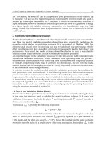
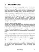
mid-term outcomes. As shown in Fig. 7, most cases had good congruity of the hip
joint as a result of both the correction osteotomy and remodeling after operation.
However, the apparent neck–shaft angle was 150° on average at the time of this inves-
tigation, thus indicating a tendency toward coxa valga (Fig. 7). There was a mean
reduction in leg length by 0.7 cm, so there is a possible influence of an altered func-
tional axis on the knee joint. Further investigation is necessary, therefore, to investi-
gate osteotomy angle, especially with respect to anterotation and valgus. Four patients
Fig. 7. A 12-year-old boy with a stable SCFE involving the left hip. A PTA was 65° at first visit
(12 years and 5 months old). B PTA was 20° immediately after operation (12 years and 6 months
old). C Good congruity of the hip joint was obtained at the final visit (18 years and 11 months
old), and neck–shaft angle was 155°
46 S. Mitani et al.
had a fracture as a result of bone fragility from long-term traction and bed rest. The
treatment scheme is under reconsideration with regard to preoperative duration of
traction, based also on the recent medical care situation.
Intertrochanteric osteotomy in the Imhäuser treatment system is considered a
useful procedure because it is relatively simple in technique and involves no develop-
ment of avascular necrosis of the femoral head. As Schai et al. [16] reported that
results of treatment with the Imhäuser method were superior to those by other pro-
cedures but entailed development of arthrosis in 45% of cases, it seems that matters
relating to treatment of this disorder are yet to be resolved. Indeed, there are problems
peculiar to this treatment method that remain to be solved, as has been disclosed by
the present study; further long-term follow-up for treated joints is needed.
References
1. Imhäuser G (1986) Spontane Epipyhsendislokation am koxalen Femurende. Orthopäde
in Praxis und Klinik, vol VII. Thieme, Stuttgart, pp 115–148
2. Imhäuser G (1977) Spätergebnisse der sog. Imhäuser-Osteotomie bei der Epiphysen-
lösung. Z Orthop 115:716–725
3. Oda K, Mitani S (1998) Slipped capital femoral epiphysis (in Japanese). Orthop Surg
Traumatol 41:439–448
4. Loder RT, Aronsson DD, Dobbs MB, et al (2001) Slipped capital femoral epiphysis.
Instr Course Lect 50:555–570
5. Canal ST (2003) Fractures and dislocations in children. Slipped capital femoral epi
physis. In: Campbell’s operative orthopaedics, 10th edn. Mosby, Philadelphia,
pp 1481–1483
6. Loder RT, Richards ABS, Shapiro PS, et al (1993) Acute slipped capital femoral epiphy-
sis: the importance of physeal stability. J Bone Joint Surg 75A:1134–1140
7. Jones JR, Paterson DC, Hillier TM, et al (1990) Remodelling after pinning for slipped
capital femoral epiphysis. J Bone Joint Surg 72B:568–573
8. Rab GT (1999) The geometry of slipped capital femoral epiphysis: implications for
movement, impingement, and corrective osteotomy. J Pediatr Orthop 19:419–424
9. Carney BT, Weinstein SL, Noble J (1991) Long-term follow-up of slipped capital
femoral epiphysis. J Bone Joint Surg 73A:667–674
10. Peterson MD, Weiner DS, Green NF, et al (1997) Acute slipped capital femoral epiphy-
sis: the value and safety of urgent manipulative reduction. J Pediatr Orthop
17:648–654
11. Otani T, Saito M, Kawaguchi Y, et al (2004
) Short-term clinical results of manipulative
reduction for acute-unstable slipped capital femoral epiphysis (in Japanese). Hip Joint
30:223–225
12. Fish JB (1994) Cuneiform osteotomy of the femoral neck in the treatment of slipped
capital femoral epiphysis. A follow-up note. J Bone Joint Surg 76A:46–59
13. DeRosa GP, Mullins RC, Kling TF Jr (1996) Cuneiform osteotomy of the femoral neck
in severe slipped capital femoral epiphysis. Clin Orthop 322:48–60
14. Crawford AH (1996) Role of osteotomy in the treatment of slipped capital femoral
epiphysis. J Pediatr Orthop 5B:102–109
15. Kartenbender K, Cordier W, Katthagen BD (2000) Long-term follow-up study after
corrective Imhäuser osteotomy for severe slipped capital femoral epiphysis. J Pediatr
Orthop 20:749–756
16. Schai PA, Exner GU, Hänsch O (1996) Prevention of secondary coxarthrosis in slipped
capital femoral epiphysis: a long-term follow-up study after corrective intertrochan-
teric osteotomy. J Pediatr Orthop 5-B: 135–143
47
Slipping of the Femoral Capital
Epiphysis: Long-Term Follow-up
Results of Cases Treated with
Imhaeuser’s Therapeutic Principle
Muroto Sofue
1
and Naoto Endo
2
Summary. Slipping of the femoral capital epiphysis is a common problem in growing
children. For the treatment of this disease, it is of the utmost importance to prevent
complications that would adversely affect normal development of the hip joint.
Therefore, it is absolutely necessary to choose a treatment that will allow the hip joint
to develop normally and which will prevent osteoarthritic changes in the future. The
long-term results of cases treated with Imhaeuser’s method [1,2] are reported here.
The results were very satisfying, and this treatment should be continued in the
future.
Key words. Slipping of the femoral capital epiphysis, Aseptic necrosis of the femoral
head, In situ pinning, Imhaeuser’s osteotomy [1,2], Three-dimensional osteotomy
Introduction
Slipping of the femoral capital epiphysis (SFCE) has recently become more common-
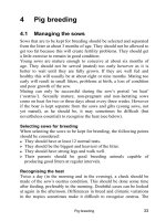
place in Japan. Figure 1 shows a patient with SFCE who was treated in the 1960s in
Niigata University Hospital. At that time, manual reduction followed by pinning was
common in Japan. However, by the age of 31, a severe arthritic change occurred in
this patient.
Authors [3,4,5] reviewed the cases in the hospitals associated with Niigata Univer-
sity and found that of fi ve cases that underwent manual reduction, unfortunately four
of them had femoral head necrosis, which resulted in osteoarthritic change at an early
age. Therefore, forceful reduction is contraindicated.
The aim of the treatment for SFCE is fi rst to improve joint incongruity and correct
the range of motion (ROM) without complications. This procedure will prevent the
development of osteoarthritis in the hip joint. With these points in mind, we chose
Imhaeuser’s method and treated the patients according to his principles. This chapter
is the report of the treatment of those patients along with their long-term follow-up.
1
Department of Orthopaedic Surgery, Nakajo Central Hospital, 12-1 Nishihoncho, Tainai,
959-2656 Niigata, Japan
2
Division of Orthopaedic Surgery, Department of Regenerative and Transplant Medicine,
Niigata University Graduate School of Medical and Dental Sciences, 1-757 Asahimachi-dori,
Niigata 951-8510, Japan
48 M. Sofue and N. Endo
Materials and Methods
In accordance with Imhaeuser’s principles [1,2], we have treated 76 cases, 79 joints
of SFCE, from 1976 to 2003.
In this study, the cases that were treated up to 1993 and followed over a period of
longer than 10 years are investigated. The 47 cases in all included 42 males and 5
females, ranging in age from 9 to 14 years old at the time of surgery, except for 1
patient treated at 20 years of age with endocrinopathy. Two cases were bilateral and
45 cases were unilateral. In the unilateral cases, 20 joints were right side and 25 were
left side. The type of slip was acute on chronic in 3 joints and chronic in 46 joints.
The direction of slip was posteroinferior in 48 cases, and 1 was posterosuperior
(Table 1).
The course of treatment is shown in Table 2. Forty-fi ve hips of the normal side
received prophylactic pinning, and 23 hips with less than 30° of slipping and 3 hips
with more than 30° of slipping, which were gently reduced to less than 30° by supra-
condylar skeletal traction, have been treated with in situ pinning. In total, 71 hips
have been pinned. Twenty-three hips with more than 30° of slipping, which were
not reduced to less than 30° in spite of direct traction, were treated by Imhaeuser’s
osteotomy. In all, 94 hips comprising 47 cases were clinically analyzed.
C
A
B
Fig. 1. A A 14-year-old boy, posterior tilt 65°. B Manual reduction and pinning. C Osteoarthritic
change after femoral head necrosis at the age of 31 years old
Imhaeuser’s Principle in Treatment for SFCE 49
Case Reports
Pinning Cases
Case 1: An 11-year-old boy with mild slipping of 20° on the right side (Fig. 2) was
treated with in situ pinning on the right side and prophylactic pinning on the left side
(Fig. 3). Sixteen years later, when he was 27 years old, a slight shortening of the
femoral neck with good joint congruency can be seen (Fig. 4). Clinically, he has no
problems and even plays soccer on a club team.
Case 2: A 14-year-old boy with bilateral slipping of 25° on the right and 20° on the
left (Fig. 5) was treated with in situ pinning on both sides (Fig. 6). Seventeen years
later, at 28 years old, there is some tendency of coxa vara in the X-ray findings, but
joint congruency is very good (Fig. 7). Clinically, he has no problems and enjoys
early-morning baseball with his club team.
Case 3: A 13-year-old boy with acute on chronic slipping of 65° on the left side (Fig.
8). After applying supracondylar skeletal traction for 3 weeks, good reduction of the
epiphysis was achieved (Fig. 9B), and in situ pinning was performed (Fig. 9C). At the
25-year postoperative follow-up examination, when he was 37 years old, very good
joint congruency can be seen (Fig. 10). He works as a long-distance driver and does
not have any complaints about his hip joints.
Table 1. Cases treated with Imhaeuser’s method [1,2],
1976–1993
Total cases: 47 (42 boys, 5 girls)
Follow-up: 10 years or more
Age: 9–14 years (except for 1 case of a 20-year-old)
Slip side: 2 bilateral, 45 unilateral (20 right, 25 left)
Slip type: 3 acute on chronic, 46 chronic
Slip direction: 1 posterosuperior, 48 posteroinferior
Table 2. Course of treatment
Normal side prophylactic nailing (45 joints)
Slip less than 30° (23 joints)
in situ nailing (26 joints)
reduced less than 30°
(3 joints)
Slip more than 30° traction (71 joints)
(26 joints)
not reduced
Imhaeuser’s osteotomy (23 joints) [1,2]
Total, 94 joints
50 M. Sofue and N. Endo
Fig. 2. An 11-year-old boy, right chronic slip, posterior tilt 20°
Fig. 3. An 11-year-old boy. Right, in situ pinning; left, prophylactic pinning
Imhaeuser’s Principle in Treatment for SFCE 51
Fig. 4. A 27-year-old man, 16 years after surgery, with good joint congruity
Fig. 5. A 14-year-old boy, bilateral chronic slip, posterior tilt: right, 25°, left, 20°
52 M. Sofue and N. Endo
Fig. 6. A 15-year-old boy, bilateral in situ pinning, 1 year after surgery
Fig. 7. A 28-year-old man, 17 years after surgery. X-ray findings show coxa vara but good joint
congruity
Imhaeuser’s Principle in Treatment for SFCE 53
Fig. 8. A 13-year-old boy, left acute on chronic slip, posterior tilt 65°
B
C
A
Fig. 9. Progression of treatment. A Slipping with posterior tilt 65°. B After 3 weeks of skeletal
traction, slipped epiphysis was gently reduced. C In situ pinning
54 M. Sofue and N. Endo
Fig. 10. A 37-year-old man, 25 years after surgery. Bilateral hips show good joint congruity
Three-Dimensional Osteotomy
(Imhaeuser’s Osteotomy) Cases
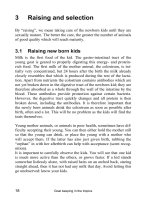
Imhaeuser’s osteotomy [1,2] consists of the following elements (Table 3):
1. Internal rotation to correct the external rotated midpoint.
2. Valgisation of 20° to 30°.
3. Flexion to correct the posterior tilting angle to a maximum permissible angle of
30°.
The valgus element (2) is necessary, because this osteotomy is performed at the inter-
trochanteric region of the femur, which has a neck-shaft angle of about 140°. Figure
11 shows an example case with external rotation from 10° to 70° (midpoint, 40°).
Case 4: A 13-year-old girl with right hip slipping of 60° (Fig. 12). In spite of direct
traction, the slip could not reduced. Imhaeuser’s osteotomy was performed. Figure
13 shows the patient’s postoperative findings with good progression. Twenty-one
years later, she is 34 years of age. The X-ray findings show good joint congruency
Table 3. The elements of Imhaeuser’s osteotomy [1,2]
1. Internal rotation to correct the external rotated midpoint
2. Valgisation of 20° to 30°
3. Flexion to correct the posterior tilting of epiphysis to maximum permissible angle of 30°
Imhaeuser’s Principle in Treatment for SFCE 55
Case with external rotation
from 10 to 70
o
( midpoint 40
o
)
1. Internal rotation
2.Valgization
( 20 to 30
o
)
3.Flexion
( Tilt minus
30
o
)
Imhaeuser’s osteotomy
Fig. 11. Scheme of Imhauser’s osteotomy [1,2] shown by an example case with external mid-
point of 40° (from 10° to 70° external rotation)
Fig. 12. A 13-year-old girl, right chronic slip, posterior tilt 60°
56 M. Sofue and N. Endo
DCBA
Fig. 13. Progression after Imhauser’s osteotomy. A Preoperative. B Operative. C Postoperative,
1 year. D Postoperative, 8 years
Fig. 14. A 34-year-old woman,
21 years after the osteotomy.
X-ray shows good joint
congruity
(Fig. 14). She has two children, has no clinical complaints, and lives an active life as
a housewife.
Case 5: A 13-year-old boy with slipping of 45° on the left hip (Fig. 15). Imhaeuser’s
osteotomy [1,2] was performed on the left hip and a prophylactic pinning was done
on the right hip (Fig. 16). Fifteen years later, he is 28 years of age. X-ray findings show
good joint congruity (Fig. 17), and the range of motion is free. He works in a restau-
rant as a cook and does not have any complaints about either leg.
Imhaeuser’s Principle in Treatment for SFCE 57
Fig. 15. A 13-year-old boy, left chronic slip, posterior tilt 45°
Fig. 16. A 14-year-old boy. Right, prophylactic pinning; left, Imhaeuser’s osteotomy [1,2],
1 year postoperative
58 M. Sofue and N. Endo
Fig. 17. A 28-year-old man, 15 years postoperative. X-ray shows good joint congruity
Table 4. Pinning results
Number of joints: 71
JOA hip score: 100 points for all joints
Complications (AVN, chondrolysis, etc.): None
Epiphyseal line: closed on all 71 joints
Bilateral pinning cases: 24 cases
Leg length discrepancy
No discrepancy: 20 cases
Discrepancy Ϲ1 cm: 4
Discrepancy >1 cm: 0
JOA, Japanese Orthopaedic Association; AVN, avascular necrosis
Results
The results of the 71 joints that received pinning were investigated (Table 4). In all
cases the Japanese Orthopaedic Association (JOA) hip score was 100 points of a pos-
sible 100 points. Complications such as avascular necrosis (AVN) of the femoral head
or chondrolysis were not observed. In all 71 joints, the epiphyseal lines were closed.
Leg length was examined in 24 cases that were pinned on both hips; 20 cases had no
discrepancy and 4 cases had some leg length discrepancy less than or equal to 1 cm.
There were no leg length discrepancies of more than 1 cm.
Imhaeuser’s Principle in Treatment for SFCE 59
Table 5. Imhaeuser’s osteotomy results
Number of cases (joints): 22 (23)
JOA score: >90 points
Complication (AVN, chondrolysis, etc.): none
Drehmann’s sign [6]: none
Tilt angle:
Before surgery: average 52°
After surgery: average 22°
(all cases less than 30°)
Leg length discrepancy:
<1 cm: 20 cases
м2 cm and <3 cm: 2
OA change:
(—): 15 joints
Coxa valga: 7 joints
Advanced stage: 1 joint
OA, osteoarthritis
The results of Imhaeuser’s osteotomy [1,2], which was done in 22 cases on 23 joints,
were also investigated (Table 5). The postoperative JOA hip score was more than 90
points of a possible 100 points. Early complications, including femoral head necrosis
or chondrolysis, were not observed. There was no persisting Drehmann’s sign [6] in
any of the cases. The preoperative tilt angle of epiphysis, on average 52°, was reduced
to less than 30° with an average of 22° after surgery.
As for leg length, 20 cases had a discrepancy of less than 1 cm, whereas the remain-
ing 2 cases had a discrepancy less than 3 cm. Except for 1 hip with an advanced stage
of osteoarthritic (OA) change, 15 hips developed normally. Although 7 hips showed
coxa valga, there was good joint congruity and no fi ndings of OA change.
Conclusion
Long-term follow-up of SFCE, treated in accordance with Imhaeuser’s principle,
showed satisfying results. This treatment should be continued in the future.
References
1. Imhaeuser G (1962) Ueber Dislokation der proximalen Femurepiphyse durch Schae-
digung der Wachstumzone (Dislokation der Hueftkopfepiphyse nach vorn-unten).
Z Orthop 96:265–276
2. Imhaeuser G (1977) Spaetergebnisse der sog. Imhaeuser Osteotomie bei der Epiphy-
senloesung. Z Orthop 115:716–725
3. Sofue M, Endo N (1993) Slipping of the femoral capital epiphysis (in Japanese). In:
Yamamuro T, Inoue S (eds) Comprehensive textbook of orthopaedic operations, vol
11. Kanahara, Tokyo, pp 145–175
4. Sofue M, Endo N (1997) The results of epiphyseal slipping of femoral head treated
with Imhaeuser’s method (in Japanese). Cent Jpn J Orthop Traum 40:821–822
60 M. Sofue and N. Endo
5. Sofue M, Hatakeyama S, Endo N, et al (2005) Imhaeuser’s three dimensional osteot-
omy for slipped femoral capital epiphysis (in Japanese). J Joint Surg 24:82–88
6. Drehmann F (1979) Das Drehmannsche Zeichen. Eine klinische Untersuchungs-
methode bei Epiphyseolysis capitis femoris. Zeichenbeschreibungen, aetiopathogene-
tische Gedanken, klinische Erfahrungen. Z Orthop 117:333–344
61
In Situ Pinning for Slipped Capital
Femoral Epiphysis
Satoshi Iida and Yoshiyuki Shinada
Summary. We reviewed retrospectively 28 hips of 25 patients (22 boys and 3 girls)
after in situ pinning for slipped capital femoral epiphysis. The mean follow-up period
was 5 years (range, 1.5–17). The mean age at surgery was 12.1 years (range, 10–14).
Twenty-four hips were stable slips and 4 hips were unstable. Fourteen hips were mild
slips (lateral head–shaft angle less than 30°), 10 hips were moderate (30°–59°), and 4
hips were severe (60° or greater). All patients had no hip pain at the latest follow-up;
however, the range of internal rotation was mildly limited in 11 hips. Osteonecrosis
and chondrolysis were not detected radiographically. Remodeling occurred in 21 of
23 hips (91%) and was not dependent on the degree of slip. The mean period from
surgery to physeal closure was 16.1 months (range, 3–57). Progressive slippage
occurred in 1 patient after pinning with a single screw. The patient (an 11-year-old
boy with a mild chronic slip) started to do hard activities before the physeal closure,
and an additional surgery was performed 29 months after the initial pinning. Moder-
ate and severe slips can be treated by in situ pinning; however, careful postoperative
management will be required.
Key words. Slipped capital femoral epiphysis, In situ pinning, Lateral head–shaft
angle, Progressive slippage, Remodeling
Introduction
Pinning in situ for slipped capital femoral epiphysis (SCFE) is generally considered
to produce satisfactory results in cases of mild slip. Recently, the use of fluoroscopic
imaging and improved cannulated screw technique makes percutaneous screw fixa-
tion the treatment of choice for most cases of SCFE. On the other hand, progressive
slippage has been reported in the literature [1,2]. The best method of treatment for
moderate and severe slip remains controversial.
Remodeling after in situ pinning has been reported in the literature. Jones et al.
advocated a new classification of remodeling and demonstrated the frequency and
what factors would influence it [3].
Department of Orthopaedic Surgery, Matsudo City Hospital, Kamihongou 4005, Matsudo,
Chiba, 271-0064, Japan
62 S. Iida and Y. Shinada
We have assessed the radiographic and clinical results after in situ pinning for SCFE
and evaluated the extent of remodeling at follow-up.
Materials and Methods
Between July 1983 and July 2003, 40 hips of 35 patients were treated at Matsudo City
Hospital for SCFE. Of these, 12 hips of 12 patients were treated with gently manipula-
tive reduction and pinning [4]. One hip with an unstable and severe slip demonstrated
osteonecrosis after the manipulative reduction and pinning. Thereafter, we have not
performed manipulative reduction intentionally and also have not done primary
osteotomy [5].
Twenty-eight hips of 25 patients that were treated with in situ pinning attended
this review. There were 22 boys and 3 girls. The mean age at surgery was 12 years
(range, 10–14). The mean follow-up period was 5 years (range, 1.6–17.1 years). One
hip was an acute slip (onset within 3 weeks), 8 hips were acute on chronic slips and
19 hips were chronic slips. The distinction between a stable and an unstable slip was
the ability to bear weight according to the classification of Loder et al. [6]. Five
patients had bilateral slips. Of these, 2 had manipulative reduction in the contralateral
hips, and they were free of complications. Another patient received manipulative
reduction on the contralateral hip at a previous hospital and had already demon-
strated osteonecrosis at the initial visit to our hospital.
All patients were treated with pinning on a fracture table under general anesthesia.
Intraoperative fluoroscopy was used. No attempts at manipulative reduction intraop-
eratively were performed. Several K-wires or Knowles pins were used in 6 hips before
1992 and one or two SCFE screws (Depuy Orthopaedics, Warsaw, IN, USA) in 22 hips
after 1992.
Clinical and radiographic examinations were undertaken in all patients. Clinically,
we reviewed the pain and the range of motion (ROM) in the involved hips. The clinical
results were classified according to the criteria of Heyman and Herndon [7]. For an
excellent result, the patient had to have a normal ROM, no hip pain, and no limp; for
a good result, slight limitation of internal rotation, no pain, and no limp; for a fair
result, limitation of abduction and internal rotation but no pain and no limp; for a
poor result, mild limp, slight pain after strenuous exercise, and slight limitation of
abduction, internal rotation, and flexion; and for a failed result, pain with activity,
limp, and marked limitation of motion that would lead to a subsequent reconstructive
procedure.
The lateral head–shaft angle was measured on the frog-leg lateral radiograph of the
hips on preoperative, postoperative, and follow-up studies. This angle served as a
comparison for the severity of the slip and a measurement of the presence or absence
of slip progression. Severity of the slip was grouped as mild, 0° to 29°; moderate, 30°
to 59°; and severe, 60° or greater. Serial follow-up radiographs were evaluated for
physeal closure, and the time from the surgery to fusion was documented. Proximal
capital femoral physeal fusion was determined to have occurred when 50% or more
of the physis had undergone linear closure. Remodeling was assessed on lateral radio-
graphs according to the classification of Jones et al. [3], as follows. Type A has a
normal configuration with the convexity of the anterior margin of the femoral head.
In Situ Pinning for SCFE 63
In type B, the anterior outline of the head and neck appears as a straight line and the
anterior margin of the femoral head and neck are the same line. In type C, the profile
is convex, the anterior margin of the femoral head is posterior to the anterior margin
of the neck, and there is a prominence in the midregion of the neck. Types A and B
were defined as remodeled, and type C represented failure of remodeling. We assessed
osteonecrosis, chondrolysis, and the difference of articulotrochanteric distance from
the contralateral normal hip in the patients whose hip was involved unilaterally.
Postoperatively, the patients with mild slip were advised to walk with partial
weight-bearing on crutches for 3 months. Patients who had moderate and severe slips
were advised to use long-leg non-weight-bearing apparatus until physeal closure was
completed radiographically.
For statistical analysis, Fisher’s exact test was performed using StatView version
4.0 software (Abacus, Berkley, CA, USA).
Results
Fifteen hips were mild slips, 8 hips moderate slips, and 5 hips severe slips. Twenty-
four hips were classified as a stable slip and 4 hips as an unstable slip. All patients
had no hip pain at the latest follow-up. Seventeen hips had an excellent result with
the criteria of Heyman and Herndon, and 11 hips had a good result. These patients
with good results showed mild limitations of internal rotation; however, no patients
revealed Drehman’s sign or walking disturbance associated with external rotation
contracture.
Radiographically, no evidence of osteonecrosis or chondrolysis was seen during
the course of this study. Two hips with unstable slip showed an improvement of the
slip intraoperatively in positioning on a fracture table, and one hip had been treated
in direct traction with improvement of the slip. These patients were free of complica-
tions. The mean period from surgery to physeal closure was 16.1 months (range, 3–57
months). All patients, except 1, showed physeal closure without slip progression. The
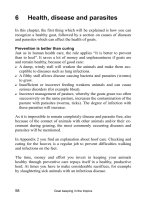
patient with slip progression was an 11-year-old boy who demonstrated a stable slip
in the left hip at presentation. Five months before the onset of pain in the left hip, he
suffered from a moderate slip in the right hip. In situ pinning with a single screw was
performed in the right hip, and in the left hip a similar procedure was done. We
advised him not to engage in any sports activities; however, despite our admonition
he discarded the crutch and began to play basketball before physeal closure. The
head–shaft angle of the left hip changed from 20° immediately after surgery to 45° at
29 months after the primary pinning. The radiograph showed a radiolucency around
the screw in the anterolateral metaphysis and maintenance of screw position in the
femoral head. We performed an additional surgery with two cannulated screws.
Ultimately, in this patient it took 4 years to demonstrate physeal closure from
the time of initial pinning (Fig. 1). In 18 patients with unilateral involvement, the
mean difference of articulotrochanteric distance was 8.8 mm (range, 3–15 mm).
Remodeling occurred in 21 hips (91%) of 23 hips in which the frog-leg lateral
radiograph was available. According to Jones’s classification, 16 hips were grouped
in type A, 5 hips in type B, and 2 hips in type C (Fig. 2). In 13 hips with moderate
and severe slips, 12 hips showed remodeling and 9 hips showed remodeling in
64 S. Iida and Y. Shinada
a b
c d
ef
Fig. 1. An 11-year-old boy. a,b Stable slip with 20° head–shaft angle at presentation. c,d Pinning
with single cannulated screw in good position. e,f Progressive slippage 2 years and 5 months
after the surgery. g,h Additional surgery with two cannulated screws. i,j Physeal closure 4 years
and 4 months after the initial surgery. (From [5], with permission)
In Situ Pinning for SCFE 65
g h
i j
Fig. 1. Continued
ab
Fig. 2. An 11-year-old girl. a,b Stable slip with 60° head–shaft angle at presentation. c,d Imme-
diately after in situ pinning with single cannulated screw. e,f At 4 years and 2 months after the
surgery. Clinical result was excellent, and the radiograph showed type A remodeling. (From [5],
with permission)
66 S. Iida and Y. Shinada
cd
ef
Fig. 2. Continued
Table 1. Remodeling and degree of slip
Head–shaft angle Remodeled Not remodeled
Type A Type B Type C
0°–29° 90 1
30° or more 75 1
Between remodeled and not remodeled, Fisher’s exact probability =
0.69; Between type A and type B, Fisher’s exact probability = 0.039
10 hips with mild slips. Remodeling was not dependent on the degree of slip
(Table 1). Excluding two hips that showed no remodeling (type C), mild slips
demonstrated significantly better remodeling than moderate or severe slips. There
was no significant correlation between triradiate cartilage status and remodeling
(Table 2).
In Situ Pinning for SCFE 67
Discussion
The indication of in situ pinning for SCFE remains controversial. O’Brien and Fahey
reported that in situ pinning might give satisfactory results even when the difference
between the two lateral head–shaft angles approached 55° to 60°, and they advocated
that if two or three pins could be inserted into the femoral epiphysis from the lateral
aspect of the femoral shaft, then in situ pinning would be indicated [8]. Recently, the
use of cannulated screws and pinning from the anterolateral aspect of the proximal
femur makes in situ pinning an acceptable alternative in some patients who have
rather advanced slipping. Aronson and Carlson [9] and Ward et al. [10] described
satisfactory results that were obtained with in situ pinning for slips greater than
70°.
Several authors have reported that satisfactory results were obtained after intertro-
chanteric osteotomy for moderate and severe slips. Intertrochanteric osteotomy was
regarded as a safe and effective procedure. Osteonecrosis and chondrolysis, however,
were described to occur after intertrochanteric osteotomy [11].
Treatment for SCFE must be aimed at minimizing osteonecrosis and chondrolysis,
which are the two main complications. To perform the safest procedure for SCFE, in
situ pinning has been selected for most slips. In these series, in situ pinning gave sat-
isfactory results for SCFE with a head–shaft angle less than 60°. Moreover, remodeling
after slipping of the epiphysis has been reported, and the inherent capacity of remod-
eling makes in situ pinning the treatment of choice for more-advanced slips. O’Brien
and Jones reported that remodeling occurred frequently after in situ pinning for SCFE
[3,8]. Jones et al. reported that remodeling was dependent on the degree of the slip
and that no hip with a head–shaft angle greater than 46° showed remodeling [3]. In
this series, 6 hips remodeled among 7 hips with a head–shaft angle greater than 40°.
Jones et al. also reported that remodeling was significantly more likely to occur if the
triradiate cartilage was open at presentation [3]. However, we did not find a signifi -
cant correlation between remodeling and triradiate status. It is necessary to evaluate
what factors would influence the remodeling after in situ pinning.
In situ pinning is considered to be a less-invasive procedure. On the other hand,
careful postoperative management is necessary, especially for moderate and severe
slips. Carney et al. and Saunders et al. reported that in several cases slippage have
progressed after in situ pinning [1,2]. We also experienced one patient with progres-
sive slippage. The patient showed a stable and mild slip at presentation and pinning
was performed in good position, but he started to play basketball without medical
permission. In this patient, time to physeal closure from the initial pinning was pro-
longed (4 years and 4 months). It should be considered that slip progression may
Table 2. Remodeling and triradiate cartilage
Triradiate Remodeled Not remodeled
cartilage
Type A Type B Type C
Open 10 3 1
Fusion 62 1
Between remodeled and not remodeled, Fisher’s exact probability
= 0.64
68 S. Iida and Y. Shinada
occur after in situ pinning until the accomplishment of physeal closure because the
epiphysis continues to slip and shear stress may act on the proximal physis. There-
fore, we recommend a long-leg non-weight-bearing apparatus for the patients with
head–shaft angle greater than 30°. Moreover it is expected that reducing the mechani-
cal stress on the physis may promote better remodeling. It should be evaluated if
careful postoperative management with limitation of weight-bearing can influence
remodeling.
In situ pinning in our institute for slip with head–shaft angle less than 60° showed
satisfactory clinical results and revealed good remodeling radiographically for short-
and midterm periods. Taking into account that all the patients are adolescent, a
longer follow-up is needed.
References
1. Carney BT, Birnbaum P, Minter C (2003) Slip progression after in situ single screw
fixation for stable slipped capital femoral epiphysis. J Pediatr Orthop 23(5):584–589
2. Saunders JO, Smith WJ, Stanley EA, et al (2002) Progressive slippage after pinning for
slipped capital femoral epiphysis. J Pediatr Orthop 22:239–243
3. Jones JR, Paterson DC, Hillier TM, et al (1990) Remodelling after pinning for slipped
capital femoral epiphysis J Bone Joint Surg 72B:568–573
4. Iida S, Shinohara H, Fujitsuka M, et al (1992) Manual reduction for slipped capital
femoral epiphysis (in Japanese). Rinsho Seikei Geka 27:771–777
5. Iida S, Shinada Y (2005) The indication and the limitation of in situ pinning for slipped
capital femoral epiphysis (in Japanese). J Joint Surg 24:76–81
6. Loder RT, Richards AABS, Shapiro PS, et al (1993) Acute slipped capital femoral
epiphysis: the importance of physeal stability. J Bone Joint Surg 75A:1134–1140
7. Heyman CH, Herndon CH (1954) Epiphyseodesis for early slipping of the upper
femoral epiphysis. J Bone Joint Surg 36A:539–554
8. O’Brien CE, Fahey JJ (1977) Remodeling of the femoral neck after in situ pinning for
slipped capital femoral epiphysis. J Bone Joint Surg 59A:62–69
9. Aronson DD, Carlson WE (1992) Slipped capital femoral epiphysis. A prospective
study of fixation with a single screw. J Bone Joint Surg 74A:810–819
10. Ward WT, Stefko J, Wood KB, et al (1992) Fixation with a single screw for slipped
capital femoral epiphysis. J Bone Joint Surg
74A:799–809
11. Jerre R, Hansson G, Wallin J, et al (1996) Long-term results after realignment opera-
tions for slipped capital femoral epiphysis. J Bone Joint Surg 78B:745–750
69
Retrospective Evaluation of Slipped
Capital Femoral Epiphysis
Meishuu Ko
1
, Kouji Ito
1
, Keiji Sano
1
, Naoki Miyagawa
1
,
Kengo Yamamoto
2
, and Youichi Katori
2
Summary. We treated 16 patients (16 hips) with slipped capital femoral epiphysis (12
boys and 4 girls) encountered during the previous 16-year period. Their age ranged
from 8 to 15 years (mean, 11.1 years), and the observation period ranged from 18 to
82 months (mean, 37 months). The evaluation items were chief complaint, mecha-
nism of injury, initial diagnosis, disease type, radiographic findings, physique and
endocrinological abnormalities, treatment methods, and complications. The disease
type was acute slip in 2 patients, chronic slip in 8, and acute on chronic slip in 6. Mild
slip was observed in 10 patients, moderate slip in 5, and severe slip in 1. Only 31.3%
of the patients were diagnosed as having slipped capital femoral epiphysis. The mean
interval from the first visit to diagnosis was 30 days. Surgery was performed in all
patients; Southwick intertrochanteric osteotomy was performed in 5 patients and in
situ pinning in 11. Concerning surgical complications, methicillin-resistant Staphy-
lococcus aureus infection developed in 1 patient and k-wire breakage in 1. Most
patients had satisfactory results. No avascular necrosis occurred. Limitation of motion
remained in 6 hips, but no hip pain, and normal gait was attained.
Key words. Slipped capital femoral epiphysis, Retrospective evaluation, Osteotomy,
In situ pinning, Early diagnosis
Introduction
The report in 2004 by the Multicenter Study Committee of the Japanese Pediatric
Orthopaedic Association showed a definite increase in patients with slipped capital
femoral epiphysis during the previous 25-year period in Japan [1]. However, physi-
cians other than pediatric surgeons are infrequently aware of slipped capital femoral
epiphysis and do not include this entity in diseases for differential diagnosis; there-
fore, its diagnosis rate is low. In addition, there are no treatment methods with
established evidence at present. We encountered 16 patients with slipped capital
1
Department of Orthopedic Surgery, Tokyo Medical University Hachioji Medical Center, 1163
Tatemachi, Hachioji, Tokyo 193-0944, Japan
2
Department of Orthopedic Surgery, Tokyo Medical University, Tokyo, Japan