Treatment of Osteoarthritic Change in the Hip - part 4 docx
Bạn đang xem bản rút gọn của tài liệu. Xem và tải ngay bản đầy đủ của tài liệu tại đây (581.89 KB, 26 trang )
Slipped Capital Femoral Epiphysis Retrospective 71
with the mean statistical values, the height of the patients was −10.1 to +19.9 cm
(mean, +6.0 cm), and height below the mean was observed in only 2 patients. Com-
pared with the mean statistical values, the weight of the patients was −10.4 to +39.7 kg
(mean, +17.6 kg), and weight below the mean was observed in only 1 patient. Body
mass index was 14.2–33.4 (mean, 24.6) and ≥25 in 8 patients (50%). The underweight
patient with a body mass index of 14.2 was a 12-year-old girl who was 3 cm taller than
the mean height.
Endocrinological examination showed a low testosterone level in one patient.
However, abnormalities could not be confi rmed in any patient because they were in
the growth stage.
Surgery was performed in all patients; Southwick intertrochanteric osteotomy [2]
was performed in 5 patients and in situ pinning in 11. Contralateral preventive bone
epiphyseal fi xation was performed in all except 1 patient.
The implant used for in situ pinning was the Knewles pin in 2 patients, Kirschner
wire (k-wire) with thread in 3, and ACE(R) SCFE screw in 6. For contralateral preven-
tive pinning, the Knewles pin was used in 2 patients, k-wire with thread in 3, ACE
SCFE screw in 9, and Hannson pin in 1
. For fi xation after Southwick intertrochanteric
osteotomy, the AO double angle plate (MIZUHO, Tokyo, Japan) was used. In all
patients, epiphyseal fi xation was added, and the implants used were the same materi-
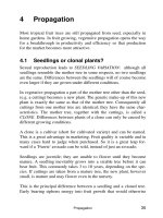
als as those used in preventive pinning. The fl exion osteotomy angle was frequently
20°–30°, although it was 50° in 1 patient. Changes in the slipping angle after osteotomy
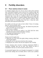
are shown in Fig. 2. Good reductions in both the posterior tilting angle and head–shaft
angle were observed.
Concerning surgical complications, methicillin-resistant Staphylococcus aureus
infection associated with Southwick intertrochanteric osteotomy developed in one
patient and k-wire breakage associated with in situ pinning in one. Leg length dis-
crepancy after Southwick intertrochanteric osteotomy until the fi nal observation was
observed in three of fi ve patients (0.5, 0.8, and 1.0 cm, respectively), but this presented
no clinical problems. Limitation in range of motion was present in six patients; only
18
20
10
8
7
19
23
59
37
29
14
54
48
12
37
78
0
10
20
30
40
50
60
06030
Posterior tiltin
g
an
g
le(de
g
ree)
Head shaft angle(degree)
Mild slip
10 cases
Moderate slip
5 cases 1 cases
Severe slip
Fig. 1. Relation between head-shaft angle and posterior tilting angle
72 M. Ko et al.
limitation in fl exion was observed in two, only that in internal rotation in two, and
that in both fl exion and internal rotation and both fl exion and internal/external rota-
tion in one each.
Concerning sequelae, one patient showed narrowing of the joint space at the initial
consultation, and although postoperative changes were negligible, the course has
been observed. No avascular necrosis of the femoral head occurred, no pain of hip,
and the patient has acquired a normal gait.
Case Presentations
Patient 1: 10-Year-Old Boy
He noticed right hip joint pain in February 2002. On March 30 of the same year, he
fell on the stairs, sustained injury, and was transported to a local hospital by ambu-
lance. A diagnosis of femoral neck fracture was made by a surgeon at the fi rst con-
sultation, and he was referred to our hospital (Fig. 3A). A diagnosis of unstable
slipped capital femoral epiphysis was made, and direct wire traction was performed
for about 2 weeks from immediately after admission. Because the slipping angle as
the posterior tilting angle was reduced from 59° to 17° by traction, in situ pinning was
performed (Fig. 3B). Five years and 4 months after operation, he has no pain or limi-
tation in the range of motion, showing a good course (Fig. 3C).
Patient 2: 12-Year-Old Girl
She noticed hip joint pain about 1 year earlier, visited a local hospital, but was told
that there was no abnormality. After an athletic meeting, her hip joint pain increased,
and she visited our hospital, was diagnosed as having slipped capital femoral
epiphysis, and admitted (Fig. 4A). Even after direct traction, adequate reduction
could not be achieved, and Southwick intertrochanteric osteotomy was performed.
The osteotomy angle was 35° in fl exion and 20° in abduction. The internal rotation
collection was 20° (Fig. 4B). Three years and 8 months after operation, remodeling of
the femoral head was good, but limitation in the range of motion in fl exion (5°)
remained (Fig. 4C).
0
20
40
60
80
0 1020304050
Head-shaft angle (degree)
Posterior tilting angle
degree
pre-operation post-operation
Fig. 2. Changes of head-shaft angle and pos-
terior tilting angle after osteotomy
Slipped Capital Femoral Epiphysis Retrospective 73
A
B
C
Fig. 3. Case 1 10-year-old boy. A Pre-operative roentgenogram of the hip. B Postoperative
roentgenogram of the hip. C Roentgenogram of the hip 64 months postoperation
74 M. Ko et al.
A
B
C
Fig. 4. Case 2 12-year-old girl. A Pre-operative roentgenogram of the hip. B Postoperative
roentgenogram of the hip. C Roentgenogram of the hip 44 months postoperation
Slipped Capital Femoral Epiphysis Retrospective 75
Discussion
In our patients, the correct initial diagnosis rate was only 31.3%, and some patients
with an incorrect diagnosis showed a change to acute on chronic slip.
The coeffi cient of the correlation between the duration until diagnosis and the
slipping angle was 0.632 (see Table 1). Saisu et al. [3] and Kocher et al. [4] reported
a signifi cant association between duration until diagnosis and slipping angle. Some
patients in this study required a considerably long time for diagnosis, increasing the
slipping angle, and thus we confi rmed the importance of early diagnosis.
Our treatment principles are as follows (Fig. 5). In patients in whom instability is
suspected at the fi rst visit and reduction can be expected, direct wire traction is per-
formed, and the severity of the disease is evaluated based on the posterior tilting
angle. In situ pinning is performed when the angle is less than 30° and Southwick
intertrochanteric osteotomy when the angle is ≥30°. Because no manual reduction is
performed either before or during operation, there is no method of confi rming insta-
bility. Therefore, we perform direct wire traction in patients with a posterior tilting
angle of ≥30° on the affected side and prophylactic pinning on the contralateral side
in principle. Castro et al. [5] stated that “close follow-up and not prophylactic pinning
was most supported by the literature.” In contrast, Schultz et al. [6] reported “a
benefi t in the long-term outcome for patients who had prophylactic of the contralat-
eral hip.” A review of the literature shows arguments both for and against prophy-
lactic pinning but no studies with a large body of evidence. We perform prophylactic
pinning because we have previously encountered children with contralateral slip and
fully realized that children at this age when this disease frequently develops do not
often follow instructions to rest.
We perform in situ pinning in patients with a posterior tilting angle of <30°.
However, some studies have shown good results after in situ pinning in patients with
an angle of ≥30°. In patients with this disease not complicated by femoral head necro-
sis or acute cartilage necrosis, short-term results are good. Even if short- or middle-
term results are good, however, because osteoarthrosis of the hip develops at middle
age or later, the expansion of the indications of this method should be carefully
evaluated.
Instability
Direct wire traction
Southwick intertrochanteric
osteotomy
In situ pinning
Skin traction
or rest
Yes
No
Slipped capital femoral epiphysis
Posterior tilting angle
30°
30°
Contralateral hip
Prophylactic
pinning
Fig. 5. Algorism of treatment for slipped capital femoral epiphysis
76 M. Ko et al.
Various osteotomy methods have also been reported. We use Southwick intertro-
chanteric osteotomy because operation-associated femoral head necrosis rarely
occurs, no high-level technique is necessary, and stable results can be expected.
References
1. Noguchi Y, Sakamaki T(2004) Epidemiology and demographics of slipped capital
femoral epiphysis in Japan. J Jpn Pediatr Orthop Assoc 13(2):235–243
2. Southwick WO (1967) Osteotomy through the lesser trochanter for slipped capital
femoral epiphysis. J Bone Joint Surg [Am] 49(5):807–835
3. Saisu T, Kamegaya M, Ochiai N, et al (2003) Importance of early diagnosis for treatment
of slipped capital femoral epiphysis. J Jpn Pediatr Orthop Assoc 12(1–2):61–64
4. Kocher MS, Bishop JA, Weed B (2004) Delay in diagnosis of slipped capital femoral
epiphysis. Pediatrics 113(4):322–325
5. Castro FP Jr, Benett JT, Doulens K (2004) Epidemiological perspective on prophylactic
pinning in patients with unilateral slipped capital femoral epiphysis. J Pediatr Orthop
20(6):745–738
6. Schultz WR, Weinstein JN, Weinstein SL (2002) Prophylactic pinning of the contralat-
eral hip in slipped capital femoral epiphysis: evaluation of long-term outcome for the
contralateral hip with use of decision analysis. J Bone Joint Surg [Am] 84A(8):
1305–1314
Part II
Avascular Necrosis of
the Femoral Head
79
Osteotomy for Osteonecrosis of the
Femoral Head: Knowledge from Our
Long-Term Treatment Experience at
Kyushu University
Seiya Jingushi
Summary. Many young patients suffer from osteonecrosis of the femoral head
(ONFH). For this reason, osteotomy is considered to be an important treatment
option, and their survival after osteotomy of the hip is expected to be of long duration.
Cases that survived more than 25 years after osteotomy were investigated to reconfirm
the principles or the indication based upon our previous experience about osteotomy
treatment for ONFH. Fifteen cases were divided into two groups with or without
advanced osteoarthritis at the last follow-up and were compared. The mean follow-up
periods were 28 and 27 years, respectively. All the cases with advanced osteoarthritis
(OA) had collapse progression. All the cases in which the preoperative stage was
advanced were included in those with advanced OA at the last follow-up. In contrast,
collapse progression was not observed in the cases without advanced OA at the
last follow-up. All these cases had minimum collapse before operation. According to
these data, we reconfirmed that collapse progression is the main cause for poor
outcome after osteotomy, and that cases operated on at an early stage are apt to
experience a good prognosis. When the indication and the operation are appropriate,
osteotomy could prevent disease deterioration even more than 25 years after the
operation.
Key words. Osteonecrosis of the femoral head, Osteotomy, Transtrochanteric anterior
rotational osteotomy, Collapse, Clinical outcome
Introduction
Once collapse occurs at the necrosis area of the femoral head, it usually progresses.
Collapse causes incongruity and instability of the hip joint, and the progression of
collapse causes incongruity and instability to increase and finally results in secondary
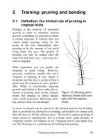
osteoarthritis (Fig. 1). The purpose of osteotomy for osteonecrosis of the femoral head
(ONFH) is to prevent the progression of collapse and secondary osteoarthritis. A
principle of osteotomy is to support weight-bearing with intact or live bone instead
Department of Orthopaedic Surgery, Graduate School of Medical Sciences, Kyushu University,
3-1-1 Maidashi, Higashi-ku, Fukuoka 812-8582, Japan
80 S. Jingushi
of the necrotic bone and to restore the subluxated femoral head (Fig. 2). In other
words, osteotomy is on-site vascularized bone grafting with articular cartilage and
with good congruency. Options of osteotomy for ONFH are transtrochanteric anterior
or posterior rotational osteotomy (ARO or PRO) developed by Sugioka et al. [1,2],
and intertrochanteric curved varus osteotomy developed by Nishio and Sugioka [3].
The treatment option is chosen depending on the lesion of osteonecrosis or on where
and how wide is the osteonecrosis area in the femoral head. Stage and age at the
operation are also considered in this choice.
Many young patients suffer from the disease. Especially for young patients, oste-
otomy is an important treatment option to be considered, and they are expected to
survive for a long time after their hip osteotomy. Osteotomy in Kyushu University
Hospital started in 1972. Sugioka developed transtrochanteric rotational osteotomy
Fig. 1. Natural course of osteonecrosis of the femoral head (ONFH)
Fig. 2. A principle of anterior rotational osteotomy for ONFH. The dashed line shows the
osteonecrosis area of the femoral head from the anterior view
Long-Term Experience of Osteotomy for Femoral Head Osteonecrosis 81
of the femoral head, so-called “rotational osteotomy” or “Sugioka’s osteotomy” [1].
Anterior rotation of the femoral head with vascularity results in weight-bearing with
the live posterior surface of the femoral head (Fig. 3).
Experience of Osteotomy in Kyushu University
Between 1972 and 1979
The cases that survived more than 25 years after the operation were investigated to
reconfirm the principles or the indication based upon our previous experience with
osteotomy treatment for ONFH [1,2,4].
Patients and Methods
Between 1972 and 1979, 128 patients with idiopathic ONFH underwent osteotomy in
our department. Fifteen hips of 9 patients, who had been visiting our outpatient office
and had their living hip joints more than 25 years after operation, were examined.
The hips were separated into two groups (Table 1). One group includes the hips that
had advanced or terminal osteoarthritis (OA) at the last follow-up. Another group
includes those that had no OA or early OA. Age at operation and period after opera-
tion were similar in both the groups. Clinical scores were assessed according to the
hip scoring system by the Japanese Orthopaedic Association.
Fig. 3. Sequential photographs of anterior rotation of the femoral head show a model of ante-
rior rotational osteotomy (ARO) with 20° varus position and indicate how ARO results in
weight-bearing with the living posterior surface of the femoral head (a–f). Hatched area indi-
cates necrotic area. All the photographs show the anterior view. According to anterior rotation,
the osteotomy line is 10° inclination away from the perpendicular to the neck (a) and 10° ret-
roversion. The result is 20° varus position after anterior rotation of the femoral head (f)
82 S. Jingushi
Results
All hips that had no or early OA at the last follow-up were at stage 3A at operation
and had no collapse progression after osteotomy (see Table 1). The average of their
clinical scores was promising. In contrast, approximately half of the hips that had
advanced or terminal OA at the last follow-up were at stage 3B at operation. Further-
more, all of them had collapse progression and had poor clinical scores at the last
follow-up.
Representative Cases
Case 1
The patient was male and had bilateral ARO at 38 years old (Fig. 4a). Preoperative
stage of the right and left hip was 3A or 3B, respectively. Twenty-eight years after
operation, collapse had progressed in the left hip, and that hip showed terminal OA
at the last follow-up (Fig. 4b). The clinical score was 34 points. The right hip had early
OA at the last follow-up, and the clinical score was 54 points, although collapse did
not progress after the operation.
Case 2
This patient was male and underwent ARO and varus osteotomy, respectively, in the
right and left hips at 33 years of age (Fig. 5a). Preoperative stage was 3A in both hips.
Twenty-seven years after the operation, collapse of the femoral head had not pro-
gressed, and OA changes were not observed (Fig. 5b). The clinical scores were 92 and
82 points, respectively. Note that good bone regeneration was observed in the osteo-
necrosis area of the bilateral femoral head.
Case 3
This patient was male and had ARO bilaterally at 24 years old at the time of operation
(Fig. 6a). Preoperative stage was 3A in both hips. Twenty-six years after the operation,
collapse of the femoral head had not progressed, and OA changes were not observed
(Fig. 6b). The clinical score was 100 points in both the joints.
Table 1. Characterization of the hips in two groups
With advanced OA Without advanced OA
Number of examined hips 96
Age at operation (years) 31 (19–52) 31 (24–38)
Involved in the contralateral side 6 (67%) 3 (50%)
Period after operation (years) 28 (25–30) 27 (26–29)
Stage 3A: 5 (56%); 3B: 4 (44%) 3A: 6 (100%)
Collapse progression 9 (100%) 0 (0%)
JOA score
a
at the last follow-up 55 (34–82) 86 (54–100)
OA, osteoarthritis
a
In the clinical scoring system for hip joints developed by the Japanese Orthopaedic Association, the
maximum score is 100 points
Long-Term Experience of Osteotomy for Femoral Head Osteonecrosis 83
Fig. 4. A representative case (case 1) that had advanced osteoarthritis (OA) 28 years after
operation. a Just after osteotomy; b 28 years after osteotomy
Fig. 5. A representative case (case 2) that had no OA changes 27 years after operation. a Just
after osteotomy; b 27 years after osteotomy
84 S. Jingushi
Fig. 6. A representative case (case 3) that had no OA changes 26 years after operation. a Just
after osteotomy; b 26 years after osteotomy
Discussion
Based on the data, we reconfirmed that progression of collapse was the main cause
of poor results after osteotomy, as previously described [1,2,4]. Cases operated on at
an early stage are apt to experience good prognosis. Stage at operation is another
important factor to influence the clinical outcome. When osteotomy is carried out at
an early stage and prevents progression of collapse, this could prevent disease dete-
rioration or maintain hip function without clinical symptoms even more than 25 years
after operation.
Experience of Osteotomy in Kyushu University
Between 1980 and 1988
Previously, we examined 125 cases that had undergone operations between 1980 and
1988 [5]. Twenty-eight hips had collapse progression more than 10 years after opera-
tion. We found that the postoperative intact ratio in the nonprogression group was
significantly larger than that in the progression group. A minimum postoperative
intact ratio to prevent collapse progression over a 10-year period was 34% (Fig. 7).
According to that study, the aim of osteotomy is to achieve more than 34% of the
Long-Term Experience of Osteotomy for Femoral Head Osteonecrosis 85
ratio of the intact area postoperatively. We try to ensure this every time during pre-
operative planning.
A Current Representative Case
Sugioka has reported good clinical outcome of osteotomy for ONFH. However, there
are many reports that show poor clinical outcome, especially as concerns rotational
osteotomy [6–8]. The most important issue about osteotomy treatment may be
whether osteotomy could be carried out successfully by others than Sugioka.
In our department, osteotomy treatment has been carried out according to the
principles based on our long experience. A current representative case is shown, a
33-year-old woman who had bilateral steroid-induced osteonecrosis. Radiographs
and magnetic resonance imaging (MRI) show a wide osteonecrosis area, and the
intact area was limited to the posterior surface of the femoral head (Fig. 8). According
to the preoperative planning, ARO with 20° varus position was expected to result in
more than 34% of the ratio of the intact articular in both the joints. The osteotomy
was carried out in the right hip joint, and then in the left hip 2 months after the first
operation. Four years after operations, collapse has not progressed in either of the
hip joints, and no OA changes are observed in the postoperative radiographs (Fig. 9).
She has no problems in walking, squatting, and going up and down the stairs (Fig.
10) at 5 years after osteotomy. Clinical scores of both hip joints are 100 points, and
she has returned to work.
Fig. 7. Kaplan–Meier survival curve of groups with a postoperative intact ratio of more than
34% and with a ratio less than 34%. The occurrence of progressive collapse is used as an end-
point. (From [5], with permission.) The figure on the left shows how to calculate the intact
ratio
86 S. Jingushi
Fig. 8. Preoperative radiographs and magnetic resonance (MR) images of a current representa-
tive case. a Anteroposterior view radiograph of bilateral ONFH. b, c Tomography of the bilateral
joints in a Lauenstein position. d Frontal section of T
1
-weighted MRI of the bilateral hip joints.
e, f View of vertical section to the femoral neck axis in right and left femoral head, respectively.
Location of the section is indicated in d
Fig. 9. Radiographs of bilat-
eral hip joints just after oste-
otomy (a) or 4 years after
osteotomy (b)
Long-Term Experience of Osteotomy for Femoral Head Osteonecrosis 87
Conclusions
Osteotomy could maintain prevention of disease deterioration of ONFH even more
than 25 years after the operation. Osteotomy is a promising treatment option for
ONFH, especially for young patients. We believe that experienced hip surgeons can
perform osteotomy, including ARO, successfully once they understand the indica-
tions and techniques.
References
1. Sugioka Y (1978) Transtrochanteric anterior rotational osteotomy of the femoral head
in the treatment of osteonecrosis affecting the hip. A new osteotomy operation. Clin
Orthop 130:191–201
2. Sugioka Y, Hotokebuchi T, Tsutsui H (1992) Transtrochanteric anterior rotational
osteotomy for idiopathic and steroid-induced necrosis of the femoral head. Clin Orthop
277:111–120
3. Nishio A, Sugioka Y (1971) A new technique of the varus osteotomy at the upper end
of the femur. Orthop Traumatol 20:381–386
4. Hosokawa A, Mohtai M, Hotokebuchi T, et al (1997) Transtrochanteric rotational oste-
otomy for idiopathic and steroid-induced osteonecrosis of the femoral head: indica-
tions and long-term follow-up. In: Urbaniak JR, Jones JP Jr (eds) Osteonecrosis, etiology,
diagnosis and treatment. The American Orthopaedic Association, Rosemont, IL, pp
309–314
Fig. 10. Activities of daily life of the representative case. a–d Walking; e, f squatting; g, h going
up stairs; i, j going down stairs
88 S. Jingushi
5. Miyanishi K, Noguchi Y, Yamamoto T, et al (2000) Prediction of the outcome of trans-
trochanteric rotational osteotomy for osteonecrosis of the femoral head. J Bone Joint
Surg 82B:512–516
6. Belal MA, Reichelt A (1996) Clinical results of rotational osteotomy for treatment of
avascular necrosis of the femoral head. Arch Orthop Trauma Surg 115(2):80–84
7. Dean MT, Cabanela ME (1993) Transtrochanteric anterior rotational osteotomy for
avascular necrosis of the femoral head. Long-term results. J Bone Joint Surg [Br]
75(4):597–601
8. Tooke SM, Amstutz HC, Hedley AK (1987) Results of transtrochanteric rotational
osteotomy for femoral head osteonecrosis. Clin Orthop 224:150–157
89
Joint Preservation of Severe
Osteonecrosis of the Femoral
Head Treated by Posterior Rotational
Osteotomy in Young Patients:
More Than 3 Years of Follow-up
and Its Remodeling
Takashi Atsumi, Yasunari Hiranuma, Satoshi Tamaoki,
Kentaro Nakamura, Yasuhiro Asakura, Ryosuke Nakanishi,
Eiji Katoh, Minoru Watanabe, and Toshihisa Kajiwara
Summary. Posterior rotational osteotomy in 48 hips of 40 young patients with femoral
head osteonecrosis with extensive and apparent collapsed lesions were reviewed with
a mean of 9.2 years of follow-up. No viable area was seen on the articular surface of
the femoral head of the loaded portion on preoperative anteroposterior radiographs
in all femoral heads. All hips had greater than 3 mm collapse; 40 hips showed no
apparent joint narrowing, and 8 hips revealed joint narrowing. Posterior viable area
of joint surface before surgery ranged from 6% to 29%, with a mean of 19%, on lateral
radiographs. Anterior viable area ranged from 6% to 42% with a mean of 21%. The
mean age of the patients was 29 years, with 13 women and 27 men. Thirty-five hips
were nontraumatic, and 13 were traumatic. Mean postoperative viable area below the
acetabular roof was 59% on anteroposterior radiographs and 54% on 45° flexed radio-
graphs. Recollapse was prevented in 44 hips (92%), with adequate viable area on the
loaded portion on final follow-up radiographs. Progressive joint narrowing was found
in 9 hips. Resphericity of the postoperative transferred medial collapsed area of the
femoral head was observed on 34 of 35 hips on final anteroposterior radiographs. The
joint space was increased in 6 of 8 hips. Posterior rotational osteotomy appeared to
be effective in delaying the progression of degeneration in young patients with exten-
sive collapsed osteonecrotic lesions.
Key words. Osteonecrosis, Osteonecrosis of the femoral head, Joint preservation, Pos-
terior rotational osteotomy, Transtrochanteric rotational osteotomy
Introduction
Nontraumatic and posttraumatic osteonecrosis involving the femoral head frequently
occurs in young patients. Preservation of the joint of the femoral head necrosis
in young patients to avoid joint replacement procedures is widely accepted for
Department of Orthopaedic Surgery, Fujigaoka Hospital, Showa University School of Medicine,
1-30 Fujigaoka Aobaku, Yokohama 227-8501, Japan
90 T. Atsumi et al.
orthopedic surgeons. However, in cases of extensive lesion and apparent collapse,
some kinds of osteotomies [1,2], with vascularized fibular grafts [3], are usually not
effective. Sugioka has reported transtrochanteric anterior rotational osteotomies for
osteonecrosis of the femoral head and described excellent follow-up results [4–6]. The
absolute indications for this operation were that the necrotic area is located on less
than the posterior one-third of the entire femoral head on correct lateral radiographs
[4]. Sugioka also mentioned indications for posterior rotational osteotomies, but he
did not report the details of this procedure [5]. We have reported on the use of pos-
terior rotational osteotomies including our modified approach, “high degree poste-
rior rotation” [7,8], for femoral head osteonecrosis with extensive lesions. The
advantages of posterior rotational osteotomies are as follows. (1) The posterior
column artery branched off from the femoral medial circumflex artery is shifted
medially and is not under tension without vascular damage by posterior rotation. We
confirmed this condition by our angiographic studies [9]. (2) The necrotic area is
transferred to the posteromedial non-weight-bearing portion. Postoperative uncol-
lapsed anterior viable areas are moved to the loaded portion below the acetabular
roof in flexed positions. After posterior rotation, congruency can be expected in a
flexed position of daily life. The purpose of this study is to investigate the effectiveness
of joint preservation by posterior rotational osteotomy for the treatment of severe
femoral head osteonecrosis with extensive collapsed lesions in patients less than 50
years old.
Materials and Methods
Between 1985 and 2002, 60 hips with apparent collapse and extensive lesions of the
femoral head in young patients (less than 50 years of age) were treated by posterior
rotational osteotomies including high-degree posterior rotation. Of these hips, 48 hips
of 40 patients with a minimum of 3 years follow-up were subjected in this study
(follow-up range, 3–20 years; mean, 9.2 years). If the patients were converted to a
prosthetic replacement, follow-up ended. The age of the patients at the time of surgery
ranged from 15 to 49 years with a mean of 29 years; 13 patients were women and 27
were men. Of the hips, 23 had a history of corticosteroid administration, 9 had a
history of alcohol abuse, 10 had a history of femoral neck fracture, and 3 had a history
traumatic dislocation; the remaining 3 hips had no apparent risk factor. We excluded
12 of 60 hips from the study because 7 hips were lost to follow-up, 4 hips were conver-
sion surgery of a prosthetic replacement less than 3 years after posterior rotational
osteotomy because of early recollapse after trauma, and 1 patient died of underlying
disease.
All 48 hips had extensive lesions from medial to lateral and from anterior to the
posterior portion of the femoral head. No viable area was seen on the articular surface
of the loaded portion of the femoral head facing the acetabular roof on preoperative
anteroposterior radiographs (type C2 of criteria of Japanese Investigations Commit-
tee) [10] in all 48 hips. On correct lateral radiographs [4], the posterior viable area of
joint surface of these hips before surgery ranged from 6% to 29% with a mean of 19%.
The anterior viable area ranged from 6% to 42% with a mean of 21%. No hips were
indicated for a traditional anterior rotational osteotomy. All 48 hips had apparent
Posterior Rotational Osteotomy in Femoral Head Osteonecrosis 91
collapse (greater than 3 mm). In these hips, 40 hips showed no apparent joint space
narrowing (stage 3B of criteria of Japanese Investigations Committee) [10]. The
remaining 8 hips revealed apparent joint space narrowing (stage 4). Twenty-five cases
were involved by osteonecrosis bilaterally on radiographs or magnetic resonance
imaging. Of these hips, 11 were treated by bilateral posterior rotational osteotomy.
Different procedures were elected for the contralateral hips of the other 14 cases: 2
anterior rotational osteotomies and 1 total hip arthroplasty. The remaining 4 cases
were not treated because of small-size lesion without symptoms. These hips were not
included in this study. All collapsed femoral heads were rotated in the posterior direc-
tion. Degrees of rotation ranged from 70° to 160° posteriorly (mean, 126°). Additional
intentional varus positioning was done from 10° to 30° (mean, 19°) in all 48 hips to
obtain an extensive noncollapsed viable articular surface of the femoral head in the
loaded portion postoperatively. A summary of the patient population is shown in
Table 1.
The rotational angle and intentional varus angle necessary for this procedure were
determined by preoperative assessment, mainly on radiographic findings. Correct
lateral radiographs (Fig. 1B) and anteroposterior radiographs in a flexed position
were taken in all hips to observe the location and extent of necrotic region. Radio-
graphs taken under these conditions can show the location and extent of the noncol-
lapsed viable articular surface of the femoral head after posterior rotation. Magnetic
resonance imaging and computed tomography can be available if the demarcation
area between living and necrotic bone is not clearly visualized on radiographs.
A modified Southern approach [7,8] was applied in 47 hips. The modified Ollier
approach as reported by Sugioka [4] was employed in 1 remaining operation. For the
fixation of osteotomy plane after femoral head rotation, we used large screws (Sugioka)
in 4 hips, an AO screw in 2, and an AO plate in 2. However, these fixation devices
were not strong enough to allow for early motion. Thereafter, the authors made and
used a customized device developed by Atsumi [7,8] in 40 hips.
Table 1. Patient population
Forty-eight hips, of 40 young patients
Age, 15–49 years old (mean, 29 years)
Sex: 13 women, 27 men
Etiological factor:
Steroid administration, 23 hips
Alcohol abuse, 9; traumatic, 13
No apparent factor, 3
Type C2: 48 hips (no viable area on articular surface of the femoral head of loaded portion on
preoperative anteroposterior radiographs)
Stage 3B, 40 hips; 4, 8 hips (all 48 showed >3 mm collapse)
Anterior or posterior viable area on correct lateral radiographs
Anterior, 6%–42% (mean, 21); posterior, 6%–29% (mean, 19)
Posterior rotational angle: 70°–160° (mean: 126°)
Additional varus position 10°–25° (mean, 19°)
Follow-up, 3–20 years (mean, 9.2 years)
Excluded cases: lost to follow-up, 7; early revised surgery, 4; died, 1
92 T. Atsumi et al.
C
A
D
B
F
E
Posterior Rotational Osteotomy in Femoral Head Osteonecrosis 93
For postoperative management, partial weight-bearing was permitted 5 to 6 weeks
after operation using two crutches. Gait with one crutch was essential for 6 months
to 1 year depending on the extent of lesion.
Radiographic outcome was influenced by the extent of the lateral noncollapsed
living area of the femoral head corresponding to the acetabular roof on postoperative
conventional anteroposterior radiographs. Extent of the noncollapsed viable area of
the loaded portion of the femoral head was measured by angle [7], and the rate of
extent was divided into three groups as follows: group A, less than the medial one-
third of the weight-bearing area is involved; group B, more than one-third but less
than two-thirds is involved; and group C, more than two-thirds is involved (Table 2).
Anteroposterior radiographs were also taken in 45° of hip flexion [(7,8)] to observe
the anterior viable portion of the femoral head. The extent of the viable area of the
anterior femoral head was also divided into three groups as well on conventional
anteroposterior radiographs. Prevention and progression of recollapse and progres-
sive joint space narrowing were observed on the follow-up radiographs, and the
relationship with the extent of viable articular surface of the femoral head was also
studied. Of the remodeling after surgery, respherical contour on the collapsed area
that moved medially and improvement of degenerative joint narrowing were investi-
gated. The necrotic focus was moved to the medial portion of the femoral head on
postoperative anteroposterior radiographs in all 48 hips. In 35 of 48 hips, collapsed
Fig. 1. A 30-year-old woman receiving high doses of corticosteroids for treatment of multiple
sclerosis. A Preoperative anteroposterior radiograph of her right hip showed extensive col-
lapsed lesion without viable area on loaded portion below the acetabular roof. B Correct lateral
radiograph showed extensively involved area. Arrows show anterior and posterior demarcation
area between necrotic and noncollapsed viable portion. Anterior viable area is 13%, posterior
viable area is 15%. C A 150° posterior rotational osteotomy with 15° varus position was per-
formed. Anteroposterior (AP) radiograph taken 3 months after operation revealed adequate
viable joint surface of the femoral head below the acetabular roof. Note the necrotic lesion is
moved to the medial area (arrow). Extent of viable femoral head was 60%. D Viable area was
82% on 45° flexion AP radiograph taken at the same time. E AP radiograph taken 11 years after
operation disclosed spherical contour of the medial femoral head (arrow). Joint space was well
maintained, and the patient was free from pain. Flexion was 80°, abduction was 30°, and Japa-
nese Orthopaedic Association (JOA) hip score was 96 points. F A 45° flexion AP radiograph
taken 11 years after operation showed sphericity of the femoral head
Table 2. Extent of viable area of femoral head on postoperative AP and 45° flexion AP
radiographs
Group A Group B Group C
м2/3 м1/3, 2/3 <1/3
Conventional AP (n = 48) 15 (31%) 27 (56%) 6 (13%)
45° Flexion AP (n = 48) 10 (21%) 33 (69%) 5 (10%)
AP, anteroposterior
94 T. Atsumi et al.
areas were clearly observed at the medial portion of the femoral head on postopera-
tive anteroposterior radiographs during less than 6 months after surgery. Respherical
contour on the medial collapsed area on final anteroposterior radiographs of 35 hips
was studied. Of the improvement of degenerative joint on 8 hips with joint space
narrowing preoperatively, observation was made for changes of acetabular subchon-
dral roof on anteroposterior radiographs at 6 months, 2 years, and final follow-up.
Clinical evaluation was assessed on JOA hip score [11].
Results
On postoperative anteroposterior radiographs taken in the short period after surgery
(less than 1 year), the lateral noncollapsed viable area of joint surface facing the ace-
tabular roof was 21% to 100% (mean, 58) in all 48 hips. In these hips, 15 hips were
group A, 27 were group B (Fig. 1C), and 6 were group C. On postoperative 45° flexion
anteroposterior radiographs, the lateral noncollapsed viable area was 11% to 100%
(mean, 54); 10 hips showed group A (Fig. 1D), 33 were group B, and 5 were group C
(Table 2); and 4 hips (8%) resulted in recollapse at final follow-up. These 4 hips were
in group C. Recollapse did not occur in 44 hips of groups A and B. Progressive joint
space narrowing was found in 9 hips. Of the extent of viable area on anteroposterior
radiographs, 3 hips were in group A, 2 were in group B, and 4 were in group A (Table
3). In 40 hips of stage 3B, recollapse was found in 3 hips and joint narrowing was
noted on 7 hips. Recollapse occurred on 1 hip and joint narrowing was seen on 2 of
8 hips with stage 4 (Table 4).
Resphericity of the medial collapsed area of the femoral head was observed in 34
of 35 hips (97%) on the final anteroposterior radiographs (Fig. 1E). Of changes of the
acetabular subchondral roof for the 8 hips with joint space narrowing before opera-
tion, atrophic changes of the acetabular subchondral roof were noted 6 months after
operation in all hips. The shape of the acetabular roof was improved and reformed
by 2 years after the procedure. The joint space was increased when comparing it
to before the surgery and was maintained at final follow-up anteroposterior
radiographs.
Table 3. Relationship between extent of viable area of femoral head after operation corre-
sponding with acetabular roof, recollapse, and progressive joint space narrowing
Conventional AP radiographs (n = 48) Total Group A Group B Group C
Recollapse 4 (8%) 004
Progressive joint space narrowing 9 (19%) 324
Table 4. Relationship between stages, recollapse, and progressive
joint space narrowing
Stage Recollapse Progressive joint
space narrowing
3B 3/40 hips (8%) 7/40 hips (18%)
41/8 hips (12%) 2/8 hips (25%)
Posterior Rotational Osteotomy in Femoral Head Osteonecrosis 95
With regard to the range of motion, in hips without recollapse or joint space nar-
rowing, the flexion angle was 60° to 130° (mean, 100°), and abduction angle was 15°
to 40° (mean, 22°). Hips with either recollapse or joint space narrowing evidenced
flexion from 40° to 100° (mean, 96°) and abduction from 5° to 25° (mean, 19°). Clinical
evaluation according to the Japanese Orthopaedic Association hip score system was
84 to 100 points (mean, 91) in hips without recollapse or 50 to 83 points (mean, 67)
in those without joint space narrowing. Two hips were revised with a total hip arthro-
plasty around 15 years after surgery. Four hips were waiting a total hip arthroplasty
due to poor results. Causes of the unsuccessful results including early failure were
postoperative inadequate viable area under the weight-bearing portion below the
acetabular roof in 3 hips, vascular impairment by operation in 2, and living bone that
fractured after a high level of activities in 2, degenerative change in 2, and challenging
procedure in 1 because of the young age of the patient.
Discussion
Several kinds of procedures for joint preservation of femoral head osteonecrosis
appear to be effective in early-stage and small or mid-sized necrosis [1–3,12]. Joint
preservation of femoral head osteonecrosis with extensive and collapsed lesions in
young patients may be an important challenge for orthopedic surgeons. The principal
concept of femoral osteotomies for joint preservation in the treatment of femoral
head osteonecrosis is that necrotic focus is moved away from the major weight-
bearing portion on the acetabulum [2,4,7]. In this situation. weight-bearing forces are
received by the transferred viable area. Reports of anterior rotational osteotomy
described by Sugioka et al. showed good results if posterior noncollapsed viable bone
remained in more than one-third of the entire femoral head and adequate viable area
could be transferred to the loaded portion opposite the acetabular roof [4–6]. However,
many young patients have extensive lesions that do not indicate anterior rotational
osteotomy is suitable.
Our previous reports of posterior rotational osteotomies including “high degree
posterior rotation” [7,8] for femoral head osteonecrosis with extensive lesions showed
good results even if patients have extensive lesions and apparent collapse. In the
present study, recollapse was prevented in cases with adequate viable area corre-
sponding to the acetabular subchondral roof on conventional anteroposterior radio-
graphs and 45° flexion on anteroposterior views. In these cases, the anterior viable
area can be moved to the loaded portion by the use of the posterior rotational oste-
otomy, including the “high degree posterior rotation osteotomy” as described. The
extent of the viable area corresponding to the weight-bearing portion below the ace-
tabular roof on conventional anteroposterior radiographs was almost equivalent to
the extent on the 45° flexion anteroposterior radiographs. Containment and congru-
ency between the femoral head and the acetabulum was improved not only in the
neutral position but also in flexion of daily activities after this posterior rotational
osteotomy.
Extended joint space and remodeling of the acetabular subchondral shape were
noted in hips with degenerative changes preoperatively. A regaining of the spherical
contour of the collapsed femoral head was also found. It was believed that the reason
96 T. Atsumi et al.
for remodeling after operation was the containment and congruency of the joint as a
result of the anterolateral adequate viable area of the femoral head. The authors
assumed that the main causes of failure with recollapse were inadequate viable area
under the weight-bearing portion below the acetabular roof, fracture of the viable
bone with mechanical weakness after a high level of activities too soon after the opera-
tion, or vascular damage caused by the operation. In conclusion, posterior rotational
osteotomy including the high degree posterior rotation appears effective for the treat-
ment of nontraumatic and posttraumatic osteonecrosis of the femoral head with col-
lapse and extensive involvement in young patients. The authors believe that this
operation may delay the progression of degeneration if adequate viable area can be
placed below the loaded portion of the acetabulum. Remodeling of the collapsed
lesion and the degenerative acetabular subchondral roof might be one of the impor-
tant factors for preserving the joints.
References
1. Kerboul M, Thomine J, Postel M (1974) The conservative surgical treatment of idio-
pathic aseptic necrosis of the femoral head. J Bone Joint Surg [Br] 56:291–296
2. Mont MA, Fairbank AC, Krackow KA et al (1996) Corrective osteotomy for osteone-
crosis of the femoral head. J Bone Joint Surg [Am] 78:1032–1038
3. Urbaniak JR, Coogan PG, Gunneson, EB, et al (1995) Treatment of osteonecrosis of
the femoral head with free vascularized fibular grafting. A long-term follow-up study
of one hundred and three hips. J Bone Joint Surg [Am] 77:681–694.
4. Sugioka Y (1978) Transtrochanteric anterior rotational osteotomy of the femoral head
in the treatment of osteonecrosis affecting the hip. A new osteotomy operation. Clin
Orthop Relat Res 130:191–201
5. Sugioka Y (1980) Transtrochanteric rotational osteotomy of the femoral head. In:
Riley LH Jr (ed) The hip. Proceedings of the eighth open scientific meeting of the Hip
Society. Mosby, St. Louis, pp 3–23
6. Sugioka Y, Hotokebuti T, Tsutsui H (1992) Transtrochanteric anterior rotational oste-
otomy for idiopathic and steroid-induced necrosis of the femoral head. Indications
and long-term results. Clin Orthop Relat Res 227:111–120
7. Atsumi T, Kuroki Y (1997) Modified Sugioka’s osteotomy. More than 130° posterior
rotation for osteonecrosis of the femoral head with large lesion. Clin Orthop Relat Res
334:98–107
8. Atsumi T, Muraki M, Yoshihara S, et al (1999) Posterior rotational osteotomy for the
treatment of femoral head osteonecrosis. Arch Orthop Trauma Surg 119:388–393
9. Atsumi T, Yamano K (1997) Superselective angiography in osteonecrosis of the
femoral head In: Urbaniak JR, Jones JP (eds) Osteonecrosis: etiology, diagnosis, and
treatment. American Academy of Orthopaedic Surgeons, Rosemont, IL, pp 247–252
10. Sugano N, Atsumi T, Ohzono K et al (2002) The 2001 revised criteria for diagnosis,
classification, and staging of idiopathic osteonecrosis of the femoral head. J Orthop
Sci 7:
801–805
11. Imura S, et al (1995) Japanese Orthopaedic Association hip score system. J Jpn Orthop
Assoc 69:860–867
12. Fairbank AC, Bhatia D, Jinnah RH, et al (1995) Long-term results of core decompres-
sion for ischemic necrosis of the femoral head. J Bone Joint Surg [Br] 77:42–49