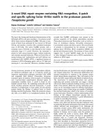
Báo cáo y học: " Near fatal posterior reversible encephalopathy syndrome complicating chronic liver failure and treated by induced hypothermia and dialysis: a case report" pps
Bạn đang xem bản rút gọn của tài liệu. Xem và tải ngay bản đầy đủ của tài liệu tại đây (275.52 KB, 4 trang )
Case report
Open Access
Near fatal posterior reversible encephalopathy syndrome
complicating chronic liver failure and treated by induced
hypothermia and dialysis: a case report
Rashmi Chawla
1
, Daniel Smith
2
and Paul E Marik
1
*
Addresses:
1
Division of Pulmonary and Critical Care Medicine, Thomas Jefferson University, Philadelphia, PA, USA and
2
Medical Intensive Care
Unit, Thomas Jefferson University Hospital, Philadelphia, PA, USA
Email: PEM* -
* Corresponding author
Published: 26 March 2009 Received: 19 September 2008
Accepted: 22 January 2009
Journal of Medical Case Reports 2009, 3:6623 doi: 10.1186/1752-1947-3-6623
This article is available from: />© 2009 Chawla et al; licensee Cases Network Ltd.
This is an Open Access article distributed under the terms of the Creative Commons Attribution License (
/>which permits unrestricted use, distribution, and reproduction in any medium, provided the original work is properly cited.
Abstract
Introduction: Posterior reversible encephalopathy syndrome is a clinico-neuroradiological entity
characterized by headache, vomiting, altered mental status, blurred vision and seizures with
neuroimaging studies demonstrating white-gray matter edema involving predominantly the posterior
region of the brain.
Case presentation: We report a 47-year-old Caucasian man with liver cirrhosis who developed
posterior reversible encephalopathy syndrome following an upper gastrointestinal hemorrhage and
who was managed with induced hypothermia for control of intracranial hypertension and continuous
veno-venous hemodiafiltration for severe hyperammonemia.
Conclusion: We believe this is the first documented case report of posterior reversible
encephalopathy syndrome associated with cirrhosis as well as the first report of the use of induced
hypothermia and continuous veno-venous hemodiafiltration in this setting.
Introduction
Posterior reversible encephalopathy syndrome (PRES), first
described by Hinchey and colleagues in 1996, is a clinico-
neuroradiological entity characterized by headache, vomit-
ing, altered mental status, blurred vision and seizures with
neuroimaging studies demonstrating white-gray matter
edema involving predominantly the posterior region of
the brain [1]. PRES is most commonly associated with
hypertensive encephalopathy, eclampsia, porphyria and
immunosuppressive or cytotoxic drugs [1,2]. Magnetic
resonance imaging (MRI) is the diagnostic test of choice,
demonstrating hyperintense echoes on T2-weighted images
of the parietal-occipital lobes; however, the cerebellar
hemispheres, basal ganglia, frontal lobes and brainstem
are often involved. While usually completely reversible, a
delay in the diagnosis and treatment may result in death or
irreversible neurological sequela [1,3]. We present a patient
with liver cirrhosis who developed PRES following an upper
gastrointestinal hemorrhage and who was managed with
induced hypothermia for control of intracranial
Page 1 of 4
(page number not for citation purposes)
hypertension and continuous veno-venous hemodiafiltra-
tion (CVVHDF) for severe hyperammonemia.
Case presentation
A 47-year-old Caucasian man with a past medical history
significant for noninsulin-dependent diabetes mellitus,
hepatitis C, past alcohol abuse and cirrhosis was admitted
to our medical intensive care unit with an upper
gastrointestinal bleed. On presentation, his blood pressure
was 104/70mmHg, heart rate 137 beats per minute,
temperature 37.2°C and respiratory rate 24 breaths per
minute. On examination, he was pale and icteric, and had
a mildly distended abdomen with no discernable organo-
megaly. Cardio-respiratory examination was normal. He
was confused and agitated with no focal neurological
signs. His white blood count was elevated at 23.7 × 10
9/
/L
with 24% bands, with a hemoglobin of 78g/L and platelets
of 83 × 10
9
/L. Albumin was 19g/dl (normal 32 to 49g/L),
total bilirubin 83.3mmol/L (normal 3.4 to 20.4mmol/L),
aspartate aminotransferase (AST) 32 IU/L (normal 7 to
42 IU/L), alanine aminotransferase (ALT) 35 IU/L (normal
1 to 45 IU/L), alkaline phosphatase (ALP) 32 IU/L
(normal 25 to 120 IU/L), international normalized ratio
(INR) of 1.84, arterial ammonia 151mmol/L (normal 11 to
35mmol/L) and lactate of 6.1mmol/L (normal 0.6 to
1.7mmol/L). His blood urea nitrogen (BUN) and creati-
nine were 9mmol/L and 124mmol/L, respectively.
The patient was electively intubated for airway protection
and to facilitate endoscopy. He was resuscitated with
crystalloids, 5% albumin, packed cells and fresh frozen
plasma and treated with vancomycin and piperacillin/
tazobactam for presumed sepsis. Ultrasound and Doppler
of his upper quadrants was consistent with cirrhosis with
normal blood flow and splenomegaly. An upper endo-
scopy revealed grade 4 esophageal varices with no active
bleed. Over the next few days, the patient became
progressively unresponsive (off sedation) with his ammo-
nia level rising above 200mmol/L despite aggr es sive
treatment with lactulose and rifaximin. Neurologic assess-
ment revealed posturing to painful stimuli with a poorly
reactive pupillary reflex. Computed tomography of the
head revealed diffuse white matter edema prominent in
the posterior temporal, parietal and occipital lobes. Brain
MRI confirmed diffuse white matter edema with temporal
and occipital lobe predominance consistent with the
diagnostic pattern for PRES (Figure 1a and 1b). His course
was complicated by the development of tonic-clonic
seizures which were controlled with intravenous levetir-
acetam. His pupils became fixed and non-responsive.
Transcranial Dopplers (TCD) of the middle and posterior
cerebral arteries demonstrated a marked reduction in
cerebral blood velocity consistent with severely increased
intracerebral pressure (ICP). As an extraordinary salvage
method to control the patient’s severe ICP, we lowered his
core body temperature to 32°C with the addition of
propofol and mannitol, titrated to keep serum osmolarity
< 310mmol/L. Induced hypothermia was maintained for
48 hours during which time he regained normal pupillary
reflexes with marked improvement in TCD velocities.
During the passive rewarming phase, the patient devel-
oped massive hematemesis. He required massive transfu-
sion and Minnesota tube placement as attempted banding
via endoscopy was unsuccessful. The patient underwent an
emergency transjugular intrahepatic portocaval shunt
(TIPS) placement followed by repeat induced hypother-
mia (32 to 34°C). Due to the anticipated increase in the
serum ammonia level following the massive gastrointest-
inal hemorrhage, we initiated high-flow continuous
venovenous hemodiafiltration (CVVHD) to facilitate
ammonia removal. The CVVHD was associated with a
fall in the ammonia level (Figure 2). At this time, the
patient was again passively rewarmed and the propofol
discontinued. His neurological status improved slowly
over the following week becoming mo re alert and
responsive and allowing extubation. A repeat MRI of the
brain showed interval improvement in extensive white
matter signal abnormality most consistent with resolving
PRES. He was discharged home with no neurological
sequela apart from amnesia for the entire hospital stay.
The patient has returned to work part-time and is currently
listed for liver transplantation.
Discussion
We believe that this is the first documented case of PRES
associated with cirrhosis as well as the first report of
induced hypothermia for the management PRES and the
use of CVVHD for control of hyperammonemia in a
cirrhotic patient. The pathogenesis of PRES remains
incompletely understood but is probably related to the
failure of cerebral autoregulation and endothelial damage
[4]. The favored pathogenetic theory suggests autoregula-
tory disturbance with hyperperfusion, resulting in blood–
brain barrier breakdown with reversible vasogenic edema
without cerebral infarction [2,4]. However, others have
suggested that the triggering event leads to cerebral
vasoconstriction, reduced brain perfusion, ischemia and
subsequent vasogenic edema [4–6]. It is not known why
the posterior circulation is preferentially affected. A
possible explanation is the lower sympathetic innervation
of the posterior cerebral arterial circulation than in the
inter nal carotid territory, with a consequent reduced
autoregulation of already impaired cerebral areas [2].
The cause of PRES in our patient and the association with
hyperammonemia is unclear. In patients with acute liver
failure and severe hyperammonemia, there is gradual
cerebral vasodilation due to loss of cerebral auto-regula-
tion resulting in increased cerebral blood flow and
vasogenic edema [7–10]. Using magnetic resonance
Page 2 of 4
(page number not for citation purposes)
Journal of Medical Case Reports 2009, 3:6623 />diffusion tensor imaging, Kale and colleagues demon-
strated an increase in interstitial brain water in patients
with cirrhosis and hepatic encephalopathy [11]. In this
study, brain water content increased with increasing grade
of encephalopathy and decreased with treatment of
hyperammonemia. Furthermore, emerging evidence indi-
cates that patients with cirrhosis may have astrocyte
swelling and low-grade cytotoxic cerebral edema which
is associated with the degree of hyperammonemia [12,13].
Our patient presented with hemorrhage and shock
complicated by systemic sepsis and required multiple
blood transfusions. Bartynski and colleagues have demon-
strated that all these factors may be implicated in the
etiology of PRES [14].
Our patient’s clinical examination, together with the
transcranial Doppler and neuroimaging studies suggested
that he had severely increased intracranial pressure (ICP)
that required aggressive treatment. Due to the patient’s
coagulopathy, direct ICP monitoring was considered
contraindicated. Moderate induced hypothermia has
been shown to reduce cerebral edema following cardiac
arrest and in patients with acute liver failure [15,16]. The
mechanisms by which hypothermia reduces neuronal
injury and cerebral edema are unknown, however,
retardation of destructive enzymatic reactions, suppres-
sion of free-radical reactions, protection of the fluidity of
lipoprotein membranes, reduction in the oxygen demand
in low flow regions, reduction in intracellular acidosis, and
inhibition of the biosynthesis, release, and uptake of
excitatory neurotransmitters have been postulated [17].
Furthermore, in patients with liver disease, hypothermia
reduces cerebral ammonia levels by decreasing brain
Figure 1.
(a) Non-contrast computed tomography showing diffuse
cerebral edema. (b) Magnetic resonance image showing
multiple cortico-subcortical areas of hyperintense signal
involving the occipital and parietal lobes bilaterally and pons.
Figure 2.
Time course of arterial ammonia concentration and core
body temperature.
Page 3 of 4
(page number not for citation purposes)
Journal of Medical Case Reports 2009, 3:6623 />ammonia uptake, decreasing ammonia production and
improving ammonia clearance [15]. We believe that the
use of induced hypothermia was life saving in our patient.
As the cause of PRES was unclear in our patient and
because hyperammonemia may cause both vasogenic and
cytotoxic edema, we elected to pre-emptively dialyze our
patient to reduce the ammonia level. Peritoneal dialysis,
hemodialysis, continuous veno-ven ous hemofiltration
(CVVH) and CVVHD have been reported to be helpful
in the treatment of hyperammonemia associated with urea
cycle disorders in neonates, children and adults. It is
unclear whether the use of CVVHDF in our patient
contributed to the favorable outcome.
Consent
Written informed consent was obtained from the patient
for publication of this case report and any accompanying
images. A copy of the written consent is available for
review by the Editor-in-Chief of this journal.
Competing interests
The authors declare that they have no competing interests.
Authors’ contributions
All authors were directly involved in the care of this
patient, were responsible for researching the literature and
writing the case report and take responsibility for the final
version.
References
1. Hinchey J, Chaves C, Appignani B, Breen J, Pao L, Wang A, Pessin MS,
Lamy C, Mas JL, Caplan LR: A reversible posterior leukoence-
phalopathy syndrome. N Engl J Med 1996, 334(8):494-500.
2. Servillo G, Bifulco F, De Robertis E, Piazza O, Striano P, Tortora F,
StrianoS,TufanoR:Posterior reversible encephalopathy
syndrome in intensive care medicine. Intensive Care Med 2007,
33:230-236.
3. Narbone MC, Musolino R, Granata F, Mazzu I, Abbate M, Ferlazzo E:
PRES: posterior or potentially reversible encephalopathy
syndrome? Neurol Sci 2006, 27(3):187-189.
4. Bartynski WS: Posterior reversible encephalopathy syndrome,
part 2: controversies surrounding pathophysiology of vaso-
genic edema. AJNR 2008, 29(6):1043-1049.
5. Weidauer S, Gaa J, Sitzer M, Hefner R, Lanfermann H, Zanella FE:
Posterior encephalopathy with vasospasm: MRI and angio-
graphy. Neuroradiology 2003, 45(12):869-876.
6. Trommer BL, Homer D, Mikhael MA: Cerebral vasospasm and
eclampsia. Stroke 1988, 19(3):326-329.
7. Larsen FS, Adel HB, Pott F, Ejlersen E, Secher NH, Paulson OB,
Knudsen GM: Dissociated cerebral vasoparalysis in acute liver
failure. A hypothesis of gradual cerebral hyperaemia. J Hepatol
1996, 25(2):145-151.
8. Blei AT, Larsen FS: Pathophysiology of cerebral edema in
fulminant hepatic failure. J Hepatol 1999, 31(4):771-776.
9. Jalan R, Olde Damink SW, Deutz NE, Davies NA, Garden OJ,
Madhavan KK, Hayes PC, Lee A: Moderate hypothermia prevents
cerebral hyperemia and increase in intracranial pressure in
patients undergoing liver transplantation for acute liver
failure. Transplantation 2003, 75(12):2034-2039.
10. Aggarwal S, Obrist W, Yonas H, Kramer D, Kang Y, Scott V, Planinsic R:
Cerebral hemodynamic and metabolic profiles in fulminant
hepatic failure: relationship to outcome. Liver Transplant 2005, 11
(11):1353-1360.
11. Kale RA, Gupta RK, Saraswat VA, Hasan KM, Trivedi R, Mishra AM,
Ranjan P, Pandey CM, Narayana PA: Demonstration of interstitial
cerebral edema with diffusion tensor MR imaging in type C
hepatic encephalopathy. Hepatology 2006, 43(4):698-706.
12. Haussinger D, Kircheis G, Fischer R, Schliess F, Vom DS: Hepatic
encephalopathy in chronic liver disease: a clinical manifesta-
tion of astrocyte swelling and low-grade cerebral edema?
J Hepatol 2000, 32(6):1035-1038.
13. Norenberg MD, Jayakumar AR, Rama Rao KV, Panickar KS: New
concepts in the mechanism of ammonia-induced astrocyte
swelling. Metab Brain Dis 2007, 22(3–4):219-234.
14. Bartynski WS, Boardman JF, Zeigler ZR, Shadduck RK, Lister J:
Posterior reversible encephalopathy syndrome in infection,
sepsis, and shock. AJNR 2006, 27(10):2179-2190.
15. Chatauret N, Rose C, Butterworth RF: Mild hypothermia in the
prevention of brain edema in acute liver failure: mechanisms
and clinical prospects. Metab Brain Dis 2002, 17(4):445-451.
16. The Hypothermia after Cardiac Arrest Study Group: Mild ther-
apeutic hypothermia to improve the neurologic outcome
after cardiac arrest. N Engl J Med 2002, 346(8):549-556.
17. Bernard SA, Buist M: Induced hypothermia in critical care
medicine: a review. Crit Care Med 2003, 31(7):2041-2051.
Page 4 of 4
(page number not for citation purposes)
Journal of Medical Case Reports 2009, 3:6623 />Do you have a case to share?
Submit your case report today
• Rapid peer review
• Fast publication
• PubMed indexing
• Inclusion in Cases Database
Any patient, any case, can teach us
something
www.casesnetwork.com
