Báo cáo y học: "Blateral synchronous occurrence of three different histological types of renal tumor: a case report" docx
Bạn đang xem bản rút gọn của tài liệu. Xem và tải ngay bản đầy đủ của tài liệu tại đây (1.4 MB, 6 trang )
Case report
Open Access
Bilateral synchronous occurrence of three different histological
types of renal tumor: a case report
Demetrios Radopoulos, Anastasios Tahmatzopoulos*, Nikolaos Kalinderis
and Georgios Dimitriadis
Address: 1st Department of Urology, Aristotle University of Thessaloniki, Ethnikis Amynis 41, 54635 Thessaloniki, Greece
Email: DR - ; AT* - ; NK - ; GD -
* Corresponding author
Published: 1 April 2009 Received: 10 December 2007
Accepted: 16 December 2008
Journal of Medical Case Reports 2009, 3:6798 doi: 10.1186/1752-1947-3-6798
This article is available from: />© 2009 Radopoulos et al; licensee Cases Network Ltd.
This is an Open Access article distributed under the terms of the Creative Commons Attribution License (
/>which permits unrestricted use, distribution, and reproduction in any medium, provided the original work is properly cited.
Abstract
Introduction: Renal cell carcinomas account for 85% of all renal neoplasms. With the introduction
of modern imaging modalities, there has been an increased diagnosis of renal tumors. Recent studies
have shown that partial nephrectomy can be as safe as radical nephrectomy for smaller renal tumors.
Renal cell carcinomas are usually unilateral, however, they can be bilateral in 2% to 4% of sporadic
cases and considerably more common in familial cases.
Case presentation: In this case report, we describe an unusual case of two bilateral synchronous
chromophobe renal cell carcinomas accompanied by an oncocytoma and an angiomyolipoma, that
were all treated by open partial nephrectomy.
Conclusions: To the best of our knowledge, this is the first case report on the synchronous
occurrence of bilateral chromophobe renal cell carcinomas associated with an oncocytoma and an
angiomyolipoma.
Introduction
Renal cell carcinoma (RCC) accounts for 85% of all renal
neoplasms. Its incidence has been rising due to the
increased use of ultrasonography and computed tomo-
graphy (CT) scans for the evaluation of patients with a
diversity of presenting symptoms. Most renal cell carcino-
mas are unilateral, but bilateral tumors, synchronous or
asynchronous, have been found in 2% to 4% of reported
sporadic cases. However, the incidence is higher among
patients suffering from Von Hippel-Lindau (VHL) disease
and other familial cases.
Radical nephrectomy is considered the standard treatment
modality for renal c ell carcinomas. However, recent data have
shown that partial nephrectomyisassafeandeffectiveas
radical nephrectomy fo r tumors s maller t han 4 cm, and mo re
recent studies support the fact that the indications for partial
nephrectomy can be safely extended to tumors up to 7cm.
Page 1 of 6
(page number not for citation purposes)
In this case report, we describe an unusual sporadic case
of bilateral synchronous RCC accompanied by an onco-
cytoma and an angiomyolipoma that were all treated by
open partial nephrectomy.
Case presentation
In February 2006, a 57-year-old man presented with vague
right upper quadrant discomfort. He had no surgical
history and was on antihypertensive (perindopril) and
antidiabetic (metformin) medication. He had a history of
acute myocardial infarction 5 years earlier and was
suffering from hypertensive ca rdiomyopathy. H e was
subjected to abdominal ultrasound which revealed bilat-
eral renal tumors, and cholelithiasis which was probably
the cause of his right upper quadrant discomfort.
In order to investigate the finding of bilateral renal tumors,
a contrast CT scan was performed which showed two
round hyperdense masses, one in each kidney, with
homogeneous contrast uptake and well defined margins,
arising from the renal cortex. The differential diagnosis
included lymphoma, atypical cysts, metastases and RCC.
Subsequent magnetic resonance imaging (MRI) confirmed
an enhancing, exophytic, well circumscribed solid tumor,
arising from the middle of the right kidney (3.7cm). On
the anterior surface of the left kidney, an enhancing,
exophytic, well circumscribed solid tumor was described
(Figures 1 and 2) and no abnormally enlarged retro-
peritoneal lymph nodes were detected. Bone scan with
99m
Tc-MDP did not show evidence of bone metastases.
In April 2006, the patient was subjected to open partial
nephrectomy on the left kidney, which revealed a 4 cm
chromophobe renal cell carcinoma (Fuhrman II), with
clear surgical margins.
In May 2006, a second open partial nephrectomy was
performed on t he ri ght k idn ey, re vealing a 3.5 cm
chromophobe renal cell carcinoma (Fuhrman II), with
clear surgical margins. A 3 cm oncocytoma and a 1.5 cm
angiomyolipoma were also detected intra-operatively,
necessitating two further partial nephrectomies on the
right kidney (Figure 3). A surgical collagen sponge with
fibrinogen and thrombin was used to aid in hemostasis in
both kidneys.
The patient did well postoperatively. Follow-up MRI at 14
months showed no evidence of recurrence (Figure 4).
Discussion
According to the literature, the synchronous presentation
of renal tumors of diverse histology and dignity is a rare
phenomenon [1,2]. The majority of published cases
describe the coincidence of angiomyolipomas and renal
cell carcinoma in otherwise healthy individuals as well as
in tuberous sclerosis (TS) patients. The latter is an
autosomal-dominant disorder characterized by mental
retardation, epilepsy, and adenoma sebaceum, a distinc-
tive skin lesion [1–5]. A few interesting reports describe the
coincidence of RCCs and oncocytomas in patients with
Birt-Hogg-Dube syndrome (BHDS), which is characterized
by the development of fibrofolliculomas, renal tumors
and spontaneous pneumothorax [6,7]. Our patient had no
known family history of renal tumors, nor did he have
clinical signs suggestive of either TS or BHDS. The patient
had no clinical evidence of VHL disease (no retinal
angioma on ophthalmologic examination, no family
history, no epididymal or pancreatic cysts, no pheochro-
mocytoma). Also, RCC in VHL patients is usually clear cell,
whereas our patient had chromophobe RCC. Taking these
facts into account, we decided not to proceed with genetic
analysis of the specimen.
Oncocytomas are the most common benign renal tumors
(3% to 7% of all solid renal tumors) [8]. They are usually
diagnosed incidentally during routine ultrasound exam-
ination and the discrimination between RCC and onco-
cytoma based solely on radiologic criteria, including CT
and/or MRI, is not always possible. Therefore, aggressive
surgical intervention is almost always warranted [9].
Angiomyolipoma (AML) is a benign clonal neoplasm that
consists of varying amounts of mature adipose tissue,
smooth muscle, and thick-walled vessels [10]. Approxi-
mately 20% of AMLs are found in patients with tuberous
sclerosis [11]. Patients typically present with abdominal or
lumbar pain as a result of m assive retro peritoneal
hemorrhage and/or a palpable abdominal mass. The
presence of fat within a renal lesion on CT scan, confirmed
by Hounsfield units ≤10, is considered diagnostic for
AML [10].
Renal cell carcinoma is the most common malignant
renal tumor and accounts for 3% of all adult
malignancies. Although potential etiologic factors have
been identified in animal models for example, viruses,
lead compounds, and more than 100 chemicals, no
specific agent has been definitively established as causative
in human RCC [10]. A familial form has been identified in
patients with VHL disease, a rare autosomal-dominant
disorder. Manifestations include the development of RCC,
pheochromocytoma, retinal angiomas, and hemangio-
blastomas of the brain stem, cerebellum, or spinal cord.
Most RCCs are asymptomatic at the time of diagnosis.
The classic triad of flank pain, gross hematuria, and
palpable abdominal mass is now rarely found. Therapy is
almost always surgical, either in the form of radical or
simple nephrectomy or in the form of nephron sparing
surgery.
Page 2 of 6
(page number not for citation purposes)
Journal of Medical Case Reports 2009, 3:6798 />Conclusion
In this paper, we present an unusual sporadic coexistence
of three different types of renal tumor. A similar case with
the concurrent occurrence of three primary neoplasms in
the same kidney - oncocytoma, chromophobe renal cell
carcinoma and angiomyolipoma - has recently been
reported [12]. It is interesting that two of the three tumors
in the right kidney were missed in pre-operative imaging
and were only detected intra-operatively. This suggests that
caution should be exercised in the interpretation of
radiological results and that the surgeon should be alert
for unexpected intra-operative findings.
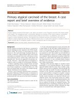
Figure 1.
Pre-operative axial magnetic resonance imaging sections showing a 3.7 cm tumor arising from the middle of the right kidney (a, b:
post contrast) as well as a 3.7 cm tumor arising from the anterior surface of the left kidney (c, d: post contrast).
Page 3 of 6
(page number not for citation purposes)
Journal of Medical Case Reports 2009, 3:6798 />We need to acknowledge that the follow-up period of 14
months is limited and that the patient is still at risk of local
tumor recurrence and disease progression. Chromophobe
RCCs, which comprise 4% to 5% of all RCCs, have an
inadequately defined clinical behavior as well as poorly
defined genetic alterations. Nevertheless, the present paper
underscores the feasibility of partial nephrectomy in the
case of multiple bilateral renal tumors.
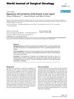
Figure 2.
Pre-operative coronal magnetic resonance imaging sections demonstrating the aforementioned right (a, c: post contrast) and left
(b, d: post contrast) renal tumors.
Page 4 of 6
(page number not for citation purposes)
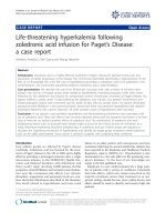
Journal of Medical Case Reports 2009, 3:6798 />Figure 3.
Intra-operative images showing (a) removal of three renal tumors in the right kidney as well as (b) intra-operative use of a
surgical collagen sponge containing the coagulation factors fibrinogen and thrombin.
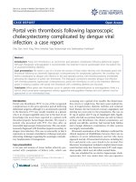
Figure 4.
Follow-up axial magnetic resonance imaging sections at 14 months postoperatively showing scar formation without local
recurrence (a, b: post contrast).
Page 5 of 6
(page number not for citation purposes)
Journal of Medical Case Reports 2009, 3:6798 />Consent
Written informed consent was obtained from the patient
for publication of this case report and any accompanying
images. A copy of the written consent is available for
review by the Editor-in-Chief of this journal.
Competing interests
The authors declare that they have no competing interests.
Authors' contributions
DR conceived the case report, performed the operation
and drafted the manuscript. GD was involved in post-
operative follow-up. AT and NK were involved in post-
operative follow-up and drafted the manuscript. All
authors read and approved the final manuscript.
Acknowledgements
Source of funding for the study: 1st Department of
Urology, Aristotle University of Thessaloniki.
References
1. Jun SY, Cho KJ, Kim CS, Ayala AG, Ro JY: Triple synchronous
neoplasms in one kidney report of a case and review of the
literature. Ann Diagn Pathol 2003, 7:374-380.
2. Billings B, Hamri ck LC, Buesch en AJ, Kenney PJ: Coexisting
angiomyolipoma and renal cell carcinoma in a kidney of an
elderly woman: case report and review of the literature.
Scientific World Journal 2004, 4(Suppl 1):27-30.
3. Jimenez RE, Eble JN, Reuter VE, Epstein JI, Folpe AL, de Peralta-
Venturina M, Tamboli P, Ansell ID, Grignon DJ, Young RH, Amin MB:
Concurrent angiomyolipoma and renal cell neoplasia: a study
of 36 cases. Mod Pathol 2001, 14:157-163.
4. Bjornsson J, Short MP, Kwiatkowski DJ, Henske EP: Tuberous
sclerosis-associated renal cell carcinoma. Clinical pathologi-
cal, and genetic features. Am J Pathol 1996, 149:1201-1208.
5. Winterkorn EB, Daouk GH, Anupindi S, Thiele EA: Tuberous
sclerosis complex and renal angiomyolipoma: case report
and review of the literature. Pediatr Nephrol 2006, 21:1189-1193.
6. Pavlovich CP, Walther MM, Eyler RA, Hewitt SM, Zbar B, Linehan
WM, Merin o MJ: Renal tumors in the Birt-Hogg-Dube
syndrome. Am J Surg Pathol 2002, 26:1542-1552.
7. Pavlovich CP, Grubb RL 3rd, Hurley K, Glenn GM, Toro J, Schmidt LS,
Torres-Cabala C, Merino MJ, Zbar B, Choyke P, Walther MM, Linehan
WM: Evaluation and management of renal tumors in the Birt-
Hogg-Dube syndrome. J Urol 2005, 173:1482-1486.
8. Dechet CB, Bostwick DG, Blute ML, Bryant SC, Zincke H: Renal
oncocytomas: Multifocality, bila teralism, metachronous
tumor development and coexistent renal cell carcinoma. J
Urol 1999, 162:40-42.
9. Licht MR: Renal adenoma and oncocytoma. Semin Urol Oncol
1995, 13:262-266.
10. Campbell SC, Novick AC, Bukowski RM: Renal tumors. In Campbell-
Walsh Urology, Volume 2. 9th edition. Edited by Wein AJ, Kavoussi LR,
Novick AC, Partin AW, Peters CA. Philadelphia: WB Saunders;
2007:1584.
11. Neumann HP, Schwarzkopf G, Henske EP: Renal angiomyolipo-
mas, cysts, and cancer in tuberous sclerosis complex. Semin
Pediatr Neurol 1998, 5:269-275.
12. Morelli L, Pusiol T, Piscioli I, Larosa M, Pozzoli GL, Monica B:
Concurrent occurrence of three primary neoplasms with
different hystotype in the same kidney, associated with an
adenoma of the omolateral adrenal gland: first case report.
Int J Urol 2006, 13:1236-1239.
Page 6 of 6
(page number not for citation purposes)
Journal of Medical Case Reports 2009, 3:6798 />Do you have a case to share?
Submit your case report today
• Rapid peer review
• Fast publication
• PubMed indexing
• Inclusion in Cases Database
Any patient, any case, can teach us
something
www.casesnetwork.com